Involvement of the receptor for advanced
glycation end products in liver transplantation
Teresa Navarra,*,*** Paolo De Simone,**,*** Serena Del Turco,* Franco Filipponi,** Giuseppina Basta*
* Institute of Clinical Physiology, National Research Council, Pisa, Italy.
** Hepatobiliary surgery and Liver Transplantation, University of Pisa Medical School Hospital, Pisa, Italy. *** Both authors contributed equally to this work.
ABSTRACT
Background and aim. Receptor for advanced glycation end products (RAGE) blockade by a soluble form of RAGE (sRAGE) appears to be protective against hepatocellular death and necrosis after I/R injury. Little is known about the role of the hepatic RAGE, its ligands, and the plasma levels of sRAGE in liver transplanta-tion (LT). Material and methods. This was a prospective study on patients (n = 28) undergoing deceased donor LT. RAGE ligands [the N(epsilon)-carboxy-methyl-lysine (CML) adduct and the high-mobility group box 1 (HMGB1) protein] and sRAGE levels were measured in donors at the time of organ procurement, while in recipients they were tested before surgery (baseline), after graft reperfusion, and on day 1 and 7 post-transplantation. Donors and recipients liver biopsies were collected to assess the transcriptional expres-sion of the full-length RAGE and of its truncated isoform, the endogenous secreted RAGE (esRAGE). Results. At baseline, CML levels were higher in LT recipients than in donors (p = 0.02), decreased immedia-tely after graft reperfusion (p < 0.0001) and returned to baseline values on day 7. Baseline HMGB1 levels (3.8 ± 2.3 ng/mL) increased after graft reperfusion (39.9±18 ng/mL, p < 0.0001), and returned to baseline values within day 1, while circulating sRAGE decreased significantly on day 7 (p < 0.0001). The graft esRAGE mRNA expression was inversely associated with bilirubin on day 7 (β = -0.62, p = 0.005). Conclusions. Early on after LT, there is accumulation of CML and a rapid increase of HMGB1 concurrent with a remarkable decline in circulating sRAGE. The RAGE-ligand axis may also be involved in early graft dysfunction.
Key words. High-mobility group box 1. N(epsilon)-carboxy-methyl-lysine. Liver transplantation. I/R injury.
Correspondence and reprint request: Giuseppina Basta, D. Biol.
CNR, Institute of Clinical Physiology.
San Cataldo Research Area. Via Moruzzi, 1. 56124 Pisa, Italy. Tel.: 0039-050-315 2216. Fax: 0039-050-315 2166
E-mail: lapina@ifc.cnr.it
Manuscript received: April 15, 2014. Manuscript accepted: September 03, 2014.
INTRODUCTION
Early allograft dysfunction (EAD) may affect up to 25% of liver transplant (LT) recipients and en-tails a sevenfold and a tenfold higher risk for graft loss and patient death, respectively.1 Both during
surgery and in the post-operative period, the liver graft may be injured by several toxic factors, such as hypotension, hypoxia, ischemia/reperfusion (I/R) injury, and use of hepatotoxic drugs. Although the mechanisms leading to EAD have not yet been en-tirely elucidated, its development is believed to be largely due to I/R injury, which is associated with
cell death, oxidative damage and a severe inflamma-tory response within the liver.2-4 In turn, the extent
of I/R injury may be related to the cold ischemia time, graft quality, donor age, the recipient’s clini-cal status at transplantation, and surgiclini-cal challeng-es. From a bio-molecular perspective, liver graft injury and EAD may result in altered expression of serum proteins associated with an inflammatory response.5 Recently, several chemokines and
cy-tokines –such as the monocyte chemotactic protein-1, interleukin-8, RANTES, interferon-γ inducible protein 10 and the interleukin-2 receptor– appear to increase in the early post-operative period of EAD recipients.6
There is experimental evidence that interaction between the receptor for advanced glycation end products (RAGE) and its ligands plays a pivotal role in liver inflammation and tissue regeneration after liver injury.7,8 RAGE interacts with
others, thus resulting in amplification of inflamma-tion and tissue injury.9,11 On the other hand, RAGE
blockade by a soluble form of RAGE significantly in-creases animal survival after extended liver resec-tion and appears to be protective against hepatocellular death and necrosis after I/R inju-ry.7,8,12 RAGE is detectable in blood as a soluble
isoform that consists of the extracellular ligand-binding domain only. The total circulating isoforms of soluble RAGE (sRAGE) include endogenous se-creted isoforms (esRAGE) generated via alternative splicing, and a truncated form generated through proteolytic cleavage of the full-length cell-surface re-ceptor.13 Both isoforms can bind RAGE ligands and
contribute to their removal by competing with the membrane-bound RAGE, thus resulting in a cytopro-tective effect.14,15
Over the last decade, clinical studies performed on patients undergoing lung, kidney, or LT have documented the involvement of the RAGE-ligand axis in the post-transplantation inflammation and regeneration processes.16-19 Cirrhotic patients
un-dergoing LT show high levels of plasma CML be-fore surgery which decrease by 50% at 3 months post-transplantation, thus confirming that the liv-er acts as a clearing organ for these adducts.18
The circulating and pro-inflammatory RAGE lig-and HMGB1 –which is present in the cell nucleus and can be released by necrotic cells or in re-sponse to hypoxia–20,21 peaks in blood after graft
reperfusion concurrent with hepatic HMGB1 ex-pression up-regulation.19
The interaction between RAGE and its ligands can thus impair liver regeneration, while sRAGE can have a protective role. Hence, we set up a pro-spective trial to evaluate the plasma levels of circu-lating RAGE ligands and/or sRAGE peri-operatively and in the early post-operative period, concurrent with assessment of the hepatic sRAGE/RAGE tran-scriptional expression in both graft recipients and their donors.
MATERIAL AND METHODS
Patient selection
This was a 12-month, prospective, single-centre study on the behaviour of the RAGE-ligand axis in 28 patients submitted to primary, whole-size, deceased donor LT performed at our institution from November 2010 to April 2012. The current report focuses on the first 7 day post-transplanta-tion.
Patients were enrolled if adults (≥ 18 years) and affected with clinically documented chronic liver disease of viral, metabolic, autoimmune, toxic and/or innate aetiology with or without hepatocel-lular carcinoma (HCC), and if they provided in-formed consent. Exclusion criteria called for: acute liver failure; a model for end-stage liver dis-ease (MELD) score ≥ 30 at transplantation; HCC beyond Milan criteria as per the pre-transplant clinical investigations; non-HCC liver neoplasms (i.e. cholangiocarcinoma, adenomatosis, heman-gioendothelioma); acute and/or chronic kidney dis-ease (defined as serum creatinine ≥ 1.5 mg/dL at transplantation); a body mass index (BMI) > 27 kg/m2; combined organ transplantation;
absti-nence from alcohol use < 6 months; autoimmune liver disease on active steroid treatment; hepatitis C virus (HCV)-related disease on treatment with interferon at the time of transplantation; history of major cardiovascular event, such as acute myo-cardial infarction, unstable angina, previous coro-nary revascularization, stroke and peripheral vascular disease grade ≥ 2; hepato-pulmonary syn-drome with arterial hypoxemia (PaO2) < 70 mmHg or an alveolar-arterial gradient > 20 mmHg; porto-pulmonary hypertension with a mean pulmo-nary arterial pressure > 25 mmHg; and mental im-pairment. At the time of transplantation, patients were re-screened for compliance with the eligibility criteria and underwent laboratory tests. The MELD score at transplantation was derived as indicated elsewhere.22 Patients were re-evaluated for
short-term complications for 7 days following LT by collecting clinical data, and routine blood parame-ters. Patients did not develop any complications within this observation period.
Approval was obtained from the institutional review board. The study was carried out in compli-ance with the principles set forth in the 2008 Seoul revision of the declaration of Helsinki.
Liver biopsies
Two biopsies were obtained from the donor liver graft during procurement and from the recipient’s liver explant, respectively, and were stored at -20°C in RNAlater (Sigma-Aldrich, St. Louis, MO, USA) for tissue stabilization.
Blood sample collection
samples were collected within 30 min after graft reperfusion, on days 1 and 7. Blood samples were centrifuged at 4 °C and plasma was immediately stored at -80 °C until analysis. All laboratory tests were performed in blinded fashion with respect to the identity of the samples. Routine biochemical analyses were determined by standard laboratory methods.
Determination of plasma sRAGE levels
Plasma sRAGE levels were determined using a double-sandwich ELISA kit (DuoSet ELISA develop-ment kit; R&D Systems, Minneapolis, MN, USA) as previously described.23 Intra-assay and inter-assay
coefficients of variation was 5.9% and 8.2%, respec-tively. The lower limit of detection of sRAGE was 21.5 pg/mL.
Determination of plasma CML levels
Plasma CML levels were measured by an in-house competitive ELISA using the mouse F(ab’)2 fragment anti-AGE monoclonal antibody (clone 6D12) (ICN Biochemical Division, Aurora, OH,USA), as previously described.24 The lower limit
of detection of CML was 0.5 μg/mL.
Determination of plasma HMGB1 levels
Plasma HMGB1 levels were determined using the double-sandwich ELISA Kit II (IBL International, Hamburg, Germany) according to the manufactur-ers’ description. Intra-assay and inter-assay coeffi-cients of variation were <8% and 10% respectively. Table 1. Donor and recipient clinical and biochemical characteristics.
• Donors (N = 28)
Age (years) 62.1 ± 17.3
• Recipients (N = 28)
Age (years) 53 ± 8.7*
Male (%) 23 (82)
Primary diagnosis, n (%)
HCC 7 (25)
HCC + HCV + cirrhosis 7 (25) HCC + HBV + cirrhosis 2 (7.1) HCV + cirrhosis 5 (17.9)
HCC + HBV 1 (3.6)
HCC + HCV 1 (3.6)
Caroli’s syndrome 2 (7.1) Primary sclerosing cholangitis 2 (7.1) Alcoholic cirrhosis 1 (3.6)
Ascites, n (%) 10 (36)
MELD score 9.48 ± 3.79
Graft cold ischemia time (min) 451.4 ± 91 Graft warm ischemia time (min) 87 ± 15
Prior LT On day 7 ALT (U/L) 69 (42-90.5) 89 (76.2-119.8) † AST (U/L) 72.5 (59-134.5) 40 (31.2-56.8)‡ GGT (U/L) 141.5 (104-197.5) 159 (106-220)
LDH (U/L) 232.7 ± 59.1 234.8 ± 39.4
Bilirubin (mg/dL) 1.51 (1.1-2.5) 3.28 (1.8-4.4)§
INR 1.27 ± 0.19 1.17 ± 0.13
Creatinine (mg/dL) 0.826 ± 0.202 0.937 ± 0.495 Antithrombin III (%) 50 (38-73)
Cholinesterase (U/L) 2663 (2,049-5,335)
Plasma samples from 30 healthy voluntaries were assayed as control (49.3 ± 12.3 years). The sensitiv-ity of the assay was 0.1 ng/mL.
RNA extraction and reverse transcription-polymerase
chain reaction (RT-PCR)
Total RNA was extracted from liver specimens by using the RNeasy Midi kit (Qiagen S.p.A, Milan, It-aly) as previously described.25 The RNA
concentra-tion was determined spectrophotometrically at 260 nm (BioPhotometer Eppendorf Italia, Milan, Italy). Integrity and purity of total RNA were checked by electrophoresis of samples on ethidium bromide 2% agarose gel and visualized at 302 nm (2UVTM Tran-silluminator, UVP, Upland, CA, USA).
Total RNA was reverse-transcribed using the iS-cript cDNA synthesis kit (Bio-Rad Laboratories, Inc,Hercules, CA, USA). The cDNA was amplified us-ing the Taq PCR Core Kit (Qiagen, Germany) with specific primers and conditions as shown below, in a GeneAmp PCR System 9700 thermal cycler (Perkin Elmer/Applied Biosystems, Foster City, CA, USA).
All PCR reactions were performed in a 50 μL to-tal volume, containing Taq polimerase (Qiagen, Ger-many) 2.5 U, deoxyribonucleotide triphosphate (dNTP) 0.2 mmol/L, MgCl2 1.5 mmol/L and 1 μmol/ L of both forward and reverse primers. The Primers, annealing temperature and cycles used were respec-tively:
• For RAGE gene, F: 5’-CAGGAATGGAAAGGA-GACCA-3’, R: 5’-CCCTTCTCATTAGGCACCAG-3’, 62°C (30 sec.) and 40 cycles.
• For esRAGE gene, F: 5’-GGGGATGGTCAACAA-GAAAGG-3’, R: 5’- AGGTTCCTCCGACTGAT-TCAGTTC -3’, 55°C (15 sec) and 40 cycles. • For GAPDH gene, F:
5'-GGTCTCCTCTGACT-TCAACAGCG-3', R: 5’-GGTACTTTATTGATGGT-ACATGAC-3’, 55° (1 min) and 30 cycles.
The amplified products were electrophoresed on ethidium bromide 2% agarose gel, in parallel with a DNA Ladder 100 bp (Qiagen, Germany). After gel image acquisition by a digital camera (Kodak DC290 Digital camera System™; Eastman Kodak, Roches-ter, NY, USA), the band intensity of PCR DNA was quantified using densitometric analysis by Scion im-age™. PCR products of esRAGE and full-length RAGE were normalized to transcript levels of the control gene glyceraldehyde 3-phosphate dehydroge-nase (GAPDH).
Figure 1. Comparison between donors and recipients (n = 28) prior to LT in plasma levels of CML (A), HMGB1 (B) and sRAGE (C) and kinetics of the same parameters in LT reci-pients at 7 days after LT. Data are presented as mean ± SD. Comparison between donors and LT recipients was evaluated by t-test while kinetics in LT recipient was evaluated by ANO-VA followed by Bonferroni test.
20
15
10
5
0
CML (
μ
g/mL)
Donors Prior LT After 1 day 7 days reperfusion
70 60 50 40 30 20 10 0
HMGB1 (ng/mL)
Donors Prior LT After 1 day 7 days reperfusion
9 8 7 6 5 4 3 2 1 0
sRAGE (Ln[pg/mL])
Donors Prior LT After 1 day 7 days reperfusion
p < 0.0001
p < 0.0001 p < 0.0001 p = 0.02
Figure 2. Simple direct correlation between baseline HMGB1 and the MELD score of the patients (n = 28) prior LT.
25
20
15
10
5
0
MELD score
0 2 4 6 8 10 12 14 Baseline HMGB-1 (ng/mL)
β = 0.45, p < 0.05
A
B
Statistical analysis
The sample size was calculated with the Stata software (version 9.2; Stata Corp. College Station, TX, USA) by the estimated power for two-sample comparison of means of CML plasma values. A sam-ple size of 56 patients (28 donors and 28 recipients) would provide 85% power to detect differences of 30% in CML value between two groups with a two-sided Student t-test at an α level of 0.05.
Data were analysed with the use of statistical software SPSS 13.0 (SPSS Inc, Chicago, IL, USA). The Kolmogorov-Smirnov test of normality was used to verify whether the distribution of variables followed a Gaussian pattern. According to the level of distribution, data are presented as mean ± stand-ard deviations (SD), medians and interquartile rang-es. Variables with a non-normal distribution were logarithmically transformed before each analysis. The unpaired Student’s t-test was used to compare normally distributed variables between donors and recipients. Multiple differences were evaluated by one-way ANOVA followed by the Bonferroni’s post-hoc test. Simple correlations were evaluated by line-ar regression analysis. A two-tailed p-value < 0.05 was considered statistically significant.
RESULTS
Donor age, cold and warm ischemia, recipient clinical and biochemical characteristics, and post-transplant markers of liver injury are illustrated in table 1. Recipients were mainly males (82%), and the most frequent indication for LT was HCC (64%). Mean recipients’ age was lower than their donors’ (53 ± 8.7 vs. 62.1 ± 17.3 years, respectively; p = 0.017).
At baseline, among the RAGE-ligands CML plas-ma levels were higher in LT recipients than donors (p = 0.02) (Figure 1A) and decreased immediately after graft reperfusion (p < 0.0001) to return to baseline values on day 1 (Figure 1A). The baseline HMGB1 levels did not differ between LT recipients and donors (3.8 ± 2.3 ng/mL vs. 3.3 ± 2.4 ng/mL, respectively) (Figure 1B). However, in both groups they were significantly (p < 0.0001) higher than in a group (30 subjects) of normal controls (0.45 ± 0.3 ng/mL). The baseline HMGB1 levels in LT recipi-ents correlated with the preoperative MELD score (β = 0.45, p < 0.05) (Figure 2). The baseline HMGB1 levels increased dramatically after graft reperfusion (39.9 ± 18 ng/mL, p < 0.0001) return-ing rapidly to baseline values one day after LT (3.6
± 2.9 ng/mL) (Figure 1B). The peak values of HMGB1 after reperfusion tended to correlate directly with the MELD score on day 7 (β = 0.42, p = 0.07).
The preoperative plasma levels of sRAGE did not differ between LT recipients and donors (Figure 1C). Plasma sRAGE levels did not change early after LT, but decreased significantly on day 7 (p < 0.0001) (Figure 1C). The decrease of sRAGE levels observed in the current patients’ population is illustrated in figure 3.
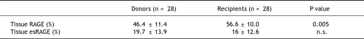
Semi-quantitative RT-PCR analysis revealed that hepatic full-length RAGE mRNA expression differed between donors and LT recipients (p = 0.005) while esRAGE mRNA did not differ between two groups (Table 2). The graft esRAGE mRNA expression was significantly and inversely associated with bilirubin levels on day 7 (β = - 0.62, p = 0.005) (Figure 4).
DISCUSSION
This study provides evidence supporting the in-volvement of RAGE and RAGE-ligands in the cas-cade of biological events following liver graft procurement and transplantation.
We found that, after transplantation, the circu-lating levels of sRAGE –which are the result of its expression in tissues and release into the blood-stream– decrease by day 7. Since sRAGE might ex-ert inhibitory effects on RAGE, acting as a decoy, but also binding to cell surface RAGE to block the formation of homodimers,26 decreased levels of
sRAGE may contribute to enhanced RAGE-mediated
Figure 3. The sRAGE plasma levels are shown for each pa-tient prior LT and 7 days after surgery. Median (95% CI) is shown. The P < 0.0001 value was evaluated by t-test on the logarithmically transformed data.
2,500
2,000
1,500
1,000
500
0
sRAGE (pg/mL)
pro-inflammatory signalling after transplantation and I/R injury. These changes in circulating sRAGE levels after LT may not only be due to decreased ex-pression/release –and possibly affected by immuno-suppression–27 but also to enhanced clearance.
However, in the current series baseline, serum cre-atinine did not change significantly vs. day 7, and the first hypothesis seems much more plausible and warrants further investigation. In agreement with our observation, previous research in kidney trans-plantation has demonstrated that circulating sRAGE levels decline after surgery and this is asso-ciated with a two- to threefold higher risk for mor-tality in kidney recipients.17
Among RAGE ligands, the most remarkable indi-cator of liver impairment is the HMGB1 protein, which is present into the cell nucleus and can be re-leased by cells undergoing necrosis or in response to hypoxia.20,21 In agreement with Ilmakunnas, et al.,19
our data confirm that HMGB1 plasma levels in-crease early on after reperfusion, probably as a re-sult of I/R injury and tended to correlate to MELD score on day 7. That is compatible with the hypoth-esis that, following ischemia-reperfusion, the early release of HMGB1 activates the RAGE and as a re-sult of this interaction the effects of the injury are amplified and revealed later.
Furthermore, we also found higher values of HMGB1 in LT recipients than in normal subjects, and a positive relationship between baseline HMGB1 protein and poperative MELD score. These re-sults underline the role of this marker in chronic liver disease and suggest that HMGB1 may in turn have a harmful impact on graft function. The LT re-cipients had higher hepatic expression of RAGE than donors. Nevertheless, we do not have a
plausi-ble explanation for this and we believe that other in-vestigations are need to know the reason of this dif-ference. We also found higher CML levels in LT recipients than in donors, possibly as consequence of reduced AGE clearance due to liver impairment. However, after a rapid drop ensuing graft reper-fusion, CML levels returned to pre-transplant val-ues on day 7, underscoring a poorly clarified reduced detoxification capacity of the liver graft early on after transplantation. An interesting relationship was the inverse association between esRAGE liver expression in donor grafts and recipients’ serum bilirubin on day 7 post-transplantation because low levels of the protective esRAGE could negatively influence the transplant in early follow-up, although the assessment of esRAGE in post-transplant biopsy (unfeasible for ethical concerns) would be of greater utility. Considering all of these results as a whole –i.e. the association between graft esRAGE and bilirubin levels on day 7, the rapid increase of HMGB1 after graft reperfusion, and the CML accu-mulation in the bloodstream followed by a dramatic decline of the protective sRAGE in the early post-operative period– we believe that these biological markers point out to metabolic events with the potential to affect graft survival and patient outcomes. The RAGE-ligand axis is a promising target of fur-ther investigation, as a biological marker of liver injury prior to and after LT. Identification of patients at risk of complications is crucial for improving post-operative care and transplant outcomes. The RAGE-ligand axis may also be used as a therapeutic target in order to enhance early graft function and improve survival.
Our study has several inherent limitations. This work is exploratory in nature and is a descriptive study, and therefore we cannot assign causality to our findings, nor can we recommend specific clinical strategies based on our conclusions. The sample size is the major limiting factor, and a prospective, larg-er validation set is needed for conclusions that are more robust. Despite these limitations, this study provides evidence for several associations between the cascade of biological events ensuing graft pro-curement, I/R, and reperfusion and the RAGE-ligands axis, and this can guide future investigation.
ABBREVIATIONS
• CML: N(epsilon)-Carboxy-Methyl-Lysine.
• EAD: early allograft dysfunction.
• esRAGE: endogenous secreted RAGE.
• HCC: hepatocellular carcinoma.
Figure 4. Simple inverse correlation between graft esRAGE mRNA expression and bilirubin levels on day 7.
14
12
10
8
6
4
2
0
Bilirubin at day 7 (mg/dL)
0 0.05 0.10 0.15 0.20 0.25 0.30 0.35 0.40 Graft esRAGE/GAPDH mRNA
Table 2. Comparison between donors and recipients in hepatic full-length RAGE and esRAGE mRNA expression.
Donors (n = 28) Recipients (n = 28) P value
Tissue RAGE (%) 46.4 ± 11.4 56.6 ± 10.0 0.005 Tissue esRAGE (%) 19.7 ± 13.9 16 ± 12.6 n.s.
Data are presented as mean ± SD.
• HMGB1: high-mobility group box 1.
• I/R: ischemia/reperfusion.
• LT: liver transplantation.
• MELD: Model for End-Stage Liver Disease.
• RAGE: receptor for advanced glycation end
products.
• sRAGE: soluble RAGE.
FUNDING SOURCES
This work was funded by institutional sources.
CONFLICT OF INTEREST
The authors have no conflicts of interest to dis-close.
ACKNOWLEDGMENTS
The authors wish to thank Alison Frank for her editorial assistance and owe a deep debt of gratitude to the nurse staff of the Hepatobiliary surgery and liver transplant Unit, University of Pisa Medical School Hospital, Pisa, Italy.
REFERENCES
1. Olthoff K M, Kulik L, Samstein B, Kaminski M, Abecassis M, Emond J, Shaked A, et al. Validation of a current definition of early allograft dysfunction in liver transplant recipients and analysis of risk factors. Liver Transpl 2010; 16: 943-9. 2. Busuttil RW, Tanaka K. The utility of marginal donors in
li-ver transplantation. Lili-ver Transpl 2003; 9: 651-63. 3. Totsukali E, Fung JJ, Ishizawa Y, Nishimura A, Ono H,
To-yoki Y, Narumi S, et al. Synergistic effect of cold and warm ischemia time on postoperative graft outcome in human li-ver transplantation. Hepatogastroenterology 2004; 51: 1413-6.
4. Briceno J, Ciria R. Early graft dysfunction after liver transplantation. Transplant Proc 2010; 42: 631-3. 5. Hassan L, Bueno P, Ferron-Celma I, Ramia J M, Garrote D,
Muffak K, Barrera L, et al. Early postoperative response of cytokines in liver transplant recipients. Transplant Proc 2006; 38: 2488-91.
6. Friedman BH, Wolf JH, Wang L, Putt ME, Shaked A, Chris-tie JD, Hancock WW, et al. Serum cytokine profiles asso-ciated with early allograft dysfunction in patients
undergoing liver transplantation. Liver Transpl 2012; 18: 166-76.
7. Cataldegirmen G, Zeng S, Feirt N, Ippagunta N, Dun H, Qu W, Lu Y, et al. RAGE limits regeneration after massive li-ver injury by coordinated suppression of TNF-alpha and NF-kappaB. J Exp Med 2005; 201: 473-84.
8. Zeng S, Feirt N, Goldstein M, Guarrera J, Ippagunta N, Ekong U, Dun H, et al. Blockade of receptor for advanced glycation end product (RAGE) attenuates ischemia and re-perfusion injury to the liver in mice. Hepatology 2004; 39: 422-32.
9. Del Turco S, Basta G. An update on advanced glycation endproducts and atherosclerosis. Biofactors 2012; 38: 266-74.
10. Hofmann MA, Drury S, Fu C, Qu W, Taguchi A, Lu Y, Avila C, et al. RAGE mediates a novel proinflammatory axis: a central cell surface receptor for S100/calgranulin polypep-tides. Cell 1999; 97: 889-901.
11. Kislinger T, Fu C, Huber B, Qu W, Taguchi A, Du Yan S, Hof-mann M, et al. N(epsilon)-(carboxymethyl)lysine adducts of proteins are ligands for receptor for advanced glycation end products that activate cell signaling pathways and mo-dulate gene expression. J Biol Chem 1999; 274: 31740-9. 12. Basta G, Navarra T, De Simone P, Del Turco S, Gastaldelli
A, Filipponi F. What is the role of the receptor for advan-ced glycation end products-ligand axis in liver injury? Li-ver Transpl 2011; 17: 633-40.
13. Raucci A, Cugusi S, Antonelli A, Barabino SM, Monti L, Bier-haus A, Reiss K, et al. A soluble form of the receptor for advanced glycation endproducts (RAGE) is produced by proteolytic cleavage of the membrane-bound form by the sheddase a disintegrin and metalloprotease 10 (ADAM10). Faseb J 2008; 22: 3716-27.
14. Yonekura H, Yamamoto Y, Sakurai S, Petrova R G, Abedin M J, Li H, Yasui K, et al. Novel splice variants of the re-ceptor for advanced glycation end-products expressed in human vascular endothelial cells and pericytes, and their putative roles in diabetes-induced vascular injury. Bio-chem J 2003; 370: 1097-109.
15. Schlueter C, Hauke S, Flohr A M, Rogalla P, Bullerdiek J. Tis-sue-specific expression patterns of the RAGE receptor and its soluble forms—a result of regulated alternative splicing? Biochim Biophys Acta 2003; 1630: 1-6.
16. Christie JD, Shah CV, Kawut SM, Mangalmurti N, Lederer DJ, Sonett JR, Ahya VN, et al. Plasma levels of receptor for advanced glycation end products, blood transfusion, and risk of primary graft dysfunction. Am J Respir Crit Care Med 2009; 180: 1010-5.
17. Gross S, van Ree RM, Oterdoom LH, de Vries AP, van Son WJ, de Jong PE, Navis GJ, et al. Low levels of sRAGE are associated with increased risk for mortality in re-nal transplant recipients. Transplantation 2007; 84: 659-63.
in patients with liver cirrhosis - amelioration by liver transplantation. J Hepatol 2002; 36: 66-71.
19. Ilmakunnas M, Tukiainen EM, Rouhiainen A, Rauvala H, Aro-la J, Nordin A, Makisalo H, et al. High mobility group box 1 protein as a marker of hepatocellular injury in human liver transplantation. Liver Transpl 2008; 14: 1517-25.
20. Xia JR, Liu NF, Zhu NX. Specific siRNA Targeting the Re-ceptor for Advanced Glycation End Products Inhibits Ex-perimental Hepatic Fibrosis in Rats. Int J Mol Sci 2008; 9: 638-61.
21. Lohwasser C, Neureiter D, Popov Y, Bauer M, Schuppan D. Role of the receptor for advanced glycation end pro-ducts in hepatic fibrosis. World J Gastroenterol 2009; 15: 5789-98.
22. Wiesner RH, McDiarmid SV, Kamath PS, Edwards EB, Malin-choc M, Kremers WK, Krom RA, et al. MELD and PELD: application of survival models to liver allocation. Liver Transpl 2001; 7: 567-80.
23. Basta G, Sironi AM, Lazzerini G, Del Turco S, Buzzigoli E, Casolaro A, Natali A, et al. Circulating soluble receptor for
advanced glycation end products is inversely associated with glycemic control and S100A12 protein. J Clin Endocri-nol Metab 2006; 91: 4628-34.
24. Kaloudi O, Basta G, Perfetto F, Bartoli F, Del Rosso A, Mi-niati I, Conforti ML, et al. Circulating levels of Nepsilon-(carboxymethyl)lysine are increased in systemic sclerosis. Rheumatology (Oxford) 2007; 46: 412-6.
25. Nannipieri M, Cecchetti F, Anselmino M, Mancini E, Mar-chetti G, Bonotti A, Baldi S, et al. Pattern of expression of adiponectin receptors in human liver and its relation to nonalcoholic steatohepatitis. Obes Surg 2009; 19: 467-74. 26. Zong H, Madden A, Ward M, Mooney MH, Elliott CT, Stitt
AW. Homodimerization is essential for the receptor for ad-vanced glycation end products (RAGE)-mediated signal transduction. J Biol Chem 2010; 285: 23137-46.