Bile Acids in Nonalcoholic Fatty Liver Disease:
New Concepts and Therapeutic Advances
Vania Cruz-Ramón, Paulina Chinchilla-López, Oscar Ramírez-Pérez, Nahum Méndez-Sánchez
Liver Research Unit, Medica Sur Clinic & Foundation, Mexico City, Mexico.
INTRODUCTION
Nonalcoholic fatty liver disease (NAFLD) is a major health burden and independent cause of cardiovascular disease. Some studies note that the worldwide prevalence of NAFLD is approximately 20%-30%1,2 with 2%-44% in Europeans and 15% in Asians.3,4 Given the lack of epidemi-ological data in the Americas, our group based on the prevalence of obesity to estimate the NAFLD prevalence in Mexico as 26% and 29% in the USA.5 Following these trends, population growth will increase the incidence of this disorder.
NAFLD is defined by the presence of steatosis, steato-hepatitis, and in some cases, the development of fibrosis and cirrhosis, including liver cancer.6 By contrast, the pathogenesis is multifactorial and not completely under-stood. Interestingly, Brunt, et al.7 proposed different key points in lipid accumulation, including weight gain, which is associated with an increase in adipose tissue and conse-quently the dysfunction and death of adipocytes. Dysfunc-tional adipocytes result in local inflammation and the upregulation of cytokines, which promote insulin
resist-ance. In turn, this condition disables the ability of adi-pocytes to store fat, resulting in the release of high levels of free fatty acids into the circulation, which will available to be captured by various organs, such as the liver. This is made possible by fatty acid transport protein 5 (FATP5/ acylCoAsynthetase) and CD36, which is a surface receptor that facilitates uptake of these fatty acids.7 An increase of FATP5/acylCoA synthase in the liver stimulates the pro-duction of triglycerides, gluconeogenesis activation, hy-perglycemia, and an increase in the synthesis of compensatory insulin. However, the excess of fatty acids put an strain on hepatocyte mitochondria, which produces a dysfunction after the hepatocyte death. Hepatocyte death can be caused through two main pathways. The first is by endoplasmic reticulum stress, which leads to mito-chondrial dysfunction. This action is regulated by a cas-pase 2-mediated cleavage excision of BH3 death agonist in an interactive domain (BID). The second is through the activation of death receptors. The most common exam-ples are the death ligands FAS and DR5, which eventually result in apoptosis and necroptosis of hepatocytes.8 In addi-tion, it is well known that participation of the liver in total
The Official Journal of the Mexican Association of Hepatology, the Latin-American Association for Study of the Liver and
the Canadian Association for the Study of the Liver
Manuscript received: Manuscript received: Manuscript received: Manuscript received:
Manuscript received: September 09, 2017.
DOI:10.5604/01.3001.0010.5498
A B S T R A C T A B S T R A C T A B S T R A C T A B S T R A C T A B S T R A C T
Nonalcoholic liver disease (NAFLD) is a major emerging health burden that is a common cause of illness and death worldwide. NAFLD can progress into nonalcoholic steatohepatitis (NASH) which is a severe form of liver disease characterized by inflammation and fibrosis. Further progression leads to cirrhosis, which predisposes patients to hepatocellular carcinoma or liver failure. The mechanism of the progression from simple steatosis to NASH is unclear. However, there are theories and hypothesis which support the link between disruption of the bile acids homeostasis and the progression of this disorder. Previous studies have been demonstrated that alterations of these pathways can lead to dysregulation of energy balance and an increase of liver inflammation and fibrosis. In this review, we summarized the current knowledge of the interaction between BA and the process related to the de-velopment of NAFLD, besides, the potential targets for novel therapies.
Key words. Key words. Key words. Key words.
Key words. Bile acids. NAFLD. Metabolism. Metabolic syndrome.
Manuscript accepted: Manuscript accepted: Manuscript accepted: Manuscript accepted:
lipids storage, through a process called de novo lipogenesis (DNL), increases the risk to develop NAFLD.8-10
This review will focus on the role of BA which are considered the newly identified players in the complex pathogenesis and treatment of NAFLD.
LIGHT AND DARK
SIDES OF BILE ACIDS IN NAFLD
A major component of bile is bile acids (BA), which are amphipathic molecules synthesized in hepatocytes from cholesterol. The process of their synthesis has been known for several decades11 (Figure 1). The hydrophobic
and hydrophilic regions of these amphipathic molecules can produce two great distinctive effects. The hydrophilic area plays a protective role for liver cells, while the hydro-phobic region can be cytotoxic and generate oxidative stress by inducing mitochondrial dysfunction and forma-tion of reactive oxygen species, ultimately leading to apop-tosis or necrosis.12
Other important functions of BA include the emulsifi-cation of dietary fats and intestinal absorption of lipids and lipophilic vitamins. BA are well known to play a critical role as regulators of hepatic lipid and glucose metabolism through farnesoid X receptor (FXR), vitamin D (NR111), and pregnane X (NR112), which are members of the nu-clear receptor superfamily (NRS), and G protein-coupled receptor 5 (TGR-5) and sphingosine-1-phosphate 2,
which are members of the G protein-coupled receptor su-perfamily.9,13 The ligands of NRS, such as peroxisome
proliferator-activated receptors (PPARs) contribute to pathogenesis in metabolic diseases. PPARs α (NR1C1), PPARs δ (NR1C2), and PPARs γ (NR1C3) are the three isotypes that are known. PPARs control the expression of a wide range of genes that maintain the metabolism of glu-cose, triglycerides (TG), and lipids, besides the synthesis, oxidation, storage, and export of BA.14 The disruption of
these metabolic processes contributes to the pathogenesis of metabolic diseases, such as obesity, metabolic syn-drome (MS), diabetes, NAFLD, and atherosclerosis.15
Current research has identified promising targets asso-ciated with BA. Specifically, through their FXR and TGR-5 receptors, BA improve lipid and glucose homeostasis and inhibit the inflammatory response.9
BILE ACIDS AND FXR ON
METABOLISM OF GLUCOSE AND LIPIDS
BA are important cell-signaling molecules that stimu-late diverse pathways to regustimu-late biological processes. The regulatory functions of BA are the result of modulation of intracellular ligand-activated nuclear receptor super-families (NRS), such as FXR and TGR-5.16-18
Additionally, FXR plays an important role in BA home-ostasis controlling the metabolism of genes such as the nu-clear receptor small heterodimer partner(SHP),19
Figure 1. Figure 1. Figure 1. Figure 1.
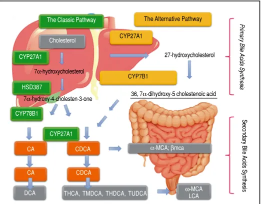
Figure 1. Pathways and Synthesis: BA Cholesterol serves as the sole substrate and is converted to cholic acid via both classic and alternative pathways. Micro-somal cholesterol 7 -hydroxylase is the ini-tial and rate-controlling enzyme for the classic pathway, whereas mitochondrial sterol 27-hydroxylase initiates the alterna-tive pathway and might be rate limiting. The product, 27-hydroxycholesterol, is then 7α-hydroxylated by microsomal oxysterol 7α-hydroxylase (27-hydroxycholesterol-7α -hydroxylase), which is different from mi-crosomal cholesterol 7α-hydroxylase. Primary bile acids are metabolized by gut bacteria to form the secondary bile acids, DCA and LCA.
Cholesterol
CDCA
CDCA The Classic Pathway
36, 7α-dihydroxy-5 cholestenoic acid The Alternative Pathway
CYP27A1
27-hydroxycholesterol
CYP27A1
HSD387
CYP27A1
Primary
Bile
Acids
Synthesi
s
Secondary
Bile
Acids
Synthesi
s
CYP7B1
CYP78B1
CA α-MCA; βmca
ω-MCA
LCA THCA, TMDCA, THDCA, TUDCA
DCA CA
7α-hydroxycholesterol
Na+-taurocholate cotransporting polypeptide (NTCP), cholesterol 7α hydroxylase (CYP7A1), and the bile salt ex-port pump (BSEP).20-22 The main functions of FXR can be summarized as follows. An increase in BA levels activates FXR, which suppresses their synthesis. This suppression occurs through induction of SHP, a nuclear receptor that binds to and interferes with the positive regulation of gene expression by other nuclear receptors, such as liver recep-tor homolog 1 (NR5A2) and liver X receprecep-tor (NR1H3).23,24 These nuclear receptors are involved in the control of genes that participate in BA synthesis and port. This transport is conducted by NTCP, which trans-ports BA from the circulation to the liver, and BSEP, which is directly activated by FXR and transports this molecule from hepatocytes to the gallbladder.25 Thus, when BA levels rise in the liver, CYP7A1, NTCP, and BA uptake from the blood is decreased, and BSEP and BA transport to the intestine is increased simultaneously (Fig-ure 2).
By contrast, in ileum enterocytes, FXR activation, by BA agonists produced in the liver, improves the transport of BA from the gut lumen to the blood by inducing ex-pression of apical sodium-BA transporters (ASBT, SLC10A2) and the organic solute and steroid transporter (OSTα , OSTβ) that convey BA from the enterocytes to the blood.26 Nevertheless, FXR has another pathway which controls the synthesis of BA in the liver. This is performed through the expression of the fibroblast growth factor 15 (FGF15) gene, designated FGF19 in humans, in enterocytes. FGF15/19 is transported to the liver and binds to and activates the hepatocyte plasma membrane re-ceptor complex FGFR4β-Klotho, resulting in the sup-pression of CYP7A1 exsup-pression27 and then a decrease of BA synthesis.27,28
In addition, LXRα, a nuclear receptor of liver, is in-volved in NAFLD pathogenesis because it has been found to be disturbed in patients with NAFLD.29 This nuclear receptor has an important role in regulation of cholesterol metabolism and hepatic free acid biosynthesis.30 In this context, it has been observed that LXRα mRNA is in-creased, in rat models of NAFLD, under a fatty diet regi-men.31,32 Therefore, a positive correlation between the expression of LXRα and the degree of NAFLD was estab-lished because of a major presence of LXR expression that will cause more DNL, which ultimately results in more infiltration of fat into the liver.
Patients with nonalcoholic steatohepatitis (NASH) are well known to have raised levels of BA, both in liver tissue and plasma,33,34 suggesting a relationship between toxic levels of BA and the development of NASH. The under-standing of the contribution of NRS and BA dysregulation for the pathogenesis and advancement of NAFLD is a principal issue in the Hepatology field.35
Patients with NASH may express different genes and proteins than patients with simple steatosis (SS). The ex-pression of FXR, LXRα, SHP, and NTCP in liver biopsy samples between patients with SS and NASH have been compared. Individuals with NASH possess a different mRNA and protein-expression profile of FXR, SHP, and NTCP versus patients with SS. An elevated mRNA ex-pression of NRS (FXR and SHP) and NTCP in liver bi-opsies from patients with NASH was observed, whereas by contrast, the protein levels were decreased in the same samples. Otherwise, LXRα gene expression and its pro-tein level between the SS and NASH groups were not found to be significantly different.36 This illustrates that mRNA expression may not always be equivalent to pro-tein level, a stark contrast to general assumption that protein levels must be correlated to the levels of their corresponding mRNAs. Many studies sustain that this general assumption may be incorrect in most cases.37-39 However, more studies are required to comprehend mo-lecular mechanisms of BA regulation more fully. In turn, this will allow us to understand the progression of NAFLD more thoroughly.40
BILE ACIDS AND TGR-5
TGR-5 plays a regulatory role in the glucose homeosta-sis through the enteroendocrine cells, which are able to induce glucagon-like peptide 1 (GLP1), a potent anti-in-flammatory molecule capable of blocking the action of cy-tokines and infiltrated macrophages by nuclear translocation of nuclear factor κB (NF-κB).41 Induction of GLP1 by TGR-5 in obese mice leads to improvement in liver and pancreatic function and consequently glucose tolerance.
Resistance to weight gain and hepatic steatosis, preser-vation of liver and pancreatic function, maintenance of glucose homeostasis and insulin sensitivity are beneficial metabolic effects produced by TGR-5 activation. These ef-fects result from improved mitochondrial function in muscle and enteroendocrine cells, leading to an increase in energy expenditure and incretin secretion.42,43
BODY MASS INDEX AND NAFLD
al-Figure 2. Figure 2. Figure 2.
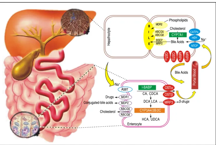
Figure 2. Figure 2. BA transporters in the hepatocytes and enterocytes the microsomal epoxide hydrolase and NTCP may be responsible for Na+-dependent uptake of conjugated BA at the basolateral membrane of the hepatocytes, whereas OATPs show substrate specificity for unconjugated BA. At the canalicular membrane of the hepatocytes, the BSEP performs a main role in biliary secretion of BA, while the MRP2 regulates secretion of organic substrates including glutathione, bilirubin, and BA. ABCG5 and ABCG8 heterodimers transport cholesterol into the bile, whereas MDR2 is responsible for biliary secretion of phospholipids. At the basolateral membrane of the hepatocytes, organic solute transporters OST and OSTβ heterodimers, MRP3, and MRP4 mediate the BA secretion into the circulation. With cholestasis, both basolateral BA efflux and renal BA excretion are increased. After BA are released from the gallbladder into the intestine, ileal BA uptake is regulated by the ASBT. Intracellular BAs are matched to the intestinal BA-binding protein (I-BABP). BA efflux is mediated by the OST and OST heterodimers at the basolateral membrane. At the apical membrane of the enterocytes, ABCG5 and ABCG8 heterodimers transport cholesterol back into the intestinal lumen, a process that confines intestinal cholesterol absorption. CYP3A4, CYP2B, and CYP2C are implicated in the metabolism and detoxification of LCA in the intestine. MDR1 effluxes drugs and MRP2 effluxes conjugated BA in the apical membrane of intestine. At the sinusoidal membrane, the MRP3 output sulfur conjugated drugs for renal excretion.
terations in BA levels, which are increased in obesity, have been observed, consequently it has suggested that these al-terations affect the energy metabolism.47
Previous studies48-50 in patients with insulin resistance
and T2D concluded that plasma levels of BA are elevated. The cause of this alteration is unknown, but some possi-bilities have been proposed. First, the hydroxylation of BA has a preference for the 12α position in patients with insu-lin resistance, resulting in relatively greater synthesis. Sec-ond, insulin reduces serum levels of BA in healthy patients; in obesity, this effect is inhibited. Third, the lev-els of BA during the meal are attenuated with respect to fasting levels. Finally, the expression of BA transporters in the liver is negatively associated with BMI.
THE ROLE OF
MICROBIOTA IN BA METABOLISM
NAFLD is a hepatic expression of MS (Figure 3), be-ing frequently related to insulin resistance, dyslipidemia, and obesity.51
A role for gut microbiota (GM) in insulin resistance, obesity, and associated metabolic disorders has been dem-onstrated, increasing the interest in GM’s relationship with NAFLD pathogenesis.52-55 Therefore, GM have
ap-peared as a potential factor involved in NAFLD.
Human GM include 10-100 trillion microorganisms56
composed of bacteria, archaea, virus, and fungi. The four main phyla of bacteria are: Firmicutes, Bacteroidetes,
Ac-Hepathoctyt
e
Phospholipids
Cholesterol
Na+
S-drugs
Enterocyte
HCA, UDCA
CYP3A4/2B/2C
DCA LCA CA, CDCA
Bile Acids Bile Acids
CYP7A1
Drugs Conjugated-bile acids
Cholesterol
CATPs mEH
NTCP
Portal
Bloo
d
OSTα
OSTβ
MRP3 I-BABP
ASBT Na+
MDR1 MDP2 ABCG5 ABCG8
BBBBB IIIII LLLLL EEEEE
MDR2 ABCG5 ABCG8 BSEP MRP2
MR
P3
MR
P4
OS
T
β
OS
T
tinobacteria, and Proteobacteria, which represent more than 95% of GM.57-59 The microbiome refers to the
col-lective genome of the GM and contains many important genes for glycan and amino-acid metabolism, xenobiotic metabolism, methanogenesis, and biosynthesis of vita-mins.58 The GM regulates body fat gain and insulin
resist-ance, so it seems to play an essential role in NAFLD through different pathways, including increasing energy harvest from diet, expression changes of genes involved in the DNL, regulation of choline metabolism, ethanol pro-duction, inflammasomes, and innate immunity and in-flammation.60
A disruption of the normal GM, dysbiosis, is linked to the pathogenesis of human liver disease. Early evidence of NAFLD associating gut dysbiosis with liver injury came from descriptive human studies of small intestinal bacteri-al overgrowth diagnosed by D-xylose and lactulose breath testing61,62 in an advanced stage of NAFLD. This early
evi-dence coincides with current data that support the role of the microbiome in human diseases, such as obesity and its related disorders. Experiments in mice provide evidence that phenotypes can be altered by transfer of GM from obese animals to lean littermates.
GM has an important function in signaling pathways and immune responses. Therefore, it plays a central role in the development and progression of NAFLD. For this reason, an understanding of GM may provide novel thera-peutic targets for improving NAFLD treatment.63
NOVEL PHARMACOLOGICAL
TARGETS FOR THE TREATMENT OF NAFLD
According to the natural history of the disease, more ag-gressive treatment is recommended for NASH than for NAFLD. In this section, we describe some of the 187 stud-ies being conducted with the aim of evaluating the efficacy and safety of NASH treatments, of which 79 are completed and 51 are still recruiting at the time of writing.64
Aramchol
In 2014, an Israeli research group developed a drug called aramchol. This drug inhibits stearoyl-CoA desaturase 1 (SCD1), which produces a decrease of fatty acids synthesis and increase in β-oxidation. Consequently, there is a decline in the storage of TG and esters of fatty acids in the liver. Safa-di and his group evaluated the effect of aramchol using spec-troscopy identifying a dose-dependent reduction in levels of hepatic steatosis from 100 to 300 mg vs. placebo.65
Elafibranor
Elafibranor is an agonist of PPARα/δ. PPARα is a lig-and-activated nuclear receptor (NR1C) expressed in the liver that adapts the rates of fatty acid catabolism and lipo-genesis in response to feeding.66 Moreover, it controls the
lipid flux in the liver by modulating fatty acid transport
Figure 3. Figure 3. Figure 3. Figure 3.
Figure 3. Metabolic Syndrome: Obesity as the critical risk factor for Nonalcoholic Liver Disease MS is one of the main risk factor for NAFLD. This illustration shows the different stages of the disease and shows a schematic representation of the action of BA as signaling molecules in NAFLD.
MS
Obesity is associated whith elevated
serum BA levels Liver uses cholesterol to
produce BA
Dybiosis intestinal has been related with
the development and progression
of liver disease
and β-oxidation, while improving plasma lipids by de-creasing TG and inde-creasing high-density lipoprotein (HDL) cholesterol. By contrast, PPARδ improves glucose homeostasis by upgrading insulin sensitivity and inhibit-ing the hepatic glucose output. This is the mechanism of elafibranor (a dual PPARα/δ agonist), which reduces the steatosis, fibrosis, and inflammation in patients with NAFLD as confirmed by Ratziu, et al.68 after assessing its safety and efficacy. Interestingly, Ratziu, et al. showed that using the highest dose (120 mg) of elafibranor did not ag-gravate the liver fibrosis, but it could reverse NASH (con-sidering “worsening” as any NAFLD stage that implicates fibrosis). The main adverse events identified in this phase 2 trial were nausea (10%), headache (7%-87%), diarrhea (5%-62%), and fatigue (5%-62%). GENFIT (Biopharma-ceutical Company) is conducting a phase 3, multicenter, randomized, double blind, placebo-controlled trial to evaluate the efficacy and safety of this drug in a bigger pop-ulation than the previous phase trials.
FXR agonists
FXR, as we mentioned before, are nuclear hormone re-ceptors expressed in several tissues (including liver, bowel, and kidney) and play a determining role in carbohydrate and lipid metabolism, such as regulation of insulin sensitivity.69
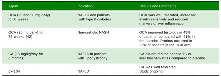
Many preclinical studies have assessed the FXR ago-nists role in the development of NAFLD and their results have shown that they cause an improvement of hyperlipi-demia, enhanced glucose tolerance, and insulin sensitivity. These results have also been compared with various other diseases of humans, mostly in patients with NAFLD or primary biliary cirrhosis (PBC) (70) (Table 1). Obeti-cholic acid (OCA), a derivative of CDCA and a first-in-class selective FXR agonist, has shown two outcomes. The first included important increases in insulin sensitivity, and reduced markers of liver inflammation and fibrosis in patients with NAFLD and with T2D.71 The second was changes in body weight, liver enzymes, serum lipids, FGF19, 7α-hydroxy-4-cholesten-3-one (C4, a cholesterol metabolite formed by the rate-limiting enzyme CYP7A1), endogenous BA, and serological measures of liver fibrosis and apoptosis. Patients in the group given an OCA dose of 50 mg experienced a significant average weight loss of 2% (P = 0.008) compared with patients given a placebo.72 In concordance with the mechanism of OCA, the FXR ago-nism resulted in an increase of FGF19 and a reduction of endogenous BA and C4 production.
Dual FXR/TGR-5 agonist
Recently, 6α-ethyl-3α,7α-dihydroxy-24-nor-5β -cho-lan-23-sulfate (INT-767), a potent dual FXR and TGR-5
agonist, has demonstrated potential for NAFLD treatment in obese diabetic mice. McMahan, et al. observed that treating diabetic (db/db) obese mice with INT-767 de-creased hepatic steatosis, and reduced proinflammatory cytokine expression, directed monocytes and macrophag-es toward an anti-inflammatory M2 phenotype. They sug-gested that the modulation of FXR and TGR-5 may be useful in NAFLD treatment by improving insulin secre-tion and sensitivity and controlling glucose, lipids, and BA homeostasis.73
In addition, the treatment of obese mice with INT-767 had shown significant reductions of total plasma choles-terol and TG levels.61 By contrast, in a MDR2-/-mouse model of chronic cholangiopathy, INT-767 was able to ameliorate hepatic injury by decreasing biliary BA output and promoting HCO-3-rich bile secretion.74
According to this, INT-767 could provide new oppor-tunities for the treatment of metabolic diseases, such as T2D and obesity, as well as chronic liver diseases.
Inhibitors of bile acid absorption
Bile acid sequestrants (BAs), such as colesevelam and cholestyramine, reduce plasma low-density lipoprotein levels and improve glycemic control. They act by promot-ing cholesterol catabolism through BA biosynthetic path-ways and improving hepatic insulin sensitivity. Thus, BA sequestration can be fruitful for NAFLD treatment.75,76
However, current experimental and clinical data do not support this argument. Le, et al. specifically designed a study to evaluate the efficiency of colesevelam in decreas-ing liver fat in patients with biopsy-proven NASH obtain-ing a small increase in liver fat detectable through magnetic resonance imaging and spectroscopy.77
Furthermore, Solis, et al. showed that BAs neither pro-duce any positive effects on liver steatosis in ob/ob mice nor histology nor hepatic triglyceride content was influ-enced by cholestyramine.
BAs might not be as helpful for the treatment of NAFLD as once was assumed. Therefore, it is important to reconsider the proposed role of BA in the treatment of diabetic patients, considering that they are a population with a high risk of liver complications such as NAFLD and its potentially progressive form NASH.78
CONCLUSION
and clarifying their safety, efficacy, and related adverse events.
ABBREVIATIONS
• ABCG5: ATP-binding cassette subfamily G member 5. • ABCG8: ATP-binding cassette subfamily G member 8. • ASBT: apical sodium-dependent bile acid transporter. • BA: bile acids.
• BAs: bile acid sequestrants. • BH3: bcl-2 homology 3.
• BID: death agonist in an interactive domain. • BMI: body mass index.
• BSEP: bile salt export pump. • C4: complement component 4. • CA: cholic acid.
• CD36: cluster of differentiation 36 or platelet glyco-protein 4.
• CDCA: chenodeoxycholic acid. • CYP3A4: cytochrome P450 3A4. • CYP2B: cytochrome P450 2B. • CYP2C: cytochrome P450 2C. • CYP7A1: cholesterol 7 hydroxylase.
• DB/DB: diabetic mice/nonfunctional leptin receptors. • DCA: deoxycholic acid.
• DR5: death receptor 5. • DNL: de novo lipogenesis
• FAS/TNFRS6: tumor necrosis factor receptor super-family member 6.
• FATP5 /acyl coA: fatty acid transport protein 5. • FGF15: fibroblast growth factor 15.
• FGF19: fibroblast growth factor 19.
• FGFR4-β β β β β Klotho: fibroblast growth factor receptor 4-beta Klotho.
• FXR: farnesoid X receptor. • GLP1: glucagon-like peptide 1.
• GM: gut microbiota.
• HDL: high-density lipoprotein.
• I-BABP: ileal bile acid-binding protein.
• INT 767: 6α-ethyl-3 ,7α-dihydroxy-24-nor-5β -cholan-23-sulfate.
• LXRααααα/NR1H3: liver X receptor α.
• LCA: lithocholic acid.
• M2: muscarinic acetylcholine receptor.
• MDR1: multidrug resistance gene associated protein 1. • MDR2: multidrug resistance gene associated protein 2. • MRP2: multidrug resistance-associated protein 2. • MRP3: multidrug resistance-associated protein 3. • MRP4: multidrug resistance-associated protein 4. • mRNA: messenger RNA.
• MS: metabolic syndrome.
• NAFLD: nonalcoholic fatty liver disease. • NASH: nonalcoholic steatohepatitis.
• NF-κκκκκB: nuclear factor kappa-light-chain-enhancer of activated B cells.
• NR1C: activated nuclear receptor.
• NR1H3: nuclear receptor subfamily 1, group H, member 3.
• NR5A2: nuclear receptor subfamily 5 group A member 2. • NR111: vitamin D receptor.
• NR112: pregnane X receptor • NRS: nuclear receptor superfamily.
• NTCP: Na+-taurocholate cotransport peptide. • OATPS: organic anion transporter.
• OCA: obeticholic acid.
• OST: organic solute transporter. • OSTααααα: organic solute transporter α.
• OSTβββββ: organic solute transporter β.
• PBC: primary biliary cirrhosis.
• PPARs: peroxisome proliferator-activated receptors α. • PPARααααα/NR1C1: peroxisome proliferator-activated
receptor α.
Table 1. Table 1. Table 1. Table 1.
Table 1. Clinical trials and ongoing studies of FXR receptor agonists in NAFLD.
Agent Indication Results and Comments
OCA (25 and 50 mg daily) NAFLD and patients OCA was well tolerated, increased
for 6 weeks with type II diabetes insulin sensitivity and reduced
markers of liver inflammation
OCA (25 mg daily) for Non-cirrhotic NASH OCA improved histology in 45%
72 weeks (42) of patients, compared with 21% in
the placebo. Pruritus occurred in 23% of patients in the OCA arm
CA (15 mg/kg/day for NAFLD in patients CA did not reduce hepatic TG or
6 months) with lipodystrophy liver biochemistries compared to placebo
CA was well tolerated.
• PPARδδδδδ/NR1C2: peroxisome proliferator-activated receptor δ.
• PPARγγγγγ/NR1C3: peroxisome proliferator-activated receptor γ.
• SCD1: stearoyl-CoA desaturase 1. • SHP: small heterodimer partner.
• SLC10A2: solute carrier family 10 member 2. • SS: steatosis simple.
• T2D: type 2 diabetes. • TG: triglycerides.
• TGR-5: G protein-coupled receptor 5.
REFERENCES
1. Sayiner M, Koenig A, Henry L, Younossi ZM. Epidemiology of nonalcoholic fatty liver disease and nonalcoholic steatohep-atitis in the United States and the rest of the world. Clin Liv-er Dis 2016; 20: 205-14.
2. Degasperi E, Colombo M. Distinctive features of hepatocellu-lar carcinoma in non-alcoholic fatty liver disease. Lancet Gastroenterol Hepatol 2016; 2: 156-64.
3. Blachier M, Leleu H, Peck-Radosavljevic M, Valla DC, Roudot-Thoraval F. The burden of liver disease in Europe: a review of available epidemiological data. J Hepatol 2013; 3: 593-608.
4. Wong VW. Nonalcoholic fatty liver disease in Asia: a story of growth. J Gastroenterol Hepatol 2013; 1: 18-23.
5. Lopez-Velazquez J, Silva-Vidal K, Ponciano-Rodrigues G, Chavez-Tapia NC, Arrese M, Uribe M, Mendez-Sanchez N. The prevalence of nonalcoholic liver disease in Americas.
Ann Hepatol 2014; 2: 166-8.
6. Almeda-Valdes P, Altamirano-Barrera A, Mendez-Sanchez
N. Insights in nonalcoholic liver disease pathophysiology with lipidomic analyses. Ann Hepatol 2015; 4: 567-9. 7. Brunt EM, Wong V, Nobili V, Day C, Sookoian S, Maher JJ,
Bugianesi E, et al. Nonalcoholic fatty liver disease. Nature Reviews 2015; 1: 1-22.
8. Wu GD, Chen J, Hoffmann C, Bittinger K, Chen YY,
Keil-baugh SA, Bewtra M, et al. Linking long-term dietary pat-terns with gut microbial enterotypes. Science 2011; 6052: 105-8.
9. Arab JP, Karpen S, Dawson P, Arrese M, Trauner M. Bile
acids and nonalcoholic fatty liver disease: molecular insights and therapeutic perspectives. Hepatology 2017; 1: 350-62. 10. Koliaki C, Szendroedi J, Schlensak M, Roden M. Adaptation
of hepatic mitochondrial function in humans with non-alco-holic fatty liver is lost in steatohepatitis. Cell Metab 2015; 5: 739-46.
11. Di Ciaula A, Garruti G, Lunardi R, Molina E, Bonfrate L, Wang D, Portincasa P. Bile acid physiology. Ann Hepatol 2017; [In press].
12. Ferslew B, Xie G, Johnston CK, Su M, Stewart PW, Jia W, Brower KL, et al. Altered bile acid metabolome in patients with nonalcoholic steatohepatitis. Dig Dis Sci 2015; 60: 3318-28.
13. Li Y, Jadhav K, Zhang Y. Bile acid receptors in non-alcoholic fatty liver disease. Biochem Pharmacol 2013; 11: 1517-24. 14. Shen F, Zheng RD, Sun XQ, Ding WJ, Wang XY, Fan JG. Gut
microbiota dysbiosis in patients with nonalcoholic fatty liver disease. Hepatobiliary Pancreat Dis Int 2017; 4: 375-81. 15. Han L, Shen WJ, Bittner S, Kraemer FB, Azhar S. PPARs:
regulators of metabolism and as therapeutic targets in
cardi-ovascular disease. Part I: PPAR-α. Future Cardiol 2017; 3: 259-78.
16. Berger J, Moller DE. The mechanisms of action of PPARs.
Ann Rev Med 2002; 53: 409-35.
17. Makishima M, Okamoto AY, Repa JJ, Tu H, Learned RM, Luk A. Identification of a nuclear receptor for bile acids. Science
1999; 5418: 1362-5.
18. Parks DJ, Blanchard SG, Bledsoe RK, Chandra G, Consler TG, Kliewer SA. Bile acids: natural ligands for an orphan nu-clear receptor. Science 1999; 5418: 1365-8.
19. Aranha MM, Cortez-Pinto H, Costa A, da Silva IB, Camilo ME, de Moura MC, Rodrigues CM. Bile acids levels are increased in the liver of patients with steatohepatitis. Eur J Gastroen-terol Hepatol 2008; 6: 519-25.
20. Wagner M, Zollner G, Trauner M. Nuclear receptors as new perspective for the management of liver diseases. Gastro-enterology 2011; 4: 1120-34.
21. Lopez-Velazquez JA, Carrillo-Cordova LD, Chavez-Tapia NC, Uribe M, Mendez-Sanchez N. Nuclear receptors in non-alcoholic fatty liver disease. J Lipids 2012; 2012: 139875. 22. Schaap FG, Trauner M, Jansen PL. Bile acid receptors as
targets for drug development. Nat Rev Gastroenterol Hepa-tol 2014; 1: 55-67.
23. Bechmann LP, Kocabayoglu P, Sowa JP, Sydor S, Best J, Schlattjan M, Beilfuss A, et al. Free fatty acids repress small heterodimer partner (SHP) activation and adiponectin counter-acts bile acid-induced liver injury in superobese patients with nonalcoholic steatohepatitis. Hepatology 2013; 4: 1394-406. 24. Zhang Y, Hagedorn CH, Wang L. Role of nuclear receptor
SHP in metabolism and cancer. Biochim Biophys Acta 2011; 8: 893-908.
25. Gonzalez FJ, Jiang C, Xie C, Patterson AD. Intestinal far-nesoid X receptor signaling modulates metabolic disease.
Dig Dis 2017; 3: 178-84.
26. Dawson PA. Role of the intestinal bile acid transporters in bile acid and drug disposition. Handb Exp Pharmacol 2011; 201: 169-203.
27. Kliewer SA, Mangelsdorf DJ. Bile acids as hormones: the FXR-FGF15/19 pathway. Dig Dis 2015; 3: 327-31.
28. Choi M, Moschetta A, Bookout AL, Peng L, Umetani M, Holm-strom SR, Suino-Powell K, et al. Identification of a hormonal basis for gallbladder filling. Nat Med 2006; 11: 1253-5. 29. Higuchi N, Kato M, Shundo Y, Tajiri H, Tanaka M, Yamashita
N, Kohjima M, et al. Liver X receptor in cooperation with SREBP-1c is a major lipid synthesis regulator in nonalcoholic fatty liver disease. Hepatol Res 2008; 11: 1122-9.
30. Janowski BA, Willy PJ, Devi TR, Falck JR, Mangelsdorf DJ. An oxysterol signalling pathway mediated by the nuclear re-ceptor LXRα. Nature 1996; 1: 728-31.
31. Ai ZL, Chen DF. [The significance and effects of liver X re-ceptor alpha in nonalcoholic fatty liver disease in rats.]
Zhonghua Gan Zang Bing Za Zhi 2007; 2: 127-30 [article in Chinese].
32. Huang YY, Gusdon AM, Qu S. Nonalcoholic fatty liver dis-ease: molecular pathways and therapeutic strategies. Lipids Health Dis 2013; 12: 171.
33. Zhang Y, Castellani LW, Sinal CJ, Gonzalez FJ, Edwards PA. Peroxisome proliferator-activated receptor-γ coactivator 1α (PGC-1α) regulates triglyceride metabolism by activation of the nuclear receptor FXR. Genes Dev 2004; 2: 157-69. 34. Dasarthy S, Yang Y, McCullough AJ, Marczewski S, Bennet
35. Ali A, Carey E, Lindor K. Recent advances in the
develop-ment of farnesoid X receptor agonists. Ann Transl Med
2015; 1: 5.
36. Aguilar-Olivos N, Carrillo-Córdova D, Oria-Hernández J, Sánchez V, Ponciano-Rodríguez G, Ramírez- Jaramillo M, Chablé-Montero F, et al. The nuclear receptor FXR, but not LXR, up regulates bile acid transporter expression in non-al-coholic fatty liver disease. Ann Hepatol 2015; 4: 487-93. 37. Gry M, Rimini R, Stromberg S, Asplund A, Ponten F, Uhlén M,
Nilsson P, et al. Correlations between RNA and protein ex-pression profiles in 23 human cell lines. BMC Genomics
2009; 10: 365.
38. Wilhelm M, Schlegl J, Hahne H, Gholami AM, Lieberenz M, Savitski MM, Ziegler E, et al. Mass-spectrometry-based draft of the human proteome. Nature 2014; 7502: 582-7.
39. Fagerberg L, Hallstrom BM, Oksvold P, Kampf C, Djureinovic D, Odeberg J, Habuka M, et al. Analysis of the human tissue-specific expression by genome-wide integration of
tran-scriptomics and antibody-based proteomics. Mol Cell
Proteomics 2014; 2: 397-406.
40. Vogel C, Marcotte EM. Insights into the regulation of protein abundance from proteomic and transcriptomic analyses. Nat Rev Genet 2012; 4: 227-32.
41. Kumar DP, Asgharpour A, Mirshahi F, Park SH, Liu S, Imai Y, Nadler JL, et al. Activation of transmembrane bile acid recep-tor TGR5 modulates pancreatic islet alpha cells to promote glucose homeostasis. J Biol Chem 2016; 13: 6626-40. 42. Charles T, Gioiello A, Noriega L, Strehle A, Oury J, Rizzo G,
Macchiarulo A, et al. TGR5-mediated bile acid sensing con-trols glucose homeostasis. Cell Metabol 2009; 3: 167-77. 43. Kumar DP, Rajagopal S, Mahavadi S, Mirshahi F, Grider JR,
Murthy KS, Sanyal AJ, et al. Activation of transmembrane bile acid receptor TGR5 stimulates insulin secretion in pan-creatic cells. Biochem Biophys Res Commun 2012; 3: 600-5.
44. Rtveladze K, Marsh T, Barquera S, Sánchez Romero M, Levy D, Melendez G, Webber L, et al. Obesity prevalence in Mexico: Impact on health and economic burden. Public Health Nutr 2014; 1: 233-9.
45. Almeda-Valdés P, Aguilar-Olivos N, Uribe M, Mendez-Sanchez N. Common features of the metabolic syndrome and nonalcoholic fatty liver disease. Rev Recent Clin Trials
2014; 3: 148-58.
46. Leite NC, Salles GF, Araujo ALE, Villela-Nogueira CA, Car-doso CR. Prevalence and associated factors of non-alcohol-ic fatty liver disease in patients with type-2 diabetes mellitus.
Liver Int 2009; 1: 113-19.
47. Schaap FG, Trauner M, Jansen PL. Bile acid receptors as targets for drug development. Nat Rev Gastroenterol Hepa-tol 2014; 1: 55-67.
48. Prinz P, Hofmann T, Ahnis A, Elbelt U, Goebel-Stengel M, Klapp B, Rose M, et al. Plasma bile acids show a positive correlation with body mass index and are negatively associ-ated with cognitive restraint of eating in obese patients.
Front Neurosci 2015; 9: 199.
49. Noel OF, Still CD, Argyropoulos G, Edwards M, Gerhard GS. Bile acids, FXR, and metabolic effects of bariatric surgery. J Obesity 2016; 2016: 4390254.
50. Monte M, Marin JG, Antelo A, Vazquez-Tato J. Bile acids: Chemistry, physiology, and pathophysiology. World J Gas-troenterol 2009; 7: 804-16.
51. Haeusler RA, Camastra S, Nannipieri M, Astiarraga B, Castro J, Xie D, Liangsu W, et al. Increased bile acid synthesis and impaired bile acid transport in human obesity. J Clin Endo-crinol Metab 2016; 5: 1935-44.
52. Vernon G, Baranova A, Younossi ZM. Systematic review: the epidemiology and natural history of non-alcoholic fatty liver disease and non-alcoholic steatohepatitis in adults. Ali-ment Pharmacol Ther 2011; 3: 274-85.
53. Ley RE, Bäckhed F, Turnbaugh P, Lozupone C, Knight RD, Gordon JI. Obesity alters gut microbial ecology. Proc Natl Acad Sci USA 2005; 31: 11070-5.
54. Ley RE, Turnbaugh PJ, Klein S, Gordon JI. Microbial ecology: human gut microbes associated with obesity. Nature 2006; 7122: 1022-3.
55. Vijay-Kumar M, Aitken JD, Carvalho FA, Cullender TC, Mwangi S, Srinivasan S, Sitaraman SV, et al. Metabolic syn-drome and altered gut microbiota in mice lacking toll-like re-ceptor 5. Science 2010; 5975: 228-31.
56. Bäckhed F, Ding H, Wang T, Hooper LV, Koh GY, Nagy A, Gordon JL. The gut microbiota as an environmental factor that regulates fat storage. Proc Natl AcadSci USA 2004;44: 15718-23.
57. Ursell LK, Clemente JC, Rideout JR, Gevers D, Caporaso JG, Knight R. The interpersonal and intrapersonal diversity of hu-man-associated microbiota in key body sites. J Allergy Clin Immunol 2012; 5: 1204-8.
58. Lagier JC, Million M, Hugon P, Armougom F, Raoult D. Human gut microbiota: repertoire and variations. Front Cell Infect Microbiol 2012; 2: 136.
59. Gill SR, Pop M, Deboy RT, Eckburg PB, Turnbaugh PJ, Samuel BS, Gordon JI, et al. Metagenomic analysis of the human dis-tal gut microbiome. Science 2006; 5778: 1355-9.
60. Lau E, Carvalho D, Freitas P. Gut microbiota: association with NAFLD and metabolic disturbances. Bio Med Res Int
2015; 2015: 979515.
61. Khalid Q, Bailey I, Patel V. Non?alcoholic fatty liver disease: the effect of bile acids and farnesoid X receptor agonists on pathophysiology and treatment. Liver Res Open J 2015; 2: 32-40.
62. Wigg A, Roberts-Thomson IC, Dymock RB, McCarthy PJ, Grose RH, Cummins AG. The role of small intestinal bacterial overgrowth, intestinal permeability, endotoxaemia, and tu-mour necrosis factor α in the pathogenesis of non-alcoholic steatohepatitis. Gut 2001; 2: 206-11.
63. Perino A, Schoonjans K. TGR5 and immunometabolism: in-sights from physiology and pharmacology. Trends Pharma-col Sci 2015; 12: 847-57.
64. US National Institutes of Health. Clinicaltrials.gov. NASH. [ht-t p s : / / c l i n i c a l [ht-t r i a l s . g o v / c [ht-t 2 / r e s u l [ht-t s ? c o n d = N A S H & [ht-t e r m = &cntry1=&state1=&recrs=]
65. Safadi R, Konikoff FM, Mahamid M, Zelber-Sagi S, Halpern M, Gilat T, Oren R, et al. The fatty acid-bile acid conjugate ara-mchol reduces liver fat content in patients with non-alcoholic fatty liver disease. Clin Gastroenterol Hepatol 2014; 12: 2085-91.
66. Pawlak M, Lefebvre P, Staels B. Molecular mechanism of PPARα action and its impact on lipid metabolism, inflammation and fibrosis in non-alcoholic fatty liver disease. J Hepatol
2015; 3: 720-33.
67. Wong R, Aguilar M, Cheung R, Perumpail R, Harrison S, You-nossi Z, Ahmed A. Nonalcoholic steatohepatitis is the sec-ond leading etiology of liver disease among adults awaiting liver transplantation in the United States. Gastroenterology
2015; 3: 547-55.
69. Rotonya M, Reid A. FXR agonists as therapeutic agents for non-alcoholic fatty liver disease. Curr Atheroscler Rep
2015; 4: 500.
70. Ali A, Carey E, Lindor K. Recent advances in the
develop-ment of farnesoid X receptor agonists. Ann Transl Med
2015; 1: 5.
71. Mudaliar S, Henry RR, Sanyal AJ, Morrow L, Marshall HU, Kipness M, Adorini A, et al. Efficacy and safety of the far-nesoid X receptor agonist obeticholic acid in patients with type 2 diabetes and nonalcoholic fatty liver disease. Gastro-enterology 2013; 3: 574-82.
72. McMahan RH, Wang XX, Cheng LL, Krisko T, Smith M, El Kasmi K, Prunzanski M, et al. Bile acid receptor activation modulates hepatic monocyte activity and improves nonalco-holic fatty liver disease. J BiolChem 2013: 17: 11761-70. 73. Rizzo G, Passeri D, De Franco F, Ciaccioli G, Donadio L,
Riz-zo G, Orlandi S, et al. Functional characterization of the semisynthetic bile acid derivative INT-767, a dual farnesoid X receptor and TGR5 agonist. Mol Pharmacol 2010; 4: 617-30. 74. Baghdasaryan A, Claudel T, Gumhold J, Silbert D, Adorini L, Roda A, Vecchiotti S, et al. Dual farnesoid X receptor/TGR5 agonist INT-767 reduces liver injury in the Mdr2-/- (Abcb4-/-)
mouse cholangiopathy model by promoting biliary HCO-3 out-put. Hepatology 2011; 4: 1303-12.
75. Turley SD, Daggy BM, Dietschy JM. Effect of feeding psylli-um and cholestyramine in combination on low density lipo-protein metabolism and fecal bile acid excretion in hamsters with dietary-induced hypercholesterolemia. J Cardiovasc Pharmacol 1996; 1: 71-9.
76. Tagawa H, Irie J, Itoh A, Kusumoto Y, Kato M, Kobayashi N, Tanaka K, et al. Bile acid binding resin improves hepatic in-sulin sensitivity by reducing cholesterol but not triglyceride levels in the liver. Diabetes Res Clin Pract 2015; 1: 85-94. 77. Le TA, Chen J, Changchien C, Peterson MR, Kono Y, Patton
H, Cohen BL, et al. Effect of colesevelam on liver fat quanti-fied by magnetic resonance in nonalcoholic steatohepatitis: a randomized controlled trial. Hepatology 2012; 3: 922-32. 78. Smith BW, Adams LA. Nonalcoholic fatty liver disease and
diabetes mellitus: pathogenesis and treatment. Nat Rev En-docrinol 2011; 8: 456-65.
Correspondence and reprint request: Prof. Nahum Méndez-Sánchez, M.D., MSc, PhD. Liver Research Unit, Medica Sur Clinic & Foundation, Puente de Piedra 150, Col. Toriello Guerra, Z.P. 14050,