Expression of Natural Killer Cell Inhibitory
Receptors is Associated with Significant Liver Injury in
Chronic Hepatitis C in Children
Anna Mania,* Mariusz Kaczmarek,** Pawel Kemnitz,* Magdalena Figlerowicz,* Jan Sikora,** Wojciech Sluzewski,* Jan Zeromski**
* Department of Infectious Diseases and Child Neurology, University of Medical Sciences Poznan, Poland. ** Chair of Clinical Immunology. University of Medical Sciences Poznan, Poland.
July-August, Vol. 16 No. 4, 2017: 521-529
INTRODUCTION
Chronic hepatitis C (CHC) is considered to have a rel-atively slowly progressing clinical course in children, giv-ing mild to moderate liver lesions.1 Nevertheless, some patients may develop serious complications in childhood including liver cirrhosis.2 Determination of such patients is extremely important for a proper clinical management. Therefore, a search for the risk factors of the aggravation of the clinical course seems to enable the determination of patients that are at greatest risk to develop significant com-plications as liver cirrhosis.3
Natural killer (NK) cells play an important role in in-nate immune response in the course of viral infection as important anti-viral effectors. Their high proportion is sit-uated in the liver, where they respond to a large number of antigens that are transported to this organ from the
gas-trointestinal tract.4 They provide anti-viral protection through direct killing of infected cells or the production of immunoregulatory cytokines influencing adaptive im-mune response. NK cells express a large number of recep-tors on their surface, which activation leads to inhibition or stimulation of their killing potential.5 Numerous fami-lies of NK cell receptors were described so far. Three most important groups include: natural cytotoxicity re-ceptors, killer cells immunoglobulin-like receptors (KIR), lectin-like receptors type C.6 Various ligands bind to the receptors: viral particles, MHC class I proteins, other proteins and glicosaminoglicans. KIR receptors are marked as CD158 particles with subsequent letters a-k.7
In chronic HCV infection, NK cells have been shown to kill infected hepatocytes. Furthermore, they may express var-ied phenotype and function. In general, data concerning the number of circulating NK cells in CHC are conflicting. The Official Journal of the Mexican Association of Hepatology,
the Latin-American Association for Study of the Liver and the Canadian Association for the Study of the Liver
Manuscript received: Manuscript received:Manuscript received:
Manuscript received:Manuscript received: September 28, 2016. Manuscript accepted:Manuscript accepted:Manuscript accepted:Manuscript accepted:Manuscript accepted: January 18, 2017.
DOI: 10.5604/01.3001.0010.0281
A B S T R A C T A B S T R A C T A B S T R A C T A B S T R A C T A B S T R A C T
Introduction and aim. Introduction and aim.Introduction and aim. Introduction and aim.
Introduction and aim. Natural Killer (NK) cells play an important role in innate immune response to viral infections and their high proportion is situated in the liver. The aim of this study was to analyze possible relation between the expression of NK cell receptors and varied intensity of liver lesions in chronic hepatitis C (CHC) in children. Material and methods.Material and methods.Material and methods.Material and methods.Material and methods. Study included 105 children with CHC - 54 boys and 51 girls, age 13.62 ± 3.48 years. Blood specimens were taken at the day of the liver biopsy. Histological evaluation was performed according to METAVIR scoring system. Circulating NK cells were evaluated by flow cytometry. The re-sults were shown as a proportion of cells expressing evaluated receptor and its' mean fluorescent intensity (MFI). Rere-sults.Results.Results.Results.Results. In 58 children with CHC (55.2%) significant liver fibrosis was observed ( ≥F2). Higher proportion of cells expressing CD158e inhibitory re-ceptors was observed in the group of children with ALT > 2UNL (21.11 ± 14.60 vs. 12.22 ± 8.99%; p = 0.037). While higher propor-tion of cells expressing inhibitory CD158b receptor was observed in children with significant fibrosis (F ≥ 2) compared to minimal fibrosis (F < 2) - (34.14 ± 12.44 vs. 27.48 ± 8.71%; p = 0.049). Children with advanced fibrosis (F ≥ 3) had higher MFI of NK cell CD 158b receptor than children with fibrosis scored F < 3 - (5344.20 ± 3407.49 vs. 2979.67 ± 1190.64; p = 0.049). Proportion of NK cells expressing CD158b was found a predictor of significant fibrosis in univariate analysis - [OR 1.065; 95%CI (1.07-1.15); p = 0.046]. Conclusions. Conclusions. Conclusions. Conclusions. Conclusions. Higher proportion of NK cells expressing inhibitory CD158b and CD158e receptors is associated with signifi-cant liver injury.
Key words. Key words.Key words. Key words.
Key words. Liver inflammation. Fibrosis. Steatosis. Innate immunity.
Golden-Mason, et al. reported decreased number of NK cells compared to healthy individuals in the study conducted on the group of HCV infected women.8 Varchetta, et al. men-tioned altered cytotoxic function of these cells in CHC.9 Furthermore, Ahlenstiel, et al. observed that patients with normal ALT activity had normal NK cell function, while those with higher ALT levels displayed higher cytotoxicity against target cells.10 Moreover, activated NK cell pheno-types were linked to the spontaneous HCV clearance, while the expression of inhibitory receptors was related to poorer treatment outcome.8,11 NK cells are considered to have in-hibitory potential regarding liver fibrosis. Their role in chronic infection is, however very complex and not entirely elucidated.11 Reports regarding this subject are conflicting and data regarding children are limited.6,7
What is known
• Important role of Natural Killer (NK) cells in antiviral response.
• High proportion of NK cells situated in the liver. • Alteration in the number, phenotype and function in
hepatitis C (HCV)-infected adults.
What is new
• The expression of NK cell inhibitory CD158b and CD158e receptors related to the liver injury expressed as higher aminotransferase activity, more advanced liv-er fibrosis and the presence of steatosis in children with chronic hepatitis C (CHC).
• The phenomenon may be a result of direct interaction of NK cells with HCV or an underlying cause of seri-ous liver injury.
AIM
The aim of this study was to analyze possible relation be-tween the expression of NK cell receptors and varied inten-sity of liver lesions in the course of CHC in children.
MATERIALS AND METHODS
Study included 105 children with CHC - 54 boys and 51 girls, mean age 13.62 ± 3.48 years with a diagnosis of CHC based on the presence of HCV-RNA in blood for a period of time longer than 6 months. Children infected with HBV, CMV, EBV and HIV were excluded from the study as well as children with other chronic liver diseases. Duration of infection was counted from the moment of diagnosis. Children with the history of malignancy were included in the study at least 5 years after completion of oncological treatment. In 53 leukemias or lymphomas were diagnosed (chemotherapy), 17 children were treated
because of solid tumors (surgical treatment). Children with CHC who underwent antiviral treatment with re-combinant interferon (IFN) and ribavirin were included in the study 2 years after cessation of the therapy. All of them were non-responders. Informed consent was ob-tained from parents or legal guardians. Blood samples for biochemical, virological and cytometric testing was taken prior to the liver biopsy. Biochemical tests were per-formed on standard laboratory analyzer. HCV-RNA was detected with RTPCR test (Amplicor HCCTM test -sensitivity level 50 IU/mL). Genotype was evaluated with Versant HCV genotype test.
Biopsy specimens were taken in 105 children in local anaesthesia with Menghini needle (Braun). Histological evaluation was preformed according to METAVIR scoring system.12 Pathologist was blinded from history and clini-cal data of the patient.
Cytometric testing was performed at the time of liver biopsy NK cells were identified in patient PBMC as CD3-/CD56+, using monoclonal antibodies: anti-CD3 peridinin-chlorophyll-protein complex (PerCP, Becton Dickinson (BD) Biosciences, USA) and anti-CD56-allo-phycocyanin (APC Mouse anti-Human CD56, BD Bio-sciences, USA). The expression of NK cells surface antigens was evaluated using patient PBMC incubated with the following antibodies to inhibitory and activating receptors: anti CD158b (KIR2DL2/DL3)-phycoerythrin (PE) (human; clone DX27), anti-CD158e (KIR3DL1)-PE (human; clone DX9) and anti-CD314 (NKG2D)-PE (hu-man; clone BAT221) (Miltenyi Biotec Inc.) Mouse IgG1-PE, Mouse IgG2a-IgG1-PE, Mouse IgG2a-APC (Miltenyi Biotec Inc.) were used as control kits. Background levels of staining was determined by isotype-matched controls. FACS lysing solution was used to lyse red cells and the lymphocytes were washed three times before further anal-ysis on a FACS Canto flow cytometer (Becton Dickinson, USA). NK cell receptors were identified by flow cytome-try and the results were presented as proportion of cells and mean fluorescence rate (MFI).
The study received the approval of the Bioethical Committee of the University of Medical Sciences in Poznan, Poland (no. 234/08 from September 4, 2008).
For the purpose of analysis children were divided into different subgroups according to ALT activity (≥ 2 UNL or < 2UNL), presence of significant fibrosis (assessed ≥ 2 or < 2), presence of advanced fibrosis (assessed ≥ 3 or < 3), presence of steatosis. Control group consisted of 23 healthy children - 12 boys and 11 girls, age 13.48 ± 5.14 years (p = 0.98). In whom only blood for hematological parameters (WBC, HGB, PLT) cytometry and ALT and AST activity was taken.
the Shapiro-Wilk test. Consequently, Mann-Whitney test or Student’s t test was used where appropriate. Categorical variables were expressed as frequency and percentage. They were compared by the chi-square or Fisher exact test where appropriate. Comparison of multiple groups was performed using Kruskal-Wallis test. Factors associated with liver injury were sought by univariate analysis and multivariate analysis. Features found significant in the comparatory tests were included univariate analysis. Mul-tivariate analysis was performed by a model including fac-tors found significant in univariate analysis. Values with p < 0.05 or with confidence interval (CI) not including 1.0 were defined as statistically significant.
RESULTS
Baseline group characteristic has been presented in ta-ble 1. Ninety-nine children had risk factors of pareneteral infection while six children of mother-to-child transmis-sion. Seventy children had a history of childhood malig-nancy - 53 with leukemias or lymphomas (chemotherapy), 17 children with solid tumors (surgical treatment with prior chemotherapy). Moreover, fifty children underwent former antiviral treatment with null response. In 58 chil-dren with CHC (55.2%) significant liver fibrosis was ob-served assessed at least F2 in METAVIR scoring system. In
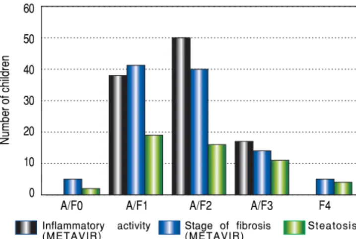
18 children (17.2%) advanced liver fibrosis was found. Figure 1.Figure 1.Figure 1. Histopathology assessment - inflammatory activity, liver fibrosisand steatosis in children with CHC, n = 105.Figure 1.Figure 1. 60
50
40
30
20
10
0
Inflammatory activity
(METAVIR) Stage of fibrosis(METAVIR) Steatosis
A/F0 A/F1 A/F2 A/F3 F4
Only 5 children had no fibrotic lesions in the whole study group. Inflammatory activity was assessed A1 in 38 chil-dren (36.2%) and A2 in 50 chilchil-dren (47.6%). Seventeen pa-tients had high inflammatory activity assessed A3 (16.2%). Fifty two patients had fatty liver. The results of histologi-cal assessment and were presented in figure 1. Figure 2 shows an example of advanced liver fibrosis in Syrius red staining.
Number of children with malignancy in the history did not differ significantly in the groups of children with
var-Table 1. Clinical and laboratory group characteristic.
Parameter Number Percent X ± SD M Range
Age (years) - - 13.62 ± 3.48 15.00 2-17
Gender: M/F 54/51 50/49
-Age at diagnosis (years) - - 6.04 ± 4.23 5.00 0-16
Duration of infection - - 7.58 ± 4.30 7.00 1-16
(years)
Probable route 99/6 94/6 - -
-of infection: parenteral/vertical
History of malignancy 70/35 60/34
Y/N
Former antiviral 50/55 47/53 - -
-treatment Y/N
Weight (kg) - - 50.62 ± 15.38 53.00 12.00-88.00
Body mass index - - - 19.68 ± 3.39 19.45 9.92-29.41
BMI (kg/m2)
HCV genotype 1b/1a/3a 19/80/6 19/75/6 - -
-HCV-RNA (IU/mL) - - 1.31x10⁶ ± 5.75x10⁶ 2.89x10⁵ 1x 10²-5.63x10⁷
ALT (IU/l) - - 64.42 ± 74.57 39 6-486
AST (IU/l) - - 52.95 ± 43.86 41 20-261
GGTP (IU/l) - - 35.25 ± 51.86 22 8-311
WBC (G/l) 6.45 ± 2.10 6.03 6.50-15.90
Inflammatory activity (METAVIR) - - 1.85 ± 0.85 2.00 1-3
Fibrosis (METAVIR) - - 1.71 ± 0.94 2.00 0-4
Liver steatosis Y/N 52/53 49/51 - -
-X: mean. SD: standard deviation. M: median. BMI: body mass index. Y: yes. N: no. G: 109. IU: international unit. kg: kilogram.
ied ALT activity, liver fibrosis and liver steatosis. The re-sults were presented in figure 3.
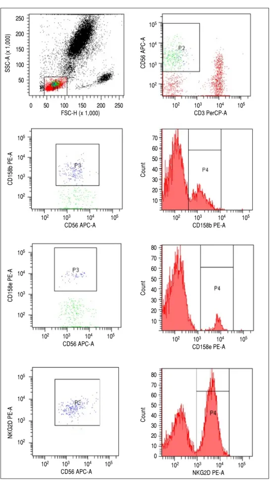
The strategy used to identify the expression of NK cell receptors was presented in figure 4. The number and pro-portion of NK cells was comparable in the group of treat-ment naïve and treattreat-ment experienced children. The density of CD158e expression was significantly higher in treatment naïve patients (16,990 ± 7,305 vs. 10799 ± 9,358 (MFI); p = 0.036). Number of NK cells was comparable in the subgroups of children with varied ALT activity (313 ± 212 vs. 466 ± 344 cells/μL; p = 0.123), presence of sig-nificant (342 ± 259 vs. 361 ± 246 cells/μL p = 0.822) and advanced fibrosis (334 ± 225 vs. 249 ± 210 cells/μL; p = 0.442) and liver steatosis (370 ± 309 vs. 315 ± 146 cells/μL,
Figure 2. Figure 2. Figure 2. Figure 2.
Figure 2. Liver specimen showing fibrotic lesions surrounding liver regener-ating nodule. Syrius red staining. Magnification 40x.
Figure 3. Figure 3. Figure 3. Figure 3.
Figure 3. Number of children with childhood malignancy in the groups with varied ALT activity, liver fibrosis and liver steatosis.
70
60
50
40
30
20
10
0
ALT ALT F < 2 F ≥2 F < 3 F ≥ 3 No Steatosis
< 2UNL > 2UNL steatosis
p = 0.50) (Tables 2 and 3). No statistically significant cor-relation were noted between the groups.
Higher proportion of cells expressing CD158e inhibi-tory receptors was observed in the group of children with ALT > 2UNL compared to patients with low ALT activi-ty (21.11 ± 14.60 vs. 12.22 ± 8.99%; p = 0.037) (Table 2). Moreover, higher proportion of cells expressing inhibito-ry CD158b receptor was observed in children with signif-icant fibrosis (F ≥ 2) compared to minimal fibrosis (F < 2) - (34.14 ± 12.44 vs. 27.48 ± 8.71%; p = 0.049) (Table 3.). Proportion of cells with the expression of NKG2D was relatively high in all compared subgroups of children and no statistically significant differences were observed in re-lation to significant fibrosis.
Children with advanced fibrosis (F ≥ 3) developed higher density of CD 158b expression presented as MFI values compared to those with fibrosis assessed F < 3 (5344 ± 3407 vs. 2980 ± 1190; p = 0.049). Furthermore, children with coexisting steatosis had higher expression of CD158e (16908 ± 8705 vs. 10877 ± 8245; p = 0.035) and
lower expression of NKG2D (4157.50 ± 593.34 vs.
5176.75± 972.41; p = 0.003).
Proportion of NK cells with the expression of CD158b was found a predictor of significant fibrosis in univariate analysis - (OR 1.065; 95%CI (1.07-1.15); p = 0.046). The density of CD158b expression was, however not found to be a predictor of advanced fibrosis in univariate analysis (OR 0.99; 95%CI (0.99-1.01); p = 0.28).
DISCUSSION
No child with CHC in this study had normal liver his-tology. Although liver fibrosis is a slow process, the rate of progression is difficult to predict and may be accelerated
P = 0.55
P = 0.47 P = 0.40 P = 0.58
59
30
11 6
32
15 38
20 60
37
10 8
36
16 34
19
Number of children
Malignancy No malignancy 59
30
11 6
32
15 38
20 60
37
10 8
36 34
19 16
Figure 4. Figure 4. Figure 4.
Figure 4. Figure 4. Cytometric assessment of the proportion of NK cells (AAAAA-BBBBB) and the approach to evaluate the expres-sion of selected receptors using anti-CD158b (CCCCC) anti CD158e (EEEE) andE antiNKG2D (GGGGG) antibodies with ac-companying histograms (DDDDD,FFFFF,HHHHH).
P3
P2
102 103 104 105 CD3 PerCP-A
CD56 APC-A
105
104
103
102
SSC-A (x 1,000)
250
200
150
100
50
0 50 100 150 200 250
FSC-H (x 1,000)
CD158b PE-A
105
104
103
102
102 103 104 105 CD56 APC-A
Count
70 60 50 40 30 20 10
102 103 104 105 CD158b PE-A
Count
80 70 60
50 40 30 20 10
102 103 104 105 CD158e PE-A
CD158e PE-A
105
104
103
102
102 103 104 105 CD56 APC-A
NKG2D PE-A
105
104
103
102
102 103 104 105 CD56 APC-A
Count
80 70 60 50 40 30 20 10 0
Mania A, et al.
, 2017; 16 (4): 521-529
Table 3. NK cell number and expression of selected NK cell receptors in children with varied liver fibrosis and liver steatosis.
Feature Fibrosis score Fibrosis score Liver steatosis
F < 2 F ≥ 2 p F < 3 F ≥ 3 p Steatosis Steatosis p
N = 47 N = 58 N = 97 N = 18 Absent Present
N = 52 N = 53
NK (cells/μL) 342 ± 259 361 ±246 0.82 334 ± 225 249 ± 210.93 0.44 370 ± 309 315 ± 146 0.50 NK cel (%) 13.74 ± 5.34 13.82 ±7.84 0.97 14.15 ± 5.13 15.24 ± 13.05 0.74 13.66 ± 5.81 14.39 ± 7.79 0.74 CD158b (%) 27.48 ± 8.71 34.14 ±12.44 0.049* 30.21 ± 11.37 35.88 ± 13.64 0.42 30.08 ± 13.07 31.79 ± 8.10 0.64 CD158b MFI 3,154.94 ± 1,323.62 3,615.00 ±2,401.73 0.48 2,979.67 ± 1,190.64 5,344.20 ± 3,407.49 0.049* 3079 ± 1,377 3,774.00 ± 2,412.20 0.27
CD158e (%) 13.24 ± 8.91 14.28 ±12.55 0.76 13.44 ± 9.63 15.50 ± 15.94 0.98 13.85 ± 10.24 14.38 ± 10.24 0.83 CD158e MFI 13,156.17 ± 8,154.41 13,986.89 ±9,935.34 0.78 13,631.48 ± 8,497.39 13,631.60 ± 12,692.32 0.99 10,877 ± 8245 16,907.6 ± 8,704.55 0.04*
NKG2D (%) 94.94 ± 6.12 96.88 ±2.65 0.27 95.99 ± 5.01 95.83 ± 3.40 0.86 95.30 ± 6.06 96.94 ± 1.97 0.329 NKG2D 4,449.80 ± 984.13 4,642.80 ±854.65 0.60 4,640.82 ± 920.12 3,580.00 ± 920.92 0.12 5,177 ± 972 4157 ± 593 0.003*
Results presented as X ± SD; values with p < 0.05 are considered statistically significant.
Feature CHC group Control Former antiviral treatment ALT activity
N = 105 group p Treatment Treatment p ALT< 2 xUNL ALT ≥ 2xUNL p
N = 23 naïve experienced N = 88 N = 17
N = 50 N = 55
NK (cells/μL) 345 ± 248 340 ± 164 0.93 398 ± 297 278 ± 162 0.12 313 ± 212 466 ± 344 0.12 NK cel (%) 13.79 ± 5.29 13.66 ± 5.62 0.96 13.59 ± 5.97 14.61 ± 7.27 0.65 13.83 ± 5.72 14.57 ± 9.98 0.78 CD158b (%) 30.84 ± 11.02 29.87 ± 11.18 0.74 32.74 ± 11.19 28.88 ± 11.94 0.29 29.77 ± 10.95 34.88 ± 11.03 0.25 CD158b (MFI) 3,390 ± 1914 2,962 ± 1,542 0.37 3,834 ± 1,802 3,039 ± 2012 0.22 3,277 ± 1,581 3,815 ± 2,958 0.49 CD158e (%) 14.09 ± 10.82 15.16 ± 13.32 0.73 17.28 ± 11.19 11.33 ± 10.20 0.10 12.22 ± 8.99 21.11 ± 14.60 0.04*
CD158e (MFI) 13,575 ± 8,874 9,882 ± 8,907 0.12 16,990 ± 7,305 10,799 ± 9,358 0.036* 12,790 ± 8,365 16,519 ± 10,667 0.30 NKG2D (%) 96.15 ± 4.44 96.70 ± 1.81 0.60 96.73 ± 3.44 95.75 ± 5.16 0.59 95.86 ± 4.87 97.06 ± 2.73 0.54 NKG2D (MFI) 4,628 ± 931 4828 ± 1402 0.56 4,503 ± 726 4,660 ± 1075 0.69 4,775 ± 930 4228 ± 874 0.19
by coexisting chronic diseases and obesity. Therefore, dis-tinguishing the group of patients with accelerated liver disease out of the whole group with moderately or slowly progressing CHC seems to be very important.2 Various factors were evaluated in children with CHC and com-pared to the factors established in adults. Children have relatively lower number of coexisting diseases related to sedentary lifestyle. Nevertheless, it has to be stressed, that some patients are cancer survivors which may also influ-ence the liver histology.14 In current study, children fin-ished their oncological treatment at least five years prior to the inclusion to the trial, which should reduce the in-fluence of chemotherapy on liver histology.
The study group consisted of Polish children, who were mostly nosocomialy infected with HCV but were not coinfected with other viruses. They were inhomoge-neous as far as clinical, virological and histological find-ings were concerned. However, established alterations enabled detailed comparable analysis of various subgroups of children. Since significant differences were detected, relation between factors related to liver injury and the ex-pression of selected receptors could be found. Significant proportion of the study group were childhood malignancy survivors, which could influence the outcome of the study. However in order to decrease the influence of un-derwent oncological treatment (mostly chemotherapy), children were included in the study five years after com-pletion of the therapy. Nevertheless, it has to be stressed that in compared groups with varied liver injury, children with malignancy in the history were present with no sta-tistically significant difference.
The innate immune response play an important role in liver infection since this organ contains larger number of NK cells than circulating peripheral blood pool. NK cells respond to antigens that are brought to the liver by blood from gastrointestinal tract. However, the intrahepatic pool needs to be evaluated directly after the biopsy. Therefore, in this study peripheral NK cells were evaluated and com-pared with clinical and histological findings. In the cur-rent study no significant differences were found in the number of NK cells in the subgroups of children with varied ALT activity, intensity of liver fibrosis and the pres-ence of steatosis. Detected differpres-ences concerned howev-er, varied expression of NK cell receptors in these groups of patients. Indolfi et al. report decreased number of CD56+CD3-NK cell in chronically HCV infected chil-dren.15 Current study consisted of treatment naive and ex-perienced patients in whom lower expression of CD158e was detected. Pegylated IFN was found to activate NK cells inducing their cytotoxic function, which however correlated to the treatment response.16,17 Lower expression of the CD158e inhibitory receptor was found in treatment experienced group in spite of the lack of response to IFN
used in the past. NK cells are regulated by numerous lig-ands through their inhibitory and activating receptors. Moreover, in the current study NK cells displayed inhib-itory potential in relation to liver injury - higher propor-tion of cells with the expression of CD158b and CD158e receptors. The differences were statistically significant comparing groups of children with significant and mild fibrosis. Furthermore, the expression of CD158b receptor was found a predictor of significant fibrosis in univariate analysis. Although data concerning intra-hepatic NK cells are limited, Fugier, et al. report, however, similar findings - significantly impaired degranulation activity of intrahe-patic pool of these cells in adult CHC patients with
high-est inflammation and fibrosis scores.18 Regarding
inhibitory influence of NK cells on the liver fibrosis, it seems obvious that in the circumstances of the inhibitory phenotype of NK cells, more advanced fibrosis of the liver was observed.19,20
Alterations in NK cell phenotype may be therefore a consequence of HCV infection, as well as a predisposing factor to develop CHC. Data confirming both explana-tions are available. HCV directly influences the grade of NK cell activation by increased expression of MHC class I proteins on the surface of the infected cells. MHC class I receptors are the ligands of NK cell inhibitory receptors, which results in the suppression of these cells. Further-more, HCV stabilizes ligands to inhibitory receptors of NK cells, which leads to even greater inhibition and therefore, lower potential to slow down the progression of liver fibrosis.12,14 It has been proven that HCV directly inhibits immune cells by a contact with HCV-infected hepatocytes.20 On the other hand, altered NK cell pheno-types could cause susceptibility to viral infections, when the advantage of inhibitory receptors is observed. Studies showed that various sets of KIR receptors predispose to the development of chronic HCV infection.21
In the subgroup of children with liver steatosis signifi-cantly lower expression of activating receptor NKG2D was present. In other words, lack of steatosis was associat-ed with higher expression of the receptor with activating potential. Liver steatosis may result in the decrease of NK cell activity through the higher secretion of proinflamma-tory cytokines. The studies conducted on animal models revealed inversely proportional relation between the liver steatosis and the number of NK cells.22
inhibition of liver fibrosis, while the presence of steatosis influence on the inhibition of their activity and progres-sion of inflammatory leprogres-sions. Previous studies confirmed that children with steatosis in the course of CHC have significantly higher inflammatory activity in histological evaluation.24
Number of studies suggest that although number of NK cells may be unchanged, differences between NK cell phenotype in patients with CHC and healthy controls may be noticed.8,9,25 Few consistent finding exist in terms of change in the expression of specific receptors. Depending on the study, natural cytotoxicity receptors are being re-ported as either up, down or not altered,25,10 which is a re-sult of the heterogeneity of patients and viral factors. Although in the current study alterations between CHC patients and healthy controls were not observed, our re-sults showed that the NK cell phenotype and possibly NK cell function may be altered in HCV-infected children in relation to various stages of liver disease, either being a cause, or a result of the phenomenon. It was suggested that, if inflammatory status remains at lower levels, NK cells display more active functions. However, if the in-flammatory status of the liver increases, these functions may be significantly suppressed. It has been proposed that, either a part of NK cells become non-reactive due to the prevention of their activation in the state of inflammation, or NK cells adapt themselves to the circumstances of pro-longed exposition to HCV, which leads to a decline in cy-tokine production and cytotoxicity in order to reduce tissue damage.25 Animal models show that NK cells reac-tivity depends on the time of exposure to inflammatory state.26 Therefore in CHC a decline in NK cell activity may favor the development of liver fibrosis. This study seem to prove this suggestions.
CONCLUSIONS
Higher proportion of NK cells with the expression of inhibitory CD158b and CD158e receptors is associated with liver injury expressed as higher aminotransferase ac-tivity, more advanced liver fibrosis and the presence of st-eatosis.
ABBREVIATIONS
• ALT: alanine aminotransferase. • AST: aspartate aminotransferase. • CD: cluster of differentiation. • CHC: chronic hepatitis C. • CI: Confidence interval. • HCV: hepatitis C virus. • HGB: hemoglobin. • IFN: interferon.
• KIR: killer cells immunoglobulin-like receptors. • MFI: mean fluorescence intensity.
• NK: natural killer. • N: no.
• PLT: platelets.
• TGF-βββββ: transforming growth factor beta. • UNL: upper normal limit.
• WBC: white blood count. • Y: yes.
CONFLICT OF INTEREST STATEMENT
The authors declare no conflict of interest regarding to the subject of the study.
FUNDING
The study was supported by Polish National Center of Science grant to AM - NN 407012036.
COMPETING INTERESTS
On behalf of all authors, the corresponding author states that there is no conflict of interest
ETHICAL APPROVAL
The study was approved by local ethical committee of the University of Medical Sciences
AKNOWLEDGEMENTS
The authors would like to thank all patients and their legal guardians for the participation in the study.
REFERENCES
1. Bortolotti F, Verrucchi G, Camma C, Cabibbo G, Zancan L, In-dolfi G, Giacchino R, et al. Italian Observatory for HCV Infec-tion and Hepatitis C in Children. Long-term course of chronic hepatitis C in children: from viral clearance to end-stage liver disease. Gastroenterology 2008; 134: 1900-7.
2. Mania A, Kemnitz P, Figlerowicz M, Mazur-Melewska K, Mozer-Lisewska I, Kowala-Piaskowska A, Wo?niak A, et al. Clinical picture and liver histology of chronic hepatitis C in children. Inf Dis Clin Pract 2012; 2: 141-7.
3. European Paediatric Hepatitis C Virus Network. Three broad modalities in the natural history of vertically acquired hepati-tis C virus infection. Clin Infect Dis 2005; 41: 45-51. 4. Zeromski J, Mozer-Lisewska I, Kaczmarek M,
Kowala-Pi-askowska A, Sikora J. NK cells prevalence, subsets and function in viral hepatitis C. Arch Immunol Ther Exp 2011; 59: 449-55.
6. Vilches C, Parham P. KIR: diverse, rapidly evolving receptors of innate and adaptive immunity. Ann Rev Immunol 2002; 20: 217-51.
7. Mondelli MU, Varchetta S, Oliviero B. Natural killer cells in vi-ral hepatitis: facts and controverisies. Eur J Clin Invest 2010; 40: 851-63.
8. Golden-Mason L, Madrigal-Estebas L, McGarth E, Conroy MJ, Ryan EJ, Hegarty JE, O'Farrelly C, et al. Altered natural killer cell subset distributions in resolved and persistent hepatitis C virus infection following single source exposure. Gut 2008; 57: 1121-8.
9. Varchetta S, Mele D, Mantovani S, Oliviero B, Cremonesi E, Ludovisi S, Michelone G, et al. Impaired intrahepatic natural killer cell cytotoxic function in chronic hepatitis C virus infec-tion. Hepatology 2012; 56: 841-9.
10. Ahlestiel G, Titerence RH, Koh C, Edlich B, Feld JJ, Rotman Y, Ghany MG, et al. Natural killer cells are polarized toward cytotoxicity in chronic hepatitis C in interferon-alfa-depend-ent manner. Gastroenetrology 2010; 138: 325-35.
11. Golden-Mason L, Bambha KM, Cheng L, Howell CD, Taylor MW, Clark PJ, Afdhal N, et al. Virahep-C Study Group. Natu-ral killer inhibitory receptor expression associated with treat-ment failure and interleukin-28B genotype in patients with chronic hepatitis C. Hepatology 2011; 54: 1559-69.
12. Gardiner C. NK cell function and recepetor diversity in the context of HCV infection. Front Microbiol 2015; 6: 1061-71. 13. Anonymous. Intraobserver and interobserver variations in
liver biopsy interpretation in patients with chronic hepatitis C. The French METAVIR Cooperative Study Group. Hepatology 1994; 20: 15-20.
14. Cesaro S, Bortolotti F, Petris MG, Brugiolo A, Guido M, Carli M. An updated follow-up of chronic hepatitis C after three decades of observation in pediatric patients cured of malig-nancy. Pediatr. Blood Cancer 2010; 55: 108-112
15. Indolfi G, Mangone G, Moriondo M, Serranti D, Bartolini E, Az-zari C, Resti M. Altered natural killer cells subsets distribu-tion in children with hepatitis C following vertical transmission. Aliment Pharmacol Ther 2016; 43: 125-33. doi: 10.1111/apt.13430. Epub 2015 Oct 16.
16. Nakamura I, Asano T, Asabe S, Ando M, Sano T, Miyata Y, Taira J, et al. Restoration of natural killer cell activity by pegylated interferon-alpha/ribavirin therapy in chronic hepa-titis C patient. Hepatol Res 2015; 45: 107-12. doi: 10.1111/ hepr.12322.
17. Ahlenstiel G, Edlich B, Hogdal LJ, Rotman Y, Noureddin M, Feld JJ, Holz LE, et al. Early changes in natural killer cell function indicate virologic response to interferon therapy for hepatitis C. Gastroenterology 2011; 141: 1231-9, 1239.e1-2. doi: 10.1053/j.gastro.2011.06.069.
18. Fugier A, Marche H, Thelu MA, Macek Jílková Z, Van Campenhout N, Dufeu-Duchesne T, Leroy V, et al. Functions of liver natural killer cells are dependent on the severity of liver inflammation and fibrosis in chronic hepatitis C. Plos One 2014; 9:doi:10.1371/journal.pone.0095614.
19. Poynard T, Bedossa P, Opolon P. Natural history of liver fi-brosis progression in patients with chronic hepatitis C. The OBSVIRC, METAVIR, CLINIVIR, and DOSVIRC groups. Lancet 1997; 349: 825-32.
20. Yoon JC, Lim JB, Park JH, Lee JM. Cell-to-cell contact with hepatitis C virus-infected cells reduces functional capacity of natural killer cells. J Virol 2011; 85: 12557-69.
21. Romero V, Azocar J, Zúñiga J, Clavijo OP, Terreros D, Gu X, Husain Z, et al. Interaction of NK inhibitory receptor genes with HLA-C and MHC class II alleles in Hepatitis C virus in-fection outcome. Mol Immunol 2008; 45: 2429-36.
22. Konjevic G, Mirjacic Martinovic K, Vuletic A, Babovic N. Nov-el aspects of in vitro IL-2 or IFN-alpha enhanced NK cytotox-icity of healthy individuals based on NKG2D and CD161 NK cell receptor induction. Biomed Pharmacother 2010; 64: 663-71.
23. Kremer M, Hines IN, Milton RJ, Wheeler MD. Favored T helper 1 response in a mouse model of hepatosteatosis is associ-ated with enhanced T cell-mediassoci-ated hepatitis. Hepatology 2006; 44: 216-27.
24. Mania A, Kaczmarek M, Kemnitz P, Mozer-Lisewska I, Figle-rowicz F, Wo?niak A, Mazur-Melewska K, et al. Alterations in NK cell phenotype in relation to liver steatosis in children with chronic hepatitis C. Inflammation 2013; 36: 1004-12. 25. Alter G, Jost S, Rihn S, Reyor LL, Nolan BE, Ghebremichael
M, Bosch R, et al. Reduced frequencies of NKp30+NKp46+, CD161+, and NKG2D+ NK cells in acute HCV infection may predict viral clearance. J Hepatol 2011; 55: 278-88. 26. Mondelli MU, Oliviero B, Mele D, Mantovani S, Gazzabin C,
Varchetta S. Natural killer cell functional dichotomy: a fea-ture of chronic viral hepatitis. Front Immunol 2012; 3: 351. 27. Jaeger BN, Donadieu J, Cognet C, Bernat C, Ordoñez-Rueda
D, Barlogis V, Mahlaoui N, et al. Neutrophil depletion impairs natural killer cell maturation, function and homeostatis. J Exp Med 2012; 209: 565-80.
Correspondence and reprint request: Anna Mania, Ass. Prof. MD, PhD
Department of Infectious Diseases and Child Neurology University of Medical Sciences
Ul. Szpitalna 27/33 60-572 Poznañ. Tel. +48 61 8491 657. Mobile: +48 605034122