Cholesterol cholelithiasis in pregnant women:
pathogenesis, prevention and treatment
Ornella de Bari,* Tony Y. Wang,*,** Min Liu,*** Chang-Nyol Paik,* Piero Portincasa,**** David Q.-H. Wang*
* Department of Internal Medicine, Division of Gastroenterology and Hepatology, Saint Louis University School of Medicine, St. Louis, USA. ** Department of Biomedical Engineering, Washington University, St. Louis, USA.
*** Department of Pathology and Laboratory Medicine, University of Cincinnati College of Medicine, Cincinnati, USA. **** Clinica Medica “A. Murri”, Department of Biomedical Sciences and Human Oncology, University of Bari Medical School, Bari, Italy.
ABSTRACT
Epidemiological and clinical studies have found that gallstone prevalence is twice as high in women as in men at all ages in every population studied. Hormonal changes occurring during pregnancy put women at higher risk. The incidence rates of biliary sludge (a precursor to gallstones) and gallstones are up to 30 and 12%, respectively, during pregnancy and postpartum, and 1-3% of pregnant women undergo cholecys-tectomy due to clinical symptoms or complications within the first year postpartum. Increased estrogen levels during pregnancy induce significant metabolic changes in the hepatobiliary system, including the for-mation of cholesterol-supersaturated bile and sluggish gallbladder motility, two factors enhancing choleli-thogenesis. The therapeutic approaches are conservative during pregnancy because of the controversial frequency of biliary disorders. In the majority of pregnant women, biliary sludge and gallstones tend to dis-solve spontaneously after parturition. In some situations, however, the conditions persist and require cos-tly therapeutic interventions. When necessary, invasive procedures such as laparoscopic cholecystectomy are relatively well tolerated, preferably during the second trimester of pregnancy or postpartum. Although laparoscopic operation is recommended for its safety, the use of drugs such as ursodeoxycholic acid (UDCA) and the novel lipid-lowering compound, ezetimibe would also be considered. In this paper, we sys-tematically review the incidence and natural history of pregnancy-related biliary sludge and gallstone for-mation and carefully discuss the molecular mechanisms underlying the lithogenic effect of estrogen on gallstone formation during pregnancy. We also summarize recent progress in the necessary strategies re-commended for the prevention and the treatment of gallstones in pregnant women.
Key words. Bile acids. Biliary lipids. Biliary sludge. Estrogen. Ezetimibe. Gallstones. Progesterone.
Correspondence and reprint request: David Q.-H. Wang, M.D., Ph.D.
Department of Internal Medicine, Division of Gastroenterology and Hepatology, Saint Louis University School of Medicine, St. Louis, MO 63104, USA. Ph.: (314) 977-8737, Fax: (314) 977-9909
E-mail: dwang15@slu.edu
Manuscript received: March 30, 2014. Manuscript accepted: July 22, 2014. INTRODUCTION
Epidemiological and clinical studies showed that at all ages, women are twice as likely as men to form cholesterol gallstones. This gender difference begins since puberty and continues through the childbearing years. Most, but not all, studies have found that the risk of developing cholesterol gall-stones is markedly increased by oral contraceptive steroids and conjugated estrogens in premenopausal women.1-7 Estrogen therapy to postmenopausal
women and to men with prostatic carcinoma also in-duces similar lithogenic effects.8-13 These findings
underscore the importance of female sex hormones on the pathogenesis of gallstones. Furthermore, hormonal changes that occur during pregnancy put women at even higher risk for gallstone forma-tion.14-17 Biliary cholesterol concentrations in
gall-bladder bile increase gradually from the first to the third trimester of pregnancy, along with a progres-sive increase in the incidence of biliary sludge (a pre-cursor to gallstones) and gallstones.18-20 Clinical
studies in the USA and Europe have found that the incidence rates of biliary sludge and gallstones are up to 30 and 12%, respectively, during pregnancy and postpartum.20-28 Although most women remain
asymptomatic, 1-3% of pregnant women undergo cholecystectomy due to clinical symptoms or compli-cations within the first year postpartum.20-28
annually in the USA, it is estimated that at least 40,000 young healthy women require postpartum cholecystectomy each year. Thus, pregnancy-associ-ated gallbladder disease is a significant cause of morbidity in young healthy women.
Parity and length of the fertility period increase the incidence of gallstones,29 as well as both the
fre-quency and number of pregnancy are important risk factors for gallstone formation.14,16,30-34 Of note,
gallbladder disease is the most common non-obstet-rical cause of maternal hospitalization in the first year postpartum,35,36 with 30% attacks of biliary
col-ic in women with gallstones.23 Following acute
ap-pendicitis, acute cholecystitis is the second most common indication for non-obstetric surgery in pregnant women.37 Again, it indicates that
gallblad-der disease is a significant cause of morbidity for pregnant women.27 Although pregnancy constitutes
a defined period of metabolic stress, a subclinical trend to form gallstones clearly exists.27 Pregnant
women with gestational diabetes or high blood pres-sure are at risk of later developing diabetes mellitus or hypertension.38,39 A similar phenomenon could
happen with biliary sludge and gallstones. Thus, ele-vated estrogen levels are a critical risk factor for de-veloping gallbladder disease during pregnancy. Moreover, biliary sludge and gallstones can sponta-neously disappear after parturition in approximately 60% of cases mostly due to a sharp decrement in es-trogen levels.20 It is important in clinical practice,
therefore, to find a way to reduce the risk of chole-lithiasis in pregnant women.
Human and animal studies have found that estro-gen increases susceptibility to cholesterol cholelithi-asis by promoting hepatic secretion of biliary cholesterol,18,19,40-47 enhancing bile lithogenicity and
impairing gallbladder motility function.10,48,49 Such
alterations, in turn, lead to a dramatic increase in cholesterol saturation of bile and gallbladder stasis, thereby enhancing cholelithogenesis.1,50-52 We have
found that the hepatic estrogen receptor α (ERα), but not ERβ, plays a major role in cholesterol gall-stone formation in mice exposed to high doses of 17β-estradiol.53 A novel concept has been proposed
that high levels of estrogen promote the formation of cholesterol gallstones through the ERα signaling cascade in the liver and higher risks for gallstone formation in women than in men are related to dif-ferences in how the liver handles cholesterol in re-sponse to estrogen.54
In this paper, we systematically review the inci-dence and natural history of pregnancy-related bil-iary sludge and gallstones and carefully discuss the
molecular mechanisms underlying the lithogenic ef-fect of estrogen on gallstone formation during preg-nancy. We also summarize recent advances in the prevention and the treatment of cholesterol gall-stones in pregnant women.
CHANGES IN PLASMA ESTROGEN LEVELS DURING THE MENSTRUAL CYCLE
AND DURING PREGNANCY
The menstrual cycle is essential for the produc-tion of eggs and for the preparaproduc-tion of the uterus for pregnancy.55 It consists of:
• The ovarian cycle that describes changes in the follicles of the ovary and
• The uterine cycle that outlines changes in the en-dometrial lining of the uterus.
Both the ovarian and the uterine cycles are divided into three phases. The ovarian cycle is composed of:
• Follicular phase, • Ovulation, and • Luteal phase.
Whereas the uterine cycle is divided into:
• Menstruation,
• Proliferative phase, and • Secretory phase.56
The stages of the menstrual cycle are character-ized by fluctuations of the four major circulating hormones: follicle-stimulating hormone (FSH), luteinizing hormone (LH), estradiol and progester-one.57,58 Although plasma concentrations of these
hormones vary substantially on an hourly basis, their daily profiles provide characteristic changes during the menstrual cycle.57 The follicular phase is
defined by a preovulatory estrogen peak in the ab-sence of progesterone.8
During the luteal phase, following ovulation, a si-multaneous increase of both estradiol and progester-one occurs.56,58-60
Plasma concentrations of unconjugated estrone (E1), estradiol (E2) and estriol (E3) increase steadily as pregnancy progresses.3,4,19 Estradiol levels in
concen-trations range from 6 to 30 ng/mL.3,29 At 9 weeks of
pregnancy, i.e., 23% of gestational length, the pla-centa becomes the primary source of estrogen.61 In
the late stage of pregnancy, plasma concentrations of estradiol and estrone are approximately 100-fold higher than the respective average values in the menstrual cycle.4,23 These alterations greatly
en-hance susceptibility to cholesterol gallstones in pregnant women.
GALLSTONE PREVALENCE IN PREGNANT WOMEN
During pregnancy, elevated estrogen levels are of-ten associated with a significant increase in hepatic secretion of biliary cholesterol. As a result, bile be-comes supersaturated with cholesterol and is more lithogenic. Additionally, high levels of estrogen and progesterone could impair gallbladder motility func-tion by inhibiting gallbladder smooth muscle con-tractile function, thus leading to gallbladder stasis.10,49 Such abnormalities greatly promote the
formation of biliary sludge and gallstones in preg-nant women. The incidence of disease appears to be increased in the last two trimesters of pregnancy. However, approximately one third of pregnant wom-en with gallstones is asymptomatic.21,23,24 When
symptoms do occur in pregnant women, however, the most common clinical presentations are biliary colic, acute cholecystitis, gallstone pancreatitis, and jaundice.62
As listed in table 1, the incidence rates of gall-stones range from 1.2 to 6.3% during pregnancy.21-25
Since the concentration of plasma female sex hor-mones increases proportionally with duration of ges-tation, the risk of gallstone formation is especially hazardous in the third trimester of pregnancy. Fur-thermore, increasing parity could be a significant risk factor for gallstones, especially in younger women.61,63,64 Clinical studies have observed that
approximately 5.1% of women developed gallbladder disease after one pregnancy, 7.6% after two preg-nancies, and 12.3% after 3 or more pregnan-cies.14,27,30-32 A study in Chile reported that the
incidence of gallstones was 12.2% of multiparous women compared to 1.3% of nulliparous women within the same age.65 Another study has reported
that women under the age of 25 years with ≥ 4 preg-nancies were 4 to 12 times more likely to develop cholesterol gallstones compared to nulliparous wom-en of the same age and body weight.66
It has been shown that the prevalence of gall-stones in women who had two to three or more pregnancies was 2- to 3-fold higher compared with nulliparous women.67 In the Italian GREPCO study,
the prevalence of gallstones positively correlated with the number of pregnancies and age.68 The
fre-quency appears to increase after two, three or more pregnancies, and this trend was more marked in younger women at the ages of 25 to 30 years vs. 35 to 40 years.61,64 Several population studies attempted
to quantify the risk of gallstones caused by multiple pregnancies, adjusting for at least some known con-founding factors. However, the number of multiple pregnancies and the adjustment for confounding fac-tors were not uniform. The results from the
Sirmi-Table 1. Gallstones formation during pregnancy and after delivery.
Incidence of gallstones
Authors Subjects (n) Nulliparous First Second Third Postpartum
trimester trimester trimester (weeks)
Maringhini, et al. (1993)20 272 - 6.0 2.0a - 2.4b (2-4)
Bolukbas, et al. (2006)21 97 - 6.3 - -
-Maringhini, et al. (1987)22 298 - - - - 5.2 (1)
Valdivieso, et al. (1993)23 980 1.3 - - - 12.2 (1)
Basso, et al. (1992)24 521 - - 2.7 -
-Stauffer, et al. (1982)25 313 - - 3.5c -
-Tsimoyiannis, et a. (1994)61 669 - - 1.2 1.9 2.1 (24)
Ko, et al. (2005)27 3,254 - - 1.9 1.8d 2.8e (4-6)
Glasinovic, et al. (1989)28 259 - 3.1 - - 11.2
a The value shows the incidence of gallstones that were found after the first trimester. b The value represents the incidence of
gallstones that were detected at 2 to 4 weeks after delivery from the first trimester. c The value indicates the incidence of
gall-stones that were observed during gestation. d The value shows the cumulative incidence of gallstones by the third trimester. e
one study,14 the Framingham study16 and the
MI-COL study69 were surprisingly similar, and the
rela-tive risks were 1.7, 1.6 and 1.7, respecrela-tively. These and other studies provided definitive evidence that multiple pregnancies are a critical risk factor for the development of gallstones.
Biliary sludge in pregnancy is attributed to two major mechanisms: estrogen-induced metabolic ab-normalities in the formation of lithogenic bile and progesterone-induced smooth muscle relaxation with subsequent gallbladder hypomotility.51,52,70-72 As
shown in tables 1 and 2, the frequency of biliary sludge has been observed to be higher than that of gallstones (~15.0% vs. ~6.0%) during pregnan-cy.23,73 Although not all cases of biliary sludge
evolve to gallstones, the presence of sludge is a nec-essary precursor involved in the formation of gall-stones.27,51,52,74 Clinical studies have reported that
biliary sludge can spontaneously disappear and re-cur over time, or it can proceed to gallstones. Bil-iary sludge is mostly asymptomatic and disappears when the primary condition resolves; however, in some cases it can progress into gallstones or mi-grate to the biliary tract, occluding the cystic duct or the common bile duct and causing biliary colic, cholangitis or pancreatitis. It should be noted that biliary sludge in the majority of women usually dis-appears a few weeks after the pregnancy concludes/ terminates/ends.66 However, only a fraction of
preg-nant women with sludge develop gallstones.72
Gall-stones and biliary sludge can spontaneously resolve in most women during the first year after delivery. However, it is considered that women with multiple or closely spaced pregnancies may form gallstones as sludge recurs or persists.75
Based on ultrasonographic assessment of the gall-bladder in 298 women in the immediate postpartum period, the incidence of biliary sludge was found to be 26.2% and the incidence of gallstones was 5.2%.
After 1 year of follow-up, only 2 out of 45 patients with biliary sludge, but 13 out of 15 patients with gallstones still had abnormal ultrasonographic find-ings.22 In another report, the prevalence of biliary
sludge in pregnant women ranged from 5 to 36% and the prevalence of gallstones from 2 to 11%.63 A
pro-spective study of 272 women recruited in the first trimester of pregnancy and followed for almost one year postpartum found that new biliary sludge de-veloped in 67 women and gallstones in 6 women in pregnancy. Whereas, women with biliary sludge re-mained generally asymptomatic, 28% of women with gallstones developed biliary pain during pregnancy. About half of the study population (115 women) was reexamined in the postpartum period. Of these, 92 had biliary sludge and 23 had gallstones. Biliary sludge disappeared in 56 affected women (61%) and gallstones were dissolved in 6 women (28%) by sev-eral months post-partum.20
It has been observed that over a 7- to 8-month pe-riod from the first trimester to the early postpar-tum, the cumulative incidence of biliary sludge was 5%, whereas an additional 5% developed incident gallstones or progressed from baseline sludge to gall-stones. Overall, 4.2% of women had newly formed biliary sludge or gallstones that were found by ul-trasonography in the early postpartum.27 Several
groups performed abdominal ultrasound examina-tions in women in the immediate postpartum period comparing the same examinations performed in the same women at the beginning of pregnancy or in age matched nulliparous women. In two large Chilean studies, approximately 12.2% of women were found to have gallstones in the postpartum period com-pared with 3.1% in the same women at the begin-ning of pregnancy and 1.3% in nulliparous women.23,72
When gallbladder motility is restored in the post-partum period, biliary sludge and gallstones could
Table 2. Biliary sludge formation during pregnancy and after delivery.
Incidence of biliary sludge
Authors Subjects (n) Nulliparous First Second Third Postpartum
trimester trimester trimester (weeks)
Maringhini, et al. (1993)20 272 - 15.0 14.0a - 29.6b (2-4)
Bolukbas, et al. (2006)21 97 - 10.9 -
-Maringhini, et al. (1987)22 298 - - - - 26.2 (1)
Ko, et al. (2005)27 3,254 - - 3.2 4.5c 5.1d (4-6)
a The value shows the incidence of biliary sludge that was found after the first trimester. b The value represents the incidence of
biliary sludge that were detected at 2 to 4 weeks after delivery from the first trimester. c The value shows the cumulative
inci-dence of biliary sludge by the third trimester. d The values indicates the incidence of new formed biliary sludge that was
pass into the cystic duct or the common bile duct, and this is a condition at risk of biliary colic or oth-er complications such as obstructive jaundice or pancreatitis. Alternatively, because of a sharp reduc-tion in estrogen concentrareduc-tions after childbirth, postpartum changes in lipid composition of bile could favor dissolution of biliary sludge and gall-stones. Maringhini, et al. reported that both biliary sludge and gallstones disappeared in 61 and 28%, re-spectively, during the first year after delivery.20 This
suggests that pregnancy-associated biliary litho-genicity and gallbladder hypomotility may be a cru-cial factor in the formation of biliary sludge and gallstones during gestation.51
Ko, et al. evaluated the incidence and natural his-tory of pregnancy-related biliary sludge and gall-stones and reported an incidence of biliary sludge or gallstones of 5.1% by the second trimester, 7.9% by the third trimester, and 10.2% by 4 to 6 weeks post-partum.27 Twenty-eight women (0.8%) underwent
cholecystectomy within the first year postpartum.27
Biliary diseases at delivery or hospitalization within 1 year postpartum were documented in ap-proximately 0.5% of all births in Washington State between 1987 and 2001, with cholecystectomy per-formed within 1 year postpartum for approximately 0.4% of births during this time period. A population-based study found that 2.1% of women aged 20-29 year and 4.9% of women aged 30-39 year have un-dergone cholecystectomy.30,35 Because many of these
women would have undergone cholecystectomy in the first postpartum year, these findings suggest that the postpartum period is a time of high risk for symptomatic gallstone disease requiring further treatment. Notably, hospitalizations resulting from gallstone-related disease in the first year postpartum period incur median charges of $5,759 in the mid-1990s, with a median length of stay of 3 days.35
MOLECULAR MECHANISMS UNDERLYING THE LITHOGENIC EFFECT OF ESTROGEN ON THE FORMATION OF CHOLESTEROL GALLSTONES IN PREGNANT WOMEN
Pregnancy and parity are two important risk fac-tors for the formation of cholesterol gallstones in women.23,29,31,62,76 As shown in figure 1, increased
levels of female sex hormones during pregnancy in-duce a variety of metabolic changes in the hepatobil-iary system, which ultimately cause bile to become supersaturated with cholesterol and enhance chole-lithogenesis.10 Moreover, biliary lithogenicity occurs
as a result of estrogen-induced increase in cholester-ol biosynthesis, providing excess amounts of chcholester-oles- choles-terol for biliary hypersecretion during pregnancy.10
So, cholesterol saturation index of bile increases es-pecially during the second and third trimesters of pregnancy. The secretion rate of cholesterol relative to that of bile acids and phospholipids is increased markedly.77 Moreover, hepatic secretion of biliary
cholesterol has been found to be increased by 40% in nonpregnant women treated with estrogen.8 In
addi-tion, the gallbladder becomes sluggish and enlarged and empties incompletely, which further increases the risk of gallstone formation.77
Estrogen could promote cholesterol cholelithogen-esis by enhancing functions of estrogen receptors in the liver and gallbladder.54 ERα and ERβ are both
expressed in the hepatocytes although they have overlapping but not identical tissue expression pat-terns.53,78-80 However, gene expression levels of ERα
are ~50-fold higher compared with ERβ. These find-ings imply that ERα is a major steroid hormone re-ceptor producing the biological effects of estrogen in the liver.10 We investigated the molecular
mecha-nisms of how estrogen influences cholesterol gall-stones formation in a mouse model of gonadectomized gallstone-resistant AKR mice.53
Ex-pression levels of ERα were significantly increased after its activation by 17β-estradiol (E2), the ERα -se-lective agonist propylpyrazole (PPT), and tamoxifen. However, the E2 effects on expression of the hepatic ERα mRNA was blocked by the anties-trogenic ICI 182,780. The ERβ-selective agonist dia-rylpropionitrile (DPN) did not influence gene expression of ERα, but up-regulates the expression of ERβ in the liver.53 Although the prevalence of
gallstones displayed a dose-dependent increase in go-nadectomized mice treated with various doses of ex-ogenous E2, all mice treated with E2 at 6 μg/day plus ICI 182,780 were gallstones free. These findings indicate that the lithogenic effects of E2 are blocked by this antiestrogenic agent. Moreover, 75% of the mice treated with PPT formed gallstones, suggest-ing its strong lithogenic effect. Tamoxifen promoted gallstone formation with a prevalence rate of 50%, indicating its estrogen-like effects on biliary lipid metabolism. By contrast, DPN did not influence gallstone formation. Thus, the receptor-dependent effects of E2 contribute to biliary cholesterol hyper-secretion and lithogenicity of bile, promoting gall-stone formation through the hepatic ERα, but not ERβ pathway.
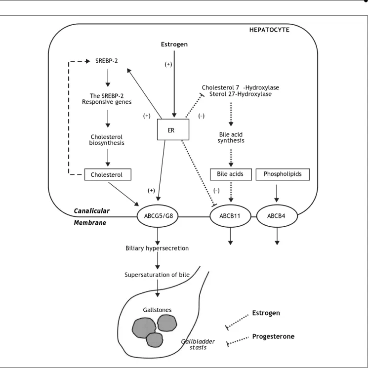
3-Figure 1. The proposed working model underlying the potential lithogenic mechanisms of estrogen through the estrogen re-ceptor a (ERα) pathway in the liver during pregnancy. In the liver, there is an “estrogen-ERα-SREBP-2” pathway promoting cho-lesterol biosynthesis and hepatic hypersecretion of biliary chocho-lesterol in response to estrogen. The negative feedback regulation of cholesterol biosynthesis (as shown in a dashed line) is inhibited by ERα that is activated by estrogen, mostly through stimula-ting the activity of sterol regulatory element-binding protein-2 (SREBP-2) with the resulstimula-ting activation of the SREBP-2 responsive genes for the cholesterol biosynthetic pathway. Consequently, these alterations induce excess secretion of newly synthesized cholesterol and supersaturation of bile that predisposes to cholesterol precipitation and gallstone formation. By contrast, es-trogen could reduce bile acid biosynthesis by inhibiting cholesterol 7α-hydroxylase in the classical pathway and sterol 27-hydroxylase in the alternative pathway through the ERα signaling cascade (as shown in a dotted line). Moreover, the hepatic ERα activated by estrogen could stimulate the activity of ATP-binding cassette (ABC) transporters ABCG5/G8 on the canalicular membrane of the hepatocyte and promote biliary cholesterol hypersecretion. It is likely that expression levels of ABCB11 are inhibited by estrogen through the ERα pathway, inducing biliary bile acid hyposecretion (as shown in a dotted line). In addition, high levels of estrogen and progesterone induce smooth muscle relaxation with subsequent impaired gallbladder motility func-tion, leading to gallbladder stasis (as shown in dotted lines). All of these alterations promote the formation of biliary sludge and gallstones in pregnant women exposed to high levels of estrogen.10
Estrogen
HEPATOCYTE
Cholesterol 7α-Hydroxylase Sterol 27-Hydroxylase SREBP-2
The SREBP-2 Responsive genes
Cholesterol biosynthesis
Cholesterol
Bile acid synthesis
Bile acids Phospholipids
ABCG5/G8 ABCB11 ABCB4
Canalicular Membrane
Biliary hypersecretion
Supersaturation of bile
Gallstones
Gallbladder stasis
Estrogen
Progesterone
(+) (–)
(+) (–)
hydroxy-3-methylglutaryl coenzyme A (HMG-CoA) reductase, the rate-limiting enzyme in hepatic cho-lesterol biosynthesis, is enhanced, followed by in-creased delivery of cholesterol from hepatic de novo
synthesis to bile.1,53,81-83 Estrogen could increase the
capacity of dietary cholesterol to induce cholesterol supersaturation of bile.1,53,81,82,84 To elucidate the
molecular mechanism by which E2 increases hepatic output of biliary cholesterol, we quantitated the con-tribution of newly synthesized cholesterol to biliary output in gonadectomized AKR mouse treated with high doses of estrogen and fed a high-cholesterol diet.83 Compared with control mice (i.e., female AKR
mice with intact ovaries), E2-treated mice with gona-dectomy displayed a significantly higher hepatic out-put of biliary total and newly synthesized cholesterol, regardless of whether chow or the high-cholesterol diet was fed. These biological effects of E2 were abolished by ICI 182,780. Thus, the origin of biliary cholesterol possibly comes mostly from the high cholesterol diet and partly from lipoproteins such as HDL carrying cholesterol from extrahepatic tissues via a reverse cholesterol transport pathway. To regulate the hepatic cholesterol biosynthesis, E2 could activate ERα which, in turn stimulates mRNA expression of sterol regulatory element binding pro-teins-2 (SREBP-2) and five major SREBP-2-respon-sive genes (HMG-CoA synthase, HMG-CoA reductase, farnesyl diphosphate synthase, squalene synthase, and lathosterol synthase) in the liver.83
Compared to the chow diet in control mice, expres-sion levels of SREBP-2 were significantly reduced by the high dietary cholesterol. These findings suggest that cholesterol biosynthesis may be inhibited by a negative feedback regulation through the SREBP-2 pathway in response to high dietary cholesterol. By contrast, E2-treated mice still showed significantly higher expression levels of SREBP-2 and the SREBP-2-responsive genes, even under high dietary cholesterol loads. These results indicate that with high levels of E2, there is a continuous cholesterol synthesis in the liver because the negative feedback regulation of its synthesis by the SREBP-2 pathway may be inhibited by E2 through the hepatic ERα. Likely, under the normal physiological conditions, there is a negative feedback regulation of cholesterol biosynthesis by cholesterol. With increased E2 lev-els, however, there is a possible “estrogen-ERα -SREBP-2” pathway for the regulation of hepatic cholesterol biosynthesis. This suggests a loss in the negative feedback regulation of cholesterol biosyn-thesis, which, in turn, results in hepatic secretion of excess amounts of newly synthesized cholesterol and
supersaturation of bile that predisposes to the pre-cipitation of solid cholesterol crystals and the forma-tion of gallstones.
It is well known that progesterone is a potent in-hibitor of hepatic coenzyme A:cholesterol acyl-transferase (ACAT), thereby decreasing hepatic synthesis of cholesteryl esters and presumably al-lowing more free cholesterol to enter an intrahepat-ic pool for biliary secretion.85
Pregnancy also induces changes in bile acid syn-thesis characterized by a decreased proportion of chenodeoxycholic acid (CDCA), thereby reducing the ability to solubilize cholesterol and favoring the pre-cipitation of solid cholesterol crystals.77 These
find-ings could be explained by changes in function of the pumps of the enterohepatic circulation due to gall-bladder stasis induced by impaired motility function by estrogen and progesterone. Female sex hormones in pregnancy may directly regulate hepatic synthe-sis of individual bile acids. This interpretation is based on the studies of individual hormones. Pro-gestins, when administrated in the absence of estro-gen, exert little or no effect on bile acid composition or bile acid kinetics.50,86 By contrast, estrogen
de-creases the synthesis of CDCA but have little or no effect on cholic acid (CA) synthesis.1 These results
imply the presence of an alteration in coupling of bil-iary lipids, consistent with the secretion of more lithogenic bile. In addition, alteration of bile acid pool may influence the physical-chemical properties of the bile acid pool and alter cholesterol absorption, cholesterol synthesis and bile acid synthesis.87-90 The
cholesterol saturation index of fasting hepatic and gallbladder bile is increased during the second and the third trimesters of pregnancy.77 Hepatic
hyper-secretion of cholesterol and hydrophobic bile acids also favor cholesterol crystallization.91,92
After the first trimester of pregnancy, real-time ultrasonography shows that fasting gallbladder vol-ume and postprandial residual volvol-ume are twice as large as in control healthy subjects, a condition pointing to gallbladder stasis. In early pregnancy, there is a 30% decrease in emptying rate, and in late pregnancy, incomplete gallbladder emptying leads to a large residual volume that causes biliary sludge and the retention of solid cholesterol crystals.52
trapped cholesterol crystals and microlithiasis with-in the gallbladder mucwith-in gel (Figure 1).93
Progester-one also causes a sluggish enterohepatic circulation of bile acids with secondary hyposecretion.77 In
addi-tion, gallbladder stasis progresses during the first 20 weeks of pregnancy and an increase in fasting gallbladder and postprandial residual volumes is di-rectly related to duration of gestation and circulat-ing progesterone concentrations. Gallbladder volumes rapidly return to control values after deliv-ery.8 Lastly, studies of muscle strips isolated from
the gallbladders of a variety of species found that progesterone inhibits smooth muscle contraction.94-96
Increased levels of progesterone block the G-protein function in the gallbladder smooth muscle cells. In pregnancy, this effect is responsible for signal-transduction decoupling and impaired gallbladder contractility to cholecystokinin.97 Inhibition of
gallbladder contraction could be due to the specific effect of progesterone on the availability of calcium for excitation-coupled contraction.
Although a critical role for estrogen in enhancing cholelithogenesis by activating classical ERα, but not ERβ in the liver has been established, the mech-anisms mediating estrogen’s lithogenic actions on gallstone formation have become more complicated with the identification of a novel estrogen receptor, the G protein-coupled receptor 30 (GPR30). Fur-thermore, GPR30 has been mapped to mouse chro-mosome 5 and is co-localized with a new gallstone gene Lith.18 However, identifying the lithogenic
mechanisms of GPR30 has been a focal point of in-terest because it remains unknown whether GPR30
plays a major role in estrogen-induced gallstones and whether it acts independently of or in conjunc-tion with ERα on inducing gallstone formation. Ob-viously, more studies are needed to distinguish the lithogenic actions of GPR30 from those of ERα in the future.
EFFECT OF OTHER
RISK FACTORS ON THE FORMATION OF CHOLESTEROL GALLSTONES
IN PREGNANT WOMEN
During the period of a normal, healthy pregnan-cy, besides elevated levels of estrogen, the body un-dergoes substantial hormonal, immunological, and metabolic changes.98-101 Some of these alterations
could become risk factors contributing to the for-mation of cholesterol gallstones in pregnant wom-en, which include a high-cholesterol and high-fat diet, weight gain, and insulin resistance, as well as
altered gut microbiota and immune function.102-108
Because many excellent review articles have sum-marized these topics in detail, we will briefly dis-cuss it here. Obviously, to keep the fetus growing, pregnant women usually consume large amounts of nutrient food containing high calorie and high die-tary fats, cholesterol, carbohydrates and proteins, thereby leading to a marked increase in body fat and a reduction in insulin sensitivity. Of special note is that in contrast to the obese state where they are detrimental to long-term health, excess ad-iposity and loss of insulin sensitivity are beneficial in the context of a normal pregnancy, as they sup-port growth of the fetus and prepare the body for the energetic demands of lactation.109-111 Although
reduced insulin sensitivity in pregnancy is still un-known, it has been correlated with changes in im-mune status in pregnancy, including elevated levels of circulating cytokines (e.g., TNFα and IL-6) that are thought to drive obesity-associated metabolic inflammation.112,113 In pregnancy, immunological
changes occur at the placental interface to inhibit rejection of the fetus, while at the mother’s mucos-al surfaces, elevated inflammatory responses often result in exacerbated bacterially mediated infec-tious diseases. These may increase a risk for bil-iary infection, gallbladder sludge, and cholesterol crystallization. Furthermore, bacterial load in the intestine is reported to increase over the course of gestation. Although these alterations may be bene-ficial in pregnancy, they could further enhance adi-posity and worsen insulin insensitivity.114-118
Insulin resistance is a risk factor for incident gall-bladder sludge and stones during pregnancy, even after adjustment for body mass index.106,119-121 It
could promote hepatic cholesterol secretion, in-crease degrees of biliary cholesterol saturation, and impair cholecystokinin-stimulated gallbladder motility. Moreover, a recent large-scale human study has found that gut microbiota dysbiosis and bacterial community assembly are associated with the formation of cholesterol gallstones.122 Gut
microbiota may have a role in the regulation of bile acid metabolism by reducing bile acid pool size and composition.123 Furthermore, human-associated
microbial communities are linked with a variety of diseases, e.g., obesity, diabetes, and nonalcoholic fat-ty liver disease,124,125 all of which are risk factors for
DIAGNOSIS OF
BILIARY SLUDGE AND GALLSTONES IN PREGNANT WOMEN
While pregnant women display a high incidence of biliary sludge, many of them are asymptomatic throughout the pregnancy and postpartum peri-od.23,24,27 Because biliary sludge is often diagnosed
incidentally by antenatal ultrasonography conduct-ed for other reasons, regularly monitoring the devel-opment of gallstones in asymptomatic patients is worthwhile be performed, especially in high-risk women with parity. If biliary sludge is suspected in a pregnant woman, transabdominal ultrasonogra-phy is the first diagnostic test.130 Real-time
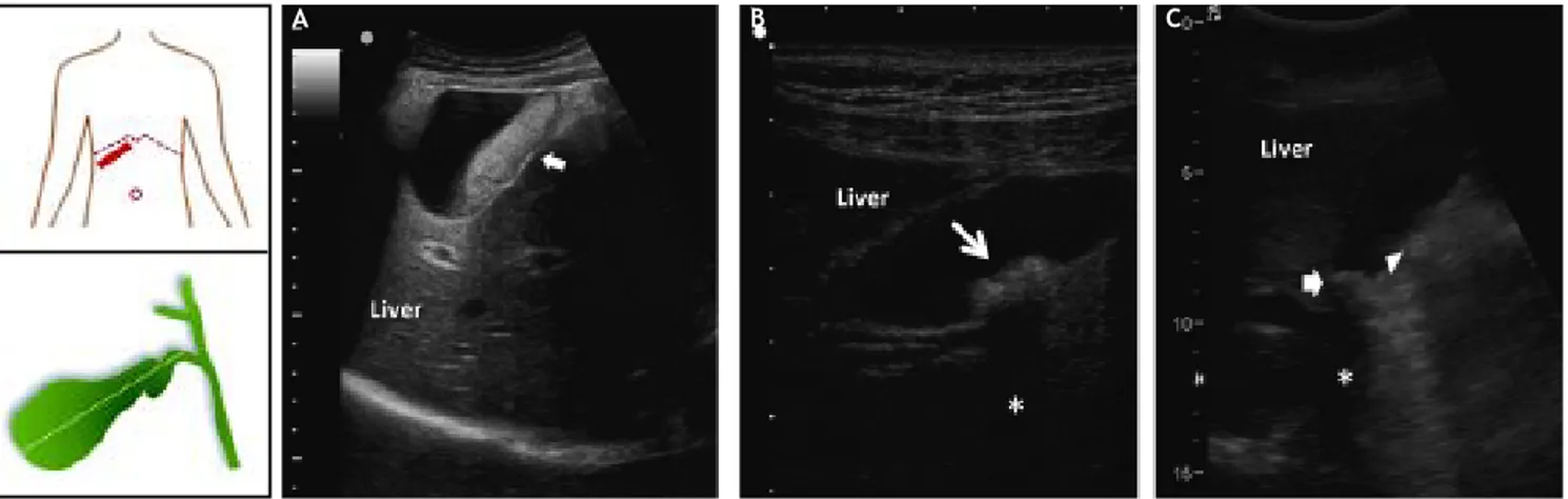
ultra-sonography can identify gallstones as small as 2 mm, with sensitivity > 95%. This technique is rap-id, noninvasive and relatively low cost, as well as can be performed at the bedside and does not involve ionizing radiations. As shown in figure 2, biliary sludge is characterized by low amplitude, gravity-de-pendent sonographic echoes seen in the gallbladder, which does not cast an acoustic shadow.72,131 By
contrast, ultrasound image can show a gallstone(s) in the gallbladder with typical acoustic shadow. As seen by polarizing light microscopy, biliary sludge consists mainly of plate-like cholesterol monohy-drate crystals and calcium bilirubinate granules em-bedded in strands of mucin gel.75 However, because
the sensitivity of ultrasonography for biliary sludge alone is only about 60%, further imaging testing and
bile examination should be considered if the result of ultrasonography is negative and clinical suspi-cion remains high, for example, in a pregnant wom-an with recurring attacks of biliary colic.75
For a pregnant woman with suspected biliary sludge and/or gallstones who have a negative result from transabdominal ultrasonography, further diag-nosing depends on the clinical symptoms, i.e., the presence or the absence of biliary colic. It is recom-mended examining gallbladder bile samples by polar-izing light microscopy for pregnant women in whom the clinical suspicion of biliary sludge is high and in whom further treatment such as laparoscopic or elective cholecystectomy would be considered.132-134
If an upper gastrointestinal endoscopy is required, gallbladder bile could be harvested during this pro-cedure and analyzed by polarizing light microscopy. If an upper gastrointestinal endoscopy is not re-quired, a gallbladder bile sample could be obtained through duodenal intubation.75 Pregnant women
with recurrent episodes of idiopathic acute pancrea-titis generally undergo endoscopic retrograde cholangiopancreatography (ERCP) (with shielding of the abdomen), and gallbladder bile can be collect-ed from the duodenum or the common bile duct dur-ing this procedure.72,135,136 Pregnant women who
have an indication for endoscopic ultrasonography, such as evaluation of abnormalities seen on previ-ous imaging studies, should undergo this procedure first. Endoscopic ultrasonography is not recom-mended for pregnant women without another
indica-Figure 2. Ultrasonographic appearance of biliary sludge, gallstones, and gallstone plus biliary sludge in the gallbladder. The left car-toons depict the site of the oblique ultrasonographic scan at the right hypochondrium (top) and the resulting longitudinal section of gall-bladder. A. A finely echogenic, dense, gravity-dependent, slowly mobile biliary sludge is seen occupying about 40% of the gallbladder lumen (thick arrows). The thickness of the gallbladder wall is not increased. B. Two mobile echogenic gallstones are indicated within the gallbladder lumen, layering on the distal wall. Each stone size is about 0.5 cm in diameter. C. A 1.5 cm gallstone in diameter (arrow) is detected in the gallbladder infundibulum and is surrounded by biliary sludge (triangle). The asterisk indicates the posterior acoustic shadowing typical of gallstones.
tion for the diagnosis. If biliary sludge is not identi-fied on these imaging examinations, gallbladder bile samples can be collected for polarizing light micros-copy. Further imaging examination such as compu-terized tomographic (CT) scanning is relatively high cost, low accurateness and availability.
During pregnancy, when common bile duct stones are suspected, radiographic imaging of the bile duct can be performed as long as the mother’s pelvis is shielded, the fetus is monitored, and the fetal dose of radiation is less than 5 radiation-absorbed dos-es.137,138 However, the safety of radiographic
imag-ing is limited to women in their second and third trimesters. Endoscopic retrograde cholangiography could be safely performed if necessary during preg-nancy, with a premature delivery rate of less than 5%.139-141 Indications for this procedure include
acute cholangitis, persistent jaundice, or severe pan-creatitis with suspected choledocholithiasis. Con-traindications include uncontrolled bleeding tendencies or inability to tolerate sedation or an-esthesia.35 In addition to fetal risks, potential
ma-ternal complications include pancreatitis, biliary tract infection, and gastrointestinal bleeding and perforation. The fetus should be shielded from radi-ation and fluoroscopy time minimized. In addition, fetal monitoring during any invasive procedures should be considered.142
PREVENTION AND TREATMENT OF BILIARY SLUDGE AND GALLSTONES IN
PREGNANT WOMEN
Following acute appendicitis, gallbladder disease is the second most common indication for non-obstetric surgical intervention during pregnancy.63,143
Gall-stones and biliary sludge are the most common causes of gallbladder disease in pregnant women. Pregnant women should be evaluated specifically for biliary sludge, microlithiasis, or gallstones only after they develop symptoms (Figure 3). If a specific cause lead-ing to biliary sludge is detected, attempts should be made to eliminate it. Biliary sludge and gallstones should be considered similar in almost all respects.
Prevention
Prevention of biliary sludge and gallstones in high-risk pregnant women has a rationale per se,
since preventive measures may dramatically reduce the risk of cholecystectomy in pregnant and post-partum women.128 Potentially useful general
meas-ures might include constant physical activity
although the ultimate beneficial role during nancy is controversial. Thus, asymptomatic preg-nant women with biliary sludge should undergo careful follow-up and managed expectantly.75
No indication exists for drug prescription as pre-ventive measure of biliary sludge and gallstones in pregnancy. This caution includes also cholecystoki-nin octapeptide (CCK-8) which has been used as prokinetic agent on the gallbladder and prevention of biliary sludge and gallstones in patients with gall-bladder stasis because of prolonged total parenteral nutrition.70,144,145
Treatment during pregnancy
Guidelines should be applied for optimal manage-ment of biliary sludge and gallstones in the preg-nant woman. The Food and Drug Administration (FDA) has developed a general rating system to pro-vide therapeutic guidance based on potential bene-fits and fetal risks, and drugs have been classified into categories A, B, C, D and X based on this sys-tem of classification.146
In asymptomatic pregnant women with biliary sludge and gallstones, expectant management is the general rule and litholysis is not indicated. Howev-er, in symptomatic pregnant women, there is a con-sensus concerning medical management of gallstones during pregnancy.72,128,135,147,148 Pain
con-trol is mandatory during pregnancy.
In a pregnant woman presenting with abdominal pain, true biliary colic should be distinguished from nonspecific abdominal discomfort. A laparoscopic or open cholecystectomy performed for true biliary col-ic is usually curative, but symptoms often persist if the procedure is performed in pregnant women with nonspecific dyspepsia and gallstones. Nevertheless, despite the availability of many imaging techniques that can be used to detect biliary sludge, microlithi-asis, and gallstones in the gallbladder and the com-mon bile duct, the diagnosis of biliary colic is ultimately based on clinical judgment.
Supportive management is highly recommended if possible, reserving definitive treatment after delivery. Women with uncomplicated biliary colic can be man-aged with intravenous hydration and narcotic pain control.149,150 Nevertheless, women who receive
sup-portive treatment are prone to symptomatic relapses, which might increase the likelihood of premature de-livery.149,150 No prospective data exist, moreover,
Figure 3. Flow-chart depicting the standard therapy of biliary sludge and gallstones during pregnancy. For management pur-poses, biliary sludge and gallstones should be considered similar in almost all respects. Surgery generally is reserved for preg-nant women with recurrent or unrelenting biliary pain refractory to medical management or with complications related to gallstones, including obstructive jaundice, acute cholecystitis, gallstone pancreatitis, or suspected peritonitis. For its safety, laparoscopic or elective cholecystectomy is one of the most common treatments for gallbladder gallstones in pregnant women, but it is recommended after the second trimester in order to reduce the rate of spontaneous abortion and preterm labor. If cho-lecystectomy is required during pregnancy, laparoscopic surgery is first recommended because of its relative safety. Of special note is that the timing of surgery is important. The effect of laparoscopic surgery on a developing fetus in the first trimester is unknown and surgery is more difficult in the third trimester with uterine enlargement. The second trimester, therefore, is belie-ved to be the optimal time for cholecystectomy. A supportive management is highly recommended if possible, delaying more de-finitive treatment until after childbirth. See text for details.
or complicated biliary tract disease, management becomes more controversial.
Lu, et al. have studied 76 women with 78 pregnan-cies admitted with biliary tract disease.149 Of the 63
women who presented with symptomatic cholelithia-sis, 10 underwent surgery while pregnant. There were no deaths, preterm deliveries, or intensive care unit admission. Fifty-three patients were treated medically and their clinical courses were complicated by symptomatic relapse in 20 patients (38%), by la-bor induction to control biliary colic (8 patients), and by premature delivery in 2 patients. Each relapse in the medically managed group led to an additional five days stay in hospital. These findings suggest that
surgical management of gallstones in pregnancy is safe and decreases days in hospital, as well as reduc-es the rate of labor induction and preterm delivery.149
The use of analgesics has successfully ameliorated biliary symptoms in 64% of symptomatic pregnant women.147 Surgery is generally reserved for
preg-nant women with recurrent or unrelenting biliary pain refractory to medical management or with gall-stone-related complications.151 Elective laparoscopic
cholecystectomy is relatively safe and is the first-line option, but it is recommended after the second tri-mester in order to reduce the rates of spontaneous abortion and preterm labor.82,147,152-155 Papillotomy
for choledocholithiasis yields relatively good results
Asymptomatic Biliary sludge Symptomatic patients
patients gallstones
Expectant Analgesics
management
Spontaneous
disappearance Complicated
of biliary sludge and gallstones
dissolution of gallstones
Supportive Poor healthy
management condition
After childbirth Percutaneous
gallstones still exist cholecystostomy
with drainage
Laparoscopic or open cholecystectomy
No Yes
No Yes
for mother and fetus.140 Selection criteria for
sur-gery need to be carefully considered in pregnant women, as illustrated in figure 3.
These with only one episode of uncomplicated bil-iary colic are at moderate risk for future recurrence of pain and more serious complications, and up to 30% of women will not develop further clinical symptoms.75 Expectant management is still an
op-tion; in principle, oral litholysis is contraindicated in pregnancy. If symptoms or complications of bil-iary sludge or gallstones do recur, however, surgery should be considered. The indication for surgery is even stronger if more serious complications develop, such as acute cholecystitis, sepsis, gangrene, ob-structive jaundice and suspected peritonitis, as well as acute pancreatitis induced by biliary sludge or microlithiasis.149,156-158 Surgery should be
consid-ered during initial hospitalization.
During pregnancy and when required, the first choice remains the laparoscopic, rather than the laparotomic cholecystectomy. Timing should be carefully considered as well.75 The effect of
laparo-scopic surgery on a developing fetus in the first tri-mester of pregnancy is unknown and surgery is more difficult in the third trimester with uterine enlargement. The second trimester, therefore, is believed to be the optimal time for cholecystecto-my.154,159-162 Laparoscopic cholecystectomy is
per-formed safely with minimal fetal morbidity during this period.147,149,152,153 The risks of preterm labor or
premature delivery in each trimester of pregnancy, however, are not clearly defined in the literature. If acute cholecystitis or cholangitis develops, earlier cholecystectomy should be considered.163
However, if a pregnant woman is under a poor healthy condition for surgery, percutaneous cholecys-tostomy with drainage should be considered.75,164-166
The long-term efficacy of these methods has not been proven by clinical trials and these approaches should be used only in pregnant women who require emergency therapy but are not good candidates for cholecystectomy. The efficacy of percutaneous chole-cystostomy with drainage in the treatment of biliary sludge has not been well established.75
Ursodeoxycholic acid (UDCA) is a litholytic bile salt (see below) and is used in a subgroup of symp-tomatic gallstone patients. Although UDCA is the treatment of choice for intrahepatic cholestasis of pregnancy167,168 and is classified as B drug by FDA
(i.e., no evidence of risk in studies), it is not used during pregnancy for biliary sludge and gallstone dissolution. Same would apply to ezetimibe, another agent acting on gallstone dissolution (see below).
Management post pregnancy
Therapeutic options of biliary sludge and gall-stones after pregnancy should follow the general guidelines, including expectant management in asymptomatic patients (no consensus about the need for prophylactic cholecystectomy), and chole-cystectomy in previously symptomatic patients or those who will become symptomatic after pregnancy. In a small subgroup of women, oral litholysis might have a role, and might include:
• Administration of the cholelitholytic agent UDCA;75 and
• Administration of the potent intestinal cholester-ol absorption inhibitor ezetimibe.128,169,170
The effect of UDCA, a hydrophilic bile acid, has been extensively studied for the dissolution of cho-lesterol gallstones in patients.171 UDCA has been
recommended as first-line pharmacological thera-py in a subgroup of symptomatic patients with small, radiolucent cholesterol gallstones, and its long-term administration has been shown to pro-mote the dissolution of cholesterol gallstones and to prevent the recurrence of gallstones.92,172 The
potential cholelitholytic mechanisms of UDCA in-volve the formation of a liquid crystalline mes-ophase.173 It favors the formation of numerous
vesicles that are composed predominantly of phos-pholipids in bile so that the growth of liquid crys-tals on the cholesterol monohydrate surface and their subsequent dispersions might occur during gallstone dissolution. Consequently, liquid crys-talline dissolution allows the transport of a great amount of cholesterol from stones, which is ex-creted into the duodenum and eventually in the faces.173 In patients with a rapid loss of body
weight, UDCA reduces the incidence of gallstones by 50 to 100%.174-176 In addition, in patients with
biliary sludge and idiopathic pancreatitis, after in-itial treatment with UDCA to dissolve cholesterol monohydrate crystals, ongoing maintenance ther-apy successfully prevents the recurrence of biliary sludge and pancreatitis.26
(NPC1L1) pathway, possibly a transporter-facili-tated mechanism.169 It could reduce cholesterol
concentrations of the liver, which in turn diminish the bioavailability of cholesterol for biliary se-cretion.169 Ezetimibe has been found to induce
a striking dose-dependent decrease in intestinal cholesterol absorption efficiency, coupled with a significant dose-dependent reduction in biliary cholesterol output and gallstone prevalence rate in gallstone-susceptible mice, even under high dietary cholesterol loads. Of note is that ezetimibe pro-motes the dissolution of cholesterol gallstones by forming an abundance of unsaturated micelles. Ezetimibe could protect gallbladder motility func-tion by desaturating bile.177 Furthermore, in a
small number of patients with gallstones, ezetimibe has been revealed to significantly reduce biliary cholesterol saturation and retard cholesterol crys-tallization in bile.177 These observations clearly
demonstrate that ezetimibe is a novel and potential cholelitholytic agent for preventing or treating cholesterol gallstone disease not only in mice but also in humans. To evaluate treatment time, re-sponse rate and overall cost-benefit analysis, a more detailed, long-term human study needs to be performed.
More recently, we found from a preliminary study that ezetimibe prevents the formation of E2-induced cholesterol gallstones by inhibiting intestinal cho-lesterol absorption. The bioavailability of intestinal source of cholesterol for biliary secretion is marked-ly reduced and bile is desaturated in mice even on the lithogenic diet. Also, ezetimibe does not influ-ence mRNA levels of the Erα, Erβ and Gpr30 genes in the liver. Therefore, these results show that ezetimibe is a potential cholelitholytic agent for pre-venting or treating E2-induced gallstones. These findings may provide an effective novel strategy for the prevention of cholesterol gallstones, particularly in women and patients exposed to high levels of es-trogen. Further clinical studies are warranted in this respect.
Because ezetimibe and UDCA promote the disso-lution of cholesterol gallstones by two distinct mech-anisms via the formation of an unsaturated micelle and a liquid crystalline mesophase, respectively, it is highly likely that biliary sludge could be prevent-ed and cholesterol gallstones could be dissolvprevent-ed fast-er by a combination thfast-erapy of ezetimibe and UDCA even in pregnant in postpartum women. A major limitation of oral litholysis, however, is the high re-currence rate of gallstones (10% per year and up to 45-50% by 5 years).178,179
CONCLUSIONS AND FUTURE DIRECTIONS
Obviously, during pregnancy, bile becomes lith-ogenic because of a significant increase in estro-gen levels, which lead to hepatic cholesterol hypersecretion and biliary lithogenicity. In addi-tion, increased progesterone concentrations im-pair gallbladder motility function, with the resulting increase in fasting gallbladder volume and bile stasis. Such abnormalities greatly pro-mote the formation of biliary sludge and gall-stones. Because plasma concentrations of female sex hormones, especially estrogen, increase linear-ly with the duration of gestation, the risk of gall-stone formation becomes higher in the third trimester of pregnancy. Increasing parity is also a risk factor for gallstones, especially in younger women. Similarly to non-pregnant women, preg-nant women are indeed exposed to the whole spec-trum of the natural history of biliary sludge and gallstones. Clinical features include no symptoms, one or more episodes of biliary colicky pain and sludge/gallstone-related complications. Due to its noninvasive features, transabdominal ultrasonog-raphy is the first-line diagnostic approach in asymptomatic and symptomatic pregnant pa-tients. All available therapeutic options (apart from oral litholysis with UDCA) must be kept ac-cessible in symptomatic patients (i.e. supportive care in patients with only one episode of biliary colic, laparoscopic cholecystectomy followed by open cholecystectomy in high-risk cases, and ERCP if biliary pancreatitis, choledocholithiasis, and cholangitis develop). Open issues for future research agenda must include ways to prevent the formation of biliary sludge and gallstones before and during pregnancy, the role of lifestyles, novel therapeutic medical agents to be tested in larger groups of patients (e.g., ezetimibe), and safe cholelitholytic agents during pregnancy, as well as ways to better prevent and treat gallstone com-plications.
ACKNOWLEDGMENT
CONFLICT OF INTEREST
None of the authors has any financial or any conflict of interest related to the content of this manuscript.
REFERENCES
1. Everson GT, McKinley C, Kern F, Jr. Mechanisms of gallsto-ne formation in women. Effects of exogenous estrogen (Premarin) and dietary cholesterol on hepatic lipid meta-bolism. J Clin Invest 1991; 87: 237-46.
2. Boston Collaborative Drug Surveillance Programme. Oral contraceptives and venous thromboembolic disease, sur-gically confirmed gallbladder disease, and breast tumours. Lancet 1973; 1: 1399-404.
3. The Boston Collaborative Drug Surveillance Program, Bos-ton University Medical Center. Surgically confirmed gall-bladder disease, venous thromboembolism, and breast tumors in relation to postmenopausal estrogen therapy. N Engl J Med 1974; 290: 15-9.
4. The Coronary Drug Project Research Group. Gallbladder disease as a side effect of drugs influencing lipid metabo-lism. Experience in the Coronary Drug Project. N Engl J Med 1977; 296: 1185-90.
5. Cirillo DJ, Wallace RB, Rodabough RJ, Greenland P, LaCroix AZ, Limacher MC, Larson JC. Effect of estrogen therapy on gallbladder disease. JAMA 2005; 293: 330-9.
6. Honore LH. Increased incidence of symptomatic choleste-rol cholelithiasis in perimenopausal women receiving estro-gen replacement therapy: a retrospective study. J Reprod Med 1980; 25: 187-90.
7. Thijs C, Knipschild P. Oral contraceptives and the risk of gallbladder disease: a meta-analysis. Am J Public Health 1993; 83: 1113-20.
8. Henriksson P, Einarsson K, Eriksson A, Kelter U, Angelin B. Estrogen-induced gallstone formation in males. Rela-tion to changes in serum and biliary lipids during hormonal treatment of prostatic carcinoma. J Clin Invest 1989; 84: 811-6.
9. Grodstein F, Colditz GA, Stampfer MJ. Postmenopausal hor-mone use and cholecystectomy in a large prospective stu-dy. Obstet Gynecol 1994; 83: 5-11.
10. Wang HH, Liu M, Clegg DJ, Portincasa P, Wang DQ. New in-sights into the molecular mechanisms underlying effects of estrogen on cholesterol gallstone formation. Biochim Bio-phys Acta 2009; 1791: 1037-47.
11. Angelin B, Olivecrona H, Reihner E, Rudling M, Stahlberg D, Eriksson M, Ewerth S, et al. Hepatic cholesterol metabo-lism in estrogen-treated men. Gastroenterology 1992; 103: 1657-63.
12. Novacek G. Gender and gallstone disease. Wien Med Wo-chenschr 2006; 156: 527-33.
13. Diehl AK. Epidemiology and natural history of gallstone di-sease. Gastroenterol Clin North Am 1991; 20: 1-19. 14. Barbara L, Sama C, Morselli Labate AM, Taroni F, Rusticali
AG, Festi D, Sapio C, et al. A population study on the pre-valence of gallstone disease: the Sirmione Study. Hepato-logy 1987; 7: 913-7.
15. Layde PM, Vessey MP, Yeates D. Risk factors for gall-bladder disease: a cohort study of young women atten-ding family planning clinics. J Epidemiol Community Health 1982; 36: 274-8.
16. Friedman GD, Kannel WB, Dawber TR. The epidemiology of gallbladder disease: observations in the Framingham Stu-dy. J Chronic Dis 1966; 19: 273-92.
17. Scragg RK, McMichael AJ, Seamark RF. Oral contraceptives, pregnancy, and endogenous oestrogen in gall stone disea-se—a case-control study. Br Med J 1984; 288: 1795-9. 18. Dhiman RK, Chawla YK. Is there a link between oestrogen
therapy and gallbladder disease? Expert Opin Drug Saf 2006; 5: 117-29.
19. Kern F Jr., Everson GT, DeMark B, McKinley C, Showalter R, Braverman DZ, Szczepanik-Van Leeuwen P, et al. Bilia-ry lipids, bile acids, and gallbladder function in the human female: effects of contraceptive steroids. J Lab Clin Med 1982; 99: 798-805.
20. Maringhini A, Ciambra M, Baccelliere P, Raimondo M, Orlan-do A, Tine F, Grasso R, et al. Biliary sludge and gallstones in pregnancy: incidence, risk factors, and natural history. Ann Intern Med 1993; 119: 116-20.
21. Bolukbas FF, Bolukbas C, Horoz M, Ince AT, Uzunkoy A, Oz-turk A, Aka N, et al. Risk factors associated with gallstone and biliary sludge formation during pregnancy. J Gas-troenterol Hepatol 2006; 21: 1150-3.
22. Maringhini A, Marceno MP, Lanzarone F, Caltagirone M, Fusco G, Di Cuonzo G, Cittadini E, et al. Sludge and stones in gallbladder after pregnancy. Prevalence and risk fac-tors. J Hepatol 1987; 5: 218-23.
23. Valdivieso V, Covarrubias C, Siegel F, Cruz F. Pregnancy and cholelithiasis: pathogenesis and natural course of gallstones diagnosed in early puerperium. Hepatology 1993; 17: 1-4. 24. Basso L, McCollum PT, Darling MR, Tocchi A, Tanner WA. A
study of cholelithiasis during pregnancy and its relations-hip with age, parity, menarche, breast-feeding, dysmeno-rrhea, oral contraception and a maternal history of cholelithiasis. Surg Gynecol Obstet 1992; 175: 41-6. 25. Stauffer RA, Adams A, Wygal J, Lavery JP. Gallbladder
di-sease in pregnancy. Am J Obstet Gynecol 1982; 144: 661-4.
26. Ros E, Navarro S, Bru C, Garcia-Puges A, Valderrama R. Occult microlithiasis in ‘idiopathic’ acute pancreatitis: prevention of relapses by cholecystectomy or ursodeoxy-cholic acid therapy. Gastroenterology 1991; 101: 1701-9. 27. Ko CW, Beresford SA, Schulte SJ, Matsumoto AM, Lee SP.
In-cidence, natural history, and risk factors for biliary sludge and stones during pregnancy. Hepatology 2005; 41: 359-65. 28. Glasinovic JC, Mege R, Ferreiro O, Rodríguez N, Marinovic I, Villarroel L, Vela P. Cholelithiasis in Chilean female popu-lation. Prevalence and associated risk factors. Gastroen-terology 1986; 96: A601.
29. Van Bodegraven AA, Bohmer CJ, Manoliu RA, Paalman E, Van der Klis AH, Roex AJ, Kruishoop AM, et al. Gallbladder contents and fasting gallbladder volumes during and after pregnancy. Scand J Gastroenterol 1998; 33: 993-7. 30. Everhart JE, Khare M, Hill M, Maurer KR. Prevalence and
ethnic differences in gallbladder disease in the United Sta-tes. Gastroenterology 1999; 117: 632-9.
31. Thijs C, Knipschild P, Leffers P. Pregnancy and gallstone disease: an empiric demonstration of the importance of specification of risk periods. Am J Epidemiol 1991; 134: 186-95.
32. The Rome Group for Epidemiology and Prevention of Chole-lithiasis (GREPCO).The epidemiology of gallstone disease in Rome, Italy. Part II. Factors associated with the disease. Hepatology 1988; 8: 907-13.
34. Lindseth G, Bird-Baker MY. Risk factors for cholelithiasis in pregnancy. Res Nurs Health 2004; 27: 382-91.
35. Ko CW. Risk factors for gallstone-related hospitalization during pregnancy and the postpartum. Am J Gastroente-rol 2006; 101: 2263-8.
36. Lydon-Rochelle M, Holt VL, Martin DP, Easterling TR. Asso-ciation between method of delivery and maternal rehospi-talization. JAMA 2000; 283: 2411-6.
37. Printen KJ, Ott RA. Cholecystectomy during pregnancy. Am Surg 1978; 44: 432-4.
38. Carpenter MW. Gestational diabetes, pregnancy hyper-tension, and late vascular disease. Diabetes Care 2007; 30 (Suppl. 2): S246-S250.
39. Valdiviezo C, Garovic VD, Ouyang P. Preeclampsia and hy-pertensive disease in pregnancy: their contributions to cardiovascular risk. Clin Cardiol 2012; 35: 160-5.
40. Bennion LJ, Ginsberg RL, Gernick MB, Bennett PH. Effects of oral contraceptives on the gallbladder bile of normal women. N Engl J Med 1976; 294: 189-92.
41. Lynn J, Williams L, O’Brien J, Wittenberg J, Egdahl RH. Effects of estrogen upon bile: implications with respect to gallstone formation. Ann Surg 1973; 178: 514-24.
42. Pertsemlidis D, Panveliwalla D, Ahrens EH, Jr. Effects of clofibrate and of an estrogen-progestin combination on fasting biliary lipids and cholic acid kinetics in man. Gas-troenterology 1974; 66: 565-73.
43. Kern F Jr., Everson GT. Contraceptive steroids increase cholesterol in bile: mechanisms of action. J Lipid Res 1987; 28: 828-39.
44. Bennion LJ, Mott DM, Howard BV. Oral contraceptives rai-se the cholesterol saturation of bile by increasing biliary cholesterol secretion. Metabolism 1980; 29: 18-22. 45. van der Werf SD, van Berge Henegouwen GP, Ruben AT,
Pals-ma DM. Biliary lipids, bile acid metabolism, gallbladder motor function and small intestinal transit during ingestion of a sub-fifty oral contraceptive. J Hepatol 1987; 4: 318-26. 46. Heuman R, Larsson-Cohn U, Hammar M, Tiselius HG.
Effects of postmenopausal ethinylestradiol treatment on gallbladder bile. Maturitas 1980; 2: 69-72.
47. Anderson A, James OF, MacDonald HS, Snowball S, Taylor W. The effect of ethynyl oestradiol on biliary lipid compo-sition in young men. Eur J Clin Invest 1980; 10: 77-80. 48. Vazquez MC, Rigotti A, Zanlungo S. Molecular mechanisms
underlying the link between nuclear receptor function and cholesterol gallstone formation. J Lipids 2012; 2012: 547643.
49. Portincasa P, Di Ciaula A, Wang HH, Palasciano G, van Er-pecum KJ, Moschetta A, Wang DQ. Coordinate regulation of gallbladder motor function in the gut-liver axis. Hepato-logy 2008; 47: 2112-26.
50. Everson GT. Pregnancy and gallstones. Hepatology 1993; 17: 159-61.
51. Everson GT, McKinley C, Lawson M, Johnson M, Kern F, Jr. Gallbladder function in the human female: effect of the ovulatory cycle, pregnancy, and contraceptive steroids. Gastroenterology 1982; 82: 711-9.
52. Braverman DZ, Johnson ML, Kern F, Jr. Effects of preg-nancy and contraceptive steroids on gallbladder function. N Engl J Med 1980; 302: 362-4.
53. Wang HH, Afdhal NH, Wang DQ. Estrogen receptor alpha, but not beta, plays a major role in 17beta-estradiol-indu-ced murine cholesterol gallstones. Gastroenterology 2004; 127: 239-49.
54. Wang HH, Portincasa P, Wang DQ. Molecular pathophysio-logy and physical chemistry of cholesterol gallstones. Front Biosci 2008; 13: 401-23.
55. Silverthorn DU. Human Physiology: An Integrated Appro-ach. 6th ed. Glenview, IL: Pearson Education, Inc.; 2013. 56. Speroff L GR, Kase NG. Clinical gynecologic endocrinology
and infertility. 6th Ed. Baltimore; 1999.
57. Ferin M JR, Warren M. The menstrual cycle. In Physiology, Reproductive Disorders, and Infertility. New York; 1993. 58. Senie RT, Tenser SM. The timing of breast cancer surgery
during the menstrual cycle. Oncology 1997; 11: 1509-17. 59. Pike MC, Spicer DV, Dahmoush L, Press MF. Estrogens,
progestogens, normal breast cell proliferation, and breast cancer risk. Epidemiol Rev 1993; 15: 17-35.
60. Chikazawa K, Araki S, Tamada T. Morphological and endocri-nological studies on follicular development during the human menstrual cycle. J Clin Endocrinol Metab 1986; 62: 305-13. 61. Tsimoyiannis EC, Antoniou NC, Tsaboulas C, Papanikolaou
N. Cholelithiasis during pregnancy and lactation. Prospec-tive study. Eur J Surg 1994; 160: 627-31.
62. Hay JE. Liver disease in pregnancy. Hepatology 2008; 47: 1067-76.
63. Mendez-Sanchez N, Chavez-Tapia NC, Uribe M. Pregnancy and gallbladder disease. Ann Hepatol 2006; 5: 227-30. 64. Scott LD. Gallstone disease and pancreatitis in
pregnan-cy. Gastroenterol Clin North Am 1992; 21: 803-15. 65. Trotman BW, Soloway RD. Pigment vs cholesterol
choleli-thiasis: clinical and epidemiological aspects. Am J Dig Dis 1975; 20: 735-40.
66. Neil Bajwa RB, Ambrish Ghumman, Agrawal R. M. . The Galls-tone Story: Pathogenesis and Epidemiology. Practical gas-troenterology 2010; XXXIV.
67. The epidemiology of gallstone disease in Rome, Italy. Part I. Prevalence data in men. The Rome Group for Epidemiolo-gy and Prevention of Cholelithiasis (GREPCO). HepatoloEpidemiolo-gy 1988; 8: 904-6.
68. Rome Group for the Epidemiology and Prevention of Chole-lithiasis (GREPCO). Prevalence of gallstone disease in an Italian adult female population. Am J Epidemiol 1984; 119: 796-805.
69. Attili AF, Capocaccia R, Carulli N, Festi D, Roda E, Barbara L, Capocaccia L, et al. Factors associated with gallstone disease in the MICOL experience. Multicenter Italian Stu-dy on Epidemiology of Cholelithiasis. Hepatology 1997; 26: 809-18.
70. Pazzi P, Gamberini S, Buldrini P, Gullini S. Biliary sludge: the sluggish gallbladder. Dig Liver Dis 2003; 35(Suppl. 3): S39-S45.
71. Davis M, Ryan JP. Influence of progesterone on guinea pig gallbladder motility in vitro. Dig Dis Sci 1986; 31: 513-8. 72. Gilat T, Konikoff F. Pregnancy and the biliary tract. Can J
Gastroenterol 2000; 14(Suppl. D): 55D-59D.
73. Acalovschi M. Cholesterol gallstones: from epidemiology to prevention. Postgrad Med J 2001; 77: 221-9.
74. Cohen S. The sluggish gallbladder of pregnancy. N Engl J Med 1980; 302: 397-9.
75. Ko CW, Sekijima JH, Lee SP. Biliary sludge. Ann Intern Med 1999; 130: 301-11.
76. Sali A, Oats JN, Acton CM, Elzarka A, Vitetta L. Effect on pregnancy on gallstone formation. Aust N Z J Obstet Gy-naecol 1989; 29: 386-9.
77. Kern F, Jr., Everson GT, DeMark B, McKinley C, Showalter R, Erfling W, Braverman DZ, et al. Biliary lipids, bile acids, and gallbladder function in the human female. Effects of pregnancy and the ovulatory cycle. J Clin Invest 1981; 68: 1229-42.