Efficacy of Probiotics and Smectite in Rats with
Non-Alcoholic Fatty Liver Disease
Nazarii Kobyliak,* Ludovico Abenavoli,** Tetyana Falalyeyeva,*** Tetyana Beregova***
* Bogomolets National Medical University, Kyiv, Ukraine. ** University Magna Graecia, Catanzaro, Italy. *** Taras Shevchenko National University of Kyiv, Kyiv, Ukraine.
January-February, Vol. 17 No. 1, 2018: 153-161
INTRODUCTION
Non-alcoholic fatty liver disease (NAFLD) is
current-ly a leading cause of chronic liver disease1,2 affecting
ap-proximately 20-40% of adults in developed countries3 and
recognized as the accumulation of lipids within the hepa-tocytes exceeding 5% of liver weight in the absence of ex-cessive alcohol intake and secondary causes of liver
diseases.4 NAFLD ranges from simple steatosis to
non-al-coholic steatohepatitis (NASH) that can have different de-grees of fibrosis and progress to liver cirrhosis and
hepatocellular carcinoma,5 which has resulted in
signifi-cant health concerns such as morbidity, mortality, and
liv-er transplants.6
Current conception of NAFLD pathogenesis evolved
from “two hit” 7 to recent “multiple hit” theory.8
Accord-ing to formerly proposed two hit model insulin resistance acts as first hit and can promote intra-hepatic fat accumula-tion and lipotoxicity; second, development of cellular in-sults which caused by oxidative stress, lipid oxidation, chronic inflammation, apoptosis and fibrogenesis that
de-termine the progression of the disease.7,9
As for nowadays the “two hit” hypothesis is outdated, as it does not fully account for several molecular and meta-bolic changes that occur in NAFLD development. The “multiple hit” hypothesis considers multiple insults acting together on genetically predisposed subjects to induce NAFLD and provides a more accurate explanation of NAFLD pathogenesis. Such hits include insulin resist-ance, disbalance of adipose tissue hormonal activity, low-grade chronic systemic inflammation, increased intestinal permeability with subsequent “metabolic endotoxemia”, The Official Journal of the Mexican Association of Hepatology,
the Latin-American Association for Study of the Liver and the Canadian Association for the Study of the Liver
Manuscript received: Manuscript received:Manuscript received:
Manuscript received:Manuscript received: March 01, 2016. Manuscript accepted:Manuscript accepted:Manuscript accepted: June 03, 2017.Manuscript accepted:Manuscript accepted:
DOI:10.5604/01.3001.0010.7547
A B S T R A C T A B S T R A C T A B S T R A C T A B S T R A C T A B S T R A C T
Introduction and aim. Introduction and aim.Introduction and aim. Introduction and aim.
Introduction and aim. Today probiotics have been suggested as a treatment for the prevention of non-alcoholic fatty liver disease (NAFLD). Smectite is a natural silicate that binds to digestive mucous and has the ability to bind endo- and exotoxins. The present study was designed to determine whether probiotics plus smectite is superior to probiotic alone on the monosodium glutamate (MSG) induced NAFLD model in rats. Materials and methods.Materials and methods.Materials and methods.Materials and methods.Materials and methods. We included 60 rats divided into 4 groups 15 animals in each. Rats of group I were intact. Newborns rats of groups II-IV were injected with MSG. The III (Symbiter) group received 2.5 ml/kg of multiprobiotic “Symbiter” containing concentrated biomass of 14 probiotic bacteria genera. The IV (Symbiter+Smectite) groups received “Symbiter Forte” combination of probiotic biomass with smectite gel (250 mg). Results. Results. Results. Results. Results. In both interventional groups reduction of total NAS score as compared to MSG-obesity was observed. Indeed similar values of steatosis score (0.93 ± 0.22 vs. 0.87 ± 0.16) in both treatment groups, we observed that lower total score for Symbiter+ Smectite are associated with more pronounced reduction of lobular inflammation (0.13 ± 0.09 vs. 0.33 ± 0.15) as compared to administration of probiotic alone. This data accompanied with significant reduction of IL-1 and restoration of IL-10 between these 2 groups. Conclusions. Conclusions. Conclusions. Conclusions. Conclusions. Additional to alive probiotic administration of smectite gel due to his absorbent activity and mucus layer stabilization properties can impact on synergistic enhancement of single effect which manifested with reduction of lobular inflammation and at list partly steatohepatitis prevention.
Key words. Key words. Key words. Key words.
nutritional factors, gut microbiota and genetic and
epige-netic factors.8,10
Among multiple hits, increasing clinical and experi-mental evidence indicates that alteration of gut-liver axis is implicated in NAFLD onset and progression, and the gut microbiota has been recognized as the key player in the
gut-liver cross talk.11,12 Thus, the modification of intestinal
bacterial flora by prebiotics and specific probiotics has been proposed as a therapeutic approach for the treatment
and/or prevention of NAFLD.13,14
Smectite is a natural silicate clay belonging to the dioc-tahedral smectite class and consists of a double aluminium
and magnesium silicate arranged in parallel leaflets.15
Smectite has recently been recommended by several
repu-table organization for treatment of acute diarrhea.16,17
It is well known that “metabolic endotoxemia” due to “multiple hit” theory is one of the most powerful triggers strongly associated with NAFLD development. Metabolic endotoxemia is defined as increase in plasma bacterial li-popolysaccharide (LPS) concentration increasing by two
to three times.18 We proposed that smectite, due to its
ability to bind endo- and exotoxins and capacity to restore the barrier properties of human intestinal cell monolay-ers, may be beneficial when supplemented with probiot-ics for NAFLD/NASH development.
Therefore, the present study aims to determine wheth-er probiotics plus smectite is supwheth-erior to probiotic alone on the monosodium glutamate (MSG) induced NAFLD model in rats.
MATERIAL AND METHODS
Study design
This study was carried out in strict accordance with the recommendations in the Guide for the Care and Use of Laboratory Animals of the National Institutes of Health and the general ethical principles of animal experiments, approved by the First National Congress on Bioethics Ukraine (September 2001). The protocol was approved by the Committee on the Ethics of Animal Experiments of the Taras Shevchenko National University of Kyiv (Pro-tocol number: 19/2015). The rats were kept in collective cages under controlled conditions of temperature (22 ± 3 °C), light (12 h light/dark cycle) and relative humidity (60 ± 5%). The animals were fed laboratory chow (PurinaW)
and tap water ad libitum.
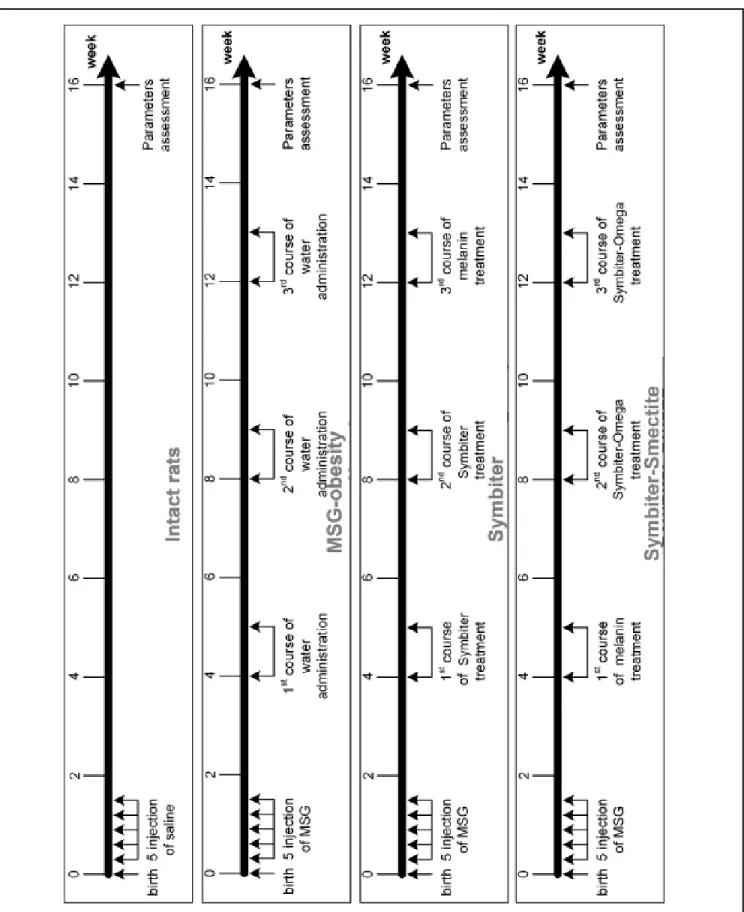
We included 60 newborn Wistar male rats divided into 4 groups 15 animals in each: intact (I), MSG-obesity (II), Symbiter (III) and (Symbiter+Smectite) (IV) groups. Newborns rats of intact group were subcutaneously (s.c.)
injected with saline in the volume of 8 μL/g and animals of
groups II-IV received MSG solution (4.0 mg/g of body
weight) s.c. at 2nd, 4th, 6th, 8th and 10th postnatal days. Neonatal administration of MSG causes the significant ac-cumulation of abdominal fat with subsequent develop-ment of NAFLD and severe visceral obesity in adult
rats.19,20 This happens because of the neurotoxicity effects
on the arcuate and ventromedial nuclei of the
hypothala-mus.21
During the first 4 months after birth rats had a normal diet. The III (Symbiter) group received 2.5 mL/kg of mul-tiprobiotic “Symbiter” containing concentrated biomass of
14 probiotic bacteria genera Bifidobacterium, Lactobacillus,
Lactococcus, Propionibacterium. The IV (Symbiter+Smectite) groups received “Symbiter Forte” combination of probi-otic biomass with smectite gel (250 mg). Administration was started at the age of 4 weeks just after the wean and continued for 3 month intermittently alternating two-week course of introduction with two-two-week course of break (Figure 1).
Sample collection and blood biochemistry analysis
Rats of all groups were fasted for approximately 12 h prior sacrifice by cervical dislocation under urethane an-esthesia. Blood was drawn from the apex of the cardiac ventricle and few blood drops were collected into a mi-crocentrifuge tube containing a mixture of NaF and EDTA at a 2:1 (w/w) ratio. Blood sample was collected into a sterile tube and centrifuged at 3,500 rpm (2260 g) for 15 min. After centrifugation serum supernatant for further analysis was aliquoted into microcentrifuge tubes and stored at -80 °C. Bilirubin, activity of alanine (ALT) and aspartate aminotransferase (AST) in serum were deter-mined by the standard biochemical methods.
The contents of interleukins (ILs) 1β, 4, 10, 12B p40,
interferon (INF) γ, and transforming growth factor
(TGF) β in rat serum were measured by ELISA using
spe-cific mono- and polyclonal antibodies (Sigma) to these
proteins. Monoclonal antibodies to ILs 4, 10 and TGF-β
were mice-produced. Polyclonal antibodies to IL-12B p40 were produced in rabbits and polyclonal antibodies to
IL-1β and INF-γ were produced in goats. Studied molecules
were immobilized in 96-well plates with sorption surface. Then primary and secondary antibodies labeled with en-zyme were added to wells. Adding of substrate led to the development of colored reaction. The optical density of the solution in each well after the addition of substrate is proportional to the content of studied cytokines. The con-tent was expressed in absorbance units of optical density.
Liver histology assessment
hepat-Figure 1. Figure 1. Figure 1.
ic lobes were taken (sample size 0.5 x 0.5 cm) for histolog-ical analysis. After being fixed for 24 h in a liquid Buena, fragments of liver were dehydrated in alcohols of increas-ing concentrations (from 70° to 96°), embedded in paraffin and then sliced into 5-6 micron thick pieces and stained with hematoxylin-eosin. A pathologist blinded to group distributions performed the histological analyses of slides using light microscopy (“Olympus”, Japan). To assess morphological changes in liver we used NAS (NAFLD activity score), which includes histological features and has been defined as unweighted sum of scores for steatosis (0-3), lobular inflammation (0-3) and ballooning (0-2).
Ac-cording to NAS scores, ≥ 5 are diagnosed as non-alcoholic
steatohepatitis (NASH), and cases with a NAS < 3 are
mentioned as not NASH.22 Lipid extraction from the liver
was performed according to Folch, et al.23
Statistical analysis
Statistical analysis performed by using SPSS-21 soft-ware. All data were expressed as mean ± standard error (M±SEM) or %. Data distribution was analyzed using the Kolmogorov-Smirnov normality test. Continuous varia-bles with parametric distribution were analyzed using Analysis of Variance (one-way ANOVA) and if the results were significant, a post-hoc Turkey’s test was performed. For data with non-parametric distribution Kruskall-Wallis and post-hoc Tukey’s test were conducted for multiple comparisons. For comparisons of categorical variables we
conducted χ2 test. The difference between groups was
de-fined to be statistically significant when a p-value was less than 0.05.
RESULTS
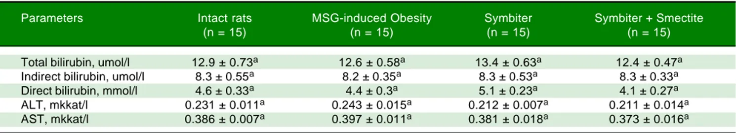
We didn’t find any significant changes in liver serum (ALT, AST, bilirubin) function parameters between in-tact, MSG-obesity and both interventional groups (Table 1).
Histological analysis of liver micropreparations con-firmed the development of NAFLD in rats. It was
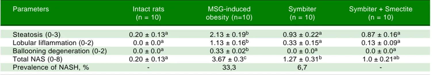
regis-tered in MSG-obesity group typical for NAFLD histolog-ical pattern: microvesicular steatosis (Figure 2A), mild lobular inflammation in zone 3 and ballooning degenera-tion (Figure 2B), which were also confirmed by total NAS score increasing approximately by 18 times (p < 0.001) compared with values of intact rats (Table 2). The degree of steatosis was increased by 10 times (p < 0.001) in MSG-rats as compared with intact MSG-rats (Table 2). Intact MSG-rats did not display inflammation in liver, at the same time lobular inflammation in MSG-group reached 1.13 ± 0.16 points (Table 2). Similar results were recorded for ballooning degeneration (Table 2).
In both interventional groups reduction of total NAS score as compared to MSG-obesity was observed. Howev-er only in SymbitHowev-er+Smectite group we didn't find
sig-nificant changes as compared to intact rats (1.0 ± 0.21 vs.
0.20 ± 0.13, p = 0.223). Indeed similar values of steatosis score (0.93 ± 0.22 vs. 0.87 ± 0.16, p = 0.994) in both treat-ment groups, we observed that lower total score for Symbiter+Smectite are associated with more pronounced
reduction of lobular inflammation (0.13 ± 0.09 vs. 0.33 ±
0.15, p = 0.695) as compared to administration of probiot-ic alone. Furthermore, in both interventional groups, lob-ular inflammation was remarkably reduced, we determined predominantly focal steatosis, the fat droplet number significantly decreased and the hepatic lobules were clearly delineated as compared with MSG group (Figures 2C, D).
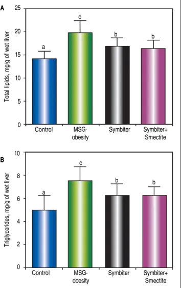
Administration of both Symbiter+Smectite or probiot-ic mixture alone led to signifprobiot-icant decrease of total lipids and triglycerides content in liver as compared to the MSG-obesity group (Figures 3A, B). Nevertheless, the differences of liver lipid contents between both interven-tional groups were insignificant.
Immunoassay analysis has shown the intensification of inflammatory processes in conditions of MSG-obesity.
The increase content of pro-inflammatory cytokines IL-1β
and IL-12B p40 in obese rat serum was found. The IL-1β
level raised by 37.1% (p < 0.001) and IL-12B p40 - by 50.7% (p < 0.001) as compared to intact rats (Figures 4 A,
Table 1. Liver function tests of rats under MSG-induced obesity and after administration of probiotic and their combination with smectite.
Parameters Intact rats MSG-induced Obesity Symbiter Symbiter + Smectite
(n = 15) (n = 15) (n = 15) (n = 15)
Total bilirubin, umol/l 12.9 ± 0.73a 12.6 ± 0.58a 13.4 ± 0.63a 12.4 ± 0.47a
Indirect bilirubin, umol/l 8.3 ± 0.55a 8.2 ± 0.35a 8.3 ± 0.53a 8.3 ± 0.33a
Direct bilirubin, mmol/l 4.6 ± 0.33a 4.4 ± 0.3a 5.1 ± 0.23a 4.1 ± 0.27a
ALT, mkkat/l 0.231 ± 0.011a 0.243 ± 0.015a 0.212 ± 0.007a 0.211 ± 0.014a
AST, mkkat/l 0.386 ± 0.007a 0.397 ± 0.011a 0.381 ± 0.018a 0.373 ± 0.016a
Figure 2. Figure 2. Figure 2.
Figure 2. Figure 2. Light microscopic micrographs of the rat liver tissue stained with hematoxylin and eosin, x400. A, B. A, B. A, B. A, B. A, B. MSG-induced obesity group (II). In micrographs observed perivascular leukocyte infiltration at zone 3 (mild lobular inflammation) (A) (A) (A) (A) (A) and predominantly microvesicular pronounced total steatosis (B).
(B). (B).
(B). (B). In micrographs mainly observed focal mild microvesicular steatosis (C, D). C. (C, D). C. (C, D). C. (C, D). C. (C, D). C. Symbiter group (III). D. D. D. D. D. Symbiter-Smectite (IV) group.
B). In contrast, we did not reveal difference in the level of
INF-γ between obese and intact rats (Figure 4C). In the
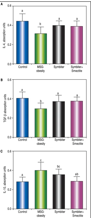
conditions of obesity, anti-inflammatory system was down-regulated. That was evident because of the
reduc-tion of IL-4 by 29.0% (p < 0.001) (Figure 5A) and TGF-β
by 27.2% (p < 0.05) (Figure 5B) in MSG-group as
com-pared to control. As opposed to this IL-10 was elevated in obese rats by 55.4% (p < 0.001) that may suggest compen-satory effect of anti-inflammatory system (Figure 5 C).
Administration of probiotic alone or in combination with smectite gel led to similar decrease of IL-12 (Figure 4 B) and significant elevation of the IL-4 (Figure 5 A) and
Table 2. Morphological changes of the liver tissue under MSG-induced obesity and after administration of probiotic and their combination with smectite.
Parameters Intact rats MSG-induced Symbiter Symbiter + Smectite
(n = 10) obesity (n=10) (n = 10) (n = 10)
Steatosis (0-3) 0.20 ± 0.13a 2.13 ± 0.19b 0.93 ± 0.22a 0.87 ± 0.16a
Lobular Iiflammation (0-2) 0.0 ± 0.0a 1.13 ± 0.16b 0.33 ± 0.15a 0.13 ± 0.09a
Ballooning degeneration (0-2) 0.0 ± 0.0a 0.33 ± 0.02b 0.0 ± 0.0a 0.0 ± 0.0a
Total NAS (0-8) 0.20 ± 0.13a 3.67 ± 0.3c 1.27 ± 0.31b 1.0 ± 0.21ab
Prevalence of NASH, % - 33,3 6,7
-Data are presented as the M ± SEM. One-way ANOVA with post hoc Tukeys test for multiple comparisons were performed for data analysis. a, b, c Values at
the same row with different superscript letters show significant differences at p < 0.05. A
AA
AA BBBBB
C CC
TGF-β (Figure 5 B) approximately on 25-35% (p < 0.001) as compared to MSG-obese rats. Furthermore, significant activation of the anti-inflammatory system was accompa-nied by restoration of cytokines levels to levels in intact
rats. The reduction of IL-1β (Figure 4A) and IL-10
(Fig-ure 5C) also at least partially achieve values of intact rats but was most pronounced in Symbiter+Smectite group as compared to probiotic alone and to MSG-obese rats.
DISCUSSION
The use of probiotics in the NAFLD treatment and prevention has dramatically increased over the last decade. Probiotic supplements are well known ingredients of functional foods and nutraceuticals and may provide
bene-Figure 4. Figure 4. Figure 4.
Figure 4. Figure 4. Serum proinflammatory cytokines content of rats with the MSG-induced obesity and after administration of probiotic and their combination with smectite. Data are presented as the M ± SEM. One-way ANOVA with post hoc Tukeys test for multiple comparisons were performed for data analysis. a,b,c Values at the same row with different superscript letters show significant differences at p < 0.05, a.u. - absorption units.
0.8
0.6
0.4
0.2
0.0
IL-1
β,
Absorption units
Control MSG- Symbiter Symbiter+
obesity Smectite
a
c
b
a
1.5
1.0
0.5
0.0
IL-12B, p40 absorption units
a
b
c
Control MSG- Symbiter Symbiter+
obesity Smectite
c
0.4
0.3
0.2
0.1
0.0
INF-γ
absorption units
a a a
Control MSG- Symbiter Symbiter+
obesity Smectite
a
Figure 3. Figure 3. Figure 3. Figure 3.
Figure 3. Liver total lipids and triglycerides content of rats with the MSG-induced obesity and after administration of probiotic and their combination with smectite. Data are presented as the M ± SEM. One-way ANOVA with post hoc Tukeys test for multiple comparisons were performed for data analysis. a,b,c Values at the same row with different superscript letters show significant differences at p < 0.05.
10
8
6
4
2
0
Triglycerides, mg/g of wet liver
Control MSG- Symbiter Symbiter+
obesity Smectite
a
c
b b
25
20
15
10
5
0
Total lipids, mg/g of wet liver
a
c
b
Control MSG- Symbiter Symbiter+
obesity Smectite
b
A AA AA
B BB BB
C C C C C A
A A A A
ficial health effects because they can influence the
intesti-nal microbial ecology and immunity.24
The previous animal studies demonstrated strain-spe-cific potential of probiotics for NAFLD development. The most favorable effects on NAFLD are associated with
Lactobacillus and Bifidobacterium strains and appear with
res-toration of gut barrier function,25 improvement of insulin
sensitivity,14 reduction of LPS absorption,26 serum
choles-terol level,27 liver fat accumulation,25-28 prevention from
lipid peroxidation, NFκB activation28 and subsequent
at-tenuation of liver inflammation and steatosis in distinct animal models of diet- and genetically determined
obesi-ty. Cani, et al. summarized these findings and showed that
at least 15 different strains of Lactobacillus and 3 strains of Bifidobacterium do not equally influence on hepatic lip-ids and NAFLD manifestation. Remarkably, 12 strains de-creased hepatic tissue inflammation and 11 reduced the hepatic triglyceride content when given as a single
treat-ment.29 Interesting data reported by these authors.30 They
find failure of NAFLD prevention with lyophilized
monoprobiotics B. animalis VKL, B. animalis VKB, L.casei
IMVB-7280 strains. Nevertheless, multiprobiotic cocktails due to formation of mutualistic interactions lead to signif-icant reduction of hepatic steatosis, total lipids and triglyc-erides content in the liver and finally prevent the development of NAFLD in animals as compared to MSG-obesity littermates. Remarkably that more pronounced changes were admitted after administration of probiotic mixture preferably containing alive strains as compared to lyophilized cocktails.
Smectite is a natural silicate that binds to digestive mu-cous and has the ability directly to absorb bacterial toxins,
bacteria, viruses and bile salts.31,32 On the other hand, when
administered orally, smectite is not absorbed and elimi-nated unchanged directly with faeces within sixteen hours. Smectite increases water and electrolyte absorption, may affect intestinal permeability, formes multilayer structure with high plastic viscosity and powerful coating proper-ties hence preserving integrity of the mucus layer and ren-dering the intestinal epithelium more resistant to endogenous (bile salts) or exogenous (such as bacterial
toxins and alcohol) impacts.31,33,34 Diosmectite also has a
protective effect against intestinal inflammation34 hence
suppressing production of cytokines such as interleukin-8
from secretory epithelial cells35in vitro and to attenuating
the proinflammatory action of TNFα.36 We suggested that
all these pharmacological properties may be beneficial for the treatment of NAFLD.
According to this suggestion we firstly designed the study to test the hypothesis that whether probiotics plus smectite is superior to probiotic alone on NAFLD pre-vention in rats. The reduction of total NAS score in both nutraceuticals groups as compared to MSG-obesity was
Figure 5. Figure 5. Figure 5.
Figure 5. Figure 5. Serum anti-inflammatory cytokines content of rats with the MSG-induced obesity and after administration of probiotic and their combination with smectite. Data are presented as the M ± SEM. One-way ANOVA with post hoc Tukeys test for multiple comparisons were performed for data analysis. a,b,c Values at the same row with different superscript letters show significant differences at p < 0.05, a.u. - absorption units.
0.6
0.4
0.2
0.0
IL-4, absorption units
Control MSG- Symbiter Symbiter+
obesity Smectite
a
b
a a
0.6
0.4
0.2
0.0
TGF-β
absorption units
a
b
a
Control MSG- Symbiter Symbiter+
obesity Smectite
a
0.6
0.4
0.2
0.0
IL-10, absorption units
a
c
bc
Control MSG- Symbiter Symbiter+
obesity Smectite
ab A
A A A A
B B B B B
observed. However only for Symbiter+Smectite group, indeed similar values of steatosis score, more pronounced but insignificant reduction of lobular inflammation (0.13
± 0.09 vs. 0.33 ± 0.15) as compared to administration of
probiotic alone was demonstrated. The reduction of IL-1β
and restoration of IL-10 also at least partially achieved val-ues of intact rats but was most pronounced in Symbiter+Smectite group as compared to probiotic alone and to MSG-obese rats.
CONCLUSION
Due to smectite gel’s absorbent property and its ability to stabilize mucus layer, combining it with administration of alive probiotics can have an impact on synergistic en-hancement of single effect manifested in reduction of lob-ular inflammation and at least partly NASH prevention.
In summary, our study creates conditions for finding new combinations of nutraceuticals that may enhance or summarize single effects of separate composition, that may be more beneficial for treatment or prevention of dif-ferent metabolic disturbances.
ABBREVIATIONS
• ALT: alanine aminotransferase.
• AST: aspartate aminotransferase.
• ILs: interleukins.
• INF: interferon.
• LPS: lipopolysaccharide.
• MSG: monosodium glutamate.
• NAFLD: non-alcoholic fatty liver disease.
• NAS: NAFLD activity score.
• NASH: non-alcoholic steatohepatitis.
• NFκκκκκB: nuclear factor kappa-light-chain-enhancer of activated B cells.
• SC: subcutaneously.
• TGF: transforming growth factor.
ACKNOWLEDGMENTS
The authors express their sincere thanks to Dr. Yanko-vsky Dmitro Stanislavovych for the help, advice and finan-cial support of this work.
CONFLICT OF INTERESTS
The authors report no conflict of interests.
REFERENCES
1. Farrell GS, Larter CZ. Nonalcoholic fatty liver disease: from steatosis to cirrhosis. Hepatol 2006; 43: 99-112.
2. Weston SR, Leyden W, Murphy R, Bass NM, Bell BP, Manos MM, Terrault NA. Racial and ethnic distribution of nonalcohol-ic fatty liver in persons with newly diagnosed chronnonalcohol-ic liver disease. Hepatol 2005; 41: 372-9.
3. Vernon G, Baranova A, Younossi ZM. Systematic review: the epidemiology and natural history of non-alcoholic fatty liver disease and non-alcoholic steatohepatitis in adults. Ali-ment Pharmacol Ther 2011; 34: 274-85.
4. Nascimbeni F, Pais R, Bellentani S, Day CP, Ratziu V, Loria P, Lonardo A. From NAFLD in clinical practice to answers from guidelines. J Hepatol 2013; 59: 859-71.
5. Kobyliak N, Abenavoli L. The role of liver biopsy to assess non-alcoholic fatty liver disease. Rev Recent Clin Trials 2014; 9: 159-69.
6. Musso G, Gambino R, Cassader M, Pagano G. Meta-analy-sis: natural history of nonalcoholic fatty liver disease (NAFLD) and diagnostic accuracy of non-invasive tests for liver disease severity. Ann Med 2011; 43: 617-49.
7. Day CP, James OF. Steatohepatitis: a tale of two “hits”? Gastroenterol 1998; 114: 842-5.
8. Tilg H, Moschen AR. Evolution of inflammation in nonalcohol-ic fatty liver disease: the multiple parallel hits hypothesis. Hepatol 2010; 52: 1836-46.
9. Mykhalchyshyn G, Kobyliak N, Bodnar P. Diagnostic accura-cy of aaccura-cyl-ghrelin and it association with non-alcoholic fatty liver disease in type 2 diabetic patients. J Diabetes Metab Disord 2015; 14: 44.
10. Buzzetti E, Pinzani M, Tsochatzis EA. The multiple-hit patho-genesis of non-alcoholic fatty liver disease (NAFLD). Me-tabolism 2016: article in press, dx.doi.org/10.1016/ j.metabol.2015.12.012.
11. Kobyliak N, Virchenko O, Falalyeyeva T. Pathophysiological role of host microbiota in the development of obesity. Nutr J 2016: 15: 43.
12. Federico A, Dallio M, Godos J, Loguercio C, Salomone F. Tar-geting gut-liver axis for the treatment of nonalcoholic steato-hepatitis: translational and clinical evidence. Transl Res 2016: 167: 116-24.
13. Kobyliak N, Conte C, Cammarota G, Haley AP, Styriak I, Gaspar L, Fusek J, et al. Probiotics in prevention and treat-ment of obesity: a critical view. Nutr Metab (Lond) 2016; 13: 14.
14. Savcheniuk O, Kobyliak N, Kondro M, Virchenko O, Falaly-eyeva T, Beregova T. Short-term periodic consumption of multiprobiotic from childhood improves insulin sensitivity, pre-vents development of non-alcoholic fatty liver disease and adiposity in adult rats with glutamate-induced obesity. BMC Complement Altern Med 2014: 14: 247.
15. Bailey SW. Summary of recommendations of AIPEA nomen-clature committee on clay minerals. Clay Minerals 1980; 65: 1-7.
16. Guarino A, Albano F, Ashkenazi S, Gendrel D, Hoekstra JH, Shamir R, Szajewska H. European Society for Paediatric Gastroenterology, Hepatology, and Nutrition/European Socie-ty for Paediatric Infectious Diseases evidence-based guide-lines for the management of acute gastroenteritis in children in Europe: executive summary. J Pediatr Gastroenterol Nutr 2008; 6: 619-21.
17. Faure C. Role of Antidiarrhoeal Drugs as Adjunctive Thera-pies for Acute Diarrhoea in Children. Int J Pediatr 2013; 2013: 612403.
18. Cani PD, Amar J, Iglesias MA, Poggi M, Knauf C, Bastelica D, Neyrinck AM, et al. Metabolic endotoxemia initiates obesity and insulin resistance. Diabetes 2007; 56: 1761-72. 19. Kobyliak N, Abenavoli L, Falalyeyeva T, Virchenko O, Natalia
devel-opment in rats with obesity via the improvement of pro/anti-oxidant state by cerium dioxide nanoparticles. Clujul Med 2016; 89: 229-35.
20. Kondro M, Mykhalchyshyn G, Bodnar P, Kobyliak N, Falaly-eyeva T. Metabolic profile and morpho-functional state of the liver in rats with glutamate-induced obesity. Curr Issues Pharm Med Sci 2013; 26: 379-81.
21. Nakagawa T, Ukai K, Ohyama T, Gomita Y, Okamura H. Ef-fects of chronic administration of sibutramine on body weight, food intake and motor activity in neonatally monoso-dium glutamate-treated obese female rats: relationship of an-tiobesity effect with monoamines. Exp Anim 2000; 49: 239-49.
22. Kleiner DE, Brunt EM, Van Natta M, Behling C, Contos MJ, Cummings OW, Ferrell LD, et al. Design and validation of a histological scoring system for nonalcoholic fatty liver dis-ease. Hepatol 2005; 41: 1313-21.
23. Folch J, Lees M, Stanley GHS. A simple method for the iso-lation and purification of total lipids from animal tissues. J Biol Chem 1956; 226: 497-509.
24. Bermudez-Brito M, Plaza-Díaz J, Muñoz-Quezada S, Gómez-Llorente C, Gil A. Probiotic Mechanisms of Action. Ann Nutr Metab 2012; 61: 160-74.
25. Ritze Y, Bárdos G, Claus A, Ehrmann V, Bergheim I, Schwi-ertz A, Bischoff SC. Lactobacillus rhamnosus GG protects against non-alcoholic fatty liver disease in mice. PLoS One 2014; 9: e80169.
26. Plaza-Diaz J, Gomez-Llorente C, Abadia-Molina F, Saez-Lara MJ, Campaña-Martin L, Muñoz-Quezada S, Romero F, et al. Effects of Lactobacillus paracasei CNCM I-4034, Bifidobac-terium breve CNCM I-4035 and Lactobacillus rhamnosus CNCM I-4036 on hepatic steatosis in Zucker rats. PLoS One 2014; 9: e98401.
27. Yin YN, Yu QF, Fu N, Liu XW, Lu FG. Effects of four Bifido-bacteria on obesity in high-fat diet induced rats. World J Gastroenterol 2010; 16: 3394-401.
28. Reichold A, Brenner SA, Spruss A, Förster-Fromme K, Bergheim I, Bischoff SC. Bifidobacterium adolescentis
pro-tects from the development of nonalcoholic steatohepatitis in a mouse model. J Nutr Biochem 2014; 25: 118-25.
29. Cani PD, Van Hul M. Novel opportunities for next-generation probiotics targeting metabolic syndrome. Curr Opin Biotech-nol 2015; 32: 21-7.
30. Kobyliak N, Falalyeyeva T, Virchenko O, Mykhalchyshyn G, Bodnar P, Spivak M, Yankovsky D, et al. Comparative experi-mental investigation on the efficacy of mono- and multiprobi-otic strains in non-alcoholic fatty liver disease prevention. BMC Gastroenterol 2016; 16: 34.
31. Dupont C, Vernisse B. Anti-diarrheal effects of diosmectite in the treatment of acute diarrhea in children: a review. Pediatric Drugs 2009; 11: 89-99.
32. Weese JS, Cote NM, de Gannes RVG. Evaluation of in vitro properties of di-tri-octahedral smectite on clostridial toxins and growth. Equine Veterinary Journal 2003; 35: 638-41. 33. Dahan R, Schatz B, Isal JP, Caulin C. Effects of smectite on
the gastric difference induced by aspirin in man. Gastroen-terol Clin Biol 1984; 8: 878-9.
34. Faure C. Role of Antidiarrhoeal Drugs as Adjunctive Thera-pies for Acute Diarrhoea in Children. Int J Pediatr 2013; 2013: 12403.
35. González R, de Medina FS, Martínez-Augustin O. Anti-inflam-matory effect of diosmectite in hapten-induced colitis in the rat. Br J Pharmacol 2004; 141: 951-60.
36. Mahraoui L, Heyman M, Plique O, Droy-Lefaix MT, Desjeux JF. Apical effect of diosmectite on damage to the intestinal barrier induced by basal tumour necrosis factor-α. Gut 1997; 40: 339-43.
Correspondence and reprint request:
Kobyliak Nazarii, M.D., Ph.D. Endocrinology Department, Bogomolets National Medical University Kyiv, 01601, Pushkinska 22a str., Ukraine.