Multiple venous and arterial thromboses of the gallbladder
causing acute cholecystitis. A previously undescribed
complication of essential thrombocythemia
(†)
Gabriela Picón-Coronel,*,** Fredy Chablé-Montero,*,*** Arturo Ángeles-Ángeles,* Jorge Albores-Saavedra*,***
* Pathology Department, Instituto Nacional de Ciencias Médicas y Nutrición Salvador Zubirán, México City, México
** Pathology Department, Instituto del Cáncer SOLCA-Cuenca, Ecuador. *** Pathology Department, Hospital Médica Sur, México City, México.
ABSTRACT
Well established complications of essential thrombocythemia are multiple thrombohemorrhagic phenomena in various abdominal organs. We describe the case of a 22 year old man with essential thrombocythemia and thrombosis of the mesenteric and splenic veins as well as cavernomatous transformation of the portal vein. The patient also had a splenic infarction and a subphrenic hematoma. Additionally, he developed signs and symptoms of acute cholecystitis which in turn led to an open cholecystectomy. The gallbladder had a markedly thickened wall due to multiple recent and recanalized thrombi predominantly in subserosal veins. Only a few arteries were occluded by thrombi. A marked vascular proliferation in the subsero-sal connective tissue mimicking a hemangioma was most likely the result of collateral circulation. There was also a mild acute and chronic inflammatory infiltrate and edema in the lamina propria of the gall-bladder. Hyperplasia of interstitial cells of Cajal in the lamina propria and between smooth muscle cells and proliferation of nerve trunks in the subserosal connective tissue adjacent to the thrombosed veins and arteries was also noted. To our knowledge this unique gallbladder thrombotic complication of essen-tial thrombocythemia has not been previously reported.
Key words. Thrombosis. Gallbladder. Essential thrombocythemia.
Correspondence and reprint request: Dr. Jorge Albores Saavedra Instituto Nacional de Ciencias Médicas y Nutrición Salvador Zubirán Vasco de Quiroga Núm. 15. Col. Sección XVI. C.P. 14000
Tel.: (0052) 5487-0900 Ext. 2184 E-mail: alboresjorge@yahoo.com
(†)Supported in part by Fundación Clínica Médica Sur.
Manuscript received: February 03, 2011. Manuscript accepted: February 12, 2011. INTRODUCTION
Essential thrombocythemia (ET) is a rare myelo-proliferative neoplasm that involves primarily the
megakacaryocytic lineage.1 It is characterized by
ex-cessive platelet production, sustained thrombocyto-sis in the peripheral blood and recurrent thrombohemorragic phenomena in arteries and
veins of various tissues and organs.2-4 Thromboses
of mesenteric, splenic and portal veins, with or without cavernomatous transformation of the portal
vein are well documented complications of ET.3-9
However, the development of thrombosis of large
subserosal veins and arteries in the wall of the gall-bladder causing acute cholecystitis has not been re-corded to date.
We describe here the case of a 22 year old man who presented with signs and symptoms of acute cholecystitis caused by thrombosis of subserosal gallbladder veins and arteries. The patient had a history of ET of 5 years duration complicated with thrombosis of mesenteric and splenic veins, caverno-matous transformation of the portal vein, a splenic infarction and a subphrenic hematoma.
CLINICAL HISTORY
frontal headache and right amaurosis. On physical examination there was intense pain on palpation of the right hypochondrium and right upper quadrant abdominal pain.
An abdominal ultrasound and angiography revea-led an enlarged gallbladder and cavernomatous transformation of the portal vein. Laboratory data showed normocytic hypochromic anemia (Hb 11.6 g/ dL; MCV 83.8 mm, MCHC 27.1 g/dL), leukocytosis (19,000/mm) and thrombocytosis (2,060,000/mm). Serum glucose and aminotransferases were within normal values.
The bone marrow karyotype was normal. The fluorescent in situ hybridization did not show t (9; 22). A JAK2 V617F mutation test is not available in our institution and therefore, was not performed in the patient. A bone marrow biopsy showed a hy-percellular marrow due to a marked proliferation of megakaryocytes most of which were large or giant with abundant eosinophilic cytoplasm and lobulated or multilobulated nuclei. There was no increase in the number of erythroid precursors, reticulin or collagen fibers. A few days after admission to the hospital, the patient underwent an open cholecystec-tomy.
Gross and microscopic pathology
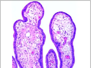
The gallbladder showed a markedly thickened wall but no gallstones (Figures 1 and 2). Grossly, the mucosa was edematous but otherwise appeared normal. However, on microscopic examination most of the mucosal folds had an expanded lamina pro-pria by edema, a mild predominantly acute inflam-matory infiltrate and numerous small blood vessels, some with recanalized thrombi (Figures 3 and 4).
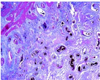
The subserosa was expanded and contained nume-rous medium sized and large veins with recent and old recanalized thrombi (Figures 5, 6 and 7). Only a
Figure 2. A mount section of the gallbladder shows that the wall is markedly thickened secondary to multiple vascular thromboses.
Figure 1. Gross photograph of the gallbladder with a mar-kedly thickened wall and thrombosed vessels.
Figure 4. A mucosal fold shows an edematous lamina pro-pria and a small blood vessel with a recanalized trombus (Masson´s trichrome stain, X100).
few arteries were occluded by recent thrombi. There was also marked proliferation of small and medium sized blood vessels mimicking a hemangioma (Figu-re 8). Likewise, numerous nerve trunks adjacent to the veins and arteries and hyperplasia of interstitial cells of Cajal between smooth muscle cells and in the lamina propria were also present (Figure 9).
DISCUSSION
Thrombohemorrhagic phenomena are relatively
common complications of ET.2-9 Mesenteric, splenic
and portal vein thrombosis with or without caver-nomatous transformation have been well
documen-ted in association with ET.3-4 However, to our
knowledge, large vein and arterial thrombosis in the Figure 5: Another mount section of the gallbladder shows
some dilated and thrombosed veins and arteries in the subse-rosa, as well as secondary vascular proliferation (Masson´s trichrome stain).
Figure 7. Subserosal vein with a recanalized thrombus
(H&E stain, X100).
Figure 6: Nerve trunk proliferation and recanalyzed
thrombi in subserosal veins. The artery shows a patent lumen
(Masson´s trichrome stain, X20). Figure 9. Hyperplasia of interstitial cells of Cajal between smooth muscle cells (positive CD117 immunostain, X120).
subserosa of the gallbladder causing acute cholecys-titis, as in our patient, has not been previously re-ported.
The marked proliferation of blood vessels in the
subserosa mimicking a hemangioma10 most likely
resulted from collateral circulation, a compensa-tory mechanism to minimize tissue damage due to ischemia, which might have been caused by blockage of venous and arterial circulation. Likewise, the ede-ma of the lamina propria and the mild acute inflam-matory infiltrate was also the result of the vein thrombosis. In addition to the newly formed blood vessels in the subserosa, the open cholecystectomy performed soon after the appearance of symptoms of acute cholecystitis contributed to the lack of necro-sis of the gallbladder wall.
We have no explanation for the hyperplasia of
in-terstitial cells of Cajal10,11 and the proliferation of
nerve trunks, a reactive change that has been docu-mented in inflammatory conditions of the
gall-bladder and extrahepatic bile ducts.12 Although
characteristic, venous and arterial thromboses in the wall of the gallbladder are not specific of ET; they have also been reported in the
catastro-phic antiphospholipid syndrome.13 Acalculous
chole-cystitis and arterial thromboses due to vasculitis has been reported in patients with systemic lupus
erythematosus and polyarteritis nodosa.14
Gangat, et al.6 reported a prevalence of 4% for
ab-dominal vein thrombosis among 460 consecutive pa-tients with ET. A possible risk factor for vein thrombosis in ET include elevated platelet counts. However, it has been recognized that vein thrombo-sis may occur at relatively low platelet counts. On the other hand, abdominal vein thrombosis has been
identified as a risk factor for poor survival.6
The true incidence of ET is unknown. However, it has been estimated that the overall incidence rate
varies from 0.6 to 2.5 per 100,000 person per year.7
The median age at diagnosis is 67 years with a fema-le preponderance, which is in contrast with our pa-tient. Of interest is that 52% of patients displayed no ET related symptoms and are diagnosed fortui-tously by routine platelet count.
JAK2 is an obligatory kinase primarily for the proliferation and differentiation of erythroid cells
and megakaryocytes.5 In recent years the JAK2
V617F mutation has been identified in patients with
ET.15 This mutation was initially thought to be
use-ful in the molecular diagnosis of ET. Unfortunately, it lacks specificity since it has also been detected in
polycythemia vera and primary myelofibrosis.5,9,15
Moreover, only 50% of patients with ET have the
mutation. Therefore, the diagnosis of ET should be based on clinicopathological features.
CONCLUSION
Acute cholecystitis caused by recent and old thrombosis predominantly of large subserosal veins of the gallbladder should be added to the list of ab-dominal vein and arterial thrombotic phenomena which are relatively common complications of ET. The diagnosis of acute cholecystitis due to thrombo-sis of subserosal veins and arteries of the gall-bladder should be suspected in patients with ET who develop intense right upper quadrant abdominal pain and the ultrasound reveals a thickened gall-bladder wall but no gallstones. It is important to keep in mind that some patients with ET may have low platelet counts in the peripheral blood.
REFERENCES
1. Thiele Y, Kvasnicka HM, Orazi A, et al. Essential thrombo-cythaemia. In: Swerdlow SH, Campo E, Harris NL, Jaffe ES, Pi-leri SA, Stein H, et al. (eds.). WHO classification of tumours of hematopoietic and lymphoid tissues IARC. Lyon; 2008. 2. Milovanovic M, Lotfi K, Lindahl T, et al. Platelet density
distribution in essential thrombocythemia. Pathophysiol Haemost Thromb 2010; 37: 35-42.
3. Cai XY, Zhou W, Hong DF, Cai XJ. A latent form of essen-tial thrombocythemia presenting as portal cavernoma.
World J Gastroenterol 2009; 15: 5368-70.
4. Rosu A, Searpe C, Sbarcea V, et al.A case of portal caver-noma associated essential thrombocythemia. J Gastroin-testin Liver Dis 2007; 16: 97-100.
5. Spivak JL. Thrombocytosis, polycythemia vera and JAK2 mutations. The phenotypic mimicry of chronic myeloproli-feration. Ann Intern Med 2010; 152: 300-6.
6. Gangat N, Wolanskyj AP, Tefferi A. Abdominal vein throm-bosis in essential thrombocythemia. Prevalence, clinical correlates, and prognostic implications. Eur J Haematol
2006; 77: 327-33.
7. Johansson P. Epidemiology of the myeloproliferative disor-ders polycythemia vera and essential thrombocythemia.
Semin Thromb Hemost 2006; 32: 171-3.
8. Petrides PE, Siegel F. Thrombotic complications in essen-tial thrombocythemia (ET). Clinical facts and biochemical riddles. Blood Cell Mol Dis 2006; 36: 379-84.
9. Landolfi R, Di Gennaro L, Falanga A. Thrombosis in myelo-proliferative disorders. Pathogenetic facts and specula-tion. Leukemia 2008; 22: 2020-8.
10. Albores-Saavedra J, Henson DE, Klimstra DS. Tumors of the gallbladder, extrahepatic bile ducts and ampulla of Vater. Atlas of Tumor Pathology. 3rd series. Fascicle 27. Was-hington, D.C.: Armed Forces Institute of Pathology; 2000. 11. Ortiz-Hidalgo C, de-Leon-Bojorge B, Albores-Saavedra J.
Stromal tumor of the gallbladder with phenotype of inters-titial cells of Cajal. A previously unrecognized neoplasm.
Am J Surg Pathol 2000; 24: 1420-3.
13. Dessailloud R, Papo T, Vaneeclo S, et al. Acalculous ische-mic gallbladder necrosis in the catastrophic antiphospholi-pid syndrome. Arthritis Rheum 1998; 41: 1318-20. 14. De-Leon-Bojorge B, Zaltzman-Girsevich S, Ortega-Salgado
A, et al. Thrombotic microangiopaty involving the gall-bladder as an unusual manifestation of systemic lupus
erythematosus and antiphospholipid syndrome. Case re-port and review of the literature. World J Gastroenterol
2006; 12: 7206-9.