FUNGAL PHYLOGENOMICS. A GLOBAL ANALYSIS OF FUNGAL
GENOMES AND THEIR EVOLUTION
Marina Marcet Houben
ISBN: 978-84-693-6431-4 Dipòsit Legal: T-1633-2010
ADVERTIMENT. La consulta d’aquesta tesi queda condicionada a l’acceptació de les següents
condicions d'ús: La difusió d’aquesta tesi per mitjà del servei TDX (www.tesisenxarxa.net) ha estat autoritzada pels titulars dels drets de propietat intel·lectual únicament per a usos privats emmarcats en activitats d’investigació i docència. No s’autoritza la seva reproducció amb finalitats de lucre ni la seva difusió i posada a disposició des d’un lloc aliè al servei TDX. No s’autoritza la presentació del seu contingut en una finestra o marc aliè a TDX (framing). Aquesta reserva de drets afecta tant al resum de presentació de la tesi com als seus continguts. En la utilització o cita de parts de la tesi és obligat indicar el nom de la persona autora.
ADVERTENCIA. La consulta de esta tesis queda condicionada a la aceptación de las siguientes
condiciones de uso: La difusión de esta tesis por medio del servicio TDR (www.tesisenred.net) ha sido autorizada por los titulares de los derechos de propiedad intelectual únicamente para usos privados enmarcados en actividades de investigación y docencia. No se autoriza su reproducción con finalidades de lucro ni su difusión y puesta a disposición desde un sitio ajeno al servicio TDR. No se autoriza la presentación de su contenido en una ventana o marco ajeno a TDR (framing). Esta reserva de derechos afecta tanto al resumen de presentación de la tesis como a sus contenidos. En la utilización o cita de partes de la tesis es obligado indicar el nombre de la persona autora.
WARNING. On having consulted this thesis you’re accepting the following use conditions:
Marina MARCETHOUBEN
F
UNGAL
P
HYLOGENOMICS
AGLOBAL ANALYSIS OF FUNGAL GENOMES AND THEIR EVOLUTION
D
OCTORALT
HESISSupervised by Dr. Juan Antonio Gabaldón Estevan
Department of Biochemistry and Biotechnology
July - 2010
Tarragona
Departament de Bioquímica i Biotecnologia c/ Marcel·lí Domingo s/n
Campus Sant Pere Sescelades 43007 Tarragona
Telèfon: 977 559 521 Fax: 977 558 232
I, Juan Antonio Gabaldón Estevan, group leader of the Comparative ge-nomics group, in the Department of Bioinformatics and Gege-nomics of the Center for Genomic regulation,
CERTIFY:
That the present study, entitled "Fungal Phylogenomics. A global analysis of fungal genomes and their evolution", presented by Marina Marcet Houben for the award of the degree of Doctor has been carried out under my supervision at the Department of Bioinformatics of the Centro de Investigación Principe Felipe, València, and the Department of Bioinformatics and Genomics of the Center for Genomic Regulation, Barcelona, and that it fulfils all the requirements to be eligible for the European Doctorate Label.
Barcelona, 1th of June 2010
A
GRAÏMENTSEstic davant de l’ordinador, com cada dia, escrivint el que serà l’última part d’aquesta tesis i casi no em puc creure que ja estigui. En aquest moment miro enrere i no puc evitar recordar moltíssims moments, bons i dolents, que han sigut part d’aquests anys d’aprenentatge. Ja us aviso per avançat que aquests agraïments seran llargs, ja que han sigut molts llocs on he treballat i molta gent a la que he conegut i a la que ara m’agradaria recordar.
Abans que res vull dedicar unes paraules al Toni, la persona que ha estat el meu supervisor, mentor i amic durant aquests últims anys. Hi han moltes característiques que fan del Toni un dels millors supervisors que un doctorant pot tenir. Des de la seva visió científica, al seu caràcter obert i alegre, i les seves idees i ganes d’innovar, ha sigut en moltíssimes ocasions una font d’inspiració i de suport. Moltes gràcies Toni per estar sempre aquí, per ajudar en els moments bons i especialment en els moments difícils, per donar-me a mi, i sospito que a tots els meus companys, la seguretat de saber que passi el que passi estas amb nosaltres i que aquesta feina és dels dos.
Tot i que en aquest volum només es recull la meva feina durant els últims tres anys, no crec que pugui fer uns agraïments complerts sense incloure els meus primers passos en aquest meravellós camp de la genòmica i la bioinformàtica. Us convido ara a que m’acompanyeu en un viatge en el temps, i recordeu amb mi aquells primers dies on fer un arbre filogenètic o un senzill script en perl era feina de dies i no de minuts.
Tarragona, Maig 2005- Agost 2006.
Si hi havia una cosa que tenia molt clara quan vaig acabar la carrera era que em volia dedicar a la bioinformàtica. Amb aquesta idea en ment va començar la meva aventura en el grup de genòmica comparativa de la URV. Moltes gràcies Santi per guiar-me durant tot el temps que vaig estar allí i fins i tot ara, quan ja fa anys que no estic a la URV, gràcies per fer-me de tutor i interessar-te per la meva feina.
Quan vaig entrar en el laboratori de bioinformàtica ja hi havia allí un grup molt ben avingut (i heu de reconèixer tots que una mica esbojarrat) que em va fer sentir benvinguda des del primer moment. Recordo moltíssims moments plens
Agraïments
de rialles i bon humor, si és que amb persones com l’Albert, el Pere, l’Esther i la Montserrat, com pots esperar altra cosa? Gràcies Pere per les moltes discussions que hem tingut durant els anys, i compte, que un dia acabaràs trencant una taula!! Albert, crec que només t’he de dir una cosa: "Visca el Barça!!". Montserrat, moltes gràcies per ser una gran amiga i meravellosa persona, en la que puc confiar i se que sempre estarà allí. Esther, no canviïs mai, la teva capacitat per alegrar el dia a la gent es increïble... i aixo ho escric cinc minuts després de que m’hagis fet riure en mig del laboratori amb un missatge pel gtalk!! També vull agrair a tots els demés membres del lab (o encara és labo?) de bioinfo, Gerard, Pep, Eduard, moltes gracies pels bons moments que vam passar. Laura, tot i que no vam arribar a coincidir en el laboratori, moltes gracies pels molts sopars, festes i demés que hem compartit. Ja em perdonareu els respectius per posar-vos aquí, amb els "frikis". Gràcies Lidia per la teva franquesa i amistat, i Jordi Puxeu per la teva paciència i simpatia.
També vull recordar aquí tots als demés membres del departament de bioquímica, no els anomenaré a tots, ja que segur que me’n deixaria. De totes maneres hi ha algunes persones amb les que he passat molt bons moments. Per exemple aquells que vaig conèixer abans fins i tot de començar el doctorat. Recordes Gemma totes aquelles pràctiques de bioquímica? Sembla que a tu et van agradar més que a mi!! Si més no, moltes gràcies pels molts anys d’amistat i companyerisme, un d’aquests dies hem de fer un dinar virtual, pero aquest cop amb web-cam, sinó no té gràcia! David! Seguramente la persona dentro de estos agradecimientos a la que más tiempo hace que conozco. Cuanto nos reimos durante la carrera! Y que lástima que ya no estuviera en Tarragona cuando empezaste el doctorado, pero bueno, igualmente creo que lo hemos pasado muy bien! Sabina, Helena, Anabel, Montse, i molts dels altres dels experimentals, moltes gràcies pels bons moments i ànims per aquells que acabeu!
Si en una cosa s’ha caracteritzat la meva tesis ha sigut en els canvis. El primer potser no va ser molt important. De la facultat de química a la facultat de medicina. De Tarragona a Reus. De bioinformàtica... bé, a bioinformàtica, que us pensaveu? Tot i que estava camuflada en un laboratori experimental!
Reus, Septembre 2006- Març 2007
Reus va ser el lloc on vaig estar menys temps, pero no per això vaig deixar de coneixer a gent important alli. Vull agrair als que van ser els meus directors, l’Anton Romeu i José Luís Paternain, l’oportunitat que em van donar de treballar allí. També agrair als membres del departament el temps que vaig passar amb ells.
Però si amb una persona vaig compartir, i segueixo compartint ara, una amistat especial va ser amb la Tania. Quantes hores vam passar les dues, en un laboratori preparat per allotjar com a minim sis persones, ella amb els
Agraïments
seus ratolins i jo amb el meu ordinador. Sempre pots esperar de la Tania que t’aparegui amb un comentari de lo més inversemblant, i no pots evitar pensar: en quin món he anat a parar ara? Moltes gràcies Tania per la teva companyia i els bons (i mals) moments que vam passar a Reus. També recordar a Espe i a la Marta, de fàrmaco.
Hi han moments en que penses: aquesta decisió pot canviar la meva vida. I en molts aspectes així va ser quan vaig deixar enrera Reus per anar a València. Però molts cops els canvis son bons i pots mirar enrera sense lamentar-te.
València, Abril 2007 - Agost 2008
Aquí es donde empezó el trabajo que presento en esta tesis. Durante mi estancia en el departamento de Bioinformática del CIPF conocí a muchísimas personas, y no es para menos dado el tamaño del departamento (debíamos oscilar entre los 20-30 bioinformáticos). Muchos son los que compartieron horas de trabajo conmigo y quiero agradecerles a todos el tiempo que pasé allí. Especialmente quiero recordar a Paco, que con su buen humor era capaz de alegrar el día a cualquiera y a Eva, por su caracter directo y simpático. También a la “invasión” de italianos: Giuseppe, Giulia, Daniela y Mariella, con los que compartí muchos momentos divertidos y a los que recuerdo con mucho cariño.
Y no, Salva y Jaime, no me olvido de vosotros (como podría?) pero puesto que me acompañasteis en la última etapa de mi pequeño viaje os invito ahora a venir a nuestro destino final.
Barcelona, Septembre 2008 - ?
És justament des d’aquest laboratori des d’on estic escrivint aquests agraiments, amb Salva i Diego treballant al meu darrera i el Toni sortint i entrant del seu despatx.
Muchas gracias Salva, por ser un buen compañero y maravilloso amigo. Recuerdas ese primer dia en Barcelona, tu en un hotel de mala muerte y yo en un piso vacío, cuando cogimos y nos fuimos a cenar por allí a pesar de que apenas nos conocíamos. Y las muchas veces que se ha repetido eso durante el último año y medio, donde hemos compartido trabajo y diversiones en miles de momentos.
Jaime, que pasa tronco? Que bien lo hemos pasado, tanto aquí como en Valencia. Muchas gracias por iniciarme en el mundo de los filomas y enseñarme muchas de las cosas que ahora se.
I des d’aquells primers moments el grup ha anat creixent: Javi, Diego (no se si llegarás a leer esto puesto que nos dejas en unos dias, pero muchas gracias por todo) i Leszek (thanks for all the fun moments, and for the ones that I hope will come in the future!).
Agraïments
I would also like to thank all the members of the FunPath consortium, some of which I have only met at the FunPath meetings (thanks, they were very interesting) and some of them which I know a little bit better. Thanks Karl, Walter and Tobby for the month I spent in Viena, it was very insightful.
A special thanks goes to Christophe and all the members of his group for the four months I spent in Paris, I had a great time even though it was freezing cold! I will always remember fondly how Christophe would come into the lab saying: “I have an idea”, it still makes me laugh even now. Thanks Ute for the many days we spent wandering around Paris, we’ll have to repeat it some time.
I aquí arribo, al final d’aquesta aventura anomenada tesis, i mirant enrera veig que a través de tots els canvis hi han constants, persones que sempre han estat amb mi, i és a ells als que dedico aquestes últimes paraules. A la meva germana, que encara que no ho sàpiga va ser un dels factors que em va animar a anar a València i que tot i la distància que ens separa ara, continua sent per mi una persona molt important. Al Vicent, que tot i a penes coneixe’m em va ajudar moltíssim durant tot el temps que vaig passar a València.
I molt especialment agraeixo als meus pares tot el seu suport i els seus consells. Per estar sempre allí quan els necessito, durant els moments bons i els dolents. Que al final ja ni s’inmutaven quan començava una conversa amb la frase: “Crec que en uns mesos me’n vaig a...”. Gràcies per l’interès que sempre heu mostrat en tot el que faig, encara que a vegades us soni a xino! Pares, moltes gràcies per tot, us estimo.
Cada amigo representa un mundo dentro de nosotros, un mundo que tal vez no habría nacido si no lo hubiéramos conocido.
Harcourt Brace
Als meus pares
C
ONTENTSTHESIS OUTLINE 1
I INTRODUCTION 3
1 The Fungi: overview of an essentially unexplored kingdom. 5
2 A brief introduction to phylogenomics 19
AIMS 26
II RESULTS 29
3 The tree versus the forest: The fungal tree of life and the
topological diversity within the yeast phylome 31
4 TreeKO: a duplication-aware algorithm for the comparison of
phylogenetic trees 49
5 Acquisition of prokaryotic genes by fungal genomes 61
6 Phylogenomics of the oxidative phosphorylation in fungi reveals extensive gene duplication followed by functional
divergence 69
7 FunPath Consortium 85
III SUMMARIZING DISCUSSION 97
8 Summarizing discussion 99
Contents
CONCLUSIONS 106
APPENDICES 109
A List of Publications 111
Bibliography 113
T
HESIS OUTLINEThis PhD thesis focusses on the use of phylogenomics to elucidate the evolution of fungal species. Here I outline the different parts that can be found in this volume.
The introductory part of the thesis is formed by Chapter 1 andChapter 2. In Chapter 1, an overview of the current knowledge on fungi is displayed with an emphasis on fungal evolution. The second chapter focuses on the main phylogenomic methods that have been applied throughout the present thesis. These two chapters are not essential for the understanding of the thesis but will provide sufficient background information for readers that are not familiar with some of the topics treated in this thesis.
Chapter 3 and Chapter 4 are focused on the analysis of species trees.
Chapter 3 presents an analysis about the robustness of the fungal species tree. This analysis is based on the use of extended phylogenetic methods to identify nodes in the fungal species tree that are in conflict with gene trees and that can therefore be source of errors in posterior analyses. The yeast phylome was used as the source of gene trees for this comparison. In addition, a comparison between two phylogeny-based orthology prediction methods (tree reconciliation and species overlap) is presented in this chapter. In Chapter 4, treeKO will be presented. TreeKO is a tree comparison program that, contrary to existing software, has been specifically designed for the comparison of multi-gene trees, and is therefore able to deal with pairs of trees that have different species content and that contain multiple duplications.
Chapter 5andChapter 6deal with two phylogenomic analyses concerning fungal species. Chapter 5 presents a high throughput search for inter-kingdom horizontal gene transfer events in 60 fully sequenced fungal genomes. We were able to identify more than 235 cases of horizontal gene transfer events, involving more than 700 genes, from prokaryotes to eukaryotes using an automatic pipeline. Additionally we observed that these events were unevenly distributed through the different fungal groups. Chapter 6 focuses on the evolution of the oxidative phosphorylation pathway in fungi. The analysis identified many duplications that occurred during the evolution of this metabolic pathway in fungi. Both chapters focus on events that may influence the gene tree topology
2 Outline
and render it different from the species tree.
Chapter 7 is dedicated to the work done within the FunPath Consortium.
The FunPath is european consortium created to elucidate the virulence mech-anisms of the human fungal pathogen, Candida glabrata. The chapter also contains some of the work done in collaboration with Christophe d’Enfert’s lab, which is part of the FunPath consortium, and will deal with the application of tree reconstruction methods using SNPs detected in several newly sequenced strains
ofCandida albicans.
The last chapter,Chapter 8, provides a summarizing discussion of the main points treated during this thesis.
Part I
Introduction
CHAPTER1
T
HEF
UNGI:
OVERVIEW OF AN ESSENTIALLYUNEXPLORED KINGDOM
.
Introduction
Fungi is one of the eukaryotic groups with the highest number of fully sequenced genomes. This kingdom comprises a large diversity of species, including mushrooms, yeasts, molds, rusts, smuts, puffballs, truffles, morsels and other less well-known organisms. The exact number of fungal species is unknown but estimates set this value around 1.5 million (Hawksworth, 1991) being 700.000 a conservative, lower estimate (Schmit and Mueller, 2007). Currently, only 70,000 species have been described, thus representing a mere 5-10% of the total diversity (Mueller and Schmit, 2007). While fossil records are scarce, data suggests that fungi already existed around 400 to 500 million years ago, though their exact origins may be more ancestral (Taylor and Berbee, 2006).
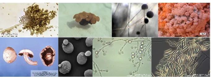
Fungi are ubiquitous and are able to colonize a broad diversity of habitats, in-cluding extreme environments such as deserts and deep sea sediments. Although a higher diversity of fungal species is found in tropical regions, the optimal growth conditions tend to vary across different species making fungi adaptable to numerous environments. As seen in figure 1.1, fungal species can adopt multiple morphologies.
One of the main roles performed by fungi within their ecosystems is that of decomposers of organic matter, thus being an important part of the carbon cycle. Fungi decompose biopolymers in order to absorb their constituents as nutrients. They usually live on their own food source and are able to grow into new sources when their first location becomes insufficient. When a food source is depleted, sporulation is triggered.
Many fungi display both sexual and asexual reproductive mechanisms. In sexual reproduction, sex determination is controlled by a small genomic region. In these regions specific gene combinations account for different mating types (Fraser and Heitman, 2003). Despite initial thoughts that the use of mating-type loci was limited to Ascomycota and Basidiomycota, recent studies have identified homologous regions to these loci in Zygomycota and Microsporidia (Idnurmet al., 2008; Leeet al., 2009). This supports the hypothesis that sex
6 Chapter 1
Figure 1.1: Example of fungal species. Images were taken from the Dr. Fungus webpage
(http://www.doctorfungus.org/). The images represent, at the top, from left to right:Aspergillus
flavus,Sclerotinia sclerotiorum,Rhizopus oryzaeandNeurospora crassa. At the bottom, from left
to right:Agaricus bisporus,Saccharomyces cerevisiae,Candida albicans,Fusarium oxysporum.
genes could have been a trait found in ancient fungi, which subsequently got lost in some lineages (Idnurmet al., 2008). Fungi can also reproduce asexually. Yeast species, for instance, preferentially reproduce through budding, and in some species the sexual cycle has never been observed. For instance,Candida glabrata
possesses all the genes that are known to be involved in sexual reproduction in yeast (Wonget al., 2003), but only asexual reproduction of this species has been observed so far.
Fungal species can adopt numerous different life styles. Many of the known fungi are free-living saprobes, which obtain nutrients and other molecules from dead or decaying organic material in woods, soils, leafs, dead animals or animal excrements. Other species are symbionts that live in direct association with plants, animals or prokaryotes (i.e. lichens, mycorrhizae or endophytes). A large number of pathogenic species can also be found within the fungi. They possess different mechanisms that enable them to infect numerous animals and plants. While not primarily pathogenic, other fungi are able to synthesise secondary metabolites that can be toxic or carcinogenic, this is the case of aflatoxins secreted by Aspergillus species. While some fungal species cause important economical damage due to their pathogenesis of humans and crops, some others are beneficial and are exploited by biotechnological industries. For instance,
Saccharomyces cerevisiaeandKluyveromyces lactisare used in the food industry
while other species, such asYarrowia lipolytica, are used in the production of antibiotics and other chemical compounds.
Some fungal species have been extensively studied and are considered model organisms. Probably the foremost species in this sense is the baker’s yeast,S.
cerevisiae. Indeed, this yeast is the first eukaryotic species for which a complete
nuclear genome sequence was obtained (Goffeauet al., 1996), and many efforts are still being done in order to fully comprehend the mechanisms that rule its biological processes. Other model organisms include the ascomycetes
Chapter 1 7
rospora crassa, Aspergillus nidulansandSchizosaccharomyces pombe, as well
as the basidiomycotaUstilago maydis. Results found in these species concerning genetics, physiological or morphological processes, are often extended to other fungal organisms, and are also the main source of functional information used for the annotation of newly sequenced genomes.
Fungal systematics: what do we know?
The advent of the genomic era led to many important discoveries regarding fungal evolution, which caused a complete revamping of the evolutionary history of these species. Morphological data was the main basis of taxonomic classification before the use of DNA and many of its postulates have been discarded since then. Probably the most relevant change was the discovery that fungi are more closely related to animals than to plants, to which they had been traditionally associated. The monophyly of fungi and metazoans within a clade known as opistokhonta is now strongly supported by both, phylogenetic and phylogenomic data (Wainrightet al., 1993; Brunset al., 1992; Langet al., 2002; Steenkampet al., 2006). Another important discovery was the fact that the group of Fungi, as described by morphological data was in fact polyphyletic and that the similarities in morphological characters found between the different groups were due to convergent evolution rather than because of a common ancestry. This led to the division between the “true fungi” and two other groups which included slime molds and oomycetes. Nowadays, the “true fungi” group holds the rank of Kingdom within the eukaryotic taxonomy, at the same level of animals and plants, being a sister group to the former. The phylogenetic position within the eukaryotes of the remaining two groups derived from the primary fungal group is not well defined yet, although slime molds are generally considered a sister branch of opisthokonts, forming a larger group known as unikonta (Keelinget al., 2005).
The fungal kingdom can be further divided into several groups. Probably the broadest division is the one that separates fungi into the early diverging groups and the dikarya. The so called early diverging fungi traditionally included Zygomycota and Chitridiomycota. Nowadays these two groups are thought to be polyphyletic. Two new groups were also included with the advent of the genomic era. Genomic data precipitated the inclusion of Microsporidia into the fungi (Keelinget al., 2000) and the separation of Glomeromycota from Zygomycota. The exact positions of these two last groups with respect to the others is still not clear. Microsporidia tend to adopt a basal position in phylogenetic analysis but it is unknown whether this results from methodological artifacts caused by their highly diverging sequences. The exact position of Glomeromycota in reference to Zygomycota and Dykaria is also still controversial (Redecker and Raab, 2006).
8 Chapter 1
Dikarya is the fungal subkingdom that contains Ascomycota and Basid-iomycota, the two best studied fungal groups. These have traditionally been considered sister groups based on their morphological characteristics (i.e. they have regularly septate hyphae and a dikaryotic stage in their life cycle), an association that has also been supported by numerous phylogenetic and phylogenomic studies (Jameset al., 2006).
Phylogenomics and the fungal species tree:
Since the start of the genomic era an effort has been made in order to derive fully resolved phylogenies, including as many of the known species as possible. Methods for tree concatenation and super-tree construction are nowadays used in an attempt to elucidate the evolution of a growing number of species (Wolf
et al., 2001; Snel et al., 2005). The fungal kingdom has been the focus of
many phylogenetic studies for several reasons. One of the most important is that this kingdom is the best sampled eukaryotic group in terms of fully sequenced genomes. The reason for this can be found in the inherently smaller size of their genomes and their importance for human health and industry. The large amount of available sequence data has opened the doors for the use of phylogenomics to address the reconstruction of the still elusive fungal species tree, or Fungal Tree of Life. The difficulties in rendering the fungal species tree reside in part in the lack of knowledge regarding most of the fungal diversity. As mentioned above, only 5% of the estimated total of fungal species has been described so far. An even smaller percentage has information on the usual genomic markers while around 100 genomes have been sequenced, which represents 0,006% of the estimated number of fungal species. In light of this, any attempt at taxonomic classification will be tampered with the knowledge that it is limited to, and biased towards, our current awareness on fungi, and may change as more species are discovered.
Several species trees have been reconstructed over the last few years and many changes have been introduced to the previous, morphology based, taxonomic classification. Some of the largest analyses, in terms of number of species, include the work of (Lutzoniet al., 2004) and (Jameset al., 2006). The first one reconstructed several species trees using different loci. The largest of these trees, with 558 taxa, is based on two loci (nucSSU and nucLSU rDNA), whereas the smallest, including 103 species, was built on information provided by four loci. Representatives for the main known fungal groups were included. The tree presented by James et al., is based on 6 loci and shows the phylogenetic relationships between 170 species. This tree, unlike the one presented by Lutzoni et al., is fully resolved and offers insights into some problematic nodes such as the position of microsporidia or the lack of monophyly in Chitrids. The use of
Chapter 1 9
completely sequenced genomes in phylogenetic reconstructions allows for the use of larger amounts of data from a limited number of species which is often biased towards Ascomycotina. Even so, several large phylogenies have been reconstructed using data from fully sequenced genomes (Marcet-Houben and Gabaldón, 2009; Wanget al., 2009; Fitzpatricket al., 2006). These trees, while slightly different in some nodes, mostly support a single topology (see chapter 3).
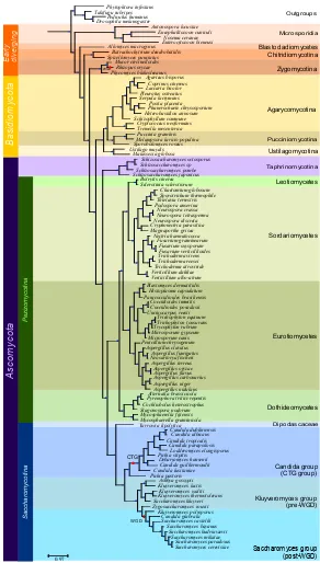
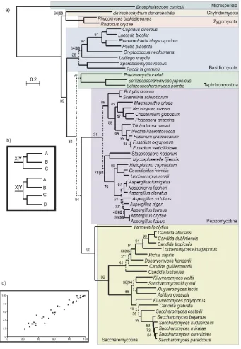
In Figure 1.2, we present the largest tree that has been reconstructed so far based on phylogenomics data. The tree represents the evolution of 102 different fungal species. It was built by concatenating 47 wide-spread proteins that displayed one-to-one orthology relationships and that were present in at least 90 species. Four outgroups were included: Takifugu rubripes, Pediculus
humanus,Drosophila melanogaster andPhytophtora infestans. The outgroups
were selected by searching for the candidate proteins in a database made of 206 completely sequenced eukaryotes using a Smith-Waterman search (Altschul
et al., 1997), we chose those outgroups that contained hits in most proteins and
minimized the number of duplications.
The resulting tree is similar to the ones published before (Fitzpatricket al., 2006; Marcet-Houben and Gabaldón, 2009; Robbertseet al., 2006; Wanget al., 2009). Microsporidia, represented by four species here, occupy a basal position in fungi with a long branch that shows how divergent these species are compared to the other fungal species. Blastocladiomycetes, which was untill recently considered a Chitrid, does appear as an independent group in our tree, having diverged before the speciation of Chitrids. Even so, the low number of fully sequenced species representing these groups makes it difficult to infer any robust conclusions on their evolution. This is reflected in the many clades that result from the analysis performed by James et al. Jameset al. (2006) where Chitrids, Zygomycota, Blastocladiales and Microsporidia do not present clear monophyletic separations. We hope that in the future, new genomes will be sequenced within the early divergent fungi and that the relationships between them will be clearly elucidated.
There are three main groups recognized in Basidiomycota: Pucciniomy-cotina, Ustilagomycotina and AgaromyPucciniomy-cotina, though their exact phylogenetic classification is still uncertain. The most supported hypothesis places Agaromy-cotina and UstilagomyAgaromy-cotina as sister groups (Lutzoniet al., 2004; Jameset al., 2006; Marcet-Houben and Gabaldón, 2009). In contrast with this hypothesis, Ustilagomycotina are basal in Basidiomycota in our current tree. While aLRT support is high for this node, in chapter 3 we show that this position has a low phylome support and that this lack of support is often translated in difficulties in establishing unequivocally how a group of species has evolved.
Another debated distribution is that of the four Pezizomycotina groups. It is well established that Leotiomycetes and Sordariomycetes are sister groups
10 Chapter 1 Saccharomyces group (post-WGD) Kluyveromyces group (pre-WGD) Candida group (CTG group) Dipodascaceae Eurotiomycetes Dothideomycetes Sordariomycetes Leotiomycetes Taphrinomycotina Pucciniomycotina Agarycomycotina Ustilagomycotina Zygomycotina Chitridiomycotina Blastocladiomycetes Microsporidia Outgroups P ez iz om yc ot in a S ac ch ar om yc ot in a A sc o m yc o ta B as id io m yc ot a E ar ly di ve rg in g WGD CTG 0.97 WGD Phytophtora infestans Takifugu rubripes Pediculus humanus Drosophila melanogaster Antonospora locustae Encephalitozoon cuniculi Nosema ceranae Enterocytozoon bieneusi Allomyces macrogynus Batrachochytrium dendrobatidis Spizellomyces punctatus Mucor circinelloides Rhizopus oryzae Phycomyces blakesleeanus Agaricus bisporus Coprinus cinereus Laccaria bicolor Pleurotus ostreatus Serpula lacrymans Postia placenta Phanerochaete chrysosporium Heterobasidion annosum Schizophyllum commune Cryptococcus neoformans Tremella mesenterica Puccinia graminis Melampsora laricis-populina Sporobolomyces roseus Ustilago maydis Malassezia globosa Schizosaccharomyces octosporus Schizosaccharomyces sp Schizosaccharomyces pombe Schizosaccharomyces japonicus Botrytis cinerea Sclerotinia sclerotiorum Chaetomium globosum Sporotrichum thermophile Thielavia terrestris Podospora anserina Neurospora crassa Neurospora tetrasperma Neurospora discreta Cryphonectria parasitica Magnaporthe grisea Nectria haematococca Fusarium graminearum Fusarium oxysporum Fusarium verticillioides Trichoderma virens Trichoderma reesei Trichoderma atroviride Verticillium dahliae Verticillium albo-atrum Blastomyces dermatitidis Histoplasma capsulatum Paracoccidioides brasiliensis Coccidioides immitis Coccidioides posadasii Uncinocarpus reesii Trichophyton equinum Trichophyton tonsurans Trycophyton rubrum Microsporum gypseum Microsporum canis Penicillium chrysogenum Aspergillus clavatus Aspergillus fumigatus Neosartorya fischeri Aspergillus terreus Aspergillus oryzae Aspergillus flavus Aspergillus carbonarius Aspergillus niger Aspergillus nidulans Alternatia brassicicola Pyrenophora tritici-repentis Cochliobolus heterostrophus Stagonospora nodorum Mycosphaerella fijiensis Mycosphaerella graminicola Yarrowia lipolytica Candida dubliniensis Candida albicans Candida tropicalis Candida parapsilosis Lodderomyces elongisporus Pichia stipitis Debaryomyces hansenii Candida guilliermondii Candida lusitaniae Pichia pastoris Ashbya gossypii Kluyveromyces lactis Kluyveromyces waltii Kluyveromyces thermotolerans Saccharomyces kluyveri Zygosaccharomyces rouxii Kluyveromyces polysporus Candida glabrata Saccharomyces castellii Saccharomyces bayanus Saccharomyces kudriavzevii Saccharomyces mikatae Saccharomyces paradoxus Saccharomyces cerevisiae
Chapter 1 11
but no consensus has been reached regarding the positions of Eurotiomycetes and Dothideomycetes. James et al. for instance presented Dothideomycetes species in a basal position for Pezizomycotina. In a concatenated tree presented by Fitzpatrick et al. (Fitzpatrick et al., 2006) Eurotiomycetes were basal to Pezizomycotina while trees reconstructed using the super tree methodology presented Dothideomycetes and Eurotiomycetes as sister groups. This last distribution has also been found in other trees (Wang et al., 2009; Marcet-Houben and Gabaldón, 2009). In our current tree, the first hypothesis, in which Dothideomycetes is the first diverging group of species, is supported. In recent years numerous genomes belonging to Dothideomycetes species have been sequenced, increasing the representation of organisms within this clade from one to six, this may confer more reliability to the phylogeny represented in figure 1.2 as it is made with a larger representation of species within the clade of interest. Even so, sequencing members of other, unrepresented, clades in Pezizomycotina may offer further resolution to this matter.
Yet another discussed node in the phylogeny of fungi is the relative positions
ofC. glabrata and S. castellii. We will address this matter further in chapter
4, but the distribution found in this tree is in line with most other phylogenetic methods whereC. glabrataappears to have diverged from the Saccharomyces clade beforeS. castellii, which is in direct contradiction to data inferred from gene order conservation (Gordonet al., 2009).
Completely sequenced fungi
The early diverging species
Very few species of fungi belonging to the early diverging groups, or lower fungi, have been sequenced so far. For several years, the only available genomes have been the microsporidian Encephalitozoon cuniculi, the two zygomycota Rhizopus oryzae and Phycomyces blakesleeanus and the chitrid,
Batrachochytrium dendrobatidis. Interest in the exact phylogenetic location
of Microsporidia and the project centered on the Origin of Multicellularity conducted by the Broad Institute (http://www.broadinstitute.org/) have attracted the attention of the scientific community to this group of species, resulting in a slow but steady growth in the number of sequenced genomes available.
Microsporidia are obligate, intracellular parasites that affect a wide variety of animals, ciliates and some apicomplexa. They lack typical mitochondria which is one of the reasons they were thought to be ancient eukaryotes pre-dating the mitochondrial endosymbiosis. However, it is now recognized that they possess highly derived forms of mitochondria (mitosomes), which are the result of secondary adaptations to anaerobic environments (Gabaldón and Huynen, 2004). Their adaptation to parasitic life-style has difficulted the elucidation of their
12 Chapter 1
phylogenetic position. Two studies involving alpha- and beta-tubulins placed the Microsporidia within the Fungal group (Edlind et al., 1996; Keeling and Doolittle, 1996). Since then, additional evidence has been found that supports this relationship (Keeling et al., 2000). The first, and for a long time only, complete genome available for this group is that ofE. cuniculi(Katinka et al., 2001), an unicellular parasite that infects numeruos animals. Several additional microsporidian genomes have been published recently (Cornman et al., 2009; Corradi et al., 2009). The scientific community hopes that the additional information will finally resolve the exact phylogenetic location of this interesting group, whether it is within any of the existing fungal groups (Keelinget al., 2000; Jameset al., 2006) or as a sister clade to fungi (Tanabeet al., 2005). Currently in our lab we are undertaking a phylogenomic analysis to address this issue.
The two groups that have traditionally been regarded as early diverging within the fungal kingdom are the Chitrids and the Zygomycetes. Chitrids have very characteristic morphological traits. For instance they are mainly aquatic and possess flagelated zoospores. From an economic standpoint, they are not very interesting, which results in a lack of financing towards genome projects. For a long time the only genome sequenced was that ofB. dendrobatidis. This species has gained importance in recent years due to its role in the eradication of amphibian populations (Poundset al., 2006). Two other groups are part of the early diverging fungi. The Glomales, which were traditionally considered part of the Zygomycota, are now thought to have evolved independently. Currently, they are considered to be more closely related to dikarya and are given the category of a new phylum: Glomeromycota, although some doubts remain about this assignment. Some of the most well known Zygomycota are molds that can be found in spoiling bread or fruits. The genomes of three species are currently available. R. oryzae, is the principal causative agent of mucoromycosis, which is lethal in immunocompromised hosts (Roden et al., 2005; Ma et al., 2009).
Mucor circinelloides, closely related toR. oryzae, has recently been sequenced
due to the interest generated to its use in the biotechnological industry. This species is able to generate large quantities of lipids that can be easily converted into biodiesel and may one day replace the use of plant oils for the production of this compound. In contrast, the interest inP. blakesleeanusis centered on its ability to respond to environmental stimulus like light, chemicals, touch, gravity and others. In this sense this species has served as a model organism for the investigation of genes that regulate responses to stimulus (Idnurmet al., 2006).
Basidiomycota
Basidiomycota comprise nearly 40% of the described fungi. Within this group we find those species that are able to produce mushrooms (fruiting bodies) as well as numerous plant symbionts. Mushroom producing fungi are probably
Chapter 1 13
among the most well known representatives of this kingdom. While the genomic sequence of the common mushroom, Agaricus bisporus, has only recently been made available at the Joint Genome Institute (http://www.jgi.doe.gov/), the sequences of other mushrooms have been available for several years. For instance, Coprinus cinereus is a saprophytic mushroom with a limited edible value, but which has been used as a model for sexual reproduction in Basidiomycota, with the aim of extrapolating the results found in this mushroom to other, more economically important, species (Kües, 2000; Kamada, 2002). On the other hand, Schizophyllum commune, belongs to the same group as the common mushroom and has been the focus of much research due to its unusual sexuality pattern, as it contains thousands of different mating types (Fowleret al., 2001). This species is one of the few known filamentous basidiomycetes that cause infections in immunocompromised humans. It can also play an important role in bioremediation by uptaking minerals, for instance uranium and cadmium (Mertenet al., 2004).
The economic importance of Basidiomycota resides not only in the com-mercialization and consumption of mushrooms, but also in their ability to produce economically important products. Numerous pathogenic species can also be found within this group. Cryptococcus neoformansis an opportunistic pathogen that can cause central nervous system and pulmonary diseases in inmunocompromised hosts and the yeastMalassezia globosahas been associated with dandruff and can cause more serious skin problems. In plants, the Ustilagomycotina, are able to infect some of the most important crops including corn, barley, wheat and sugarcane (Bakkeren et al., 2008). Ustilago maydis, the corn smut pathogen, has been used as a model organism for many years in molecular plant pathogeny research. Moreover, it has been suggested that U.
maydiscould be a good model to study animal basic cell processes when baker
yeast is unable to cover them. For example, part of the DNA repair machinery in animals has no homologs in yeast, but it has been found inU. maydis(Steinberg and Perez-Martin, 2008; Münsterkötter and Steinberg, 2007).
Ascomycota
Three monophyletic groups have been described within Ascomycota: Taphri-nomycotina, Pezizomycotina and Saccharomycotina. Pezizomycotina and Sac-charomycotina are very well supported sister groups and together they represent more than 70% of the total number of sequenced species. The phylogenetic position and monophyly of Taphrinomycotina, is still under discussion (Kuramae
et al., 2006a). The clade was first defined based on the phylogenetic
reconstruc-tion using rRNA (Nishida and Sugiyama, 1993), and ended up grouping a very diverse set of species, including Schizosaccharomyces and Pneumocystis. The statistical support of the group was not very high and phylogenies derived from
14 Chapter 1
more than one gene have been contradictory. Most phylogenomics analyzes using nuclear proteins place Schizosaccharomyces as basal to Pezizomycotina and Saccharomycotina. On the other hand, phylogenies using mitochondrial sequences, tend to group Schizosaccharomyces within the Saccharomycotina. In a recently published paper, Liu and collaborators (Liu et al., 2009) pro-vide support for the monophyly of Taphrinomycotina, and place it as basal to Saccharomycotina and Pezizomycotina. Their analysis of mitochondrial sequences also showed that the addition of new Taphrinomycotina species to their trees lowered the support of the grouping of Schizosaccharomyces within the Saccharomycotina.
Currently four species belonging to this group have been sequenced. The yeast Schizosaccharomyces pombe has been used for many years as a model species for the study of cell cycle and cell biology. Schizosaccharomyces
japonicusandSchizosaccharomyces octosporusare two very close relatives of
S. pombe, which were sequenced with the aim of furthering the knowledge
on this model species. On the other hand, Pneumocystis cariniiis a pathogen that is found in the lungs of mammals and can cause serious pneumonia to inmunocompromised people.
Pezizomycotina is one of the largest groups of fungi both in terms of described species, more than 32,000, and in terms of the number of completely sequenced genomes. Nowadays, 76 genomes plus several different strains for some species have been sequenced. Pezizomycotina are mainly haploid and filamentous, though some species are known to be dimorphic. They live in a large diversity of habitats and ecological niches (Spatafora et al., 2006). The phylogenetic relationship of the classes found within Pezizomycotina remains unresolved. Only four of these classes have representatives with fully sequenced species: Leotiomycetes, Sordariomycetes, Eurotiomycetes and Dothideomycetes. One of the few well established phylogenetic relationships is the association of Leotiomycetes and Sordariomycetes as sister groups.
The Leotiomycetes is a diverse group from a morphological standpoint, with great differences in fruiting bodies, which range from the large, brightly colored one found in Cyttaria to the small, dark dots produced by Lophodermium (Wang et al., 2006b). One of the main difficulties in classifying the species within this group is the lack of a clear connection between anamorphs and teleomorphs. Many fungi are known by their teleomorphic stage but the anamorph is not known while in some cases the opposite is true (Wanget al., 2006a). Botrytis cinereaandSclerotinia scleriotiorumare both fully sequenced plant pathogens.S. scleriotiorumhas a large range of hosts, up to 400, including many economically important crops, like soybean and pea, or oil seeds, like canola and sunflower (Hegedus and Rimmer, 2005).
In contrast to Leotiomycetes, its sister group, Sordariomycetes, is one of the largest classes within the Ascomycota with 3,000 known species and 18
Chapter 1 15
completely sequenced species. Numerous well known pathogenic species can be found within this group such as the plant pathogens Magnaporthe grisea
and Fusarium or the human pathogens Chaetomium globosum and Nectria
haematococca. In addition,C. globosumcan produce mycotoxins that are lethal
to animals.
Sequencing efforts during the last few years seem to have been centered around the Dothideomycetes. All the genomes sequenced so far belong to plant pathogenic species. For instanceStagonospora nodoruminfects wheat and other related cereals (Haneet al., 2007). Many phylogenetic trees only include this representative of the Dothideomycetes. The uncertainty present in those trees has shown that more data is needed in order to fully understand the position of this group within the Pezizomycotina. More sequences are nowadays available and yet the uncertainty remains, indicating a need for sequencing species of the remaining classes of Pezizomycotina instead of increasing the sampling of already represented groups.
The last group of Pezizomycotina with sequenced species is the Euro-tiomycetes. Sequencing efforts for this group have been clearly centered around the Aspergillus species. A total of eight different Aspergillus have been sequenced, ranging from the human pathogens Aspergillus fumigatus
and Aspergillus flavus to the rarely pathogenic, but economically interesting,
Aspergillus nigerandAspergillus oryzae (Denninget al., 2002)which are used
in industry to produce cytric acid and other proteins or to produce fermented foods like sake or miso. Also enclosed in this group is the saprobeAspergillus
nidulans, who has long been one of the model organisms and was thought to
be the only Aspergillus species with a sexual cycle. Recent research has shown that other Aspergillus species, like A. fumigatus, also can reproduce sexually (O’Gormanet al., 2009).
The last large group of Ascomycota species is the Saccharomycotina, a monophyletic group that mostly comprises yeast species. They are usually found in damp or wet habitats that are rich in organic materials. They are very important in many biotechnological areas, for instance, the baker’s yeast,S. cerevisiae, is used in bread, beer and wine making. Numerous pathogens can also be found within this group, for instance the human pathogenCandida albicansor the plant pathogen,Ashbya gossypii. Many species have been described as being involved with arthropods in which the yeasts provide vitamins and enzymes in exchange of an efficient habitat.S. cerevisiaehas been often the focus of numerous studies because the basic mechanisms of DNA replication, chromosomal recombination, cell division, gene expression, and metabolism are generally conserved between yeast and higher eukaryotes (Castrillo and Oliver, 2004).
More than 20 complete genomes are available for this group. The most divergent species included is Yarrowia lipolytica, which is often used as an outgroup for studies based on Saccharomyces.Y. lipolyticacan be found in
16 Chapter 1
rich environments, it is mainly non-pathogenic and it has been used in industry for the production of citric acid (Casaregolaet al., 2000).
In this overview we will divide the remaining species of this group in three different sub-groups that are often distinguished in literature: the Candida group, the Kluyveromyces group and the Saccharomyces group. The first one, also known as CTG group, comprises all the species that are closely related to C.
albicans. The second one includes the three Kluyveromyces species in addition
toA. gossypiiandSaccharomyces kluyveri. This group is often also referred to as
the Pre-whole genome duplication group (Pre-WGD) as it is the group of species that diverged from Saccharomyces just before the whole genome duplication (WGD) (Wolfe and Shields, 1997). The last group is formed by the species that underwent a WGD, they are closely related toS. cerevisiaeand therefore have been the focus of numerous studies.
Candida or CTG group: One of the main distinctive features of the Candida
group, is the change in the codification of the CTG codon from leucine to serine. This change occurred at the base of the group formed by the candidas and is characteristic for all its members. Candida species are opportunistic pathogens that can cause important infections in inmunocompromised hosts. C.
albicans is the main causative agent for candidiasis. The effect of antifungals
that specifically target this species has caused an emergence of infections caused by non-albicans candidas in recent years. One example of this are the infections produced byC. glabrata, which is much more closely related to S. cerevisiae
than toC. albicans (see chapter 7). Also, some Candidas are rarely observed as pathogens, but under certain conditions they can be found as the prevalent strains. For instance, Candida guilliermondii, while not usually found causing infections, is considered the main cause of fungemia in cancer patients. While mostly known because of their pathogenesis, some of the species within the Candida group are non-pathogenic. Debaryomyces hansenii for instance is a marine yeast that can tolerate high salinity levels. Other non-pathogenic species within the Candida group areLodderomyces elongisporus and Pichia stipitis, which is known for its ability to ferment xylose.
Kluyveromyces group: Probably the two most well studied species within
this group are Ashbya gossypii and Kluyveromyces lactis. The first one is a plant pathogen while the second one is a saprobe which is often found in lactic products as it is able to grow on lactose. Little is known about the ecology of the three other species that conform this group (Kluyveromyces
waltii, Saccharomyces kluyveri and Kluyveromyces thermotolerans) and they
were sequenced mainly due to their proximity to S. cerevisiae. This group of species has often been used as a reference to compare to Saccharomyces genomes as they are thought to retain most of the genomic structure the species had before undergoing the whole genome duplication.
Saccharomyces clade: In nature Saccharomyces species have been found in
Chapter 1 17
the bark of some trees and in fermenting fruits or other high sugar environments. They have a high tolerance to alcohol, many of them grow anaerobically and are able to ferment glucose to alcohol. Due to the importance of S. cerevisiae
as a model species, the genomes of several closely related species have been sequenced. Probably one of the most interesting species within this group isC.
glabratawhich, unlike the others, is an opportunistic human pathogen that can
cause candidiasis in inmunocompromised hosts. The fact that this species is so closely related to numerous non-pathogenic species, coupled by the differences observed in the infection mechanism when compared toC. albicans, points to the fact thatC. glabratadeveloped its pathogenesis independently. One of the main phylogenetic questions that remains unresolved within this group is the relative position of C. glabrata andS. castellii (see chapter 4). Phylogenetic analysis usually shows a poorly supported topology in which C. glabratawas the first species to diverge from the main Saccharomyces group, but results based on synteny support that the first species to diverge wasS. castellii (Gordon et al., 2009).
Final remarks
The fungal kingdom presents a high diversity and we have barely started to discover the many entities that are part of it. Even though current estimates show that we are only aware of about 5% of the fungi, the many differences that have already been shown in terms of morphology, ecology and adaptation to different environments is amazing. The fungal kingdom is, without doubt, one of the most interesting groups of species that still remain mostly undiscovered. Fungal species will play a very important role in the genomic era. The conformation of their genome, with enough simplicity to facilitate its investiagtion but also with enough complexity to find numerous parallels to more complex organisms, conforms one of the main attractions for their use in experimental studies. The current availability of more than 100 completely sequenced genomes has also opened the doors for comparative genomics studies that will help understand how these organisms have been able to adapt to so many different environments. One of the main questions that still remains is which is the exact topology of the fungal species tree. Hopefully, our growing knowledge will help clarify those points in the fungal species tree that still remain unresolved.
CHAPTER2
A
BRIEF INTRODUCTION TO PHYLOGENOMICSFrom phylogenetics to phylogenomics
Phylogenetics aims at establishing the evolutionary relationships between or-ganisms. Since the publication of “The origin of species” (Darwin, 1859), species evolution has been represented in the form of a hierarchical tree where extant species are located at the tips of the branches whereas bifurcating nodes represent their common ancestors. Initially, phylogenies were based on morphological data, an approach that is highly susceptible to homoplasy effects. The increasing availability of biological sequences has facilitated the replacement of morphological characters as the basis for phylogenetic reconstruction. The first trees based on sequences were built using single molecular markers such as 16sRNA (Zuckerkandl and Pauling, 1965) but it was soon obvious that different markers may produce different phylogenies and, therefore, they were not suitable for establishing the evolutionary relationship between species. The possibility of combining several different markers to reconstruct a single species tree is a straightforward solution, which is now favored with the growing availability of molecular data.
Phylogenomics represents the intersection between the fields of evolution and genomics (Eisen and Fraser, 2003). Increasingly larger groups of genes are treated together in an attempt to gain a broader view of the events that have shaped the evolution of species. This incresing amount of data will enforce another way of working, with automation being a key factor in any phylogenomics study. Automatic pipelines have been designed to generate high quality phylogenetic trees with minimal manual imput. In addition, programs also need to be designed to automatically process the information present in phylogenetic trees, since the large amount of data makes manual processing unfeasible.
Some examples of phylogenomics studies comprise the construction of species trees using multiple genes (chapter 3), the establishment of orthology and paralogy relationships between genes (chapter 3) or the use of phylogenetic trees to establish evolutionary events such as duplications (chapter 6) or horizontal genes transfers (chapter 5).
20 Chapter 2
Phylogenetic reconstruction
Phylogenetic pipeline
Often, the basis of phylogenomics studies consists of phylogenetic analysis at a large scale, both in terms of number of trees and in terms of the number of species included. In order to cope with the increasing amount of data, pipelines have been designed that are able to reconstruct large amounts of phylogenetic trees (Huerta-Cepaset al., 2007; Hubbardet al., 2007; Gabaldónet al., 2008). A balance between accuracy and speed has to be achieved in order to produce accurate trees at a reasonable speed. Most phylogenetic pipelines can be divided into three steps: selection of putative homologous sequences, reconstruction of a reliable multiple sequence alignment and reconstruction of a phylogenetic tree that represents the evolutionary relationships of the sequences involved.
The pipeline we have used throughout this thesis was first applied in the reconstruction of the human phylome (Huerta-Cepas et al., 2007). First a Smith-Waterman search (Smith and Waterman, 1981) is performed using an initial protein which will be referred to as seed protein. This search is performed against a locally installed database that contains either a downloaded database (for instance from NCBI (http://www.ncbi.nlm.nih.gov/) or Uniprot (http://www.uniprot.org/)) or a manually created database. The results are then filtered, and only those hits that have an e-value below a given threshold and have a long enough continuous aligned region are taken as homologs. In the second step Muscle v3.6 (Edgar, 2004) is used to make the multiple sequence alignment. This alignment is trimmed using trimAl (Capella-Gutiérrez et al., 2009) in order to remove poorly aligned regions. PhyML (Guindon and Gascuel, 2003) is then used to derive the phylogenetic trees. In this pipeline a NJ tree is first reconstructed using BIONJ (Gascuel, 1997) as implemented in PhyML, then this tree is used as starting point to reconstruct the maximum likelihood trees. Up to four different evolutionary models are used (typically JTT, WAG, VT and Blosum62). The evolutionary model best fitting the data is then determined by comparing all the likelihoods according to the AIC criterion (Akaike, 1973).
Phylome reconstruction and phylomeDB.
While the described pipeline could be applied to one single protein family it was specifically designed to reconstruct phylomes. Phylomes are defined as the complete collection of phylogenetic trees for each gene in a genome (Sicheritz-Pontén and Andersson, 2001). They can be used for numerous studies including the prediction of orthology and paralogy relationships (Huerta-Cepas et al., 2007; Marcet-Houben and Gabaldón, 2009), the tree based inference of function for newly sequenced genomes (Consortium, 2010) and to search for poorly
Chapter 2 21
supported nodes in a species tree (Marcet-Houben and Gabaldón, 2009). The reconstruction of phylomes produces a large amount of data. In order to make this information available to the whole community, phylomeDB (www.phylomedb.org) (Huerta-Cepas et al., 2008) was designed in our lab. PhylomeDB provides users with the opportunity to actively visualize phyloge-netic trees or to download all the data for more intensive use. PhylomeDB has several browsing methods which include the search of proteins using Ids and the possibility to perform a blast search against the proteomes stored in the database. Several external Ids are covered in the database, easing the access to the data.
While the number of public phylomes is still relatively small (12), the number of proteomes included in the database is much larger (945). Even though proteomes not used as seed are not fully covered, many of their proteins can still be present in other phylogenetic trees. The work performed during this thesis has contributed to the reconstruction of 7 phylomes (four of Saccharomyces
cerevisiae, one of Candida glabrata, one of Candida albicans and one of
Schistosoma mansoni).
Combining information from various genes to derive a single phy-logeny
Phylogenetic trees based on a single gene family are useful in many areas. However, they present the drawback of displaying different topologies depending on the family used to reconstruct the tree (Castresana, 2007). This situation is especially problematic when the aim of the analysis is to define a single phylogeny that represents the evolutionary relationships among a group of species, the so called species tree. A possible solution to this problem is to integrate the phylogenetic information from various genes into a single tree (Altekar et al., 2004). We can distinguish between two kind of approaches. The first kind comprises those methods that do not use sequence data to infer the tree but rather are based on genome characteristics such as gene content or gene order (Snelet al., 2005). The second kind directly uses sequences, either to create a super alignment (Brownet al., 2001) from which to derive the tree, or to combine different trees created from individual genes into a single super-tree (Bininda-Emonds, 2004).
Here we will use the gene concatenation method as it is one of the most cited in the literature and is considered highly reliable (Delsucet al., 2005). It must be noted that comparisons between super-tree approaches and concatenations have not detected large differences in terms of topologies (Dutilhet al., 2007; Fitzpatricket al., 2006). The rationale behind gene concatenation is that genes in a genome have undergone the same evolutionary history. Therefore, if taken together, they should be able to amplify the phylogenetic signal they share. In order to use gene concatenation, each gene should, ideally, be present in single
22 Chapter 2
copy in all species considered. As a result, the number of genes that can be used in such analyses decreases as the number of species included grows. To attenuate this effect, genes absent in a few genomes can be included by introducing gaps in the missing species and methods to select one gene from few recent paralogs can be applied.
Major drawbacks of this method include a low number of appropriate genes when large groups of species are considered. Another concern is related to data heterogeneity. In this approach all genes are treated together under the same models, assuming homogeneity in the data, which is known to be untrue (Bull
et al., 1993). Additionally, events such as lineage sorting or horizontal gene
transfers can led to a different evolutionary history for a given gene. This method is very sensitive to such cases and care should be taken to exclude genes that are susceptible to have undergone such events (Wolfet al., 2001).
Interpretation of large-scale datasets
Together with the high computational costs associated to large scale phylo-genetics reconstruction, the biological interpretation of extensive collections of trees and alignments represents another important challenge in the field of phylogenomics. Inspection of thousands of phylogenies cannot be addressed manually and, therefore, automatic methods and algorithms for the interpretation of phylogenies are necessary. Here we give some examples of studies that can be undertaken and which have been used throughout the present thesis.
Phylogeny-based orthology prediction
Orthologs are genes that diverged after a speciation event (Fitch, 1970). When compared to paralogs, which evolved through a duplication event, orthologous pairs of genes show a higher tendency to perform the same function. This is the reason why predicting them correctly has become so important (Gabaldón, 2005).
There are many methods that predict whether two genes are orthologous to each other, most of them based on pair-wise similarities (Koonin, 2005). However, since the original definition of orthology is an evolutionary one (Fitch, 1970), a prediction based on phylogeny seems to be more appropriate (Gabaldón, 2008). There are two main approaches to derive orthology relationships from phylogenetic trees, namely reconciliation and species-overlap method. Reconciliation methods use a species tree as reference (Page and Charleston, 1997). When comparing a gene tree with the species tree, mismatching nodes are identified and considered to be duplication events which were subsequently followed by the necessary amount of gene loss to explain the resulting phylogeny. This approximation will render correct orthology predictions if the assumption
Chapter 2 23
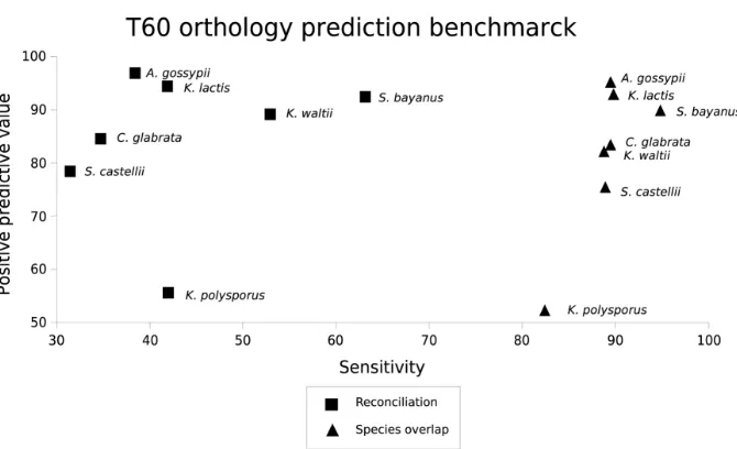
that the gene and species trees are correct holds. An alternative approach is the so-called species-overlap method. In this case duplication nodes are only considered as such when their branches have shared species. While simple, this method performs well (Huerta-Cepaset al., 2007) and has the advantage that the only information that is needed from the species phylogeny is the one used to root the tree. In a comparison between the two methods we showed that the species overlap algorithm had a higher sensitivity while the positive predictive value of both methods remained similar (Marcet-Houben and Gabaldón, 2009).
Tree comparison and topological pattern search
Natural questions that may arise when inspecting large datasets of phylogenetic trees include how similar a group of trees are from each other or which fraction of trees provide support for a specific topology. There is a large variety of programs and metrics that are able to compare two trees. Perhaps the quartet (Estabrook
et al., 1985) and Robinson and Foulds (Robinson and Foulds, 1981) distances
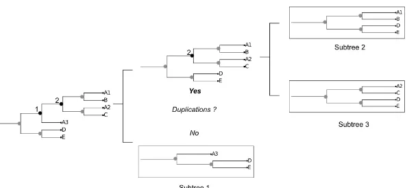
are the most commonly used. The quartet distance counts the number of sub-trees formed by four leaves that differ between two sub-trees, whereas Robinson and Foulds distance is based directly on the edge structure of the trees and their induced bipartitions. While usually distance methods are centered around the topological comparison of trees, some programs also include information regarding branch lengths (Soria-Carrascoet al., 2007). Their drawback is that they usually can not be directly applied on trees with different evolutionary rates. A serious problem involving the comparison of trees is that most algorithms are limited to trees with the same taxa. This situation is unrealistic as events such as gene loss or duplication often produce relationships between genes of different species that are not one-to-one. The straightforward solution to deal with the different amount of taxa in two trees is to prune the two trees until they contain the same amount of taxa. Then distances are corrected to take this deletion into account. The matter of duplications is slightly more complicated. In the Topd/FMTs (Puigbòet al., 2007) method they address the problem by randomly deleting duplicated genes and comparing all the resulting trees with each other. While mathematically sound, the method does not account for orthology and paralogy relationships when prunning the tree (i.e. the prunned tree may contain a mixture of orthologous and paralogous genes) and by randomly choosing the sequences to delete it risks comparing multiple different trees even if the initial trees are identical. A clear example of this problem occurs when comparing a tree with several duplications with itself. The expected distance would be 0, but due to the randomness of the prunning step, the distance can often reach near random values (unpublished observations). In chapter 4 we present our algorithm of tree comparison (treeKo, http://treeko.cgenomics.org), which was designed with this specific problem in mind.
24 Chapter 2
Related to the problem of comparing two trees is the need to develop algorithms that identify specific topological patterns within the trees. Patterns can go from the simplest case scenario in which we want to retrieve those trees in which one species is grouped specifically with another, without any other species between them, to much more complicated patterns that can even have duplication events involved. Some groups have implemented algorithms to search for specific topological patterns (Esser et al., 2004; Gabaldón and Huynen, 2003) that are based on the examination of all the possible tree partitions. Dufayard and colleagues (Dufayardet al., 2005; Gouretet al., 2009) have implemented a similar algorithm that allows the user to define specific scenarios with the help of a graphical interface.
Transference of function to newly annotated proteins
The genomics era has brought about a large amount of sequence data that needs to be processed. Even with high-throughput experiments, thousands of coding sequences remain without an experimental validation. Even in model species, such asEscherichia coliandSaccharomyces cerevisiae, on which scientists have focused their efforts for decades, the function of many proteins still remains unknown or is poorly defined (Hu et al., 2009; Peña-Castillo and Hughes, 2007). One way to overcome this lack of experimental information regarding the functionality of newly sequenced proteins is by using computational means to predict the putative function of a protein based on previous knowledge obtained in other species. Similarity between sequences, as predicted by best blast hits, was at first used to automatically transfer annotations from one protein of a species to another. While this method has been used extensively, it is full of potential pitfalls. Relationships between proteins of different species are not necessarily one to one, due to duplication events. The consequence is that if we transfer the function of a protein based only on similarity we may end up transferring the function of a paralogous protein. Another danger is the fact that a relationship of homology (or orthology) between two proteins does not necessarily mean they will have the same function at all levels. For instance there are large variations of substrate affinities within families of transporters or metabolic enzimes.
The use of phylogenies in order to transfer functional annotations is ben-eficial twofold. On one hand it clearly depicts the evolutionary relationship between proteins, easily differentiating between paralogs and orthologs. It also allows the identification of proteins that have several co-orthologs, which will immediately imply that the annotation can not be trusted without further analysis. On the other hand, it will allow the transference of functions not only from one species to another, but rather of all the species in the tree that have an annotation. This can either raise our confidence in the prediction, when all the annotations
Chapter 2 25
agree on the same function, or it can reveal when a protein changed its function over time. If we expand this to encompass a whole phylome, we will possess a useful tool which will help in the annotation of a newly sequenced genome. This methodology has already been successfully used in the annotation of the pea aphid genome (Consortium, 2010; Huerta-Cepaset al., 2010b), which has been the first newly sequenced genome to be functionally annotated with a phylogeny based strategy, and the Schistosoma mansonigenome (unpublished data). Moreover we have used it in the functional analyses of C. glabrata
genome, performed within the context of the FunPath consortium (see chapter 7).
Final remarks
Phylogenomics can be used for multiple purposes. The elucidation of the evolutionary relationship of a group of species, the transference of function from one protein to another, the detection of evolutionary important events, they are all based on the construction of one or, more often, numerous phylogenetic trees. The increase in the number of phylogenies used in a given analysis has led to the necessary automation of the whole process of tree reconstruction (Gabaldón
et al., 2008). A balance between speed and accuracy has been necessary in
order to ensure the viability of the data used in further analysis. There exist different ways in which to deal with the data produced by phylogenies. Due to the size of the datasets involved, manual exploration is often put aside in favor of an automatic approach. Several applications of phylogenomics are comprised within this volume. Each chapter will involve a study in which phylogenomics tools are applied to a group of fungal species in order to solve different biologicaly relevant questions.
A
IMSz Reconstruct a fungal species tree that reflects the evolution of all com-pletely sequenced fungal species.
z Asses, on a genome-wide basis, the robustness and congruence of each node in the fungal species tree.
z Develop novel tools that enable truly genome-wide scales in the compari-son of gene trees and species trees.
z Assess the extend of some evolutionary mechanisms that may cause topological variation in fungal gene trees (i.e. gene loss, gene duplication, horizontal gene transfer).