The n e w e n g l a n d j o u r n a l of m e d i c i n e
c l i n i c a l p r o b l e m - s o l v i n g
In this Journal feature, information about a real patient is presented in stages (boldface type) to an expert clinician, who responds to the information, sharing his or her reasoning with
the reader (regular type). The authors’ commentary follows.
Forgotten but Not Gone
Ashish K. Jha, M.D., Kaveh G. Shojania, M.D., and Sanjay Saint, M.D., M.P.H.
From the Division of General Internal Medi-cine, Brigham and Women’s Hospital and Harvard Medical School, Boston (A.K.J.); the Department of Medicine, University of Cali-fornia, San Francisco (K.G.S.); and the Vet-erans Affairs Health Services Research and Development Center of Excellence and De-partment of Medicine, University of Mich-igan, Ann Arbor (S.S.). Address reprint re-quests to Dr. Jha at the Division of General Medicine, Brigham and Women’s Hospital, 1620 Tremont St., Boston, MA 02120, or at [email protected].
N Engl J Med 2004;350:2399-404. Copyright © 2004 Massachusetts Medical Society. A 74-year-old man was brought to the emergency department after being found
con-fused and incapacitated at home. The patient lived in a residential hotel and had previ-ously been healthy and socially active. Having not seen him for three days, his friends entered his room and found him on the floor, covered in stool. The patient was conver-sant but confused, recalling only a recent fall and his inability to get up.
Causes of acute alteration in mental status fall primarily into four categories: infec-tious, neurologic, drug-related, and metabolic. In elderly patients, infections are the most common cause, although the other causes remain frequent enough to be pursued just as assiduously. Neurologic causes include hemorrhagic and nonhemorrhagic stroke, seizures, and subdural hematoma. Medications such as sedatives and opiates commonly cause confusion. Metabolic disturbances include alcohol withdrawal, sodium disorders, hyperosmolar coma, and uremia. Other conditions, such as acute myocardial infarction or an intraabdominal infection, can develop atypically in the elderly, predominantly with confusion as the main finding.
The patient’s conversation was tangential. He reported fatigue, diffuse weakness, and a minimally productive cough of four days’ duration. He reported that he had no pre-vious medical problems and had not sought medical care for many years; that he took no regular medications; and that he had not traveled outside the United States in more than 30 years. He had quit smoking 15 years earlier and reported no serious alcohol or illicit-drug use.
Pneumonia is now my leading diagnosis. Elements of the clinical assessment do not indicate which type of pneumonia, although Streptococcus pneumoniae is probably the lead-ing cause of severe community-acquired pneumonia. Pneumococcal pneumonia could certainly account for the patient’s confusion, even without overt bacteremia. Lung can-cer presenting as postobstructive pneumonia is possible, given his smoking history, as is cancer-induced hypercalcemia. An acute exacerbation of chronic bronchitis is unlikely to present in this manner.
The n e w e n g l a n d j o u r n a l of m e d i c i n e
guaiac-positive brown stool. Motor strength was 4/5 in the upper and lower extremities bilaterally. The sensory examination was normal, with sym-metric reflexes in the upper and lower extremities. The patient could recall his name and where he lived but did not know the name of the hospital, the date, or the year. He was able to recall only one of three objects mentioned.
The patient’s vital signs and lung findings are con-sistent with the presence of pneumonia, but the icterus adds additional possibilities. Though the patient reported that he did not drink heavily, his appearance obliges us to consider the possibility of alcoholic liver disease, complicated by pneumo-nia and hepatic encephalopathy. We should also consider other causes of jaundice. Acute viral hep-atitis seems unlikely without recent exposure risks. Cholecystitis or gallstone pancreatitis could cause icterus, but he reported no abdominal pain. Hemo-lytic causes of jaundice, including unrecognized chronic lymphocytic leukemia, should be consid-ered. Given the presence of a heart murmur, en-docarditis is possible. Nevertheless, community-acquired pneumonia remains the most likely diag-nosis.
Laboratory tests revealed a white-cell count of 8300 per cubic millimeter, a hematocrit of 43 per-cent with a mean corpuscular volume of 89.5 µm
3 3 3 3 , and a platelet count of 69,000 per cubic millimeter. The serum sodium concentration was 132 mmol per liter, and the serum calcium concentration was 8.1 mg per deciliter (2.0 mmol per liter); other se-rum electrolyte levels were normal. The results of other laboratory tests were as follows: blood urea nitrogen, 31 mg per deciliter (11.1 mmol per liter); aspartate aminotransferase, 256 U per liter; ala-nine aminotransferase, 77 U per liter; alkaline phosphatase, 214 U per liter; total bilirubin, 6.2 mg per deciliter (106.0 µmol per liter); direct bilirubin, 3.5 mg per deciliter (59.8 µmol per liter); and crea-tine kinase, 26 U per liter. His albumin level was 1.9 g per deciliter, and the prothrombin time was 13.4 seconds (international normalized ratio, 1.2). The partial-thromboplastin time was elevated, at 37.9 seconds. Other than revealing tachycardia, his electrocardiogram was normal. A chest radio-graph obtained with portable equipment revealed slight haziness in the left lower lobe with clearly visible borders of the left heart and diaphragm — findings that are most consistent with the
pres-ence of atelectasis. Radiographs of his hips and lower extremities revealed no fractures. Two sets of blood cultures were performed.
Despite the patient’s denial of excessive alcohol use, it is increasingly difficult to discount the pos-sibility of cirrhosis. The levels of aspartate amino-transferase and alanine aminoamino-transferase in com-bination with thrombocytopenia (not to mention the several missing teeth) all suggest the possibility of alcoholic cirrhosis. Acute causes of liver injury seem less likely, as they would leave unexplained the low albumin level, and the history was not con-sistent with chronic malnutrition. Given the hy-perbilirubinemia, I would also consider cholestatic processes, especially cancer of the gallbladder or pancreas. Intrahepatic causes of cholestasis are pos-sible, including infiltrative disorders (e.g., sarcoid-osis or amyloidsarcoid-osis), infections (e.g., abscess or tu-berculosis), and metastatic carcinoma.
Regardless of the underlying problem, an acute event has also occurred. Given the patient’s age, the absence of fever does not diminish my concern about infection; this remains the most likely cause. Although the findings on the chest radiograph ap-pear to be consistent with atelectasis, pneumonia is a reasonable diagnosis. At this point, I would err on the side of administering empirical antibi-otics to provide coverage for common bacterial causes of pneumonia and for an abdominal infec-tion such as cholangitis. I would obtain a measure-ment of the serum ammonia level to see whether hepatic encephalopathy may account for the pa-tient’s delirium.
c l i n i c a l p r o b l e m - s o l v i n g
32 mm Hg, and a pH of 7.38 while the patient was breathing 6 liters of oxygen through a nasal can-nula. A computed tomographic (CT) scan of the head, obtained without the administration of contrast material, was normal except for the find-ing of multiple small old infarcts that were con-sistent with hypertensive disease. Blood cultures showed no growth of any organisms. Intravenous fluids were stopped, and the patient was given in-travenous furosemide for presumed pulmonary edema. His mental status worsened.
Although empirical treatment for pneumonia was reasonable, the possible hepatobiliary problem seems to have been largely ignored. Abdominal im-aging to evaluate the liver, biliary tract, portal vein, and pancreas remains important. What about the lungs? Lack of improvement for 24 to 48 hours is possible with uncomplicated community-acquired pneumonia, but improvement followed by worsen-ing is a reason for concern. One possibility is that though he has pneumonia, the organism is resis-tant to the antibiotics administered. Another pos-sibility is that the primary problem is delirium and that he has aspirated and now has chemical pneu-monitis or a new pneumonia. Finally, one must con-sider the possibility that the patient mistakenly did not receive his antibiotics.
Instead of pneumonia, the patient may have car-diogenic or noncarcar-diogenic pulmonary edema. Giv-en his murmur, it is possible that he has valvular heart disease or alcoholic cardiomyopathy. Right-sided endocarditis with septic emboli to the lungs is unlikely unless he uses injection drugs. Also, the murmur’s characteristics are not suggestive of tri-cuspid regurgitation, and the negative blood cul-tures to date are reassuring. The patchy infiltrates on his chest radiograph may represent acute res-piratory distress syndrome due to worsening sep-sis rather than pneumonia. The cause of the sepsep-sis may be an intraabdominal infection (e.g., cholan-gitis) that responded partially to the treatment for community-acquired pneumonia but has now pro-gressed.
On the fourth hospital day, the patient’s condi-tion worsened, with a body temperature of 35.4°C, a respiratory rate of 24 breaths per minute, a pulse of 120 beats per minute, and a blood pressure of 85/50 mm Hg. Worsening hypoxemia necessitated intubation and transfer to the intensive care unit, where a third chest radiograph showed increased
patchy infiltrates bilaterally. Multiple cultures of blood and urine were performed, and a test for the human immunodeficiency virus (HIV) was ob-tained. His antibiotic regimen was changed to in-clude piperacillin–tazobactam and gentamicin. Pulmonary-artery catheterization revealed a cardi-ac index of more than 5 liters per minute per square meter of body-surface area, a systemic vascular re-sistance of less than 300 dyn•sec•cm¡
555 5 ,
and a
pul-monary-capillary wedge pressure of 15 mm Hg.
Although hepatic dysfunction can cause hypoten-sion, we must assume that the patient has sepsis on the basis of his hemodynamic measurements and hypothermia. The patient either has pneumo-nia as the cause of these infiltrates with sepsis sec-ondary to pneumonia or has some other cause of sepsis (probably an intraabdominal cause) with pulmonary edema. Pneumonia has been the lead-ing diagnosis from the start and remains so, with an increasing likelihood of a resistant organism. Thus, while I agree with broadening the antibiotic coverage, vancomycin is also indicated for peni-cillin-resistant pneumococcal infection. I am also concerned about legionella infection, given his nonproductive cough, hyponatremia, and poor re-sponse to b-lactam antibiotics and aminoglyco-sides. Tuberculosis or a fungal infection seems unlikely, given the acute presentation and brief im-provement with cefuroxime and doxycycline.
The n e w e n g l a n d j o u r n a l of m e d i c i n e
To look for other possible sources of sepsis, I would order an abdominal CT scan and an echocar-diogram. One additional possibility is adrenal in-sufficiency, which may complicate critical illness. In this patient with hypotension, hypothermia, and hyponatremia, I would perform a corticotropin stimulation test or consider measuring the corti-sol level in a random serum specimen, but would initiate empirical treatment with corticosteroids while awaiting the result.
The patient remained hypotensive, despite the use of dopamine and phenylephrine hydrochloride, and hypoxemic, despite 100 percent inspired ox-ygen delivered by a ventilator. All blood and urine cultures and the HIV test were negative. An echocar-diogram was notable only for hyperdynamic left ventricular function, without wall-motion abnor-malities. Abdominal ultrasonography revealed a thickened gallbladder wall with pericholecystic flu-id but no stones and normal hepatic and biliary ducts. Further radiographic studies or bronchos-copy was not performed, because of the patient’s unstable clinical condition. His hemoglobin level decreased to 6 g per deciliter. He passed large amounts of black stool that were guaiac-positive. His blood pressure continued to drop, despite ag-gressive fluid support and use of vasopressor med-ications. The patient died on the sixth hospital day. An autopsy was performed.
The patient’s subsequent deterioration did not un-dermine the initial diagnosis of pneumonia but shifted concern to a resistant organism — penicil-lin-resistant Streptococcus pneumoniae, multidrug-resistant Haemophilus influenzae,Pseudomonas aeru-ginosa, or legionella. The tempo of the illness still seemed quick for tuberculosis or fungal infection. I expect the autopsy to reveal pneumonia and hope that tissue cultures identify the organism.
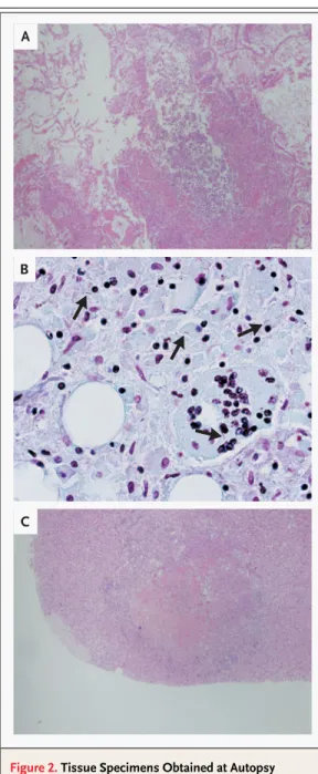
The autopsy revealed a normal gallbladder and bile ducts and a slightly enlarged liver with micro-nodular cirrhosis. Evidence of a previous myocar-dial infarction, congestive hepatopathy, and dif-fuse alveolar damage with pulmonary edema and hyaline membranes were present. There was blood in the stomach and small bowel. Pathological spec-imens from the lungs (Fig. 2A), mediastinal lymph nodes (Fig. 2B), liver, and kidney showed necro-tizing granulomas, with acid-fast bacilli apprecia-ble in some specimens, a finding that is diagnostic
of systemic tuberculosis infection. Both adrenal glands had extensive involvement of necrotizing granulomas as well (Fig. 2C). Examination of the brain and spinal cord revealed no acute abnormal-ities. Two physicians involved in the patient’s care subsequently had positive tests with purified pro-tein derivative for mycobacterium exposure. Their chest radiographs were normal, and they received six months of prophylactic therapy.
Although the severe acute respiratory syndrome (SARS), West Nile virus infection, anthrax, and in-fections with various poxviruses are claiming the attention of patients and physicians, tuberculosis remains a major cause of morbidity throughout the world. In 2002, tuberculosis was responsible for more than 2 million deaths worldwide1
and, in 2001, more than 15,000 infections in the United States.2 However, if tuberculosis is not considered, it will not be diagnosed. In retrospect, the diagnosis of tuberculosis warranted consideration in a margin-ally housed, elderly man with probable chronic liv-er disease. As the discussant points out, the lack of response to the initial antibacterial therapy also in-dicated the need for further consideration of atypi-cal infections, among them tuberculosis.
This diagnosis, however, was far from straight-forward. The original presentation did not obvi-ously indicate tuberculosis. Clinicians often try to find simple explanations to expedite the evaluation and care of elderly patients found incapacitated.3 Here, the clinicians considered common causes, such as urinary tract infection, pneumonia, electro-lyte abnormalities, and cardiovascular events, and treated the patient empirically for a bacterial pneu-monia. In one study of patients who were found helpless or dead in their homes, one out of three patients had an infection.3
Neurologic events and cardiac disorders were the next most likely causes of being found in this state. Unless the cause is ob-vious, all these possibilities should be evaluated. Aside from pneumonia, other causes, including a neurologic event, warranted greater attention in this case. Early evaluation of hepatic encephalop-athy, stroke, or a central nervous system infection (due to subacute meningitis, for example) would have been appropriate though unlikely to lead to the correct antemortem diagnosis.
Initially, the patient’s condition seemed to im-prove. Dehydration and malnutrition can
c l i n i c a l p r o b l e m - s o l v i n g
cate many disease processes, especially infections, and correction of these problems can result in ap-parent improvement, while the underlying cause remains untreated. Such improvements should not curtail the evaluation of other diagnoses. As noted by the discussant, adrenal insufficiency warranted
consideration when the patient remained hypoten-sive. The threshold for evaluating and empirically treating adrenal insufficiency in critically ill patients should be low, given an incidence as high as 40 per-cent in this population.4
Although cortisol levels were not measured, the diffuse involvement of the adrenal glands that was apparent at autopsy sug-gests that adrenal insufficiency due to tuberculosis probably complicated the patient’s illness. Tubercu-losis remains an important cause of primary adre-nal insufficiency.5
This case highlights the value of autopsy. Un-fortunately, the autopsy rate continues to decline in the United States; in 1994, the most recent year for which figures are available, autopsies were per-formed after only 6 percent of deaths in nonfo-rensic cases.6
Yet there continues to be a large dis-crepancy between clinical diagnoses and autopsy findings, with a median major-error rate of 24 per-cent.6
Without an autopsy, the patient’s clinicians would have neither recognized their exposure to tuberculosis nor had the opportunity to learn in or-der to improve the care of future patients. The pos-sibility that an autopsy will unearth important clin-ical information is sufficiently high that initiatives to improve autopsy rates are needed.
Finally, it is important to remember that tuber-culosis has varying clinical manifestations, espe-cially when infection is widely disseminated. The discussant considered but rejected tuberculosis as a diagnosis, because the clinical course was too quick. Although miliary tuberculosis commonly has a subacute presentation,7,8
it may be manifested as a syndrome of rapidly progressing multiorgan dys-function with sepsis9,10
or as the acute respiratory distress syndrome.11,12
Miliary tuberculosis is more common among immunocompromised patients, including those with the acquired immunodefi-ciency syndrome, children under the age of five years, and the elderly. The mortality associated with miliary tuberculosis approaches 50 percent. Because miliary tuberculosis reflects hematogenous spread of the mycobacterium, it typically affects multiple organs (e.g., the liver, spleen, and adrenal glands), with central nervous system involvement in up to 20 percent of patients.7,8
Though miliary tubercu-losis derives its name from the tiny, discrete gran-ulomas resembling millet seeds seen on chest ra-diography, the chest radiograph is interpreted as normal in approximately 40 percent of cases.13 Transmission to others is uncommon but may have occurred in this case.
Figure 2. Tissue Specimens Obtained at Autopsy from the Patient.
Specimens from the lung (Panel A, hematoxylin and eosin, ¬40) and adrenal gland (Panel C, hematoxylin and eosin, ¬40) contain multiple caseating granulomas. A specimen from a mediastinal lymph node (Panel B, acid-fast stain, ¬400) contains acid-fast bacilli (arrows).
A
B
c l i n i c a l p r o b l e m - s o l v i n g
Although not as headline-grabbing as SARS or West Nile virus infection, tuberculosis remains a major cause of morbidity and mortality throughout the world and in the United States. With rising num-bers of elderly people and the increasing rate of chronic immunosuppression, clinicians should con-sider the possibility of tuberculosis even if the
clini-cal manifestations and presentation are atypiclini-cal. Supported by a National Research Service Award from the Agency for Healthcare Research and Quality (to Dr. Jha) and a Career Devel-opment Award from the Health Services Research and DevelDevel-opment Program of the Department of Veterans Affairs and a Patient Safety Developmental Center Grant (P20HS11540) from the Agency for Healthcare Research and Quality (both to Dr. Saint).
We are indebted to Henry Holdt for his critical aid in obtaining pathological and radiologic data.
r e f e r e n c e s
1. Tuberculosis. Fact sheet no. 104. Rev.
Geneva: World Health Organization, August 2002.
2. Reported tuberculosis in the United
States, 2001. Atlanta: Centers for Disease Control and Prevention, September 2002.
3. Gurley RJ, Lum N, Sande M, Lo B, Katz
MH. Persons found in their homes helpless or dead. N Engl J Med 1996;334:1710-6.
4. Marik PE, Zaloga GP. Adrenal
insuffi-ciency during septic shock. Crit Care Med 2003;31:141-5.
5. Oelkers W. Adrenal insufficiency. N Engl
J Med 1996;335:1206-12.
6. Shojania KG, Burton EC, McDonald
KM, Goldman L. Changes in rates of
autopsy-detected diagnostic errors over time: a systematic review. JAMA 2003;289:2849-56.
7. Maartens G, Willcox PA, Benatar SR.
Miliary tuberculosis: rapid diagnosis, hema-tologic abnormalities, and outcome in 109 treated adults. Am J Med 1990;89:291-6.
8. Kim JH, Langston AA, Gallis HA.
Mili-ary tuberculosis: epidemiology, clinical man-ifestations, diagnosis, and outcome. Rev Infect Dis 1990;12:583-90.
9. Ahuja SS, Ahuja SK, Phelps KR, Thelmo
W, Hill AR. Hemodynamic confirmation of septic shock in disseminated tuberculosis. Crit Care Med 1992;20:901-3.
10.George S, Papa L, Sheils L, Magnussen
CR. Septic shock due to disseminated tuber-culosis. Clin Infect Dis 1996;22:188-9.
11.Dyer RA, Chappell WA, Potgieter PD.
Adult respiratory distress syndrome associ-ated with miliary tuberculosis. Crit Care Med 1985;13:12-5.
12.Heffner JE, Strange C, Sahn SA. The
impact of respiratory failure on the diagno-sis of tuberculodiagno-sis. Arch Intern Med 1988; 148:1103-8.
13.Kwong JS, Carignan S, Kang EY, Muller
NL, FitzGerald JM. Miliary tuberculosis: diagnostic accuracy of chest radiography. Chest 1996;110:339-42.
Copyright © 2004 Massachusetts Medical Society.
personal archives in the journal online
Individual subscribers can store articles and searches using a feature on the Journal’s
Web site (www.nejm.org) called “Personal Archive.” Each article and search result