R E S E A R C H A R T I C L E
Open Access
sigE
facilitates the adaptation of
Bordetella
bronchiseptica
to stress conditions and lethal
infection in immunocompromised mice
Sarah E Barchinger
1†, Xuqing Zhang
2,3†, Sara E Hester
2, Maria E Rodriguez
4, Eric T Harvill
2*and Sarah E Ades
1*Abstract
Background:The cell envelope of a bacterial pathogen can be damaged by harsh conditions in the environment outside a host and by immune factors during infection. Cell envelope stress responses preserve the integrity of this essential compartment and are often required for virulence.Bordetellaspecies are important respiratory pathogens that possess a large number of putative transcription factors. However, no cell envelope stress responses have been described in these species. Among the putativeBordetellatranscription factors are a number of genes belonging to the extracytoplasmic function (ECF) group of alternative sigma factors, some of which are known to mediate cell envelope stress responses in other bacteria. Here we investigate the role of one such gene,sigE,in stress survival and pathogenesis ofBordetella bronchiseptica.
Results:We demonstrate thatsigEencodes a functional sigma factor that mediates a cell envelope stress response. Mutants ofB. bronchisepticastrain RB50 lackingsigEare more sensitive to high temperature, ethanol, and
perturbation of the envelope by SDS-EDTA and certainβ-lactam antibiotics. Using a series of immunocompromised mice deficient in different components of the innate and adaptive immune responses, we show that SigE plays an important role in evading the innate immune response during lethal infections of mice lacking B cells and T cells. SigE is not required, however, for colonization of the respiratory tract of immunocompetent mice. ThesigEmutant is more efficiently phagocytosed and killed by peripheral blood polymorphonuclear leukocytes (PMNs) than RB50, and exhibits decreased cytotoxicity toward macrophages. These altered interactions with phagocytes could contribute to the defects observed during lethal infection.
Conclusions:Much of the work on transcriptional regulation during infection inB. bronchisepticahas focused on the BvgAS two-component system. This study reveals that the SigE regulon also mediates a discrete subset of functions associated with virulence. SigE is the first cell envelope stress-sensing system to be described in the bordetellae. In addition to its role during lethal infection of mice deficient in adaptive immunity, our results indicate that SigE is likely to be important for survival in the face of stresses encountered in the environment between hosts.
Keywords:B. bronchiseptica, Extracytoplasmic function sigma factor, Cell envelope stress, Pathogenesis
* Correspondence:eth10@psu.edu;ades@psu.edu
†
Equal contributors
2Department of Veterinary and Biomedical Sciences, Pennsylvania State
University, W210 Millennium Science Complex, University Park, PA 16802, USA
Full list of author information is available at the end of the article
Background
The cell envelope of bacterial pathogens is critical for survival both in a host during infection and in the envir-onment outside of the host. As the interface between the bacterium and the outside milieu, the cell envelope acts as a barrier protecting the cell against extracellular hazards. Cell envelope structures are also intimately involved in the formation of contacts with host tissues during infection. To safeguard this important compart-ment, gram-negative bacteria possess an array of stress responses that sense conditions in the cell envelope and alter gene expression to ensure its integrity [1,2]. In many bacterial pathogens, cell envelope stress responses play a multifaceted role. They provide protection against damage caused by components of the immune system, such as complement and antimicrobial peptides that target the cell envelope [3-5]. They regulate the expres-sion of chaperones required for proper assembly of cell envelope-associated structures, including outer mem-brane porins, pili, and fimbrae [3,6,7]. In addition, cell envelope stress responses can sense the environment around the bacterium and regulate the expression of virulence factors in response to specific cues, ensuring that these factors are expressed at the proper time and location in the host [2,8]. Despite their importance, no cell envelope stress responses have yet been identified or implicated in pathogenesis inBordetellaspecies.
Bordetella bronchisepticais a respiratory pathogen that is closely related to Bordetella pertussis and Bordetella parapertussis, the causative agents of whooping cough in humans [9,10]. B. bronchiseptica causes a range of diseases in various mammals that can be chronic, difficult to completely eradicate, and of variable viru-lence [11-13]. It is the etiological agent of atrophic rhin-itis in swine, kennel cough in dogs, and snuffles in rabbits [12,13]. Documented human infections, generally traced to an animal source, have been observed in im-munocompromised individuals, and can be serious, sys-temic infections [11,14].
TheB. bronchiseptica,B. pertussisandB. parapertussis
genomes encode a large number of putative transcrip-tion factors relative to their overall genome size [15], suggesting that these pathogens have the capacity to ex-tensively regulate gene expression in response to envir-onmental and physiological changes. Despite this finding, only a few Bordetella transcription factors have been studied in any detail [16-20]. Among the predicted transcription factors is an ortholog of the cell envelope stress response sigma factor, σE, of E. coli. In bacteria, sigma factors are the subunits of bacterial RNA poly-merases required for specific promoter recognition and transcription initiation [21]. Alternative sigma factors, likeσE, are activated in response to specific stresses and rapidly reprogram gene expression by replacing the
housekeeping sigma factor and directing RNA polymer-ase to the genes in their regulons [21,22].
σEbelongs to the RpoE-like group of extracytoplasmic function (ECF) sigma factors that have been increasingly implicated as key factors contributing to both bacterial stress responses and virulence [23,24]. These sigma fac-tors are widely distributed across bacterial phyla. Where studied, they direct a diverse set of stress responses pri-marily targeted to the cell envelope [2,8,24,25]. InE. coli
and Salmonella enterica serovar Typhimurium, σE con-trols many genes whose products are required for the proper expression of outer membrane porins and LPS [26,27]. During infection, σE of S. Typhimurium is required for survival and proliferation in epithelial and macrophage cell lines, and in the presence of antimicro-bial peptides [6,28,29]. In Pseudomonas aeruginosa, the
σEhomologue, AlgU, controls the expression of the exo-polysaccharide alginate and conversion to mucoidy. AlgU is constitutively activated in many clinical isolates from cystic fibrosis patients [30,31]. In addition, σE is required for the viability of some bacterial species, but not others. The gene encoding σE is essential in E. coli
and Yersinia enterocolitica, but is dispensable in the closely related species S. Typhimurium [6,32,33]. These observations suggest that the functions of σE orthologs have been adapted to combat the challenges each organ-ism faces in its particular environmental niche. By exploring the role of σE in diverse bacterial species, we can learn which aspects of this widespread regulatory pathway are universally conserved and which have diverged over the course of evolution.
Here we show that the B. bronchisepticaσE ortholog, encoded by the gene sigE (BB3752), is an active sigma factor that mediates a cell envelope stress response. This is the first demonstration of an envelope stress-sensing system in Bordetella species. Using a murine infection model, we demonstrate that SigE plays an important role during lethal infection in mice lacking adaptive immun-ity, but not in respiratory tract colonization. This finding has important implications for human disease, given the observation thatB. bronchiseptica can cause serious sys-temic infections in immunocompromised humans [11,14]. This study suggests that SigE is a critical factor in this process, in addition to the BvgAS master viru-lence regulatory system.
Results
sigEencodes an active sigma factor
inE. coli. This promoter shares a high degree of similarity with a consensus promoter proposed for the RpoE-like sigma factors that was determined from both experimen-tal data and predicted promoter sequences (Figure 1C)
[image:3.595.56.542.90.539.2][24,27]. ThesigEgene fromB. bronchisepticastrain RB50 was cloned into the pTrc99a expression plasmid and transformed into a derivative of E. coliMG1655 that car-ries an rpoHP3::lacZ reporter gene fusion integrated on Figure 1B. bronchisepticaSigE is a functional sigma factor.(A) Amino acid sequence alignment of RpoE- like sigma factors fromEscherichia coli(Ecoli), Vibrio cholerae(Vchol),Pseudomonas aeruginosa(Paer),Nitrosomonas europaea(Neur) andB. bronchiseptica(Bbron) using ClustalW2 (EMBL-EBI). Asterisks indicate identity, two dots indicate strong similarity, and one dot indicates weak similarity between amino acid residues. Conserved sigma factor regions 2.1-2.4 and 4.1-4.2 [22] are indicated above the alignment. Regions 2.3, 2.4, and 4.2 are responsible for promoter recognition [22]. (B)β-galactosidase activity from theE. coli rpoHP3-lacZreporter increases whenB. bronchiseptica sigEexpression is induced from plasmid pSEB006 in strain SEA5005 by the addition of IPTG. No increase is seen upon IPTG addition to the control strain, SEA008, containing the empty vector. The observed difference in the amount ofβ-galactosidase activity between the two strains in the presence of IPTG is statistically significant (P value <0.001) (C) In vitro transcription from a supercoiled plasmid template containing theE. coliσE-dependentrpoHP3 promoter
withE. colicore RNA polymerase (core), SigE alone, EσE, and ESigE (left panel). In vitro transcription from a linear template containing the
the chromosome [34]. When sigE expression was induced, LacZ activity increased, indicating that SigE can initiate transcription from this promoter (Figure 1B). Fur-thermore, we found that the gene encoding σE, rpoE, which is essential for viability inE. coli, could be deleted whensigEwas overexpressed (data not shown, see Mate-rials and Methods).
To provide additional evidence that SigE is a func-tional sigma factor, N-terminally His-tagged SigE was purified and tested for its ability to initiate transcription in vitro from theE. coli rpoHP3 promoter. Holoenzyme formed with SigE and E. coli core RNA polymerase (ESigE) was able to direct transcription and produced a transcript of equivalent length to that generated by E. coli EσE (Figure 1C). The region immediately upstream of the B. bronchiseptica rpoH homologue, encoded by the famgene, contains a sequence that is similar to the proposed σE-dependent consensus promoter, suggesting thatB. bronchiseptica rpoHis regulated by SigE. Indeed, SigE was able to direct transcription from the putative
fam promoter region in vitro (Figure 1C). Taken to-gether, these results demonstrate that SigE is a func-tional sigma factor and can initiate transcription from promoter sequences similar to those utilized by other members of the RpoE-like sigma factor family.
sigEcontributes to theB. bronchisepticastress response
To investigate the role of SigE in B. bronchiseptica, an in-frame deletion of the sigE gene was constructed in RB50 (RB50ΔsigE) that removed 176 out of 200 codons of the gene, leaving 22 and 2 codons at the 5´ and 3´ ends of the gene, respectively. The deletion was con-firmed by PCR and Southern blotting methods (data not shown). σE orthologs are essential in some bacteria, in-cluding E. coli and Y. enterocolitica [33,35], yet are not required for viability in many other species, such as S.
Typhimurium, P. aeruginosa, and Burkholderia pseudo-mallei[6,36,37]. Deletions of B. bronchiseptica sigEwere readily obtained, suggesting that it falls in the latter class, and is not essential for viability. Furthermore, RB50ΔsigE grew at a rate similar to that of RB50 under standard growth conditions (37°C in Stainer-Scholte broth) (Figure 2A).
To investigate whether SigE mediates a cell envelope stress response inB. bronchiseptica, we used disk diffu-sion assays to compare the sensitivity of RB50 and RB50ΔsigE to several chemicals that compromise cell envelope integrity and a series of antibiotics that block different steps in peptidoglycan synthesis. The sigE mu-tant was more sensitive than the wild-type strain to the detergent SDS in combination with EDTA (Figure 2B). The sigEmutant was also more sensitive than wild-type RB50 to the antibiotics mecillinam and ampicillin (Figure 2B), whereas sensitivity to meropenem, aztreonam,
and imipenem was not affected (data not shown). Unlike
σE orthologs in other bacteria, SigE was not required for resistance to the cationic antimicrobial peptide polymyxin B, which targets bacterial membranes, or to osmotic stress (Figure 2B and data not shown) [6,36,38,39]. RB50ΔsigE
and RB50 were also equally sensitive to antibiotics that inhibit cytoplasmic processes such as translation (chloram-phenicol, erythromycin, kanamycin, tetracycline), transcrip-tion (rifampicin), and cytoplasmic enzymes such as DNA gyrase (nalidixic acid), and dihydrofolate reductase (tri-methoprim) (data not shown). This lack of sensitivity to multiple antibiotics suggests that the sigE mutation does not lead to an overall increase in the permeability of the outer membrane, which would allow more of the antibiotic to enter the cell. These results show that SigE is important for survival in response to specific types of damage to the cell envelope, such as disruption of cellular membranes caused by SDS/EDTA and interference with synthesis of the peptidoglycan layer caused by ampicillin and mecillinam.
We next asked ifsigEis important for survival follow-ing a shift to high temperature, which perturbs both the cell envelope and cytoplasm. RB50 and RB50ΔsigEwere grown at 37°C to an OD600of 0.4, then shifted to 50°C, a
lethal temperature for B. bronchiseptica. Cell viability, assessed by CFU/ml, was measured after the shift to 50° C. Survival of the RB50ΔsigEstrain was lower than that of RB50 (Figure 2C). In attempting to complement this phenotype, we found that plasmid-encoded sigEdid not restore survival during heat shock (data not shown), al-though it did complement other phenotypes, as described below. Similar variability in complementation of a σE mutant by a plasmid-encoded rpoE gene has been seen in other bacteria [29,36,40,41]. Work from
Burkholderia cenocepacia showed that expressing σE
from a plasmid actually increased sensitivity to heat stress [36]. InS.Typhimurium, anrpoEmutant was sen-sitive to paraquat and did not survive in stationary phase under anaerobic conditions. Expression of rpoE from a plasmid partially complemented the former phenotype, but not the latter [29]. Because the anti-sigma factor that regulates σE activity was not included in any of these instances, it is likely that proper regulation of SigE activ-ity is required for optimal response to particular stresses, not merely excess SigE activity, complicating comple-mentation experiments.
role of SigE in this phenomenon, RB50 and RB50ΔsigE
were grown to an OD600of 0.1 at 37°C, shifted to 40°C for
90 min, then shifted to 50°C. RB50 cultures incubated at 40°C before 50°C survived better at all time points than those directly shifted from 37°C to 50°C. For example, 54% of the RB50 cells pre-adapted at 40°C survived two hours after the shift to 50°C (Figure 2C) compared to 0.1% survival for those shifted directly from 37°C to 50°C (Figure 2C). RB50ΔsigEpre-adapted at 40°C also survived better at 50°C than when directly shifted from 37°C to 50°C. However, only 38% of the RB50ΔsigE cells survived after one hour (compared to 76% of the wild-type RB50), and 5% survived after two hours at 50°C (Figure 2C). These results demonstrate thatB. bronchisepticaexhibits a classical ther-motolerance response and that SigE contributes to this response.
Both ethanol and heat shock lead to protein unfolding and membrane perturbation and often elicit similar stress responses [43]. To test the role ofsigEin response to ethanol stress, RB50 and RB50ΔsigEwere subcultured from mid-exponential-phase cultures into fresh Stainer-Scholte broth with or without 3% ethanol. Both strains grew similarly in medium without ethanol, as noted above. RB50 grew significantly slower in medium con-taining 3% ethanol than in medium without ethanol (compare the growth curve for RB50 in Figure 2D with that in Figure 2A), but eventually reached a cell density only slightly below that of cultures grown without etha-nol. In contrast, the cell density of RB50ΔsigE grown in the presence of 3% ethanol never surpassed an OD600of
[image:5.595.59.539.90.366.2]around 0.1, even after 24 hours. Expression of plasmid-encoded sigE in RB50ΔsigE complemented this Figure 2Role of SigE in response to environmental stresses.(A) RB50 (squares) and RB50△sigE(triangles) grow similarly at 37°C in Stainer-Scholte broth. (B) RB50△sigE(white bars) is more sensitive than RB50 (grey bars) to treatment with 100μg mecillinam, 10μg ampicillin, or 750μg SDS and 2.9μg EDTA, but is similarly sensitive to treatment with 300 IU polymyxin B in disk diffusion assays. The average diameters of the zones of inhibition ± SE from at least three independent experiments are shown. The disk diameter was 6 mm. The observed differences between the zones of inhibition for RB50 and thesigEmutant are statistically significant for mecillinam, ampicillin, and SDS-EDTA (* indicates a P-value of < 0.05; ** indicates a P-value < 0.01). (C) RB50△sigE(triangles) is more sensitive than RB50 (squares) to heat shock (solid line, filled symbols) caused by shifting cultures from 37°C to 50°C. RB50△sigEalso exhibits reduced thermotolerance (dashed line, open symbols), surviving less well than RB50 when adapted first to 40°C before a shift to 50°C. The mean percent survival±SE of fifteen independent experiments for each strain is shown. (D) RB50△sigEcontaining the empty cloning vector pEV (open triangles) is more sensitive to treatment with 3% ethanol than RB50 pEV (squares). Expression of plasmid-encoded SigE (RB50△sigEpSigE) restores growth in 3% ethanol (filled triangles) to near wild-type levels at the 6 and 12 hour time points and partially restores growth at the 24 hour time point. The mean OD600± SE of at least four independent experiments
phenotype, restoring growth in medium with 3% ethanol to nearly that of RB50 (Figure 2D), indicating thatsigEis required for survival during ethanol stress.
σE homologues have also been found to play a role during oxidative stress in S.Typhimurium and Burkhol-deria pseudomallei [29,41]. However, in disk diffusion assays, SigE was not required for survival in the presence of hydrogen peroxide or paraquat, two inducers of oxi-dative stress (data not shown). Either SigE is not involved in combating oxidative stress in B. bronchisep-tica, or other oxidative-stress responsive pathways com-pensate for SigE when it is absent.
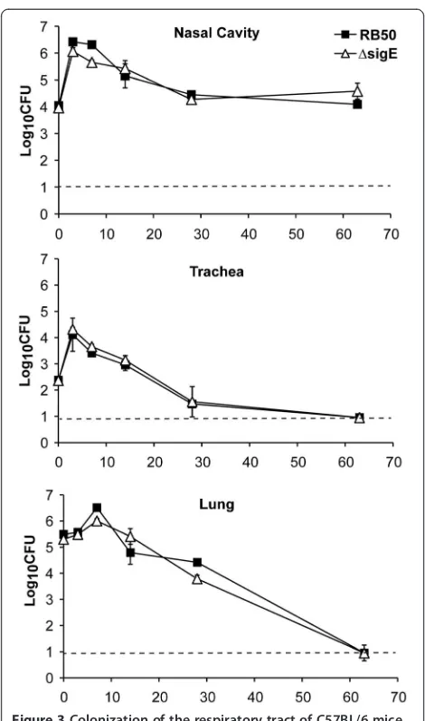
Growth in the murine respiratory tract is not affected by the lack ofsigE
B. bronchisepticaRB50 colonizes the respiratory tract of immunocompetent mice, causing an asymptomatic in-fection that is eventually cleared by the immune system. To determine whetherB. bronchisepticaSigE contributes to colonization and persistence in the respiratory tract, groups of C57BL/6 mice were inoculated with RB50 or RB50ΔsigE. Colonization was measured in the nasal cavity, trachea, and lung on days 0, 3, 7, 14, 28 and 63 post-inoculation. Both wild-type andsigE-deficient RB50 colonized the nasal cavity at comparable levels, peaking on day 3 post-inoculation, and stabilizing at about 104-5CFU by 2 weeks post-inoculation (Figure 3). Both strains also showed similar colonization kinetics in the lower respira-tory tract of C57BL/6 mice, peaking in numbers on days 3 and 7 post-inoculation in the trachea and lungs, respect-ively, and declining thereafter, with complete clearance in both organs by day 63 post-inoculation (Figure 3). These data indicate that B. bronchiseptica SigE is not required for colonization or persistence in the murine respiratory tract.
SigE contributes to lethalB. bronchisepticainfection in mice lacking B cells and T cells, but not in mice lacking TLR4 or TNF-α
B. bronchiseptica has been observed to cause a range of disease including bronchitis, lethal pneumonia, and even systemic infection [11,12]. Mice with defined immune deficiencies are particularly susceptible to different forms of disease [44-46], facilitating assessment of the roles of specific bacterial factors/functions in interac-tions with different aspects of the host immune response.
Mice lacking key components of innate immunity, either TLR4 or TNF-α, were challenged with RB50 or RB50ΔsigE and signs of severe disease were monitored. Consistent with published studies, TLR4defand TNF-α−/−
mice inoculated with 105CFU of RB50 quickly developed signs of lethal bordetellosis such as ruffled fur, hunched posture, decreased activity, and difficulty breathing, and succumbed 2 to 5 days post-inoculation [46,47]. Mice challenged with RB50ΔsigE also showed similar signs of disease and time to death (data not shown). In a separate experiment, TLR4def mice and TNF-α−/− mice infected
[image:6.595.56.291.85.484.2]with RB50 or RB50ΔsigE that were still alive by day 3 post-inoculation were dissected for bacterial enumeration in the respiratory as well as systemic organs. Both wild-type and sigE-deficient RB50 colonized the lungs of TLR4def mice at 107-8 CFU, which was almost 1000-fold higher than in the lungs of TLR4suf mice. Moreover, both strains colonized the systemic organs in TLR4def, but not TLR4suf mice (data not shown). Both strains also grew to higher numbers in the Figure 3Colonization of the respiratory tract of C57BL/6 mice
by RB50 and RB50ΔsigE.Groups of three 4–6 week-old C57BL/6 mice were inoculated with 5 × 10⁵CFU of RB50 (filled squares) and RB50△sigE(open triangles). Groups of three mice were sacrificed at each time point. The bacterial load in the indicated organ is expressed as log10CFU ± SE. The dashed line indicates the limit of
lungs of TNF-α−/− mice than in the lungs of C57BL/6
mice and were recovered from systemic organs only in TNF-α−/− mice (data not shown). These data
indi-cate that SigE is not required for B. bronchiseptica to cause lethal infection and colonize systemic organs in mice lacking TLR4 or TNF-α.
B and T cell-deficient Rag1−/− mice succumb to B.
bronchiseptica infection, and death is associated with systemic spread of the infection [48]. To assess the role of SigE during infection in hosts deficient in adaptive immunity, groups of Rag1−/− mice were inoculated with
5 × 10⁵CFU of RB50 or RB50ΔsigE. Rag1−/−mice
inocu-lated with RB50 showed symptoms of lethal bordetellosis on day 13 post-inoculation and succumbed between days 14–35 post-inoculation (Figure 4A). However, Rag1−/−
mice inoculated with RB50ΔsigE survived without any
overt signs of disease and were euthanized on day 122 post-inoculation. The nasal cavity, trachea, lungs, spleen, liver, and kidneys of these mice were excised to enumer-ate bacterial loads. Although 105-7 CFU of RB50ΔsigE
were recovered from the respiratory tract, this strain failed to colonize the spleen or kidney, and only 300 CFU were recovered from the liver (Figure 4B, dark gray bars). In a separate experiment, RB50 and RB50ΔsigE-inoculated Rag1−/− mice were sacrificed on
day 28 post-inoculation, when some of the RB50-challenged mice were still alive. The bacterial loads of RB50 and RB50ΔsigE in the respiratory tract on day 28 post-inoculation were similar, about 105-7 CFU. At this time, 104-6 CFU of RB50 were recovered from liver, spleen, and kidney (Figure 4B, white bars). RB50ΔsigE, however, failed to colonize the spleen, kidney or liver (Figure 4B, light gray bars). These results demonstrate that SigE is required for lethal infection by B. bronchi-septicain Rag1−/−mice.
The failure of RB50ΔsigE to colonize distal organs of Rag1−/−mice suggests that this mutant may be defective
in getting into or survival in the bloodstream and/or sys-temic organs. The bloodstream includes many important bactericidal factors of the host immune system, includ-ing complement and phagocytes. We first examined whether B. bronchiseptica lacking sigE is more suscep-tible to complement-mediated killing. 500 CFU of RB50, RB50ΔsigE, or RB50Δwbm, a strain lacking O-antigen, which is known to be susceptible to complement [48], were incubated at 37°C for one hour in PBS with 20% complement-active or complement-inactive serum from naïve mice. The survival of RB50ΔsigE and RB50 was not affected by the presence of either serum (data not shown). In contrast, the RB50Δwbm strain was almost completely killed by complement-active, but not complement-inactive serum (0.7% survival in the pres-ence of complement-active serum compared to 100% survival in the presence of complement-inactive serum). The observation that RB50ΔsigE survived in the pres-ence of serum without B. bronchiseptica-specific anti-bodies indicates that the defect in causing systemic infection in mice lacking B and T cells is not due to fail-ure to survive the antimicrobial components in serum, including complement.
SigE contributes to cytotoxicity to macrophages
[image:7.595.57.292.304.600.2]We further tested whether RB50ΔsigE interacts differ-ently than RB50 with another major bactericidal compo-nent in the bloodstream, phagocytes.B. bronchisepticais cytotoxic to macrophages, and this toxicity has been attributed to the activities of the type three secretion system (TTSS) [49]. To test the role of SigE in macro-phage cytotoxicity, RAW264.7 murine macromacro-phages were incubated for 4 hours at an MOI of 10 with RB50, Figure 4Survival and systemic colonization of Rag1−/−mice
following infection with RB50 and RB50ΔsigE.(A) Groups of Rag1
−/−mice (n = 6) were inoculated with 5 × 10⁵CFU of RB50 (solid line
with filled squares) or RB50△sigE(dashed line with open triangles) and monitored for survival. (B) Groups of four Rag1−/−mice were
inoculated with 5 × 10⁵CFU of RB50 (white bars) or RB50
△sigE(light grey bars) and dissected on day 28 post-inoculation for bacterial enumeration in the indicated organs. In a separate experiment, Rag1
−/−mice inoculated with RB50
△sigEwere euthanized for bacterial numbers in the indicated organs on day 122 post-inoculation (dark grey bars). The bacterial load is expressed as log10CFU ± SE. Limit of
RB50 lacking sigE, or RB50 lacking a functional TTSS (WD3). In this experiment, both the RB50 and RB50ΔsigE strains contained the empty cloning vector pEV to allow direct comparisons with the complemented strain, RB50ΔsigEpSigE. Cytotoxicity was determined by measuring LDH release from the treated macrophages. WD3 caused little cytotoxicity, similar to treatment with medium alone. RB50ΔsigE pEV caused approximately 50% less cytotoxicity than wild-type RB50 pEV (Figure 5). This defect in cytotoxicity was complemented by supply-ing thesigE gene on the plasmid pSigE (Figure 5), indi-cating that loss of sigE negatively impacts the ability of RB50 to kill macrophages.
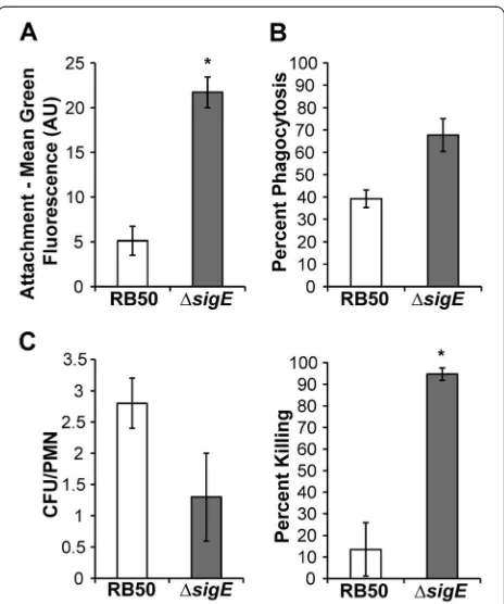
RB50ΔsigEis more efficiently phagocytosed and killed by PMNs
To test if RB50ΔsigEis more susceptible to another bac-tericidal mechanism, phagocytosis by peripheral blood polymorphonuclear leukocytes (PMNs), RB50 and RB50ΔsigE were incubated with freshly isolated human PMNs and attachment to, phagocytosis by, and killing by these cells were measured. PMNs bound RB50ΔsigE
more efficiently than RB50 (Figure 6A), and significantly more RB50ΔsigE than RB50 were phagocytosed by PMNs (Figure 6B). However, the number of viable intra-cellular RB50ΔsigE was ~50% of the numbers of viable RB50 (Figure 6C, left panel). When differences in
attachment and phagocytosis were taken into consider-ation, significantly more internalized RB50ΔsigE were killed compared to RB50 (Figure 6C, right panel). To-gether, these data indicate that SigE contributes to B. bronchiseptica resistance to phagocytosis and killing by PMNs.
Discussion
[image:8.595.306.538.87.365.2]The BvgAS system of the bordetellae plays a central role in regulating gene expression during pathogenesis [50-52]. However, other regulators may be required during the infectious disease cycle, as Bordetella gen-omes have a large number of putative sensory systems Figure 5RB50ΔsigEis less cytotoxic to macrophages than
[image:8.595.56.292.448.597.2]RB50.RAW 264.7 cells were incubated at an MOI of 10 with medium containing RB50 pEV, RB50△sigEpEV, RB50△sigEpSigE, TTSS-deficient RB50 strain WD3, or medium alone for 4 hours in the presence of 1 mM IPTG to induce expression ofsigEfrom the pLac promoter of pSigE. The average percent cytotoxicity of four wells in four separate experiments as measured by (LDH release from a well/ LDH release from the positive control well) x100 ± SE is shown. The differences in percent cytotoxicity between RB50△sigEpEV and either RB50 pEV or RB50△sigEpSigE are statistically significant (** indicates P value < 0.01), while the cytotoxicities of RB50 pEV and RB50△sigEpSigE are not significantly different.
[10,16-20]. In this study, we focused on cell envelope sensing systems and investigated the alternative sigma factor, SigE. We found that SigE of B. bronchiseptica
does indeed mediate a protective cell envelope stress re-sponse and that strains lacking SigE do not establish lethal infections in mice lacking adaptive immunity. These data suggest that the role of SigE is to combat stresses to the envelope imposed by the immune system within a host and by harsh conditions in the environ-ment outside a host. This work is the first demonstra-tion of a cell envelope sensing system in the bordetellae. The σE system has been explored in the most depth in enteric pathogens belonging to the Gammaproteobac-teria [23,25,53]. The bordetellae, members of the Betaproteobacteria, encounter distinctly different envir-onments in the respiratory tract and therefore provide an excellent model to study how the SigE system has been adapted throughout evolution to serve the needs of diverse bacterial pathogens.
The entire sigE locus (BB3752-BB3750) is identical at the amino acid sequence level among the classical bor-detellae, suggesting a conserved role in the human pathogens B. pertussis and B. parapertussis. However, the lifestyles and, therefore, conditions encountered dif-fer amongst these three species. B. bronchiseptica can live outside the host and primarily infects mammals, although it can infect immunocompromised humans [11,14]. In contrast,B. pertussisandB. parapertussis pri-marily infect humans and are directly transmitted between hosts [54,55]. As we learn more about the role of SigE in the bordetellae, it will be of interest to deter-mine whether stresses that induce the SigE system and the SigE regulon members are as highly conserved as thesigElocus itself among the bordetellae.
Our results define roles for SigE in B. bronchiseptica
that are only partially overlapping with those for σE in other pathogens. SigE was important for survival of B. bronchiseptica in the face of both global stresses to the cell envelope caused by heat shock, exposure to ethanol and detergent, and specific stresses caused by several beta-lactam antibiotics (Figure 2). Heat shock, ethanol, and detergent are classical stressors used in the labora-tory to mimic conditions that lead to unfolded proteins and disrupted lipids during infection and in the environ-ment. In contrast to the B. cenocepacia and S. Typhi-murium proteins, B. bronchiseptica SigE was not required for survival during osmotic stress [6,36]. SigE was also not required for response to oxidative stress or the antimicrobial peptide polymyxin B, unlike the S.
Typhimurium σE ortholog [6,29]. The variations among bacteria in their use of σE systems likely reflect both differences in stresses encountered in environmental reservoirs and in particular host tissues during infection, as well as differences in the arrays of additional cellular
stress responses possessed by each species. These other responses can act along with or in place ofσE. The pres-ence of other stress responses may be particularly per-tinent to B. bronchiseptica. Its genome is predicted to encode six related ECF sigma factors of unknown func-tion in addifunc-tion to SigE [24] that may have complimentary and redundant functions with SigE. Future studies defining conditions that activate other ECF sigma factors and their roles inB. bronchisepticapathogenesis will provide a more comprehensive understanding of how B. bronchiseptica
copes with extracytoplasmic stress.
Stress response systems, like theσEsystem, rapidly in-duce the expression of specialized sets of genes. These systems are often tightly regulated and expressed only when needed, because inappropriate expression of their regulons can interfere with other important cellular functions [8,56,57]. We found that SigE was not required for colonization and persistence of RB50 within the re-spiratory tract of an immunocompetent host (Figure 3), the primary niche of B. bronchiseptica. This result sug-gests that the pathogen does not encounter stresses in the respiratory tract that require a response by the SigE system. However, B. bronchiseptica encounters different challenges during infection in Rag1−/− mice lacking B
and T cells. In these mice, the infection spreads to the bloodstream, which is under greater immune surveil-lance and has a different arsenal of antimicrobial factors to attack invaders than the respiratory tract. The defect of RB50ΔsigEin lethal infection of Rag1−/− mice,
there-fore, reveals a specific function for SigE in response to an unknown stress, possibly related to the innate im-mune response, that the bacteria encounter during infec-tions that proceed beyond colonization of the respiratory tract.
The inability of RB50ΔsigEto cause lethal infections in Rag1−/− mice (Figure 4) could be due to failure to enter
or survive in the bloodstream and/or systemic organs of these mice. Since the mutation does not affect survival during incubation with serum in vitro, it is unlikely that the sigE-deficient strain is more susceptible to comple-ment or other antimicrobial components in serum. The defect in infection of Rag1−/− mice may then be related
to altered interactions of the mutant strain with phago-cytic cells in the bloodstream. RB50ΔsigE is more sus-ceptible to peripheral blood PMNs than RB50 (Figure 6), and is also less cytotoxic to macrophages than RB50 (Figure 5). Either or both of these defects could explain the failure to recover RB50ΔsigE from systemic organs of mice lacking adaptive immune responses and the decreased virulence in these mice.
Why does the RB50ΔsigE mutant spread systemically and cause lethal infection in TLR4defand
TNF-α−/−mice,
but not Rag1−/− mice? The lower cytotoxicity of thesigE
does not affect its virulence in mice lacking innate im-mune functions. This could be because bacterial numbers within the respiratory tract of TLR4defor TNF-α−/− mice
are nearly an order of magnitude higher than in the lungs of Rag1−/− mice. As such, the large number of bacteria in
TLR4defor TNF-α−/− mice may overwhelm limiting host
antimicrobial defense mechanisms that can contain the lower bacterial numbers in the lungs of Rag1−/−mice.
Al-ternatively, although the cytotoxicity of thesigEmutant is reduced, it may still be sufficient to establish lethal infec-tions in the absence of TLR4 or TNF-α. Thus TLR4- and TNF-α-dependent functions, such as efficient phagocyt-osis and killing, appear to be sufficient to prevent lethal
infection by RB50ΔsigE in Rag1−/− mice. Although the
exact role remains to be elucidated, our results clearly in-dicate that SigE is required for lethal infection of mice lacking B and T cells.
Although the B. bronchiseptica strain RB50 causes asymptomatic infections in immunocompetent mice, other strains ofB. bronchisepticacan cause a wide range of disease severity in other hosts [11-13]. In particular subsets of immunocompromised humans, such as those infected with HIV, severe systemic B. bronchiseptica
infections have been observed [14]. These facts, along with the high degree of sequence conservation for the
[image:10.595.56.538.290.732.2]sigElocus inB. pertussisandB. parapertussis, highlights
Table 1 Strains and plasmids
Strain name Genotype Source, Reference
E. coli SEA001 MG1655ΦλrpoHP3::lacZ△lacX74 [60]
SEA5036 BL21(DE3)△slyD::kan pLysS pPER76 [61]
XQZ001 BL21(DE3)△slyD::kan pLysS pXQZ001 This work
SEA4114 CAG43113△rpoE::kan△nadB::Tn10 [62]
SEA008 SEA001 pTrc99a [62]
SEA5005 SEA001 pSEB006 This work
XQZ003 DH5αpXQZ0003 This work
SS1827 DH5αpSS1827 [63]
B. bronchiseptica RB50 RB50 [58]
SEA5516 RB50△sigE This work
MER001 RB50 pCW505 This work
MER002 RB50△sigEpCW505 This work
SEA5518 RB50 pEV This work
SEA5520 RB50△sigEpEV This work
SEA5526 RB50 pSigE This work
SEA5530 RB50△sigEpSigE This work
RB50△wbm RB50△wbmBwbmCwbmDwbmE [64]
WD3 RB50△bscN [49]
Plasmid name Description Source, Reference
pTrc99a Vector, pBR322 ori, ApR Pharmacia
pSEB006 sigEin pTrc99a This work
pSEB015 isolatedrpoHP3 promoter in pRLG770, ApR [61]
pPER76 rpoEin T7 expression vector pET15b, KanR [65]
pXQZ001 sigEin T7 expression vector pET15b, KanR This work
pXQZ002 △sigEin TOPO-TA vector This work
pSS1827 helper plasmid competent for mating, ApR [63]
pSS3962 Bordetella-specific allelic exchange vector, KanR Stibitz, unpublished work
pXQZ003 △sigEin pSS3962 This work
pEV Vector pJS72,ΩSpecRcassette replaced with CmR This work
pSigE sigEin pEV This work
the importance of understanding the stressors that acti-vate SigE and how the SigE system responds to them during infection.
Conclusions
In this work, we have demonstrated that theB. bronchi-septica extracytoplasmic function sigma factor, SigE, is important for surviving global stresses that affect the whole cell, such as heat shock and ethanol stress, spe-cific stresses that target the cell envelope, such as beta-lactam antibiotics and SDS-EDTA, and in interactions with the host innate immune system, particularly phago-cytes. During infection, SigE is not required for colonization of the respiratory tract of immunocompe-tent mice. However, it is needed for a specific set of functions associated with virulence, particularly those involved in surviving the innate immune response when the infection progresses in immunocompromised mice. Although SigE systems are widely conserved, the details as to which aspects are shared and which have diverged are complex. As evidence accumulates from studies in different bacteria, it is becoming apparent that these sensory modules are important for stress survival, par-ticularly with respect to the cell envelope. However, the nature of the stresses that SigE systems combat varies. During infection, comparisons are even more difficult, since differences are seen not only amongst SigE systems from one pathogen to another, but also within different niches in the host or during the progression of disease for a single pathogen.
Methods Strains and media
A complete list of strains used in this study can be found in Table 1.B. bronchisepticastrains are derivatives of the previously described B. bronchisepticastrain RB50 [58].
B. bronchiseptica was maintained on Bordet-Gengou (BG) agar (Difco) containing 10% defibrinated sheep blood (Hema Resources) and 20 μg/ml streptomycin. In liquid culture, B. bronchiseptica was grown in Stainer-Scholte broth [59] with aeration. Chloramphenicol was used at 20 μ/ml and IPTG at 1 mM where noted. The RB50ΔsigE mutant was constructed as described below. E. coli strains used to measure SigE activity are derivatives of MG1655 that carry the σE -dependent rpoHP3::lacZreporter (strain SEA001 [34]).
E. coli strain BL21(DE3) pLysS was used to express constructs for protein purification. E. coli were grown in LB broth in a gyratory water bath with aeration. Ampicillin was used at 100 μg/ml, tetracycline at 20 μg/ml, and kanamycin at 15 μg/ml as needed for experiments with E. coli.
Plasmid constructions
All plasmids used in this study are listed in Table 1 and oligonucleotide sequences are given in Table 2. Plasmid pSEB006 was constructed to express sigEinE. coli. The
sigE gene was amplified from RB50 genomic DNA with the primers SigEF and SigER and cloned into the expres-sion vector pTrc99a under the control of the IPTG-inducible trc promoter. To facilitate purification of SigE, the plasmid pXQZ001 was constructed by amplifying the sigE gene from RB50 genomic DNA using the pri-mers HisSigEF and HisSigER. The resulting PCR product was cloned into the T7 expression vector pET-15b (Novagen), which adds a 6X-His tag to the N-terminus of recombinant proteins. To express sigEin B. bronchi-septica, sigE was amplified from RB50 genomic DNA using primers 72SigEF and 72SigER and ligated into the XbaI and XhoI sites downstream of the pLac promoter in pEV to create pSigE. The expression vector pEV was constructed from the broad host range vector pJS72 by replacing the spectinomycin resistance gene with thecat
gene encoding chloramphenicol resistance amplified from pKD3 [67] using primers 72ChlorF and 72ChlorR. The exchange of drug markers was necessary because RB50 is naturally resistant to spectinomycin. pEV and pSigE were moved into RB50 and RB50ΔsigE through tri-parental mating on BG agar with MgCl2.
Transconju-gants were selected on BG containing 60μg/ml strepto-mycin and 20μg/ml chloramphenicol. Plasmid pCW505 (kindly supplied by Dr. Alison Weiss, Cincinnati, Ohio), which induces cytoplasmic expression of GFP without affecting growth or antigen expression, was used to visualize RB50 and RB50ΔsigEin the phagocytosis assays described below [68].
Construction of RB50ΔsigEstrain
Tri-parental mating with wild-type B. bronchiseptica strain RB50, E. coli strain DH5α harboring the pXQ003 vector (strain XQ003), and DH5α harboring the helper plasmid pSS1827 (strain SS1827) [69,70] and selection of mutants were performed as previously described [69]. The deletion strain was verified by PCR using primers SigEKO_LeftF and SigEKO_RightR and by Southern blot analysis.
β-galactosidase assays
Overnight cultures were diluted into fresh medium and grown to an OD600of 0.1-0.2 at 30°C. Where indicated,
IPTG was added to a final concentration of 1 mM. Sam-ples were collected 2.5 hours later and β-galactosidase activity from the σE-dependent reporter was assayed as previously described [60,71].
Complementation ofE. coliΔrpoEbyB. bronchiseptica sigE
The ability of B. bronchiseptica sigE to suppress the le-thality caused by deletion of rpoE in E. coli was deter-mined using a cotransduction assay as described [62]. The ΔrpoE::kan ΔnadB::Tn10 allele from strain SEA4114 was moved via P1 transdution into strain SEA5005, which carries sigE on the plasmid pSEB006. Tet-resistant (tetR) transductants were selected and then screened for kanamycin resistance (kanR). Although the
nadBandrpoEalleles are tightly linked (>99%), cotrans-duction resulting in tetRkanRcolonies will only occur if
rpoE is no longer essential for viability. In transductions with E. coli expressing sigE (strain SEA5005) as the re-cipient strain, 31 out of 32 tetR transductants were also kanR. In contrast, none of the 39 tetRtransductants were kanR when E. coli carrying the empty cloning vector (strain SEA008) was the recipient strain.
Protein purification
N-terminally His-tagged B. bronchiseptica SigE and E. coliσEwere purified from strain XQZ001 and SEA5036, respectively, as previously described for E. coli σE [61]. Briefly, cells were grown at 25°C to an OD600of 0.5, at
which point IPTG was added to induce protein produc-tion. Following 1.5-3 hours of induction, cells were har-vested by centrifugation and resuspended in lysis buffer (20 mM Tris–HCl pH 8.0, 500 mM NaCl, 20 mM imid-azole, 2.5 mMβ-mercaptoethanol, 1 mM PMSF). Resus-pended cells were then lysed by sonication, and the lysate cleared by centrifugation. The supernatant con-taining soluble His-SigE was loaded onto a Ni-NTA col-umn (Qiagen). Bound proteins were eluted with a stepwise gradient of 20, 60, 100, and 200 mM imidazole in column buffer (20 mM Tris–HCl pH 8.0, 500 mM NaCl, 2.5 mMβ–mercaptoethanol). Fractions containing SigE were pooled and dialyzed into 20 mM Tris–HCl pH 8.0, 50 mM NaCl, and 2.5 mMβ-mercaptoethanol.
In vitro transcription
100 nM E. coli core RNA polymerase (Epicentre) was incubated with 400 nM His-SigE or His-σE in transcrip-tion buffer (40 mM Tris–HCl pH 8.0, 10 mM MgCl2,
[image:12.595.58.540.100.311.2]50 mM NaCl, 1 mM DTT, 0.1μ/ml BSA) for 10 min at 30°C to form holoenzyme. Multi-round transcription reactions were initiated by addition of holoenzyme at a final concentration of 40 nM sigma factor and 10 nM core RNA polymerase, to prewarmed (30°C) transcrip-tion mix containing 5.0 nM supercoiled plasmid tem-plate pSEB015 [61] or 5.0 nM linear Pfamtemplate, 5% glycerol, 200 mM ATP, 200 mM CTP, 200 mM GTP, 10 mM UTP, and 2.5 mCi [α-32P]UTP in transcription buffer. After 10 min at 30°C, reactions were stopped by
Table 2 Primer sequences
Primer name Sequence (5´ - 3´) Source or Reference
SigEF GGCGGAGAATTCAGGAGGAGGCGTCATGAGCGAACGCGATG This work
SigER GGCCTAGGATCCTTACCAGCGACGCTCGGCAT This work
HisSigEF GGCCTGGCATATGAGCGAACGCGATGTCGA This work
HisSigER GGCCTAGGATCCTTACCAGCGACGCTCGGCAT This work
72SigEF GCGCGGTCTAGAAGGAGGAGGCGTCATGAGCGAACGCGATG This work
72SigER GCCCGGCTCGAGTTACCAGCGACGCTCGGCAT This work
72ChlorF GCGGCGGGATCCTGTGTAGGCTGGAGCTGCTTC [67]
72ChlorR GCCGCCGGATCCCATATGAATATCCTCCTTA [67]
SigEKO_LeftF GGGAATTCAAGATCGAGATCGGCCTGTCGAAT This work
SigEKO_LeftR AGGGATCCGAAGGCTTTCTTGTCGCCACGTTGTA This work
SigEKO_RightF AGGGATCCTGGTAAGGAGTGGCAGTCATGCAA This work
SigEKO_RightR GCGAATTCAAAGCAACGGTGTCATCAACGTCC This work
PFamF GGGCGGGAATTCTGCCGTTCGTGGATGTCCAG This work
the addition of stop solution (80% formamide, 20 mM EDTA, 0.1% xylene cyanol, and 0.1% bromophenol blue). Samples were electrophoresed on 6% polyacrylamide gels containing 7.5 M urea, and transcripts were visua-lized by phosphorimaging. The linear Pfamtemplate was generated by amplification of the promoter region of the gene encoding σ32 in RB50, fam, using the primers PFamF and PFamR (Table 2). The sequence logo in Figure 1C was generated using WebLogo version 2.8.2 (http://WebLogo.berkeley.edu, [72]).
Disk diffusion assays
B. bronchisepticacultures in mid-log phase were diluted to 6 × 10⁸ CFU/ml and spread on Stainer-Scholte agar plates to generate a lawn of bacteria. Disks containing 300 IU polymyxin B, 10 μg ampicillin, 100 μg mecilli-nam, 750 μg sodium dodecyl sulfate (SDS) and 2.9 μg EDTA, 30 μg aztreonam, 10μg imipenem, 10 μg mero-penem, 30 μg chloramphenicol, 15 μg erythromycin, 30μg kanamycin, 30μg nalidixic acid, 150μg rifampicin, 23.75 μg sulfamethoxazole and 1.25 μg trimethoprim, 30 μg tetracycline, 3.0 μg deoxycholate, 3% hydrogen peroxide, or 2% paraquat were applied to the plates and the zones of inhibition were measured after overnight incubation at 37°C.
Temperature and ethanol stress
For temperature stress experiments, mid-log phase cul-tures of RB50 and RB50ΔsigEwere diluted to an OD600
of 0.01 in fresh Stainer-Scholte broth and incubated at 37°C in a gyratory water bath with shaking. At an OD600
of 0.1, cultures were either shifted to 40°C for adaptation or kept at 37°C. After 90 minutes, all cultures were shifted to 50°C, and survival was measured by plating and CFU counts. For ethanol stress experiments, mid-log-phase cultures of the pertinent strains were subcul-tured into fresh Stainer-Scholte broth with or without 3% ethanol and incubated at 37°C in a gyratory water bath with aeration. Bacterial growth was measured by OD600.
Complement killing assay
Complement killing assays were performed as previously described [73]. Approximately 500 CFU of RB50, RB50ΔsigE, and RB50Δwbm from mid-log phase cul-tures were incubated with 45 μl of diluted serum from C57BL/6 mice or PBS (final volume for incubation was 50μl) for 1 hour at 37°C. Bacterial numbers before and after incubation were determined by plating and CFU counts. Each strain was assayed in triplicate.
Cytotoxicity assay
Cytotoxicity assays were performed as previously described [44]. Briefly, bacteria were added to RAW
264.7 murine macrophage cells at a multiplicity of infec-tion (MOI) of 10 and incubated for four hours. Percent lactate dehydrogenase (LDH) release, a measure of cyto-toxicity, was determined by using Cytotox96 Kit (Promega) according to the manufacturer’s protocol.
Phagocytosis and killing by polymorphonuclear leukocytes
Attachment and phagocytosis of the B. bronchiseptica
strains by peripheral blood polymorphonuclear leukocytes (PMNs) were evaluated as previously described with a few modifications [74]. Briefly, GFP-expressing bacteria were incubated with PMNs at an MOI of 50 for 20 min at 37°C to allow binding. After extensive washing to remove non-attached bacteria, an aliquot was maintained on ice to be used as a bacterial attachment control. The remaining PMNs were further incubated for 30 min at 37°C to allow internalization, at which point phagocytosis was stopped by placing PMNs on ice. Bacteria bound to the cell surface in both aliquots were detected by incubation with RB50 immune serum for 30 min at 4°C, followed by incubation with R-phycoerythrin (RPE)–labeled goat F(ab')2
frag-ments of anti-mouse IgG at 4°C for 30 min. All incuba-tions were done in the presence of 25% heat-inactivated human serum to prevent nonspecific binding of anti-bodies. After washing, ten thousand cells per sample were analyzed by flow cytometry. Attachment control samples were also analyzed by fluorescence microscopy using a DMLB microscope coupled to a DC 100 camera (Leica Microscopy Systems Ltd.). Green fluorescence intensity associated with PMNs maintained at 37°C for 20 min has previously been shown to represent bacterial attachment [74]. Phagocytosis was calculated from the decrease in mean red fluorescence intensity of GFP-positive PMNs after the 30 min incubation allowing for internalization, as previously described [75]. Percent phagocytosis was calcu-lated as follows: 100 × (1-RPE2/RPE1), where RPE1 is the mean RPE-fluorescence of the GFP-positive cells after 20 min at 37°C (attachment control) and RPE2 is the mean RPE-fluorescence of the GFP-positive cells after 50 min (internalized bacteria) at 37°C.
index (1-RPE2/RPE1), N = number of viable bacteria per cell after incubation with antibiotics. Control experiments to assess the efficacy of antibiotic bactericidal activity were performed in parallel. Briefly, samples of 5 × 10⁸ bac-teria were incubated with antibiotics for 30 min at 37°C and plated. This resulted in a >99% decrease in CFU.
Animal experiments
C57BL/6J, B6.129 S-Tnftm1Gkl/J (TNF-α−/−), B6
129S7-Rag1tm1Mom/J (Rag1−/−), C3H/HeOuJ (TLR4suf) and C3H/
HeJ (TLR4def) mice were obtained from Jackson laborator-ies (Bar Harbor). All mice were bred in our Bordetella -free, specific pathogen-free breeding rooms at The Penn-sylvania State University. For inoculation, mice were sedated with 5% isoflurane (Abbott laboratory) in oxygen and 50 μl of PBS containing 105 or 5 × 10⁵ CFU of the indicated bacteria were pipeted onto the external nares [76,77]. This method reliably distributes the bacteria throughout the respiratory tract [76]. Survival curves were generated by inoculating TLR4def, TNF-α−/− and Rag1−/−
mice with either RB50 or RB50ΔsigE. Mice suffering from lethal bordetellosis as determined by severe hunched pos-ture, ruffled fur, extremely labored breathing and apathy were euthanized to prevent unnecessary suffering [47]. For quantifying bacterial load, mice were euthanized via CO2 inhalation, and lung, trachea, nasal cavity, spleen,
liver and/or kidneys were excised. Tissues were homoge-nized in PBS, aliquots were serially diluted, plated, incu-bated at 37°C for 2 to 3 days, and CFU were determined. All protocols were reviewed by the university IACUC and all animals were handled in accordance with institutional guidelines (IACUC approval number: 31297).
Statistical analysis
The mean +/−standard error (SE) of the geometric mean
was determined when appropriate and expressed as error bars. Two-tailed, unpaired Student’s T-tests were used to de-termine statistical significance between groups. All experi-ments were performed at least twice with similar results.
Authors’contributions
SB and SA conceived and designed the molecular and stress experiments, which were performed by SB. XZ and EH conceived and designed the infection studies, which were performed by XZ. SH performed the cytotoxicity experiments and MR performed the phagocytosis experiments. SB, XZ, EH, and SA wrote the manuscript. All authors have read, contributed to editing, and approved the final manuscript.
Acknowledgements
We thank Dr. Scott Stibitz (FDA) for providing the allelic exchange vector pSS3962 and the helper plasmid pSS1827. We thank Dr. Kenneth Keiler (the Pennsylvania State University) for providing the plasmid pJS72. This work was supported by NIH grant GM083113 (E.T.H), in part by NSF grant MCB-0347302 (S.E.A.) and a NSF Graduate Research Fellowship to S.E.B.
Author details
1Department of Biochemistry and Molecular Biology, Pennsylvania State
University, 406 Althouse Laboratory, University Park, PA 16802, USA. 2Department of Veterinary and Biomedical Sciences, Pennsylvania State
University, W210 Millennium Science Complex, University Park, PA 16802, USA.3current address: Department of Microbiology and Immunology, Harvard Medical School, 200 Longwood Ave, Boston, MA 02115, USA. 4
CINDEFI (UNLP, CONICET La Plata), School of Science, La Plata University, La Plata, Argentina.
Received: 7 December 2011 Accepted: 25 June 2012 Published: 16 August 2012
References
1. MacRitchie DM, Buelow DR, Price NL, Raivio TL:Two-component signaling and gram negative envelope stress response systems.Adv Exp Med Biol 2008,631:80–110.
2. Rowley G, Spector M, Kormanec J, Roberts M:Pushing the envelope: extracytoplasmic stress responses in bacterial pathogens.Nat Rev Microbiol2006,4:383–394.
3. Crouch ML, Becker LA, Bang IS, Tanabe H, Ouellette AJ, Fang FC:The alternative sigma factor sigma is required for resistance of Salmonella enterica serovar Typhimurium to anti-microbial peptides.Mol Microbiol 2005,56:789–799.
4. Ernst RK, Guina T, Miller SI:Salmonella Typhimurium outer membrane remodeling: role in resistance to host innate immunity.Microb Infect 2001,3:1327–1334.
5. Jongerius I, Ram S, Rooijakkers S:Bacterial complement escape.Adv Exp Med Biol2009,666:32–48.
6. Humphreys S, Stevenson A, Bacon A, Weinhardt AB, Roberts M:The alternative sigma factor,σE, is critically important for the virulence of Salmonella Typhimurium.Infect Immun1999,67:1560–1568.
7. Mathur J, Waldor MK:The Vibrio cholerae ToxR-regulated porin OmpU confers resistance to antimicrobial peptides.Infect Immun2004, 72:3577–3583.
8. Raivio TL:Envelope stress responses and Gram-negative bacterial pathogenesis.Mol Microbiol2005,56:1119–1128.
9. Arico B, Gross R, Smida J, Rappuoli R:Evolutionary relationships in the genus Bordetella.Mol Microbiol1987,1:301–308.
10. Parkhill J, Sebaihia M, Preston A, Murphy LD, Thomson N, Harris DE, Holden MT, Churcher CM, Bentley SD, Mungall KL,et al:Comparative analysis of the genome sequences of Bordetella pertussis, Bordetella parapertussis and Bordetella bronchiseptica.Nat Genet2003,35:32–40. 11. Goodnow RA:Biology of Bordetella bronchiseptica.Microbiol Rev1980,
44:722–738.
12. Mattoo S, Cherry JD:Molecular pathogenesis, epidemiology, and clinical manifestations of respiratory infections due to Bordetella pertussis and other Bordetella subspecies.Clin Microbiol Rev2005,18:326–382. 13. Musser JM, Bemis DA, Ishikawa H, Selander RK:Clonal diversity and host
distribution in Bordetella bronchiseptica.J Bacteriol1987,169:2793–2803. 14. Mazumder SA, Cleveland KO:Bordetella bronchiseptica bacteremia in a
patient with AIDS.South Med J2010,103:934–935.
15. Madan Babu M, Teichmann SA, Aravind L:Evolutionary dynamics of prokaryotic transcriptional regulatory networks.J Mol Biol2006, 358:614–633.
16. Brickman TJ, Vanderpool CK, Armstrong SK:Heme transport contributes to in vivo fitness of Bordetella pertussis during primary infection in mice. Infect Immun2006,74:1741–1744.
17. Conover MS, Redfern CJ, Ganguly T, Sukumar N, Sloan G, Mishra M, Deora R: BpsR modulates Bordetella biofilm formation by negatively regulating the expression of the Bps polysaccharide.J Bacteriol2012,194:233–242. 18. Jungnitz H, West NP, Walker MJ, Chhatwal GS, Guzman CA:A second
two-component regulatory system of Bordetella bronchiseptica required for bacterial resistance to oxidative stress, production of acid phosphatase, and in vivo persistence.Infect Immun1998,66:4640–4650.
19. Vanderpool CK, Armstrong SK:Integration of environmental signals controls expression of Bordetella heme utilization genes.J Bacteriol2004, 186:938–948.
20. Zimna K, Medina E, Jungnitz H, Guzman CA:Role played by the response regulator Ris in Bordetella bronchiseptica resistance to macrophage killing.FEMS Microbiol Lett2001,201:177–180.
21. Paget MS, Helmann JD:The sigma70 family of sigma factors.Genome Biol 2003,4:203.
23. Helmann JD:The extracytoplasmic function (ECF) sigma factors.Adv Microb Physiol2002,46:47–110.
24. Staron A, Sofia HJ, Dietrich S, Ulrich LE, Liesegang H, Mascher T:The third pillar of bacterial signal transduction: classification of the
extracytoplasmic function (ECF) sigma factor protein family.Mol Microbiol 2009,74:557–581.
25. Missiakas D, Raina S:The extracytoplasmic function sigma factors: role and regulation.Mol Microbiol1998,28:1059–1066.
26. Alba BM, Gross CA:Regulation of the Escherichia coli sigma-dependent envelope stress response.Mol Microbiol2004,52:613–619.
27. Rhodius VA, Suh WC, Nonaka G, West J, Gross CA:Conserved and variable functions of theσEstress response in related genomes.PLoS Biol2006,
4:e2.
28. Muller C, Bang IS, Velayudhan J, Karlinsey J, Papenfort K, Vogel J, Fang FC: Acid stress activation of theσEstress response in Salmonella enterica serovar Typhimurium.Mol Microbiol2009,71:1228–1238.
29. Testerman TL, Vazquez-Torres A, Xu Y, Jones-Carson J, Libby SJ, Fang FC: The alternative sigma factorσEcontrols antioxidant defences required for Salmonella virulence and stationary-phase survival.Mol Microbiol 2002,43:771–782.
30. Deretic V, Schurr MJ, Boucher JC, Martin DW:Conversion of Pseudomonas aeruginosa to mucoidy in cystic fibrosis: environmental stress and regulation of bacterial virulence by alternative sigma factors.J Bacteriol 1994,176:2773–2780.
31. Rowen DW, Deretic V:Membrane-to-cytosol redistribution of ECF sigma factor AlgU and conversion to mucoidy in Pseudomonas aeruginosa isolates from cystic fibrosis patients.Mol Microbiol2000,36:314–327. 32. De Las Penas A, Connolly L, Gross CA:TheσE-mediated response to
extracytoplasmic stress in Escherichia coli is transduced by RseA and RseB, two negative regulators ofσE.Mol Microbiol1997,24:373–385. 33. Heusipp G, Schmidt MA, Miller VL:Identification of rpoE and nadB as host
responsive elements of Yersinia enterocolitica.FEMS Microbiol Lett2003, 226:291–298.
34. Mecsas J, Rouviere PE, Erickson JW, Donohue TJ, Gross CA:The activity of σE, an Escherichia coli heat-inducible sigma-factor, is modulated by expression of outer membrane proteins.Genes Dev1993,7:2618–2628. 35. De Las Penas A, Connolly L, Gross CA:σEis an essential sigma factor in
Escherichia coli.J Bacteriol1997,179:6862–6864.
36. Flannagan RS, Valvano MA:Burkholderia cenocepacia requires RpoE for growth under stress conditions and delay of phagolysosomal fusion in macrophages.Microbiology2008,154:643–653.
37. Yu H, Schurr MJ, Deretic V:Functional equivalence of Escherichia coliσE and Pseudomonas aeruginosa AlgU: E. coli rpoE restores mucoidy and reduces sensitivity to reactive oxygen intermediates in algU mutants of P. aeruginosa.J Bacteriol1995,177:3259–3268.
38. Bianchi AA, Baneyx F:Hyperosmotic shock induces theσ32andσEstress regulons of Escherichia coli.Mol Microbiol1999,34:1029–1038.
39. Mathur J, Davis BM, Waldor MK:Antimicrobial peptides activate the Vibrio choleraeσEregulon through an OmpU-dependent signalling pathway. Mol Microbiol2007,63:848–858.
40. Keith LM, Bender CL:AlgT (σ22) controls alginate production and tolerance to environmental stress in Pseudomonas syringae.J Bacteriol 1999,181:7176–7184.
41. Korbsrisate S, Vanaporn M, Kerdsuk P, Kespichayawattana W, Vattanaviboon P, Kiatpapan P, Lertmemongkolchai G:The Burkholderia pseudomallei RpoE (AlgU) operon is involved in environmental stress tolerance and biofilm formation.FEMS Microbiol Lett2005,252:243–249.
42. Tomoyasu T, Mogk A, Langen H, Goloubinoff P, Bukau B:Genetic dissection of the roles of chaperones and proteases in protein folding and degradation in the Escherichia coli cytosol.Mol Microbiol2001,
40:397–413.
43. Kovacikova G, Skorupski K:The alternative sigma factorσEplays an important role in intestinal survival and virulence in Vibrio cholerae. Infect Immun2002,70:5355–5362.
44. Harvill ET, Cotter PA, Yuk MH, Miller JF:Probing the function of Bordetella bronchiseptica adenylate cyclase toxin by manipulating host immunity. Infect Immun1999,67:1493–1500.
45. Mann PB, Elder KD, Kennett MJ, Harvill ET:Toll-like receptor 4-dependent early elicited tumor necrosis factor alpha expression is critical for innate host defense against Bordetella bronchiseptica.Infect Immun2004, 72:6650–6658.
46. Mann PB, Kennett MJ, Harvill ET:Toll-like receptor 4 is critical to innate host defense in a murine model of bordetellosis.J Infect Dis2004, 189:833–836.
47. Mann PB, Wolfe D, Latz E, Golenbock D, Preston A, Harvill ET:Comparative toll-like receptor 4-mediated innate host defense to Bordetella infection. Infect Immun2005,73:8144–8152.
48. Burns VC, Pishko EJ, Preston A, Maskell DJ, Harvill ET:Role of Bordetella O antigen in respiratory tract infection.Infect Immun2003,71:86–94. 49. Yuk MH, Harvill ET, Miller JF:The BvgAS virulence control system regulates
type III secretion in Bordetella bronchiseptica.Mol Microbiol1998,28:945–959. 50. Bock A, Gross R:The BvgAS two-component system of Bordetella spp.: a
versatile modulator of virulence gene expression.Int J Med Microb2001, 291:119–130.
51. Cotter PA, Jones AM:Phosphorelay control of virulence gene expression in Bordetella.Trends Microbiol2003,11:367–373.
52. Mattoo S, Foreman-Wykert AK, Cotter PA, Miller JF:Mechanisms of Bordetella pathogenesis.Front Biosci2001,6:E168–E186.
53. Bashyam MD, Hasnain SE:The extracytoplasmic function sigma factors: role in bacterial pathogenesis.Infect Genet Evol2004,4:301–308. 54. Gerlach G, von Wintzingerode F, Middendorf B, Gross R:Evolutionary
trends in the genus Bordetella.Microb Infect2001,3:61–72. 55. Porter JF, Parton R, Wardlaw AC:Growth and survival of Bordetella
bronchiseptica in natural waters and in buffered saline without added nutrients.Appl Environ Microbiol1991,57:1202–1206.
56. Park SD, Youn JW, Kim YJ, Lee SM, Kim Y, Lee HS:Corynebacterium glutamicumσEis involved in responses to cell surface stresses and its activity is controlled by the anti-sigma factor CseE.Microbiology2008, 154:915–923.
57. Sheehan BJ, Bosse JT, Beddek AJ, Rycroft AN, Kroll JS, Langford PR:Identification of Actinobacillus pleuropneumoniae genes important for survival during infection in its natural host.Infect Immun2003,71:3960–3970.
58. Cotter PA, Miller JF:BvgAS-mediated signal transduction: analysis of phase-locked regulatory mutants of Bordetella bronchiseptica in a rabbit model.Infect Immun1994,62:3381–3390.
59. Stainer DW, Scholte MJ:A simple chemically defined medium for the production of phase I Bordetella pertussis.J Gen Microbiol1970, 63:211–220.
60. Costanzo A, Ades SE:Growth phase-dependent regulation of the extracytoplasmic stress factor,σE, by guanosine 3',5'-bispyrophosphate (ppGpp).J Bacteriol2006,188:4627–4634.
61. Costanzo A, Nicoloff H, Barchinger SE, Banta AB, Gourse RL, Ades SE:ppGpp and DksA likely regulate the activity of the extracytoplasmic stress factor σEin Escherichia coli by both direct and indirect mechanisms.Mol Microbiol2008,67:619–632.
62. Hayden JD, Ades SE:The Extracytoplasmic stress factor,σE, is required to maintain cell envelope integrity inEscherichia coli.PLoS One2008, 3:e1573.
63. Stibitz S, Aaronson W, Monack D, Falkow S:The vir locus and phase-variation in Bordetella pertussis.Tokai J Exp Clin Med1988,13(Suppl):223–226. 64. Preston A, Allen AG, Cadisch J, Thomas R, Stevens K, Churcher CM,
Badcock KL, Parkhill J, Barrell B, Maskell DJ:Genetic basis for
lipopolysaccharide O-antigen biosynthesis in bordetellae.Infect Immun 1999,67:3763–3767.
65. Rouviere PE, De Las Penas A, Mecsas J, Lu CZ, Rudd KE, Gross CA:rpoE, the gene encoding the second heat-shock sigma factor,σE, in Escherichia coli.EMBO J1995,14:1032–1042.
66. Schaeffer LM, McCormack FX, Wu H, Weiss AA:Bordetella pertussis lipopolysaccharide resists the bactericidal effects of pulmonary surfactant protein A.J Immunol2004,173:1959–1965.
67. Datsenko KA, Wanner BL:One-step inactivation of chromosomal genes in Escherichia coli K-12 using PCR products.Proc Natl Acad Sci U S A2000, 97:6640–6645.
68. Weingart CL, Broitman-Maduro G, Dean G, Newman S, Peppler M, Weiss AA: Fluorescent labels influence phagocytosis of Bordetella pertussis by human neutrophils.Infect Immun1999,67:4264–4267.
69. Buboltz AM, Nicholson TL, Weyrich LS, Harvill ET:Role of the type III secretion system in a hypervirulent lineage of Bordetella bronchiseptica. Infect Immun2009,77:3969–3977.
71. Miller JH:Experiments in molecular genetics. Cold Spring Harbor, NY: Cold Spring Harbor Laboratory Press; 1972.
72. Crooks GE, Hon G, Chandonia JM, Brenner SE:WebLogo: a sequence logo generator.Genome Res2004,14:1188–1190.
73. Goebel EM, Wolfe DN, Elder K, Stibitz S, Harvill ET: O-antigen protects Bordetella parapertussisfrom complement.Infect Immun 2008, 76:1774–1780.
74. Rodriguez ME, Hellwig SM, Hozbor DF, Leusen J, van der Pol WL, van de Winkel JG:Fc receptor-mediated immunity againstBordetella pertussis.J Immunol2001,167:6545–6551.
75. Rodriguez ME, Van der Pol WL, Van de Winkel JG:Flow cytometry-based phagocytosis assay for sensitive detection of opsonic activity of pneumococcal capsular polysaccharide antibodies in human sera. J Immunol Methods2001,252:33–44.
76. Harvill ET, Preston A, Cotter PA, Allen AG, Maskell DJ, Miller JF:Multiple roles forBordetellalipopolysaccharide molecules during respiratory tract infection.Infect Immun2000,68:6720–6728.
77. Kirimanjeswara GS, Agosto LM, Kennet MJ, Bjornstad ON, Harvill ET: Pertussis toxin inhibits neutrophil recruitment to inhibit antibody-mediated clearance ofBordetella pertussis. J Clin Invest2005, 115:3594–3601.
doi:10.1186/1471-2180-12-179
Cite this article as:Barchingeret al.:sigEfacilitates the adaptation of
Bordetella bronchisepticato stress conditions and lethal infection in immunocompromised mice.BMC Microbiology201212:179.
Submit your next manuscript to BioMed Central and take full advantage of:
• Convenient online submission
• Thorough peer review
• No space constraints or color figure charges
• Immediate publication on acceptance
• Inclusion in PubMed, CAS, Scopus and Google Scholar
• Research which is freely available for redistribution
![Figure 1 B. bronchiseptica(EMBL-EBI). Asterisks indicate identity, two dots indicate strong similarity, and one dot indicates weak similarity between amino acid residues.Conserved sigma factor regions 2.1-2.4 and 4.1-4.2 [22] are indicated above the alignm](https://thumb-us.123doks.com/thumbv2/123dok_es/4752677.60366/3.595.56.542.90.539/bronchiseptica-asterisks-indicate-similarity-indicates-similarity-conserved-indicated.webp)