PONTIFICIA UNIVERSIDAD CATÓLICA DEL ECUADOR FACULTAD DE CIENCIAS EXACTAS Y NATURALES
ESCUELA DE CIENCIAS BIOLÓGICAS
Diversidad de hongos asociados a los intestinos de escarabajos (Passalidae) que habitan en el Parque Nacional Yasuní y evaluación de degradación de celulosa in vitro
Disertación previa a la obtención del título de Licenciada en Ciencias Biológicas
SOFÍA ALEJANDRA LÓPEZ CHÁVEZ
Certifico que la disertación de Licenciatura en Ciencias Biológicas de la candidata Srta. Sofía Alejandra López Chávez ha sido concluida de conformidad con las normas establecidas; por lo tanto, puede ser presentada para la calificación correspondiente.
M.Sc. Alexandra Narváez Trujillo Directora de la Disertación
AGRADECIMIENTOS
Quiero agradecer ante todo a mis papás porque sin ellos nada de esto habría sido posible. A mi papá Rodrigo por su apoyo y amor incondicional y a mi mamá Katia por ser el ángel protector de mi familia. A mi hermana Catalina por su inmensa alegría y amor. A mis abuelitos Idalia y Fausto por su preocupación y cariño infinito.
Este trabajo de investigación se llevó a cabo gracias al financiamiento otorgado por la Universidad Pontificia Universidad Católica a la cual extiendo mis agradecimientos, así también por permitirme el inmenso aprendizaje tanto académico, profesional como personal.
A Alexandra Narváez por recibirme en el Laboratorio de Biotecnología Vegetal y permitirme ser parte de su equipo de investigadores. Le agradezco enormemente por haber dirigido este trabajo y haber contribuido con ideas constructivas para el mismo. Así mismo, agradezco a Verónica Crespo y Carolina Portero por sus comentarios y valiosas sugerencias para este trabajo.
A los miembros del Laboratorio de Biotecnología Vegetal Eliana Veloz, Fernando Marín, Berenice Benavides, Carolina Castro por su amistad y apoyo. Y, principalmente a Carolina Portero y Stephany Villota por su guía, enseñanza y amistad.
A mis amigos Jorge Castillo, Carolina Yandún, Saúl Aguirre, Andrea Villota, Alejandra Moscoso, Gloria Del Alcázar, Daniela Pareja, Daniel Rivadeneira, Rubén Abad por su preocupación, apoyo y colaboración. A la profesora Susana León por su sincera amistad y apoyo durante este proceso.
LISTA DE ABREVIATURAS
Abreviatura Significado
ANOSIM Analysis of similarities
BLAST Basic Local Alignment Search Tool CEQCA Colección de Endófitos Quito Católica CMC Carboxymethyl cellulose
DNA Deoxyribonucleic acid DNS 3,5-dinitrosalicylic acid
g Gram
h Hour
ITS Internal transcribed spacer
M Molar mass
mg Miligram
min Minute
ml Mililiter
mM Milimolar mass
Muscle Multiple sequence alignment by log-expectation NMDS Non-metric multidimensional scaling
no. Number
º C Celsius degrees
Abreviatura Significado
RAxML Randomized Axelerated Maximum Likelihood rDNA Ribosomal Deoxyribonucleic Acid
rpm Revolutions per minute
sec Second
SIMPER Similarity percentage
YM Yeast mold
μl Microliter
μM Micromolar mass
μmol Micromole
TABLA DE CONTENIDO
LISTA DE ABREVIATURAS ... vii
1. RESUMEN ... 1
2. ABSTRACT ... 3
3. MANUSCRITO PARA PUBLICACIÓN ... 4
INTRODUCTION ... 5
MATERIALS AND METHODS ... 9
Sample collection, medium, and culture conditions ... 9
Fungi DNA extraction and PCR amplification... 10
Phylogenetic analyses ... 11
Assays for cellulose degrading activity ... 11
Diversity and community composition analysis ... 13
RESULTS ... 15
Insect identification ... 15
Phylogenetic analyses ... 15
Cellulase enzymatic activity ... 16
Diversity and community composition analyses ... 17
DISCUSSION ... 19
ACKNOWLEDGMENTS ... 25
REFERENCES ... 26
4. FIGURES ... 33
6. APPENDIXES ... 55
LISTA DE FIGURAS
Figure 1. A. Adult passalid beetle, Passalus intertitialis. B. Passalus intertitialis dissected with its entire gut. ... 34 Figure 2. Maximum likehood phylogenetic tree analysis of fungi ITS sequences with emphasis on the Hypocreales order where most of the sequences were classified. Outgroup was Kluyveromyces waltii. Strains isolated in this study are shown with the CEQCA code. Numerical values indicate bootstrap percentiles from 200 replicates. ... 35 Figure 3. Orders of fungi isolated from passalid guts. Percentage of isolates classified in each order (N=98). ... 36 Figure 4. Species-accumulation curve of all host fungal species, isolated from the guts of
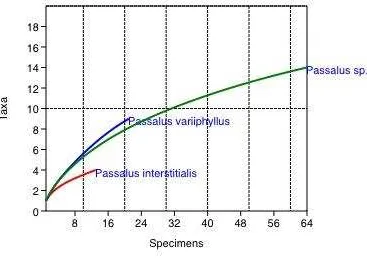
three Passalidae species (N=98). ... 37 Figure 5. Species-accumulation curve of fungal species isolated from the guts of Passalidae beetles from three decaying trees (tree no. 1 in red line (N=20), tree no. 2 in blue line (N=13) and tree no. 3 in green line (N=65)). ... 38 Figure 6. Species-accumulation curve of fungal species isolated from the guts of three
LISTA DE TABLAS
Table 1. Cellulase activities of extracts of fungi isolated from the guts of three Passalidae species beetles based on reducing sugar methodology. ... 43
Table 2. Cellulase activity of the gut of Passalidae beetles. Positive (+) cellulase activity and negative cellulase activity ... 44
Table 3. All host diversity indices and diversity indices according to decaying tree. ... 45
Table 4. All host fungal diversity indices and diversity indices according to Passalidae species. ... 46
Table 5. Analysis of similarities (ANOSIM) of fungal composition according to decaying trees (R= 0.5177, P= 0.0058). ... 47
Table 6. Analysis of similarities (ANOSIM) of fungi composition according to passalid species (R= 0.2816, P= 0.0977). ... 48
Table 7. Similarity percentage (SIMPER) analysis shows fungal species that contributed the most (as a percentage) to the total dissimilarity within all three decaying trees. ... 49
Table 8. Similarity percentage (SIMPER) analysis shows fungal species that contributed the most (as a percentage) to the total dissimilarity comparing within three decaying trees. Only fungal species showing more than 2% of contribution to dissimilarity were considered... 50
LISTA DE APÉNDICES
Appendix 1. Fungal groups according the Passalidae species from each decaying tree. Ninety-eight fungal species were identified. ... 56
Appendix 2. Internal transcribed spacer (ITS) annotated and referenced DNA sequences of fungal species used for phylogenetic analysis... 61
Appendix 3. Taxonomic identification of passalid’s gut fungi. Phylogeny based on maximum
1.
RESUMEN
importante en la alimentación de los escarabajos durante la digestión y la fermentación de la celulosa.
2.
ABSTRACT
3.
MANUSCRITO PARA PUBLICACIÓN
REVISTA
Applied and Environmental Microbiology
TÍTULO
Diversity of fungi associated with guts of Bess beetles (Passalidae) that inhabit the Yasuni National Park and in vitro evaluation of cellulose degradation
AUTORES
Sofía López-Chávez 1#, Alexandra Narváez-Trujillo1*
1Laboratory of Plant Biotechnology, School of Biological Sciences, Pontificia Universidad
Catolica del Ecuador, Quito, Ecuador1
#Present address: Address correspondence to Sofía López-Chávez, Laboratory of Plant Biotechnology, School of Biological Sciences, Pontificia Universidad Catolica del Ecuador
salopez@outlook.com
*Present address: Address correspondence to Alexandra Narváez-Trujillo, Laboratory of Plant Biotechnology, School of Biological Sciences, Pontificia Universidad Catolica del Ecuador
anarvaez@puce.edu.ec
Tel. +593 2 2991700 ext. 1810
INTRODUCTION
Soil organic matter (SOM) in forest ecosystem represents the main source of energy, especially plant litter materials. The decomposition of SOM depends on environmental factors such as pH, temperature, precipitation and oxygen supply, as well as on the action of decomposer organisms, soil fauna and microorganisms (1).
Microbial communities in soil represent a great diversity of biotic interactions (2). During organic decomposition of biomass there is a change in community dynamics, a succession of fungal species occurs and the availability of niches are altered when associations of fungal species are replaced (3). Soil fungal species such as Phoma, Cylindrocarpon, Clodosporium, Phomopsis, Trichoderma and Fusarium take part in litter decay because of their capacity to break down cellulose bonds (4, 5). As a consequence of these microbiotic interactions in the SOM decomposing process, new resources and microhabitats are also created for soil fauna, such as Collembola, Acari (3) and Passalidae (6) which also play an important role in speeding up the decomposing process.
Soil-inhabiting invertebrates are key components of the decomposer web and nutrient cycling pathways in the edaphic ecosystems. In a wide diversity of ecosystems, soil invertebrates mediate about 15% of the C and 30% of the N cycling (7), and influence physical-chemical properties of soils (8, 9). Therefore, micro-, meso- and macroinvertebrates contribution is enormous in the stabilization and destabilization of SOM (10).
colonized the decaying litter. Bacteria and fungi have developed different abilities to establish symbiotic relationships especially with its host’s digestive tract (11). This gut microbiota plays an essential role in insect growth, development and nutrition, especially with the acquisition of nutrients by fermentation and lignocellulose degradation, nitrogen fixation, amino acid biosynthesis and uric acid degradation (12, 13).
Plant litter is composed mainly by cellulose, hemicellulose and lignin (14). Cellulose is a difficult molecule to breakdown since it consists of linear polymers of β-D-glucopiranose
units (glucose) linked by β-1-4 glycosidic bonds (15). Endoglucanases, exoglucanases and
β-glucosidases (12) are enzymes required to give way to primary products such as glucose, cellobiose and cello-oligosaccharides (16). Studies have demonstrated that cellulose digestion occurs in many types of insects such as termites, ants, wood roaches, wasps, aphids and a wide range of beetles (17, 18). Some of these, like termites and longhorn beetles have the capacity to produce their own cellulase enzymes in the gut, while other insects complement their cellulose digestion with the cellulase enzymatic capacity of endosymbiont microorganisms (14, 19, 20).
This study focused on a group of passalid beetles (Passalidae: Coleoptera). Six hundred eighty species have been described within this family, distributed mainly in tropical and humid-temperate regions of the world, such as the Neotropical region (36). Their biological cycle mostly occurs in tunnels or galleries built in rotten logs preferably of dicotyledonous angiosperms (37), where they can obtain refuge, nourishment and more stable microenvironmental conditions (38). These insects are cataloged as a highly subsocial group due to their living form. Adults (parents), juveniles, larvae in different developmental stages, and eggs live together in the same tunnel system (38). Parental care creates dependence of juveniles and larvae by feeding a mixture of digested wood, salivary secretions and inoculated feces that contain microbiota with cellulase activity (13). Diet is the key to their fitness, passalids eating only rotting wood in the absence of endosymbiotic microbiota, start losing weight and die much sooner (39).
To take part in the decaying process of fallen trees, Passalidae beetles have developed interactions with other soil decomposing organisms. Harrel (1967) (40) reported a varied assemblage of parasitic and commensalistic organisms that included mites, nematodes and protozoa associated to the passalid beetle Odontotaenius disjunctus. However, it was Lichtardt (1968) (40) that reported on the numerous fungi associated with this beetle species and its mites, specifically, Trichomycetes (Zygomycota) found in the hindgut and Laboulbeniales (Ascomycota) common found externally on both the passalids and their mites. Various studies on the microbiota of passalids have revealed a vast diversity of xylose and cellulose-degrading yeast species (11, 13, 27, 29, 41); few studies have reported on other components of this microbiota.
MATERIALS AND METHODS
Sample collection, medium, and culture conditions
Passalid adults were collected from three rotting logs in the Yasuní Research Station in the Amazon Basin in Ecuador. Each decaying log was considered a sampling site. Tree no. 1 (00.67543 S 76.39896 W) and tree no. 2 (00.67542 S 76.39896 W) were approximately three meters away from each other. Tree no. 3 (00.67508 S 76.39938 W) was 100 m away. Only tree no. 1 could be identified as Inga spectabilis (Fabaceae). All beetles found inside the logs were collected in 50 ml conical centrifuge tubes and kept alive to be processed in the Laboratory of Plant Biotechnology of the Pontificia Universidad Catolica del Ecuador. Beetles were grouped according to their sizes and the tree from which they were collected.
Fungi isolates were obtained from passalid guts. Dissections were performed following the method described in Berkov, et al (2007) (19), Nguyen, et al (2006) (41), Nguyen, et al (2007) (42), and Suh & Blackwell (2004) (43). Insects were isolated in individual Petri dishes. Before dissection, insects were euthanized at -4 ºC for 15 min. Surface sterilization was accomplished by submersion in 75% ethanol for 2-3 min and washed with 0.7% saline solution. This solution was striated on petri dishes containing either potato dextrose agar (PDA) (1x) or 2% malt agar to serve as negative controls. Dissections were performed removing guts aseptically on an ice bath. Six dissected guts from tree no. 3 were taken and preserved in Eppendorf tubes at -16 ºC for posterior enzymatic gut assay. After dissection passalid exoskeletons were preserved in 75% alcohol for morphological identification.
cultures; 2) macerated guts, striated on PDA (1/10x) and 2% malt agar cultures; and 3) pooled gut contents, striated onto PDA (1/10x), 2% malt agar and yeast mold agar (YM) at pH 3.5 (adjusted with HCl). All culture media were incubated at 25 ºC for a week; daily petri dishes were examined to isolate all possible hyphae found. Morphologically different fungi were isolated, purified twice and maintained in PDA (1x) culture with streptomycin and penicillin antibiotics to eliminate any bacterial contamination. They were placed into permanent stocks and deposited in the Endophyte Collection Quito Católica (CEQCA) at -80 ºC. In order to preserve activity, agar plugs from PDA (1X) fungi chosen for enzymatic assays were stocked in cryovial tubes with autoclaved miliQ water and preserved at 4 ºC until their use. Isolated fungi were divided into 13 groups according to the different beetles species from which they were isolated and from the tree from which each beetle species was collected (Appendix 1).
Fungi DNA extraction and PCR amplification
DNA was obtained from the mycelial growth of a seven day PDA (1X) culture using 5% Chelex following Bahl (2007), Bucheli, et al. (2002) (44) and Camacho, et al. (1997). The fungal isolates were identified by sequencing the internal transcribed spacer (ITS) region of 5.8S rDNA (45). Primers used were ITS1
(5´-TCCGTAGGTGAACCTGCGG-3´) and ITS4 (5’-TCCTCCGCTTATTGATATGC-3’). Polymerase chain reaction (PCR)
was performed in a 50 μl mixture containing 10 μl of Green GoTaq Flexi Buffer 1x, 5 μl of MgCl2 (25 mM), 1 μl of dNTPs (10 mM), 2.5 μl of each primer (ITS1 and IT4) (30 μM),
holding at 4 ºC. Paired-end sequencing was performed by Macrogen (Macrogen Inc., Seoul, South Korea) using the Sanger method.
Phylogenetic analyses
Each consensus sequence was compared to ITS annotated DNA sequences of the GenBank database using the Basic Local Alignment Search Tool (BLASTn). BLASTn search resulted in consensus sequences with >99% homologies for several possible species, in these cases phylogenetic trees were constructed to confirm species. For final taxonomical identification, selected sequences from the BLAST analysis were aligned with the consensus sequences using the Muscle (Multiple sequence alignment by log-expectation) software (46). Maximum likehood trees were constructed using the RAxML (Randomized Axelerated Maximum Likelihood) software (47), applying the GTR ɣ model of nucleotide substitution and 100 boot-strapped replicates (48). Given the abundance of Trichoderma species, a separate tree was constructed using ITS annotated and referenced DNA sequences from GenBank, Hypomyces subiculosus (Appendix 2) was selected as the outgroup, as the possible sister group of Trichoderma (49). The remaining monogeneric trees were executed without outgroups. Final phylogenetic analysis was performed with 145 sequences and 200 boot-strapped replicates to give support to each node. The yeast Kluyveromyces waltii (Appendix 2) was chosen as the outgroup for the tree (50).
Assays for cellulose degrading activity
Fungi enzymatic extraction
T. hamatum, T. harzianum, T. virens, Trichoderma sp.1, T. spirale and Scytalidium sp. were selected for enzymatic assays based on previous reports of endo-β-1,4-glucanase activity (51–53). PDA (1X) media was inoculated with one 3 mm fungal plug preserved at 4 ºC in water. After ten days, the initial PDA (1X) plug inoculum was removed; the remaining culture was chopped and extracted in 25 ml of 0.05 M citrate buffer at pH 5.0. This mixture was maintained on an ice bath for 2 h to obtain a crude mixture of extracellular proteins. Enzyme extracts were obtained by centrifugation in 2 ml Eppendorf tubes at 13,000 rpm for 5 min (18). The resultant supernatants were collected and frozen in aliquots at -20 ºC.
Insect gut enzyme extraction
For gut enzymatic assays, each frozen gut was homogenized with 500 μl of citrate buffer
0.05 M at pH 5.0. Samples were centrifuged at 10,000 rpm for 10 min (12) to obtain total enzyme extract that would contain the pool of cellulose degrading enzymes. The supernatant was collected and frozen at -20 ºC.
Enzymatic assays
Cellulase activity was determined using carboxymethyl cellulose (CMC) as a substrate and the 3,5-dinitrosalicylic acid (DNS) reagent to quantify glycoside hydrolase activity. Cellulose hydrolysis assays were preformed following Ghosh (1987) (54). The reaction
mixture contained 50 μl of 2% CMC in 0.05 M citrate buffer at pH 5.0 and 50 μl of the
After each incubation time, 300 μl of DNS reagent was added. Tubes were boiled for 5 min
in a vigorously boiling water bath. After boiling, tubes were transferred to a cold-water bath and 2 ml of miliQ water was added. The absorbance was measured at 540 nm using Pharmacia Biotech Ultrospec 2000 spectrophotometer. The negative control contained
50 μl CMC 2% with 50 μl of citrate buffer boiled with DNS reagent. Enzyme blanks were
50 μl of CMC 2% with 50 μl enzyme extractions boiled with DNS reagent. A glucose standard curve was made using CMC 2% at different glucose dilutions 1:1 (1.0 mg/0.5 ml), 1:1.5 (0.67 mg/0.5 ml), 1:2 (0.5 mg/0.5 ml) and 1:4 (0.25 mg/0.5 ml) and boiled with the DNS reagent. Linear regressions of fungi enzymatic and glucose curves were made to calculate coefficients for the cellulase activity. One unit (U/ml) of enzymatic activity is defined as the amount of enzyme necessary to produce 1 μmol of glucose per min at 37 ºC.
All enzyme activities represent averages from triplicate measurements of three independent replicates.
Diversity and community composition analysis
Diversity indices and species richness estimator were calculated using fungal sequence data. Shannon’s diversity index and Fisher’sα values were used to describe fungal species
RESULTS
Insect identification
A total of 30 Passalidae beetles were collected from the galleries of three different decomposing trees (Appendix 1). Passalid beetles species identification was accomplished using morphological characters and taxonomic keys (Fig. 1) (36, 58–61). All passalid beetles collected belong to the Passalini tribe (58). Four specimens were identified as Passalus interstitialis and three as Passalus variiphyllus; the remaining individuals were classified as a single species that could not be identified and is therefore reported as Passalus sp. (Table 1).
Phylogenetic analyses
We obtained a total of one hundred sixty two fungal isolates from the guts of 30 adult passalid beetles. All guts dissected contained fungal isolates. Ninety-eight fungal isolates were sequenced representing 19 fungal species retrieved from the pasalid beetle guts (Appendix 3). One fungus identified as Gliocladium sp., isolated from the control solution, was excluded from all posterior analysis.
with 7%. Nectriaceae, another family of the Hypocreales order, represents over 7% of the sequences analyzed. No other single family accounts for more than 2% of isolates. Only three isolates could not be identified up to the species level Trichoderma sp. 1, Trichoderma sp. 2 and Scytalidium sp. The first two had >99% of homology, however Scytalidium sp. had 92% of homology according to BLAST, which could indicate a possible new species.
Cellulase enzymatic activity
The fungal cultures Campylocarpon pseudofasciculare, Chaunopycnis alba, Clonostachys rossmaniae, Epicoccum nigrum, Scytalidium sp., Trichoderma sp. 1, T. asperellum, T. atroviride, T. hamatum, T. harzianum, T. spirale, and T. virens isolated from the guts of Passalidae beetles were screened for their cellulase activity. All fungal species evaluated show varying levels of CMC hydrolysis (Table 1). The majority of fungal species selected for this assay belong to the Trichoderma genus which is known for its cellulase activity (62–65). Among the twelve fungi extracts, Clonostachys rossmaniae and Epicoccum nigrum exhibit the highest enzymatic activity followed by Trichoderma sp. 1, T. hamatum, T. spirale, Chaunopycnis alba and T. asperellum which show similar enzymatic activity. Finally, Campylocarpon pseudofasciculare, T. virens, Scytalidium sp., T. harzianum and T. atroviride present the lowest activity (Table 1).
Diversity and community composition analyses
Fisher’s α for all host fungal species identified was 7.03 and the Shannon’s diversity index
was 2.13 (Table 3). Rarefaction species-accumulation curve for fungal isolation show that the asymptote indicating saturation of sampling is not observed yet (Fig. 4). Because the number of passalid beetles analyzed for each passalid species obtained from three trees was different, terminal values of the curves cannot be directly compared. Instead, the shape of the curves was compared, with higher initial slopes of the curves representing greater richness. Diversity indexes and the rarefaction curve produced for fungal species in relation to decaying trees showed no differences of fungal diversity among decaying trees (Fig. 5; Table 3). For the fungal species among the three passalid species, diversity indexes show that the hosts Passalus sp. and Passalus variiphyllus have similar levels of diversity, regardless of their specimen number. Passalus interstitialis exhibits a considerably lower diversity of fungal species (Fig. 6; Table 4). Rarefaction analysis, based on the curve shape, was little higher for Passalus variiphyllus than Passalus sp. Passalus variiphyllus shows the lowest fungal species-accumulation curve.
Trichoderma hamatum and Fusarium solani also contributed to dissimilarities (>7%), all other fungal species were below this threshold. In the comparison for decaying trees, there is a significant dissimilarity between tree no. 3 and tree no. 1 (85.73%) and between tree no. 3 and tree no. 2 (64.35%) (Table 8). The fungal species that most contribute to the dissimilarity are Trichoderma spirale, Trichoderma harzianum, Trichoderma virens and Trichoderma atroviride.
DISCUSSION
After performing phylogenetic analyses, testing the cellulase capacity of Passalid beetle’s
guts and gut-inhabiting fungi, and analyzing fungal ecological diversity contributions, our results suggest that Passalid beetle guts works as a microecosystem with a complexity of phylogenetic composition, species richness and community configuration with enzymatic capabilities. We report on the fungal association to the wood-feeding Passalid (order Coleoptera) in the Yasuní Rainforest and the identification of cellulase activity that can contribute importantly to the degradation of organic matter.
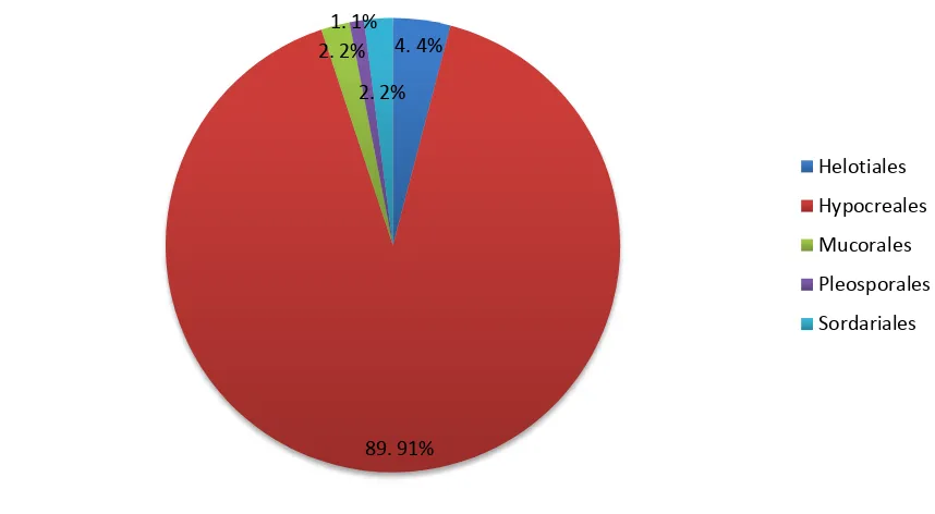
A total of 19 fungal species were isolated from Passalidae adult guts, principally from Hypocreales order which includes saprotrophic and symbiotic species associated to insects (66). As mentioned, studies regarding fungi associated to wood-decomposing insects are scarce and recent. Rojas-Jiménez et al (2015) and Vargas-Asensio et al (2014) published the results of the unique studies on the association of microbial diversity and Coleoptera families in Costa Rican tropical forests. Interestingly, Hypocreales was the most abundant fungi order isolated. These results are surprisingly similar to the results found in this present study, in which the order Hypocreales accounts for more than 88 of the 98 fungal isolates sequenced.
in the ability to survive in many ecological niches. The remaining orders isolated showed a lower abundance, being represented by a single fungal species. Other fungal genera found in common to the findings by Rojas-Jiménez et al (2015) are Scytalidium (Helotiales), Fusarium (Hypocreales) and Mucor (Mucorales). Chaunopycnis alba, isolated in this study is phylogenetically closely related to the genus Cordyceps, which is recognized as an insect parasite reported in beetle’s gut by Rojas-Jiménez et al (2015).
The fungal species isolated from passalid guts in this research, in most of the cases, take part in the decomposition process of soil and litter, because of their capabilities to degrade cello-oligosaccharides (51, 52, 66, 69). Mucor and Epicoccum belong to the initial phases as early colonizers (3, 70), while versatile fungal degraders such as Trichoderma and Fusarium (4) which have cellulase, xylase and lipase enzymes, join in this process around one or two years later, when decaying wood is almost completely decomposed. Given that fungal species of soil and litter succession were discovered in this research we suggest that different fungal species found in the gut of passalid beetles perform synergistically to complete several functions during degradation and fermentation of lignocellulosic materials (71); and contribute to satisfying the beetle nutritional needs. Fungi may create affinities for substrates, so members of the phylum Basidiomycota could degrade larger polymeric molecules, Ascomycota fungi can degrade diverse lignocellulosic constituents, while the bacteria degrade and ferment the smaller monomeric and dimeric hexoses and pentoses (71, 72).
source for growth in culture conditions plays an important role in the production of lignocellulolitic enzymes (75). In this assay, cellulose activity was not induced using a pre-media culture with cellulose as some articles suggest (76), we used a general pre-media for fungal growth, PDA (1X), so as to evaluate baseline production of cellulose degrading enzymes. A variety of substrates can be used for detecting and quantifying cellulose
enzymatic activity (74), we used CMC as a substrate to quantify exclusively endo-β-1,4-glucanase activity considering that fungal cellulose degrading activity is based
on principally endo- and exocellulases (17).
Endogenous cellulose activity has been reported in various insects that have wood habitats, as is the case of termites (14, 77). In beetles, the guts of Cerambycidae larvae and adults have been reported to have endogenous cellulase enzymes (76, 78–83), as is the case of the
Cerambycidae larvae of Anoplophora glabripennis, which exhibit endogenous endo-β-1,4-glucanase activity (15). Up to now there are no reports of endogenous
cellulases in passalid guts. The sugar reducing DNS method used in this study revelead that 67% beetle intestines had CMC degrading ability at the one time point evaluated; this could suggest the digestion of this carbon compound by endogenous enzymes of the beetle gut. However, it is evident that additional research is necessary to understand spatial digestion in the beetle intestine and determine the level of collaboration of fungal enzymes to the digestive process of the Passalidae beetle, as well as the contribution of other microorganisms to this complex process and possible endogenous production of cellulose degrading enzymes.
activity was also assayed by Rojas-Jiménez et al (2015) resulting in positive hydrolysis of all lignocellulase sources by Trichoderma. Interestingly, similar results were obtained in this study showing a stable cellulase activity during the eight-hour period of enzymatic assays using the extracts of the Trichoderma species and all other fungal extracts tested. The cellulose activity was comparably high within fungal extracts in Clonostachys rossmaniae strains cultivated under optimized conditions; the fungal isolate of this species also presented positive bioactivity in the degradation of polyurethane plastic in vitro (com. pers. Alexandra Narvaez-Trujillo). According to studies by Nilsson (1974), the fungal species Xygolone sp. and Scytalidium sp. had low enzymatic activity (69), which is also the case in this study for Scytalidium sp. that was one of the fungal extracts with the lowest CMC hydrolysis. The use of CMC for evaluation of cellulose degrading enzymes enable determining that there are endo-β-1,4-glucanase enzymes present both in the fungal species as the passalid guts; however additional assays are needed to determine other bioactive cellulose degrading enzymes in the fungal and gut extracts used.
hyperdiverse groups, such as fungi, it is not possible to carry out a complete survey of the fungal diversity at any investigation site (85). Sampling of the passalid beetles at each decaying tree uncovered a limited number of individual adults; all available individuals were taken. We consider this an exhaustive sampling of each site, despite not having an equitable number of individuals among the different passalid species identified. Additionally, the high number of fungal isolates obtained, by using different culture media, permits a robust estimate of fungal species richness associated to the passalid guts and enables differentiation among sites as well as among passalid species (71).
Our statistical analyses (NMDS, ANOSIM and SIMPER) suggest that each decaying tree could work as a microhabitat in itself, determined by spatial localization, soil composition, and species of felled tree. Tree no. 3, is the most differentiated. Tree no. 3 had the highest number of passalid specimens, so initially this could account for this differentiation, however we must also consider that this tree is spatially more distant to the other two trees sampled. Therefore, we suggest that the difference in fungal composition determined could also be accounted by different environmental conditions, chemical composition of soil and litter, time of tree decomposition and even spatial distances (67, 86). Studies on bacterial community composition in insect guts have demonstrated that host diet and taxonomy influence considerably the bacterial composition in the insect gut (12, 87, 88). Geib et al (2009) demonstrated that Cerambycidae beetles show preferences between tree species, which seem to be related to bacterial diversity (12). Environment conditions and chemical composition of soil and litter should be studied thoroughly to determine if these characteristics along with the distance affect fungal composition.
on which this beetle feeds on. Additionally, the fungal community composition analyses
revealed that there’s a significant difference of fungal composition among Passalidae species, indicating possible host specificity as observed in the study of cellobiose- and xylose-fermenting yeasts of Guatemalan Passalidae beetles’ guts carried out by Urbina et al (2013).
Our results point to an important contribution to the digestion process in Passalidae beetles and a contribution to biomass conversion in the tropical rainforest, nevertheless we cannot determine conclusively on if the fungal species found in Passalidae guts of this research are real endosymbionts or are transitory organisms inhabitants associated with decomposing trees from which Passalidae nourish. Furthermore, some of these microorganisms could perform as commensals, parasites, or facultative endosymbionts (89). They might even be using the insect as a dispersal mechanism. However, the similarity in results found in this study and the results presented by Rojas-Jiménez et al (2015) and Vargas-Asensio et al (2014) indicate that fungal species consistently isolated may have a close relationship with their insect hosts as for example the genus Trichoderma.
but for beetles fitness and evolutionary symbiotic relationships with insects. Additionally, in terms of biotechnology development, the fungal species recovered in this study have the potential to be investigated for industrial application such as the generation of biofuels based on biotic wastes.
ACKNOWLEDGMENTS
REFERENCES
1. Guggenberger G. 2005. Humification and mineralization in soils, p. 87–106. In Buscot, F, Varma, A (eds.), Microorganisms in soils: roles in genesis and functions. Springer, Berlin, Germany.
2. Dilly O. 2005. Microbial energetics in soils, p. 123–138. In Buscot, F, Varma, A (eds.), Microorganisms in soils: roles in genesis and functions. Springer, Berlin, Germany.
3. Frankland JC. 1998. Fungal succession — unravelling the unpredictable. Mycol Res 102:1–15.
4. Kjoller A, Struwe S. 2002. Fungal communities, succession, enzymes, and decomposition, p. 267–278. In Burns, R, Dick, R (eds.), Enzymes in the environment: activity, ecology and appications. Marcel Dekker, New Jersey, United States.
5. Schuster A, Schmoll M. 2010. Biology and biotechnology of Trichoderma. Appl Microbiol Biotechnol 87:787–799.
6. Cano EB, Shuster JC. 2012. La ecología de la degradación de la madera por escarabajos Passalidae (Coleoptera): simbiosis y efectos sobre el comportamiento. Rev 24 la Univ del Val Guatemala.
7. Anderson JM. 1995. Soil organisms as engineers: microsite modulation of macroscale processes, p. 94–106. In Jones, CG, Lawton, JH (eds.), Linking species and ecosystems. Chapman & Hall, London, United Kingdom.
8. Hunter MD, Adl S, Pringle CM, Coleman DC. 2003. Relative effects of macro invertebrates and habitat on the chemistry of litter during decomposition. Pedobiologia (Jena) 47:101–115.
9. Kuperman RG. 1996. Relationships between soil properties and community structure of soil macroinvertebrates in oak-hickory forests along an acidic deposition gradient. Appl Soil Ecol 4:125–137.
10. Wolters V. 2000. Invertebrate control of soil organic matter stability. Biol Fertil Soils 31:1–19.
11. Vargas-Asensio G, Pinto-Tomas A, Rivera B, Hernandez M, Hernandez C, Soto-Montero S, Murillo C, Sherman DH, Tamayo-Castillo G. 2014. Uncovering the cultivable microbial diversity of costa rican beetles and its ability to break down plant cell wall components. PLoS One 9:e113303.
13. Urbina H, Schuster J, Blackwell M. 2013. The gut of Guatemalan passalid beetles: a habitat colonized by cellobiose- and xylose-fermenting yeasts. Fungal Ecol 6:339–355.
14. Breznak JA, Brune A. 1994. Role of microorganisms in the digestion of lignocellulose by termites. Annu Rev Entomol 39:453–487.
15. Geib SM, Tien M, Hoover K. 2010. Identification of proteins involved in lignocellulose degradation using in gel zymogram analysis combined with mass spectroscopy-based peptide analysis of gut proteins from larval Asian longhorned beetles, Anoplophora glabripennis. Insect Sci 17:253–264.
16. Vatanparast M, Hosseininaveh V, Ghadamyari M, Minoo Sajjadian S. 2012. Pectinase and cellulase activity in the digestive system of the elm leaf beetle, Xanthogaleruca luteola Muller (Coleoptera: Chrysomelidae). J Asia Pac Entomol 15:555–561.
17. Martin MM. 1983. Cellulose digestion in insects. Comp Biochem Physiol 75A:313–324.
18. King BC, Donnelly MK, Bergstrom GC, Walker LP, Gibson DM. 2009. An optimized microplate assay system for quantitative evaluation of plant cell wall-degrading enzyme activity of fungal culture extracts. Biotechnol Bioeng 102:1033–
44.
19. Berkov A, Feinstein J, Small J, Nkamany M. 2007. Yeasts isolated from Neotropical wood-boring beetles in SE Peru. Biotropica 39:530–538.
20. Douglas, A E. 1989. Mycetocyte symbiosis in insects. Biol Rev Camb Philos Soc 64:409–434.
21. Watanabe H, Tokuda G. 2010. Cellulolytic systems in insects. Annu Rev Entomol 55:609–32.
22. Houseknecht JL, Hart EL, Suh S-O, Zhou JJ. 2011. Yeasts in the Sugiyamaella clade associated with wood-ingesting beetles and the proposal of Candida bullrunensis sp. nov. Int J Syst Evol Microbiol 61:1751–1756.
23. Suh S-O, Blackwell M. 2005. Four new yeasts in the Candida mesenterica clade associated with basidiocarp-feeding beetles. Mycologia 97:167–177.
24. Borges TA, de Souza AT, Squina FM, Riaño-Pachón DM, Corrêa Dos Santos RA, Machado E, Velasco De Castro Oliveira J, Damásio ARL, Goldman GH. 2014. Biochemical characterization of an endoxylanase from Pseudozyma brasiliensis sp. nov. strain GHG001 isolated from the intestinal tract of Chrysomelidae larvae associated to sugarcane roots. Process Biochem 49:77–83. 25. Delalibera I, Handelsman J, Raffa KF. 2005. Contrasts in cellulolytic activities of
Cerambycidae), and the bark beetles, Ips pini and Dendroctonus frontalis (Coleoptera: Curculionidae). Environ Entomol 34:541–547.
26. Reid NM, Addison SL, Macdonald LJ, Lloyd-Jones G. 2011. Biodiversity of active and inactive bacteria in the gut flora of wood-feeding huhu beetle larvae (Prionoplus reticularis). Appl Environ Microbiol 77:7000–7006.
27. Suh S-O, Marshall CJ, Mchugh J V, Blackwell M. 2003. Wood ingestion by passalid beetles in the presence of xylose-fermenting gut yeasts. Mol Ecol 12:3137– 3145.
28. Suh S-O, Nguyen NH, Blackwell M. 2008. Yeasts isolated from plant-associated beetles and other insects: seven novel Candida species near Candida albicans. FEMS Yeast Res 8:88–102.
29. Suh S-O, Houseknecht JL, Gujjari P, Zhou JJ. 2013. Scheffersomyces parashehatae f.a., sp. nov., Scheffersomyces xylosifermentans f.a., sp. nov., Candida broadrunensis sp. nov. and Candida manassasensis sp. nov., novel yeasts associated with wood-ingesting insects, and their ecological and biofuel implicat. Int J Syst Evol Microbiol 63:4330–4339.
30. Agbogbo FK, Coward-Kelly G. 2008. Cellulosic ethanol production using the naturally occurring xylose-fermenting yeast, Pichia stipitis. Biotechnol Lett 30:1515–24.
31. Farrell BD, Sequeira AS, O’Meara BC, Normark BB, Chung JH, Jordal BH. 2001. The evolution of agriculture in beetles (Curculionidae: Scolytinae and Platypodinae). Evolution (N Y) 55:2011–2027.
32. Graham K. 1967. Fungal-insect mutualism in trees and timber. Annu Rev Entomol 12:105–126.
33. Hulcr J, Cognato AI. 2010. Repeated evolution of crop theft in fungus-farming ambrosia beetles. Evolution (N Y) 64:3205–3212.
34. Hulcr J, Mogia M, Isua B, Novotny V. 2007. Host specificity of ambrosia and bark beetles (Col., Curculionidae: Scolytinae and Platypodinae) in a New Guinea rainforest. Ecol Entomol 32:762–772.
35. Hulcr J, Novotny V, A. Maurer B, I. Cognato A. 2008. Low beta diversity of ambrosia beetles (Coleoptera: Curculionidae: Scolytinae and Platypodinae) in lowland rainforests of Papua New Guinea. Oikos 117:214–222.
36. Jiménez-Ferbans L, Amat-García G. 2010. Clave para los géneros y especies de Passalidae (Coleoptera: Scarabaeoidea) del Caribe colombiano. Intropica 57–62. 37. Boucher S. 2005. Évolution et phylogénie des Coléoptères Passalidae
38. Reyes-Castillo P, Amat-García G. 1991. Notas sobre la taxonomía y distribución de Passalidae (Insecta: Coleoptera) en Colombia y descripción de una nueva especie. Caldasia 16:501–508.
39. Shuster JC, Shuster LC. 1985. Social behavior in passalid beetles ( Coleoptera :
Passalidae ): Cooperative brood care. Florida Entomol 68:266–272.
40. Lichtwardt RW, White MM, Cafaro MJ, Misra JK. 1999. Fungi associated with passalid beetles and their mites. Mycologia 91:694–702.
41. Nguyen NH, Suh S-O, Marshall CJ, Blackwell M. 2006. Morphological and ecological similarities: wood-boring beetles associated with novel xylose-fermenting yeasts, Spathaspora passalidarum gen. sp. nov. and Candida jeffriesii sp. nov. Mycol Res 110:1232–1241.
42. Nguyen NH, Suh S-O, Blackwell M. 2007. Five novel Candida species in insect-associated yeast clades isolated from Neuroptera and other insects. Mycologia 99:842–858.
43. Suh S-O, Gibson CM, Blackwell M. 2004. Metschnikowia chrysoperlae sp. nov., Candida picachoensis sp. nov. and Candida pimensis sp. nov., isolated from the green lacewings Chrysoperla comanche and Chrysoperla carnea (Neuroptera: Chrysopidae). Int J Syst Evol Microbiol 54:1883–1890.
44. Bucheli E, Gautschi B, Shykoff J a. 2000. Host-specific differentiation in the anther smut fungus Microbotryum violaceum as revealed by microsatellites. J Evol Biol 13:188–198.
45. White TJ, Bruns T, Lee SJWT, Taylor JW. 1998. Amplification and direct sequencing of fungal ribosomal RNA genes for phylogenetics PCR protocols: a guide to methods and applications.
46. Edgar RC. 2004. MUSCLE : multiple sequence alignment with high accuracy and
high throughput. Nucleic Acids Res 32:1792–1797.
47. Stamatakis A. 2006. RAxML-VI-HPC : Maximum likelihood-based phylogenetic analyses with thousands of taxa and mixed models. Bioinformatics 22:2688–2690. 48. Gladden JM, Allgaier M, Miller CS, Hazen TC, Vandergheynst JS, Hugenholtz
P, Simmons BA, Singer SW. 2011. Glycoside hydrolase activities of thermophilic bacterial consortia adapted to switchgrass . Appl Environ Microbiol 77:5804–5812. 49. Hoyos-Carvajal L, Orduz S, Bissett J. 2009. Genetic and metabolic biodiversity
of Trichoderma from Colombia and adjacent neotropic regions. Fungal Genet Biol 46:615–631.
51. Soares de Melo I, Montes Peral Valente A, Kavamura VN, Dias Vilela ES, Faull JL. 2014. Mycoparasitic nature of Bionectria sp. strain 6.21. J Plant Prot Res 54:327–333.
52. Pečiulytė D. 2007. Isolation of cellulolytic fungi from waste paper gradual recycling materials. Ekologija 53:11–18.
53. Ögel ZB, Yarangümeli K, Dü H, Ifrij I. 2001. Submerged cultivation of Scytalidium thermophilum on complex lignocellulosic biomass for endoglucanase production. Enzyme Microb Technol 28:689–695.
54. Ghose TK. 1987. Measurement of cellulase actitivies. Pure Appl Chem 59:257–
268.
55. Gotelli NJ, Colwell RK. 2001. Quantifying biodiversity: Procedures and pitfalls in the measurement and comparison of species richness. Ecol Lett 4:379–391.
56. Unterseher M, Schnittler M, Dormann C, Sickert A. 2008. Application of species richness estimators for the assessment of fungal diversity. FEMS Microbiol Lett 282:205–213.
57. Hammer Ø, Harper DAT, Ryan PD. 2001. Past: Paleontological Statistics Software Package for Education and Data Analysis. Palaeontol Electron 6:1–9. 58. Amat-García G, Reyes-Castillo P. 2007. Los Passalidae (Coleoptera:
Scarabaeoidea: Passalidae) del departamento del Amazonas, Colombia. Caldasia 29:329–354.
59. Reyes-Castillo P. 2000. Coleoptera Passalidae de México. PRIBES.
60. Amat-García G, Reyes-Castillo P. 2002. Los Coleoptera Passalidae de Colombia. PRIBES.
61. Schuster J, Cano E. 2005. Clave para los géneros de los Passalidae americanos. 62. Naji KM, Abdullah QYM, Al-Zaqri AQM, Alghalibi SM. 2014. Evaluating the
biodeterioration enzymatic activities of fungal contamination isolated from some ancient yemeni mummies preserved in the national museum. Biochem Res Int 2014:1–9.
63. Ghose T. 1969. Continuous enzymatic saccharification of cellulose with culture filtrates of Trichoderma viride QM 6a. Biotechnol Bioeng 11:239–261.
64. Dashtban M, Maki M, Leung KT, Mao C, Qin W. 2010. Cellulase activities in biomass conversion: measurement methods and comparison. Crit Rev Biotechnol 30:1–8.
66. Torres MS, White JF. 2009. Clavicipitaceae: free-living and saprotrophs to plant endophytes, p. 422–430. In Schaechter, M (ed.), Encyclopedia of Microbiology. Elsevier, Oxford, United Kingdom.
67. Papavizas GC. 1985. Trichoderma and Gliocladium: Biology, ecology, and potential for biocontrol. Annu Rev Phytopathol 23:23–54.
68. Shahid M, Srivastava M, Kumar V, Singh A, Sharma A, Pandey S, Rastogi S, Pathak N, Srivastava AK. 2014. Phylogenetic diversity analysis of Trichoderma species based on internal transcribed spacer (ITS) marker. African J Biotechnol 13:449–455.
69. Nilsson T. 1974. Microscopic studies on the degradation of cellophane and various cellulosic fibres by wood-attacking microfungi Studia Forestalia Suecica.
70. Webster J, Dix NJ. 1960. Succession of fungi on decaying cocksfoot culms. Trans Br Mycol Soc 43:85–99.
71. Rojas-Jiménez K, Hernández M. 2015. Isolation of fungi and bacteria associated with the guts of tropical wood-feeding Coleoptera and determination of their lignocellulolytic activities. Int J Microbiol 2015:1–11.
72. Baldrian P, Valásková V. 2008. Degradation of cellulose by basidiomycetous fungi. FEMS Microbiol Rev Rev 32:501–521.
73. Nannipieri P, Kandeler E, Ruggiero P. 202AD. Enzyme activities and microbiological and biochemical processes in soil, p. 1–33. In Burns, R, Dick, R (eds.), Enzymes in the environment: Activity, ecology and appications. Marcel Dekker, New Jersey, United States.
74. Willis JD, Oppert C, Jurat-Fuentes JL. 2010. Methods for discovery and characterization of cellulolytic enzymes from insects. Insect Sci 17:184–198.
75. Jatinder K, Chadha BS, Saini HS. 2006. Optimization of culture conditions for production of cellulases and xylanases by Scytalidium thermophilum using response surface methodology. World J Microbiol Biotechnol 22:169–176.
76. Scully ED, Hoover K, Carlson J, Tien M, Geib SM. 2012. Proteomic analysis of Fusarium solani isolated from the Asian longhorned beetle, Anoplophora glabripennis. PLoS One 7:e32990.
77. Zhang D, Lax AR, Raina AK, Bland JM. 2009. Differential cellulolytic activity of native-form and C-terminal tagged-form cellulase derived from Coptotermes formosanus and expressed in E. coli. Insect Biochem Mol Biol 39:516–522.
79. Genta FA, Terra WR, Ferreira C. 2003. Action pattern, specificity, lytic
activities, and physiological role of five digestive β-glucanases isolated from Periplaneta americana. Insect Biochem Mol Biol 33:1085–1097.
80. Dojnov B, Bozić N, Nenadović V, Ivanović J, Vujcić Z. 2008. Purification and properties of midgut alpha-amylase isolated from Morimus funereus (Coleoptera: Cerambycidae) larvae. Comp Biochem Physiol Part B 149:153–160.
81. Dojnov B, Lončar N, Božić N, Nenadović V, Jelisaveta I, Vujčić Z. 2010.
Comparison of α-amylase isoforms from the midgut of Cerambyx cerdo L.
(Coleoptera: Cerambycidae) larvae developed in the wild and on an artificial diet. Arch Biol Sci 62:575–583.
82. Dojnov B, Pavlović R, Božić N, Margetić A, Nenadović V, Ivanović J, Vujčić Z. 2013. Expression and distribution of cellulase, amylase and peptidase isoforms along the midgut of Morimus funereus L. (Coleoptera: Cerambycidae) larvae is dependent on nutrient substrate composition. Comp Biochem Physiol B Biochem Mol Biol 164:259–267.
83. Pavlović R, Grujić M, Dojnov B, Vujčić M, Nenadović V, Ivanović J, Vujčić Z. 2012. Influence of nutrient substrates on the expression of cellulases in Cerambyx cerdo L. (Coleoptera: Cerambycidae) larvae. Arch Biol Sci 64:757–765.
84. Schuster J, Schuster L. 1997. The evolution of social behavior in Passalidae (Coleoptera), p. 260–269. In Choe, JC, Crespi, BJ (eds.), The evolution of social behavior in insects and arachnids. Cambridge University Press, Cambridge, United Kingdom.
85. Unterseher M, Reiher A, Finstermeier K, Otto P, Morawetz W. 2007. Species richness and distribution patterns of leaf-inhabiting endophytic fungi in a temperate forest canopy. Mycol Prog 6:201–212.
86. Harman GE, Herrera-Estrella AH, Horwitz B a, Lorito M. 2012. Special issue: Trichoderma-from basic Biology to Biotechnology. Microbiology 158:1–2.
87. Colman DR, Toolson EC, Takacs-Vesbach CD. 2012. Do diet and taxonomy influence insect gut bacterial communities? Mol Ecol 21:5124–5137.
88. Santo Domingo JW, Kaufman MG, Klug MJ, Holben WE, Harris D, Tiedje JM. 1998. Influence of diet on the structure and function of the bacterial hindgut community of crickets. Mol Ecol 7:761–767.
A B
[image:48.595.81.483.83.461.2]
Figure 1. A. Adult passalid beetle, Passalus intertitialis. B. Passalus intertitialis dissected with its entire gut.
W
ho
le i
ntes
ti
ne
d
isse
cted
a
nd
tes
ted
f
or
ce
ll
ulo
ly
ti
c
ac
ti
vit
Figure 3. Orders of fungi isolated from passalid guts. Percentage of isolates classified in each order (N=98).
4. 4%
89. 91% 2. 2%
1. 1%
2. 2%
Helotiales
Hypocreales
Mucorales
Pleosporales
[image:52.595.95.501.227.526.2]
Figure 7. Non-metric multidimensional scaling (NMDS) ordination of fungal isolates from passalid beetles inhabiting three decaying trees en Ecuadorian Amazonia. Tree no. 1 (SLA, SLB and SLE), tree no. 2 (SLC and SLR) and tree no. 3 (SLG, SLH, SLI, SLK, SLL, SLM, SLN and SLP). See Appendix 1 for details regarding abbreviations.
SLA SLB SLE SLC SLR SLG SLH SLI SLK SLL SLM SLN SLP
-0,6 -0,5 -0,4 -0,3 -0,2 -0,1 0,0 0,1 0,2 0,3
Figure 8. Non-metric multidimensional scaling (NMDS) ordination of fungi isolated from the guts of wood-inhabiting Passalidae beetle species Passalus interstitialis (SLA and SLP), Passalus variiphyllus (SLB and SLC) and Passalus sp. (SLE, SLG, SLH, SLI, SLK, SLL, SLM, SLN and SLR). See Appendix 1 for details regarding abbreviations.
SLA SLP SLB SLC SLE SLG SLH SLI SLK SLL SLM SLN SLR
-0,6 -0,5 -0,4 -0,3 -0,2 -0,1 0,0 0,1 0,2 0,3
Table 1. Cellulase activities of extracts of fungi isolated from the guts of three Passalidae species beetles based on reducing sugar methodology.
Collection code Fungal species tested
Cellulase activity (umol/min)
Standard deviation
CEQCA-O3783 Clonostachys rossmaniae 0.08 0.040
CEQCA-O3772 Epicoccum nigrum 0.07 0.01
CEQCA-O3778 Trichoderma sp. 0.06 0.00
CEQCA-O4922 Trichoderma hamatum 0.06 0.01
CEQCA-O4879 Trichoderma spirale 0.06 0.02
CEQCA-O3794 Chaunopycnis alba 0.04 0.01
CEQCA-O4929 Trichoderma asperellum 0.04 0.01 CEQCA-O3792 Campylocarpon pseudofasciculare 0.03 0.00
CEQCA-O4950 Trichoderma virens 0.03 0.00
CEQCA-O4991 Scytalidium sp. 0.03 0.00
Table 2. Cellulase activity of the gut of Passalidae beetles. Positive (+) cellulase activity and negative (-) cellulase activity
Passalidae field codes
Passalidae species
Cellulase activity
LV4AB Passalus sp. -
LV4BA Passalus sp. +
LV4BE Passalus sp. +
LV4CD Passalus sp. -
LV4FA Passalus interstitialis
+
Table 3. Diversity indexes of fungi isolated and fungal diversity indexes according to each decaying tree.
Fungal diversity indexes
Tree no. 1 (Inga spectabilis)
Tree no. 2 Tree no. 3
Taxa S 19 7 6 13
Individuals 98 20 13 65
Shannon 2.13 1.54 1.52 1.74
Table 4. Fungal diversity indexes according to each Passalidae species.
Passsalus
variiphyllus Passalus sp.
Passalus interstitialis
Taxa S 9 14 4
Individuals 21 64 13
Shannon 1.75 1.89 1.03
Table 5. Analysis of similarities (ANOSIM) of fungal composition according to decaying trees (R= 0.5177, P= 0.0058).
Tree no. 1 Tree no. 2 Tree no. 3
Tree no. 1 - 0.40 0.01
Tree no. 2 0.40 - 0.04
Table 6. Analysis of similarities (ANOSIM) of fungi composition according to passalid species (R= 0.2816, P= 0.0977).
Passalus interstitialis
Passalus
variiphyllus Passalus sp. Passalus
interstitialis - 1.00 0.56
Passalus
variiphyllus 1.00 - 0.06
Table 7. Similarity percentage (SIMPER) analysis shows fungal species that contributed the most (as a percentage) to the total dissimilarity within all three decaying trees.
Comparing decaying trees
Dissimilarity %
Fungal species Av.
dissim Contrib. % Cumulative % Mean abund. 1 Mean abund. 2 Mean abund. 3
Tree no. 1, 74.87 Trichoderma harzianum 19.20 25.65 25.65 0.67 2.00 4.13
tree no. 2, Trichoderma spirale 19.10 25.51 51.16 3.33 2.50 0.13
tree no. 3 Trichoderma virens 6.49 8.67 59.83 0.00 0.00 1.00
Trichoderma atroviride 5.63 7.52 67.35 0.00 0.00 1.00
Trichoderma hamatum 4.33 5.78 73.12 1.00 0.00 0.00
Fusarium solani 3.84 5.13 78.25 0.00 0.50 0.50
Trichoderma strigosum 2.28 3.05 81.29 0.67 0.00 0.00
Clonostachys rossmaniae 2.05 2.73 84.02 0.33 0.00 0.00
Mucor irregularis 1.80 2.41 86.43 0.00 0.50 0.13
Epicoccum nigrum 1.51 2.02 88.45 0.00 0.50 0.00
Trichoderma asperellum 1.51 2.02 90.47 0.00 0.50 0.00
Campylocarpon pseudofasciculare 1.45 1.93 92.40 0.33 0.00 0.13
Trichoderma sp. 2 1.14 1.52 93.92 0.33 0.00 0.00
Chloridium virescens 1.13 1.51 95.43 0.00 0.00 0.25
Xylogone ganodermophthora 0.90 1.20 96.63 0.00 0.00 0.25
Scytalidium sp. 0.90 1.20 97.83 0.00 0.00 0.25
Chaunopycnis alba 0.73 0.97 98.80 0.00 0.00 0.13
Trichoderma sp. 1 0.45 0.60 99.40 0.00 0.00 0.13
Table 8. Similarity percentage (SIMPER) analysis shows fungal species that contributed the most (as a percentage) to the total dissimilarity comparing within three decaying trees. Only fungal species showing more than 2% of contribution to dissimilarity were considered
Comparing decaying trees
Dissimilarity %
Fungal species Av.
dissim Contrib. % Cumulative % Mean abund. 1 Mean abund. 2 Tree no. 1, 59.52 Trichoderma spirale 16.09 27.04 27.04 3.33 2.50
tree no. 2 Trichoderma harzianum 10.40 17.48 44.51 0.67 2.00
Trichoderma hamatum 7.04 11.82 56.33 1.00 0.00
Fusarium solani 5.21 8.75 65.09 0.00 0.50
Trichoderma strigosum 3.67 6.17 71.25 0.67 0.00
Clonostachys rossmaniae 3.37 5.66 76.91 0.33 0.00
Epicoccum nigrum 3.36 5.64 82.55 0.00 0.50
Trichoderma asperellum 3.36 5.64 88.19 0.00 0.50
Mucor irregularis 3.36 5.64 93.83 0.00 0.50
Campylocarpon pseudofasciculare 1.84 3.08 96.92 0.33 0.00
Trichoderma sp. 2 1.84 3.08 100.00 0.33 0.00
Tree no. 1, 85.73 Trichoderma harzianum 24.47 28.55 28.55 0.67 4.13
tree no. 3 Trichoderma spirale 21.83 25.46 54.01 3.33 0.13
Trichoderma virens 7.52 8.77 62.78 0.00 1.00
Trichoderma hamatum 6.53 7.62 70.39 1.00 0.00
Trichoderma atroviride 6.51 7.59 77.99 0.00 1.00
Trichoderma strigosum 3.45 4.03 82.01 0.67 0.00
Clonostachys rossmaniae 3.08 3.59 85.60 0.33 0.00
Fusarium solani 2.89 3.37 88.97 0.00 0.50
Campylocarpon pseudofasciculare 1.97 2.29 91.26 0.33 0.13
[image:64.842.118.730.148.512.2]Table 8. Continued
Comparing decaying trees
Dissimilarity %
Fungal species Av.
dissim
Contrib. %
Cumulative %
Mean abund.
1
Mean abund.
2 Tree no. 2, 64.35 Trichoderma spirale 16.15 25.09 25.09 2.50 0.13
tree no. 3 Trichoderma harzianum 14.60 22.68 47.78 2.00 4.13
Trichoderma virens 7.38 11.47 59.25 0.00 1.00
Trichoderma atroviride 6.42 9.98 69.22 0.00 1.00
Fusarium solani 4.75 7.39 76.61 0.50 0.50
Mucor irregularis 3.14 4.88 81.49 0.50 0.13
Epicoccum nigrum 3.09 4.80 86.30 0.50 0.00
Trichoderma asperellum 3.09 4.80 91.10 0.50 0.00
Table 9. Similarity percentage (SIMPER) analysis shows fungal species that contributed the most (as a percentage) to the total dissimilarity within all three Passalidae species.
Comparing Passalidae species
Dissimilarity %
Fungal species Av.
dissim Contrib. % Cumulative % Mean abund. 1 Mean abund. 2 Mean abund. 3 P. interstitialis, 72.12 Trichoderma spirale 20.82 28.87 28.87 1.50 5.00 0.33
P. variiphyllus, Trichoderma harzianum 17.86 24.76 53.63 4.00 1.00 3.22
Passalus sp. Trichoderma virens 4.73 6.56 60.19 0.50 0.00 0.78
Trichoderma atroviride 4.57 6.34 66.53 0.00 0.00 0.89
Trichoderma hamatum 3.55 4.92 71.45 0.00 1.00 0.11
Trichoderma strigosum 3.01 4.17 75.63 0.00 1.00 0.00
Fusarium solani 2.90 4.01 79.64 0.00 0.00 0.56
Mucor irregularis 2.01 2.79 82.43 0.00 0.50 0.11
Epicoccum nigrum 1.81 2.51 84.94 0.00 0.50 0.00
Trichoderma asperellum 1.81 2.51 87.45 0.00 0.50 0.00
Chaunopycnis alba 1.74 2.41 89.87 0.50 0.00 0.00
Campylocarpon pseudofasciculare 1.72 2.39 92.25 0.00 0.50 0.11
Trichoderma sp. 2 1.51 2.09 94.34 0.00 0.50 0.00
Chloridium virescens 0.94 1.30 95.64 0.00 0.00 0.22
Clonostachys rossmaniae 0.85 1.18 96.82 0.00 0.00 0.11
Xylogone ganodermophthora 0.76 1.06 97.88 0.00 0.00 0.22
Scytalidium sp. 0.76 1.06 98.94 0.00 0.00 0.22
Trichoderma sp. 1 0.38 0.53 99.47 0.00 0.00 0.11
[image:66.842.87.773.148.485.2]Table 10. Similarity percentage (SIMPER) analysis shows fungal species that contributed the most (as a percentage) to the total dissimilarity comparing within three Passalidae species. Only fungal species showing more than 2% of contribution to dissimilarity were considered.
Comparing Passalidae species
Dissimilarity %
Fungal species Av.
dissim Contrib. % Cumulative % Mean abund. 1 Mean abund. 2 P. interstitialis, 69.68 Trichoderma spirale 19.31 27.71 27.71 1.50 5.00
P. variiphyllus Trichoderma harzianum 18.76 26.93 54.64 4.00 1.00
Trichoderma strigosum 5.51 7.90 62.54 0.00 1.00
Trichoderma hamatum 5.51 7.90 70.44 0.00 1.00
Epicoccum nigrum 3.31 4.75 75.19 0.00 0.50
Trichoderma asperellum 3.31 4.75 79.94 0.00 0.50
Mucor irregularis 3.31 4.75 84.70 0.00 0.50
Campylocarpon pseudofasciculare 2.75 3.95 88.65 0.00 0.50
Trichoderma sp. 2 2.75 3.95 92.60 0.00 0.50
Chaunopycnis alba 2.58 3.70 96.30 0.50 0.00
Trichoderma virens 2.58 3.70 100.00 0.50 0.00
P. interstitialis, 62.82 Trichoderma harzianum 22.99 36.60 36.60 4.00 3.22
Passalus sp. Trichoderma spirale 14.54 23.15 59.75 1.50 0.33
Trichoderma atroviride 5.71 9.08 68.83 0.00 0.89
Trichoderma virens 5.58 8.88 77.71 0.50 0.78
Fusarium solani 3.65 5.82 83.53 0.00 0.56
Table 10. Continued
Comparing Passalidae species
Dissimilarity %
Fungal species Av.
dissim
Contrib. %
Cumulative %
Mean abund.
1
Mean abund.
2
P. variiphyllus, 81.96 Trichoderma spirale 27.44 33.48 33.48 5.00 0.33
Passalus sp. Trichoderma harzianum 12.52 15.28 48.75 1.00 3.22
Trichoderma hamatum 5.54 6.76 55.51 1.00 0.11
Trichoderma strigosum 5.46 6.67 62.18 1.00 0.00
Trichoderma atroviride 4.45 5.43 67.61 0.00 0.89
Trichoderma virens 4.37 5.33 72.94 0.00 0.78
Epicoccum nigrum 3.29 4.02 76.96 0.50 0.00
Trichoderma asperellum 3.29 4.02 80.98 0.50 0.00
Mucor irregularis 3.27 3.99 84.97 0.50 0.11
Fusarium solani 2.78 3.39 88.36 0.00 0.56
Campylocarpon pseudofasciculare 2.75 3.36 91.72 0.50 0.11
Appendix 1. Fungal groups according the Passalidae species from each decaying tree. Ninety-eight fungal species were identified.
Fungal group
no.
Fungal group
code
Passalidae field code
Passalidae species Decaying tree type CEQCA code Fungi species isolated
1 SLA LV1EA Passalus interstitialis Tree no. 1 CEQCA-O4877 Trichoderma spirale (Inga spectabilis) CEQCA-O4924 Trichoderma harzianum
CEQCA-O4878 Trichoderma spirale CEQCA-O4879 Trichoderma spirale 2 SLB LV1DA, LV1DB Passalus variiphyllus Tree no. 1 CEQCA-O4982 Trichoderma hamatum
(Inga spectabilis) CEQCA-O3779 Trichoderma spirale CEQCA-O3771 Trichoderma spirale CEQCA-O4969 Trichoderma spirale CEQCA-O4970 Trichoderma spirale
CEQCA-O3792 Campylocarpon pseudofasciculare CEQCA-O4961 Trichoderma sp.2
CEQCA-O3400 Trichoderma strigosum CEQCA-O4983 Trichoderma spirale CEQCA-O4920 Trichoderma strigosum CEQCA-O4921 Trichoderma hamatum CEQCA-O3784 Trichoderma spirale
3 SLC LV3AA Passalus variiphyllus Tree no. 2 CEQCA-O3772 Epicoccum nigrum