P
Rev Inves Clin. 2017;69:77-93
Received for publication: 13-02-2017 Approved for publication: 01-03-2017
Additionally, young patients are faced with the threat of a potentially fatal illness at the peak of their pro-fessional careers and family planning years. Further-more, the toxicity profile of the treatments they re-ceive may be much more difficult to tolerate than in older patients, with a detrimental effect on fertility and risk of premature menopause and its associated short-term and long-term risks. For these reasons, young patients tend to be less adherent to systemic adjuvant therapies, especially to endocrine therapy (ET), which can increase the chance of recurrence5.
In recent years there has been a significant improve-ment in the understanding of the biology of breast cancer in young women. The most important finding
INTRODUCTION
Breast cancer in young women is an uncommon disease. However, despite the relatively low absolute risk, it is the leading cause of death amongst women under age 45 in high-income countries, and therefore it presents a sub-stantial public health concern1. In addition, it was found that breast cancer incidence and mortality for Latin American women aged < 45 years were significantly higher in comparison to those of developed countries2.
Breast cancer in young patients represents a special entity. The main reason for this is its unique biology which, compared to older patients, is more aggressive and typically presents at a more advanced stage3,4.
Corresponding author:
Eitan Amir
Princess Margaret Cancer Centre
Division of Medical Oncology and Hematology 700 University Ave 7-721
Toronto ON M5G 1Z5, Canada E-mail: [email protected]
ABSTRACT
Breast cancer in young women is a complex disease to manage due to its biological heterogeneity and special issues related to toxicity of different treatment strategies. Defining a cut-off for young age has been challenging since it is not clear whether the prognostic effect of age is continuously variable or whether there are certain thresholds at which the prognosis changes (e.g. those < 50 years of age or ≤ 35 years of age). In this review article, we define young patients as those being premeno-pausal. In addition, we discuss the most recent data of the biological diversity of breast cancer arising in premenopausal patients and current treatment modalities in early and advanced settings. Survivorship, with special emphasis on the importance of early supportive care, is also discussed. (REV INVES CLIN. 2017;69:77-93)
Key words: Age. Biology. BRCA. Breast cancer. Menopause. Treatment. Young women.
General Overview and Treatment
Recommendations for Young Women
with Breast Cancer
Domen Ribnikar
1, Ivica Ratoša
2, Andraž Perhavec
2and Eitan Amir
1*
is that although age is a significant factor to consider, it should not be the only or the most important factor for the choice of treatment for both early and ad-vanced disease. To avoid overtreatment of young breast cancer patients, treatment planning should be made according to the biological characteristics of the tumor, its stage, and the patient’s characteristics6,7. A cut-off of age to define “young breast cancer pa-tients” cannot be determined accurately since the bio-logical, hormonal, and environmental milieu underlying tumor biology is continuously changing during a life-time8. Most research refers to young patients as those being under age 35 or 40; however, it is still unclear whether young age as a prognostic factor represents a continuous variable or may be an inherent feature of wider subgroups, i.e. premenopausal (< 50 years) or very young patients (< 35 years of age)9,10.
For this review article, we define young patients as those before menopause as we will focus on the biology and current treatment modalities in this important sub-group of breast cancer patients. Survivorship, with special emphasis on the importance of early support-ive care, is also discussed.
DEFINING MENOPAUSE
The accurate identification of a woman’s menopausal status is crucial for treatment planning. Aromatase in-hibitors (AI) are contraindicated in pre- and perimeno-pausal women as they can induce ovarian hyperstimula-tion and cause an increase in ovarian produchyperstimula-tion of estrogens, which may lead to a hypothetical increased risk of breast cancer relapse11,12. Additionally, there is an increased risk of unplanned pregnancy with their use. The term “natural menopause” is defined as the perma-nent cessation of menstruation resulting from the loss of ovarian activity. It is recognized to have occurred after 12 consecutive months of amenorrhea for which there is no other obvious pathological and physiological cause13. Since breast cancer patients can become amen-orrheic during or after adjuvant systemic chemotherapy and may receive tamoxifen as adjuvant endocrine treat-ment, it is inappropriate to use the definition for meno-pause described above. Additionally, the National Com-prehensive Cancer Network’s definition of menopause specifically notes that it is not possible to identify the menopausal status of women receiving luteinizing
hormone-releasing hormone (LHRH) analogues, and that amenorrhea after chemotherapy is not a reliable indica-tor of menopausal status14. Furthermore, the criteria for identifying patients as postmenopausal vary between trials and research groups15-17. It is important to high-light also that levels of sex hormones “in the postmeno-pausal range” can occur during the perimenopostmeno-pausal stage and additional factors such as body mass index, lifestyle, and menstrual irregularity may also lead to a misdiagnosis of menopause when based on amenorrhea alone18. Confirmatory biochemical tests can also be un-informative in patients being treated with tamoxifen since the latter is associated with a marked fall in follicle-stimulating hormone levels19. In addition, estradiol lev-els can be elevated by tamoxifen due to cross-reactiv-ity of tamoxifen and its metabolites and the estradiol assay20. In summary, the clinical significance of amen-orrhea and determination of follicle-stimulating hor-mone levels and estradiol levels as surrogates for menopause are not clear in this patient population21. Defining menopause in amenorrheic breast cancer patients after chemotherapy (CT) is also challenging. It is not adequate to use cessation of menstruation as the only surrogate marker of menopause after chemotherapy as there have been case reports of spontaneous conception in women with amenorrhea after chemotherapy22.
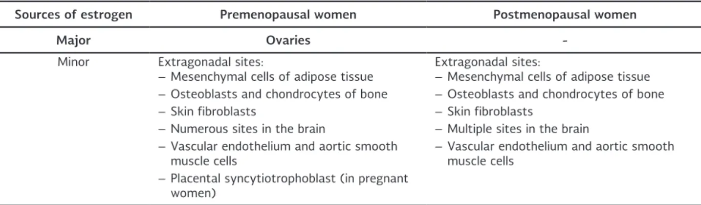
Additionally, there are data suggesting that 27% of patients achieving chemotherapy induced amenorrhea regained ovarian function after starting AI treatment at a median time of 12 months (range 4-59)12. These data suggest that an arbitrary cutoff of 12 months period of amenorrhea is not suitable for menopause determination and in such patients, AIs should not be used in the absence of concurrent ovarian function sup-pression (OFS). Consequently, we urge extreme caution in the use of AIs without ovarian function suppression in women < 50 years old with amenorrhea after che-motherapy or tamoxifen use. Table 1 represents sourc-es of sourc-estrogen in pre- and postmenopausal women.
BIOLOGY
Clinicopathological characteristics
of breast cancer in young women
population according to actual age. The first one, which included 1,427 premenopausal patients aged ≤ 50 years at the time of primary diagnosis, demonstrated signifi-cant age-related differences in aggressive features: es-trogen receptor (ER) and/or progesterone receptor (PR) negativity, human epidermal growth factor receptor 2 (HER2) positivity, presence of lymphovascular invasion, grade 3, Ki67 ≥ 20%23. These pathological features were observed more frequently in cancers arising in younger patients. Similar results were reported by the study of Han, et al., which included 9,885 premenopausal pa-tients aged ≤ 50 years at the time of primary breast cancer diagnosis24. Survival outcomes of patients aged 35-39 years were not different from those of patients aged 40-50 after adjustment for important prognostic factors. However, in patients aged < 35 years, the risk of death rose by 5% for every year of decrease in age. More recently, published data of the POSH study, the largest prospective observational study evaluating pathological characteristics of breast cancer patients under age 40, have shown that the majority of these patients had high-grade tumors (59%) of ductal his-tology (87%)25. Half of the patients had node-posi-tive disease and multifocality was observed in 27% of patients. One third of tumors were ER-negative and one-quarter HER2-positive.
The advent of genomic signatures has allowed a better understanding of the biological heterogeneity of breast cancer. Four main intrinsic subtypes of breast cancer are recognized: luminal A, luminal B, HER2-enriched, and basal-like26-28. Azim, et al. published the results of the largest study including 3,522 patients, evaluating the pattern of breast cancer molecular subtypes according to age using gene-expression profiling29. Out of 3,522
patients, 1,611 were aged ≤ 52 years at diagnosis. There were a significantly higher proportion of basal-like cancers (31%) in this cohort compared to older women (21%). Another recently reported study has found significant differences in the distribution of mo-lecular subtypes when comparing the different groups according to age30. The proportion of luminal B-like cancers was ~ 14% in patients ≤ 35 years of age31. This high proportion is consistent with the greater percent-age of high-grade cancers and the more frequent pres-ence of HER2-positive cancer in young women with hormone receptor-positive disease. Conversely, young patients were less likely to present with luminal A can-cers, but the difference was significant only when com-paring those aged ≤ 40 years to those > 40 years. Studies found that younger patients are more likely to be diagnosed with stage IV disease at first presen-tation with breast cancer32-35. A study of the inci-dence and survival rates of women with breast cancer from the SEER database found a small but statistically significant increase (~ 2% per year) in the incidence of de novo metastatic breast cancer in patients aged < 40 years35. The same study showed a higher inci-dence of de novo metastatic disease for women with positive subtypes compared to women with ER-negative subtypes35.
Young age itself, basal-like, and HER2-enriched tumors are associated with a higher risk of brain metastasis in most reported studies36,37. A population-based study reported that younger patients (< 50 years) were more likely to have liver metastases (31 vs. 20%; p < 0.001) than older patients, while frequency of bone, lung/pleural, and skin metastasis did not vary significantly38.
Table 1. Sources of estrogen in premenopausal and postmenopausal women.
Sources of estrogen Premenopausal women Postmenopausal women
Major Ovaries
-Minor Extragonadal sites:
– Mesenchymal cells of adipose tissue Extragonadal sites:– Mesenchymal cells of adipose tissue – Osteoblasts and chondrocytes of bone – Osteoblasts and chondrocytes of bone
– Skin fibroblasts – Skin fibroblasts
– Numerous sites in the brain – Multiple sites in the brain – Vascular endothelium and aortic smooth
muscle cells – Vascular endothelium and aortic smooth muscle cells – Placental syncytiotrophoblast (in pregnant
Prognostic genomic signatures in young
women with breast cancer
Currently there are several gene-expression profiles available in routine clinical practice to improve prog-nostication and aid decision-making in the adjuvant setting26. They are not used as the only parameter for decision-making about adjuvant chemotherapy, but can be integrated with classical clinicopathologi-cal features. However, there have been some concerns about whether they offer the same prognostic value in young patients as they were developed mainly in populations of postmenopausal women.
Two first-generation gene signatures, MammaPrint® and Oncotype DX®, were evaluated in young breast cancer patients in a Dutch study. Data revealed that 82% (52/63) of young patients were classified as high-risk on MammaPrint®39. Similar data were ob-served for Oncotype DX®, where the majority of pa-tients ≤ 40 years of age had a high-risk score (RS: >30; 56%). The proportion of patients aged 40-50 being classified as high risk was only 29%40.
In an analysis of 755 patients with ER-positive disease, of whom 87 were aged ≤ 40 years, each of the three genomic profiles including MammaPrint®, Genomic grade index, and GENE 76 were significantly associated with disease-free survival and added significant prog-nostic information to the clinical risk classifier, Adju-vant! Online29.
Additionally, young patients with luminal types of breast cancer have a high risk for late distant relapse, since 40-50% of recurrences occur beyond the initial five years25. Therefore, second-generation gene expression profiles (PAM50, EndoPredict® and BC Index) might serve as tools to predict the residual risk of distant re-lapse and to identify those young patients that would benefit from extended endocrine treatment. However, the late recurrence genomic signatures developed so far have not yet been validated in young patients.
Gene expression and genomic aberrations
in young women with breast cancer
One of the first analyses of the biology of breast cancer in young women using gene-expression profiling showed a higher proportion of phosphatidylinositide 3-kinase (PI3K) and Myc pathway dysregulation, but the analysis
was not adjusted for known prognostic factors and potential differences in molecular subtypes41.
More recently, a large gene-expression analysis has been performed to evaluate the association between patients’ age and nearly 50 genes that were thought to be related to early onset breast cancer. Importantly, the analysis was adjusted for differences in molecular sub-type, histological grade, tumor size, and nodal status. It was found that younger patients have higher expression of RANK-ligand, c-kit, mammary stem cell, luminal pro-genitors, and BRCA1 mutation signatures than older patients29. In addition, there were more aberrant MAPK-PI3K pathways and lower expression of many apoptosis-related genes, particularly FAS. Furthermore, the high frequency of BRCA1 mutation signature is consistent with the already known relatively high prevalence of BRCA1 mutations in younger women42. The latter are more commonly diagnosed with basal-like tumors43. More recent work has explored the prevalence of so-matic mutations and chromosomal copy number vari-ations according to age44. Data showed that GATA3 mutations were the main somatic event that charac-terized tumors arising in young women. Some preclini-cal data suggest that mutations in GATA3 affect ER binding to DNA, promote tumor growth, and may be associated with endocrine resistance45-47. These find-ings are extremely important since the worst progno-sis in young breast cancer patients has been observed mainly in ER-positive disease, especially luminal B-like tumors31. Moreover, these data clearly indicate that breast cancer in young women is a disease associated with unique molecular features that are independent of tumor stage, histology, and molecular subtyping.
Young age as prognostic
and predictive biomarker
cancer-specific survival varies by tumor subtype. Young age alone does not seem to be an independent predictor of poor outcome in triple-negative and HER2-positive disease49. However, in patients with luminal tumors, younger age has an important prognostic role. There are controversial data regarding age as a deter-minant of benefit for adjuvant chemotherapy. In the Early Breast Cancer Trialists’ Collaborative Group (EBCTCG) meta-analysis, the mean annual risk reduc-tion of relapse attributable to chemotherapy (mainly CMF and anthracyclines) was 40% in patients under age 40, 36% in those aged 40-49, and 23% in patients aged 50-5950. However, when ER status is taken into account, age loses its independent predictive utility for chemotherapy. All ER-negative patients benefit from chemotherapy to the same relative extent50. More recent data with regimens including taxanes in the adjuvant setting have been even more controversial. The results from the GeparTrio trial showed that pa-tients < 40 years of age with triple-negative disease had much higher pathologic complete response rates after neoadjuvant chemotherapy with anthracyclines and taxanes than those aged > 40 (57 vs. 34%)51. Age was the only independent predictor for chemotherapy response in the triple-negative group of patients. Con-versely, the benefit of adjuvant trastuzumab appears independent of age in studies published so far52.
TREATMENT
General considerations
Young women with breast cancer need, on average, more treatment modalities than their older counter-parts and have unique needs including fertility preser-vation, genetic counseling, and sexual health and psy-chosocial considerations. Therefore, they should be referred to specialized breast clinics and discussed within a multidisciplinary tumor board in order to avoid any gaps in management53.
Surgical treatment
Younger age has consistently been associated with high mastectomy rates. Proponents of mastectomy argue that it offers better locoregional control and that this might translate in improved survival.
However, a recent meta-analysis did not show any survival advantage amongst young breast cancer pa-tients who underwent mastectomy compared to breast-conserving surgery (BCS), which is consistent with the results of individual randomized trials54-56. Long-term results of EORTC studies, however, showed that younger age and breast conservation were inde-pendent risk factors for isolated locoregional recur-rence57. Other series also confirmed that young age is associated with increased risk for local recurrence after BCS58-60. Nevertheless, a recent population-based study from The Netherlands showed that five-year rates of developing local or regional recurrence in women < 35 years of age were only 3.5 and 3.7%, respective-ly61. These were not influenced by surgery type (BCS vs. mastectomy) and were substantially lower than report-ed in the past60,62. A trend for decreasing risk of locore-gional breast cancer recurrence has been observed among all breast cancer patients and likely represents screening-associated stage migration, with an increas-ing proportion of patients diagnosed with smaller tu-mors as well as improvements in adjuvant therapies61. Given the similar overall survival, the low risk of lo-coregional recurrence in contemporary studies, and improved cosmetic results, BCS with or without onco-plastic repair is the surgical treatment of choice whenever technically feasible, even in young breast cancer patients53,61,63.
Although BCS is the preferred option, some young breast cancer patients still need mastectomy because of multicentricity or large tumors. When mastectomy is indicated, immediate breast reconstruction should be discussed with all patients except those with in-flammatory breast cancer53. The decision regarding the type of reconstruction is influenced by patient characteristics such as body habitus and comorbidi-ties, the expertise of the reconstructive surgeon, pa-tient preferences, and indications for post-mastecto-my irradiation. The latter is not a contraindication for immediate reconstruction, but expander placement followed by implant placement or autologous tissue transplantation after the completion of irradiation is the preferred option for such patients64.
A
Subgroup Under40 40-49 50 and over
Risk Ratio [95% CI] 0.49 [0.30, 0.80] 0.47 [0.30, 0.74] 0.47 [0.42, 0.53]
0.5 0.7 1 1.5 2
B
Subgroup Under40 40-49 50 and over
Risk Ratio [95% CI] 0.79 [0.45, 1.39] 0.90 [0.60, 1.35] 0.80 [0.64, 1.00]
0.5 0.7 1 1.5 2
Favor treatment Favor control
A
Subgroup Under40 40-49 50 and over
Risk Ratio [95% CI] 0.49 [0.30, 0.80] 0.47 [0.30, 0.74] 0.47 [0.42, 0.53]
0.5 0.7 1 1.5 2
B
Subgroup Under40 40-49 50 and over
Risk Ratio [95% CI] 0.79 [0.45, 1.39] 0.90 [0.60, 1.35] 0.80 [0.64, 1.00]
0.5 0.7 1 1.5 2
Favor treatment Favor control Favor treatment Favor control
A
Subgroup Under40 40-49 50 and over
Risk Ratio [95% CI] 0.35 [0.15, 0.82] 0.75 [0.40, 0.41] 0.44 [0.32, 0.61]
0.5 0.7 1 1.5 2
B
Subgroup Under40 40-49 50 and over
Risk Ratio [95% CI] 0.92 [0.40, 2.12] 0.85 [0.58, 1.25] 0.75 [0.56, 1.00]
0.5 0.7 1 1.5 2
Favor treatment Favor control Favor treatment Favor control
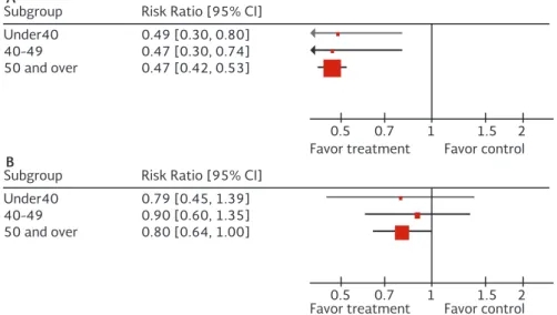
Figure 1. Forest plots for the benefit of radiation therapy after breast conserving surgery (BCS) (node negative disease;
A: Disease-free survival, B: Overall survival)
Figure 2. Forest plots for the benefit of radiation therapy after breast conserving surgery (BCS) (node positive disease;
A: Disease-free survival, B: Overall survival)
Moreover, robotic techniques are evolving in breast surgery, further improving cosmetic issues66.
The management of the axilla is similar in young women and those that are older53.
Radiotherapy
Adjuvant radiotherapy after BCS in women with pathologically node-negative disease (pN0) roughly halved the risk of first recurrence at nine years from 11.5 to 5.9% (RR: 0.52; 95% CI: 0.38-0.72) in wom-en aged < 40 and from 6.1 to 2.7% (RR: 0.48; 95% CI: 0.39-0.59) in women aged 40-49 years, as
A
Subgroup Under40 40-49 50 and over
Risk Ratio [95% CI] 0.75 [0.54, 1.04] 0.76 [0.60, 0.96] 0.70 [0.58, 0.84]
0.5 0.7 1 1.5 2
B
Subgroup Under40 40-49 50 and over
Risk Ratio [95% CI] 0.75 [0.50, 1.13] 0.76 [0.58, 1.00] 0.84 [0.75, 0.94]
0.5 0.7 1 1.5 2
Favor treatment Favor control Favor treatment Favor control
disease (one to three lymph nodes involved) (Fig. 3; forest plot for the benefit of radiation therapy after mastectomy)68,73-76. Locoregional radiotherapy is in-ternationally accepted as a recommended adjuvant treatment in locally advanced breast cancer (T3-4 and N2-3 tumors), while adjuvant radiotherapy of N1 disease is a subject of ongoing discussion and re-search64,71,77-82. Recent postmastectomy radiothera-py guidelines focusing on T1-2 and N1 tumors could not agree on specific patient subgroups in whom ra-diotherapy could be safely omitted, but recommend-ed younger age (< 45 years) as a potential factor for clinical decision-making in favor of radiation71,83,84. The absolute benefit of regional nodal radiotherapy in individual patients with 1-3 positive lymph nodes is not known accurately, and the estimation of relative risk reduction with radiotherapy is extrapolated from results of reported randomized trials in unselected pa-tients. The interaction between systemic therapy and locoregional radiation treatment is not fully under-stood and it is proposed that the two treatments may complement each other71,79. The balance between potential benefits of regional nodal irradiation and toxicity should be discussed with the patient71,78. Moderately hypofractionated radiotherapy schedules of breast/chest wall and/or lymph node areas with higher dose per fraction (> 2 Gy) and shortened overall treatment time are increasingly gaining popularity around the globe including in patients younger than 50 years85-88. Shortened treatment schedules demonstrat-ed equivalent local control and similar or lower toxicity
rates, regardless of the patient’s age87,89. Up to one third of included patients in randomized trials of hypo-fractionated radiotherapy were aged < 50 years87,89,90. Moderately hypofractionated radiation schedules in locoregional radiotherapy or in patients with recon-structed breast are still not used routinely because of normal tissue toxicity concerns as those patients were underrepresented in randomized controlled trials and toxicities were not separately reported87,89. It is not expected that a higher dose per fraction with a lower total prescribed dose and similar biologically effective dose would produce more detrimental normal tissue side effects than conventional fractionation (e.g. ra-diation-induced brachial plexopathy or lymphedema), but long-term results from clinical trials are still pend-ing87,91-93. Some authors suggest that patients with postoperative complications, such as those with large breasts for whom a maximum dose of < 107% is not achievable, or patients with breast implants who have an increased risk for late fibrosis or cosmetic deterio-ration following radiotherapy, should receive a biologi-cally less-intense dose94. Adjuvant radiotherapy after breast reconstruction (especially implant-based) is a risk factor for higher tissue toxicity95.
A schedule of 3-4 weeks seems to be more convenient than the standard five or six as fewer daily treatments mean less time and costs spent travelling and less time away from family and work85. Shorter courses of breast radiation therapy allow more women access to life-saving treatments, especially in countries where there are limited radiotherapy facilities96.
Additional dose to tumor bed for patients treated with BCS further improves local control in all age groups and all tumor types, with the largest absolute benefit in young patients, although it increases the risk of worse cosmetic results and fibrosis62,97. Guidelines on margin status after BCS suggest that, similarly to older patients, larger margin widths than no ink on tumor after BCS are not needed in younger patients98. It is important to note that positive margins are associated with an elevated risk of breast cancer recurrence re-gardless of a higher radiation boost-dose delivery, sys-temic treatments, or favorable tumor biology98,99. In a recently published randomized trial evaluating ra-diation boost after whole-breast irrara-diation, the abso-lute risk reduction in local recurrence was greatest in the youngest patient group: the addition of a radiation boost resulted in an absolute risk reduction at 20-years of 11.6% in women < 40 and of 5.9% in those aged 41-50 years97. The final results of a radiation dose-in-tensity study in young women (Young boost trial, Clini-calTrials.gov identifier: NCT00212121) will provide us more data on the local recurrence risks and cosmetic outcome100. In the future, the decision of adjuvant ra-diation treatment could be supported by individual tu-mor prognostic and predictive factors based on gene-expression profiles as some reports suggest72,101-103. In selected patients, accelerated partial breast irra-diation (APBI) is an alternative to whole breast ir-radiation, but patients < 50 years old are underrep-resented (up to 23%) in key published APBI trials, and off-protocol use of APBI in younger patients is not recommended104. Results of the phase III clinical trial NSABP B-39/RTOG 0413 are awaited and should pro-vide greater clarity regarding this treatment in younger women (ClinicalTrials.gov identifier: NCT0010318)105. Modern radiation techniques are continuously improv-ing and are based on CT delineation of target volume and adjacent organs at risk106,107. Tangential three-di-mensional conformal or intensity modulated radiother-apy planning approaches allow for more homogenous target coverage, and at the same time, dose-volume estimations of delivered dose to organs at risk. Dif-ferent strategies to reduce the dose to thoracic or-gans are being researched108-110. Image-guided deep inspiratory breath hold is a reproducible and stable radiotherapy technique, which can be implemented as a voluntary, non-computer-controlled technique in
breast/chest wall and/or locoregional radiotherapy, including its use in low-resource settings111. Deep in-spiratory breath hold has been shown to significantly reduce dose to heart, left anterior descending coro-nary artery, and lung108,112.
Contralateral breast cancers and
secondary tumors after adjuvant
radiotherapy
Adjuvant breast cancer radiotherapy is one of the fac-tors that contribute to the risk of contralateral breast cancer and secondary tumors. Younger age (< 40-45), dose of radiation, and volume of normal tissue in the radiation field all increase the risk for secondary pri-mary cancers after breast radiotherapy113,114. Women < 40 years of age who received an absorbed radiation dose > 1.0 Gy to the part of contralateral breast had a 2.5-fold greater long-term risk of devel-oping a second primary contralateral breast cancer compared to unexposed women. Interestingly, no ex-cess risk was observed in women > 40 years of age113. Radiotherapy was significantly associated with an in-creased risk of second lung and esophagus cancer or second sarcomas (RR: 1.12; 95% CI: 1.06-1.19) in a recent large meta-analysis of 762,468 patients114. The risk increased over time, and was highest 15 or more years after breast cancer diagnosis, especially for second lung and second esophageal cancer114. The absolute risk was relatively small, but special atten-tion should be paid to minimizing radiaatten-tion exposure to organs at risk as the number of long-term survivors after breast cancer radiotherapy is growing.
A
Subgroup Under40 40-49 50 and over
Risk Ratio [95% CI] 0.56 [0.40, 0.76] 0.71 [0.58, 0.89] 0.57 [0.53, 0.61]
0.5 0.7 1 1.5 2
B
Subgroup Under40 40-49 50 and over
Risk Ratio [95% CI] 0.61 [0.42, 0.89] 0.76 [0.54, 1.07] 0.68 [0.60, 0.77]
0.5 0.7 1 1.5 2
Favor treatment Favor control Favor treatment Favor control
A
Subgroup Under40 40-49 50 and over
Risk Ratio [95% CI] 0.60 [0.50, 0.72] 0.64 [0.56, 0.73] 0.84 [0.75, 0.94]
0.5 0.7 1 1.5 2
B
Subgroup Under40 40-49 50 and over
Risk Ratio [95% CI] 0.71 [0.57, 0.88] 0.70 [0.62, 0.79] 0.88 [0.82, 0.94]
0.5 0.7 1 1.5 2
Favor treatment Favor control Favor treatment Favor control
Figure 5. Forest plots for the benefit of endocrine therapy (ET) (A: Disease-free survival, B: Overall survival)
Figure 4. Forest plots for the benefit of chemotherapy (CT) (A: Disease-free survival, B: Overall survival)
Systemic treatment
General recommendations
Decisions about adjuvant systemic treatment for in-vasive breast cancer (in both early and metastatic settings) should be made according to the anatomi-cal extent of the disease, biologianatomi-cal features of the tumor, and characteristics (comorbidities and prefer-ences) of each individual patient and not based on age alone53. Figures 4 and 5 represent forest plots of the benefit of different systemic treatments (CT and ET).
In view of the longer life expectancy of young women, not only efficacy but particularly also long-term toxici-ties of systemic therapy should be taken into account. These include cardiovascular disease, bone loss, cogni-tive impairment, sexual dysfunction, secondary can-cers, and infertility. Young patients with breast cancer are more likely to have concerns about maintaining high function at home and work and are more likely to suffer from depression compared to older patients118.
menopause before starting with systemic therapy (either CT or ET) and those aged < 45 should be re-ferred to fertility clinics53.
Systemic chemotherapy
in early stage disease
There is no evidence to recommend a specific chemo-therapy regimen for young women requiring neo/adju-vant chemotherapy. In the EBCTCG meta-analysis involv-ing anthracycline- or taxane-based regimens, relative risk reductions were not influenced by age119. The landmark NSABP-B18 trial not only moved neoadjuvant chemo-therapy to the setting of operable disease, but also dem-onstrated a non-significant trend for younger women benefiting from neoadjuvant rather than adjuvant CT120. In a recent pooled analysis, including premenopausal patients enrolled in two phase III adjuvant trials, investigators reported that dose-dense scheduling of chemotherapy was associated with a significant improvement in overall survival as compared to standard-interval treatment hazard ratio [HR]: 0.71; p = 0.021)121. This may be confounded by higher proportions of triple-negative breast cancer (TNBC) in younger patients. ER-negative breast cancer has been shown to benefit from dose-dense scheduling, unlike ER-positive disease122. Additionally, there was no increased risk of chemotherapy induced amenor-rhea, suggesting the greater efficacy of dose-dense treatment does not seem to be related to a greater activity in suppressing ovarian function. This needs to be further explored in prospective randomized trials. Around 15% of unselected triple-negative breast can-cers occur in BRCA1/2 mutants123. The benefit of addi-tion of platinum compound has been observed in some randomized phase II trials and a meta-analysis in triple-negative breast cancer patients with BRCA-mutations and/or positive family history of breast or ovarian can-cer124,125. The latter tend to respond better compared with non-TNBC patients and achieve higher rates of pathologic complete response when platinum agent is added to standard anthracycline- and taxane-based therapy. However, there are limited data supporting platinum therapy in BRCA2 carriers with ER-positive dis-ease. As the true value of pathologic complete response, especially in BRCA-associated patients, is unclear and no long-term survival data are available, the use of plati-num agents is currently not recommended as a stan-dard of care in the neo/adjuvant setting.
Endocrine therapy in early stage disease
Neoadjuvant endocrine therapy (ET) should not be routinely prescribed to any young patient with breast cancer outside of clinical trials since there are insuf-ficient data regarding its efficacy in this breast cancer population126. The STAGE study, evaluating neoadju-vant ET in premenopausal breast cancer patients, was underpowered to assess long-term outcome127. All young women with invasive hormone receptor posi-tive breast cancer should be offered adjuvant ET. In the EBCTCG meta-analysis, five years of tamoxifen (TAM) compared to no ET was associated with a relative re-duction in breast cancer recurrence of 39%, which translates into a 13% absolute reduction in the risk of recurrence at 15 years (33 vs. 46%). The relative risk of breast cancer mortality was reduced by 30%, which translated into a 9% absolute reduction in breast can-cer-related death (24 vs. 33%)128. Tamoxifen has a carryover effect, meaning its efficacy after five years of treatment persists even after its discontinuation, and the mortality reduction remains significant throughout at least the first 10 years of survival128. A significant benefit was seen in both pre- and postmenopausal women with hormone receptor positive disease, regard-less of age, stage, and grade of the disease.
especially for those with high-risk tumor characteris-tics, as the reduction in risk of recurrence is observed even within the initial 2-3 years of AI therapy132. Aromatase inhibitors alone are contraindicated in pre-menopausal breast cancer patients and should not be used in those patients who develop chemotherapy in-duced amenorrhea. Two large phase III trials, TEXT and SOFT, evaluated the role of AIs and ovarian function suppression (OFS) in premenopausal patients. Patients with higher risk of relapse (those with large tumors, node-positive and high-grade disease) requiring adju-vant CT and remaining premenopausal after CT now may benefit from OFS in combination with either TAM or exemestane. The absolute improvement in the five-year breast cancer-free survival rate with exemestane and OFS (as compared with tamoxifen) OFS was 5.5% in TEXT and 3.9% in SOFT in patients who received CT. The impact of adding OFS to either TAM or exemes-tane was particularly striking in patients aged < 35; the five-year breast cancer-free rate was 67.7% for pa-tients receiving TAM alone, 78.9% for those assigned TAM/OFS, and 83.4% for those treated with exemes-tane-OFS129. The ABCSG-12 trial also explored the value of addition of OFS to an AI anastrozole (ANA) in premenopausal breast cancer patients, where the ma-jority of patients did not receive CT133. There was no difference in disease-free survival after treatment with three years of goserelin plus either TAM or ANA after 94.4 months of median follow-up. However, there was an apparent inferior overall survival in the AI arm (HR: 1.63; p = 0.030). These discordant results between the TEXT/SOFT and ABCSG/12 trials may be due to dif-ferent patient characteristics and difdif-ferent treatment durations. In addition, only 18% of women in the ABC-SG/12 trial were aged < 40 in comparison with 30% in the SOFT/TEXT trials.
While AIs in young women require concurrent LHRH analogue, the optimal duration of the latter in combi-nation with TAM remains unknown; most old genera-tion studies utilized 2-3 years of LHRH agonist with five years of TAM, while in the SOFT and TEXT trials, five years of treatment was used129,134. In a meta-analysis that analyzed the role of OFS in 11,906 pre-menopausal patients with early breast cancer, there was no significant benefit for the use of LHRH ago-nists alone, but the addition of these agents to CT, TAM, or both significantly reduced recurrence by 12.7% and death after recurrence by 15.1%135. Patients that
derived the most benefit from LHRH agonists after CT were those aged < 40 and not older premeno-pausal women.
Comprehensive OFS is not always successfully achieved with LHRH analogues; suboptimal estrogen suppression has been reported in up to one third of patients136. Despite the fact that estradiol assays are not standardized and their accuracy and interpreta-tion can be problematic in the presence of very low levels of estradiol, it is recommended to check hor-mone levels if there are concerns about suboptimal ovarian function suppression (such as in response to breakthrough vaginal bleeding in patients receiving an AI)137. In situations of doubt, OFS with TAM may be preferable to OFS and AI.
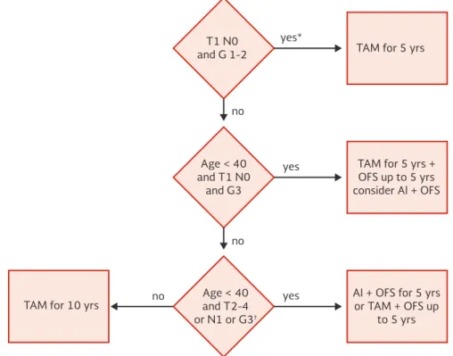
Unfortunately, we do not have any data about ex-tended ET after five years of combined OFS and either AI or TAM. Worrisome is the fact that there are no ongoing or planned trials that might give us an answer to this very important question in the near future. Figure 6 represents a suggested algorithm for adjuvant ET in premenopausal women with breast cancer.
Adjuvant anti-HER2 therapy
The benefit of adjuvant trastuzumab appears indepen-dent of age in all published studies52. As such, treat-ment of HER2-positive early breast cancer should mir-ror that of older patients with the same disease. All young women with HER2-positive early breast cancer should be treated with standard one-year adjuvant trastuzumab unless contraindicated6. Adjuvant trastu-zumab with chemotherapy is indicated for patients with tumors measuring more than 1 cm and for all patients with node-positive HER2-positive disease re-gardless of tumor size27. Consideration of adjuvant chemotherapy and trastuzumab in women with smaller (< 1 cm) node-negative tumors is appropriate. How-ever, patients should be counseled about the uncer-tainty regarding available data in these tumor types.
Adjuvant bisphosphonates
yes*
no
no
no
yes
yes
TAM for 5 yrs T1 N0
and G 1-2
Age < 40 and T1 N0
and G3
Age < 40 and T2-4 or N1 or G3†
TAM for 5 yrs + OFS up to 5 yrs consider Al + OFS
AI + OFS for 5 yrs or TAM + OFS up
to 5 yrs TAM for 10 yrs
A
Subgroup Under45 45-54 55 and older
Risk Ratio [95% CI] 1.00 [0.79, 1.26] 0.82 [0.61, 1.11] 0.72 [0.55, 0.94]
0.5 0.7 1 1.5 2
Favor treatment Favor control
showing a potential benefit was the ABCSG-12 trial, where zoledronic acid once every six months effec-tively prevented bone loss in hormone receptor posi-tive premenopausal patients whose endocrine treat-ment included LHRH analogues, or those who developed complete ovarian suppression following adjuvant CT133. Interestingly, in this trial, patients aged > 40 years had significantly improved disease-free survival in comparison to those ≤ 40 years of age. This may relate to those patients aged < 40 not achieving com-prehensive OFS and therefore not being “postmeno-pausal” (the group of patients thought to benefit from
adjuvant bisphosphonates). The characteristics of pa-tients benefiting from zoledronic acid in the ABCSG-12 study is at odds with the characteristics of those ben-efiting from OFS in the SOFT and TEXT trials. There-fore, the benefit of adjuvant bisphosphonates in wom-en receiving OFS remains uncertain.
Systemic therapy for metastatic disease
In the metastatic setting, patients’ preferences should always be taken into account as the disease is incur-able. Metastatic disease should be biopsied, whenever
Figure 7. Forest plot for the benefit of adjuvant bisphosphonates.
Figure 6. Suggested algorithm for adjuvant endocrine treatment of premenopausal breast cancer patients. *May confirm low-risk disease with molecular tests, e.g. Oncotype DX®/MammaPrint®;
†Factors demanding CT.
feasible, for confirmation and determination of recep-tor status6,139.
Cytotoxic therapy for metastatic breast
cancer in young women
General recommendations for CT should not differ from those for older women with the same biological features of metastatic disease and its extent. Young age by itself must not be an indicator for more inten-sive treatment nor use of combination CT in a meta-static setting. There is no survival advantage for com-bination CT over sequential single-agent chemotherapy and combination treatment leads to higher toxicity, which is of special importance in the treatment of advanced, incurable disease6.
Platinum compounds have been seeing a renewed in-terest in TNBC, especially those associated with BRCA1/2 mutations. The TNT trial compared stan-dard docetaxel to carboplatin in unselected TNBC patients140. Carboplatin was found to be superior among BRCA-mutated patients, but not in the un-selected TNBC population. It is important to highlight that 15% of patients did not receive prior systemic chemotherapy in early disease setting, and only 35% had received taxanes in the neo/adjuvant setting. Carboplatin was better tolerated than docetaxel.
Endocrine therapy for metastatic disease
Endocrine therapy should be the preferred systemic treatment for young patients with metastatic hormone receptor positive breast cancer, unless there is visceral crisis present or concern about endocrine resistance139. Ovarian suppression/ablation with additional endo-crine agent (TAM or an AI) remains a standard treat-ment option for first-line treattreat-ment for young meta-static breast cancer women with hormone receptor positive disease139. The choice of an additional endo-crine agent depends on the type of previous adjuvant therapy, as well as time elapsed from completion of adjuvant therapy and onset of metastatic disease. There has been increased interest in fulvestrant in the metastatic setting for postmenopausal breast cancer patients. Hypothetically, it should also be effective in pre- and perimenopausal patients. However, it has not been studied adequately in premenopausal patients.
Therefore, the use of OFS concomitantly with fulves-trant is often required when used in this group. An important advance in the management of hormone receptor positive metastatic breast cancer has been the introduction of a new class of agents, the CDK4/6 inhibitors, in combination with endocrine agents. Pal-bociclib, a CDK4/6 inhibitor, was evaluated in the phase III PALOMA 2 trial together with letrozole as first-line treatment in postmenopausal patients with hormone receptor positive metastatic breast cancer and demonstrated a 10-month improvement in pro-gression-free survival, with the main toxicity being he-matological (neutropenia)141. Its use together with an AI in pre- and/or perimenopausal patients mandates the use of ovarian suppression/ablation, but lacks a robust evidence base. The future role of mammalian target of rapamycin (mTOR) inhibitors is uncertain due to their high toxicities and lack of overall survival ben-efit. Additionally, there is a lack of data supporting the use of mTOR inhibitors after CDK4/6 inhibitors, which are increasing being used in earlier lines of therapy.
Anti-HER2 therapy for metastatic disease
Similar to HER2-positive early breast cancer, manage-ment of HER2-positive metastatic breast cancer is similar in younger and older women.
New targeted treatment options
for patients with BRCA-associated
breast cancer
An important characteristic of BRCA-mutated can-cers is defective function of one of the major DNA damage repair pathways, the homologous recombina-tion pathway142. The discovery of the family of nucle-ar enzymes poly(ADP-ribose) polymerases (PARP), and their role in DNA damage repair pathways led to de-velopment of a new class of drugs, so-called PARP inhibitors, which have the ability to interfere with the DNA damage repair systems of cancer cells. Over the past years, many different PARP inhibitors have been developed and their potential role has been evaluated especially in the treatment of TNBC and BRCA-asso-ciated tumors.
high143. There are several phase III trials currently ongoing in both the metastatic and neo/adjuvant and are lim-ited to patients with BRCA1/2-associated tumors142.
PSYCHOLOGICAL AND SOCIAL BURDEN OF
BREAST CANCER IN YOUNG SURVIVORS
Diagnosis of breast cancer can have a substantial effect on the life of a young woman. A prospective cohort study from the Netherlands demonstrated that health-related quality of life (which includes the role function-ing, emotional and cognitive functionfunction-ing, fatigue, and pain) in the first year after BCS and radiotherapy is more strongly affected in young breast cancer survivors than in older patients, but improves over time144. Emotional burden can be high for patients and family members145. A cross-sectional study from Canada found that breast cancer survivors with children ex-perience higher levels of fear of cancer recurrence and report more problems in intimacy many years after diagnosis146. It seems that parenting adolescent chil-dren can be particularly challenging and emotionally stressful for a young woman with breast cancer147. Prolonged adjuvant treatments after breast cancer surgery have a financial impact on the lives of young patients along with their extended families. Higher risk of losing paid employment and seeking of unemploy-ment and disability benefits up to 5-10 years after diagnosis are reported in young breast cancer survi-vors148. Work and home productivity losses among survivors aged 18-44 years are of considerable impor-tance and call for the support and understanding of healthcare systems around the globe149.
Rehabilitation strategies are important to improve all aspects of quality of life, including physical, psychologi-cal, social, and vocational well-being. Younger women report the need for their healthcare professionals to be more active in recommending rehabilitative care after initial breast cancer treatment has been completed150.
CONCLUSIONS
Breast cancer in young women represents a special entity of the disease. Not only do young patients face particular challenges and experience different social impacts of diagnosis and treatment, but also the
biology of their disease is distinct, even independently of stage and molecular subtyping.
Current treatment modalities for young women with breast cancer are based on biological features of an individual tumor, and the stage of the disease and should not be based on age alone. Data show that breast cancer in young women is complex and hetero-geneous, with many activated signaling pathways, which may represent therapeutic targets for future drug development. It is important to further investi-gate the reasons for a worse outcome of young breast cancer patients with luminal subtypes of the disease and find alternative treatment strategies for this im-portant subgroup of young patients.
At present, there is a lot of uncertainty regarding which young breast cancer patients need OFS as ad-juvant endocrine treatment, since the trials demon-strated discordant results. The decision is important from both the perspective of efficacy and toxicity as young women have a lower adherence with adjuvant therapy than older patients.
Early supportive care with special focus on fertility issues and psychosocial morbidity is crucial for opti-mal management of these patients.
Despite some advances, only few data still exist re-garding the management of young breast cancer pa-tients, especially in advanced disease setting, there-fore prospective randomized trials designed in this breast cancer population are desirable.
REFERENCES
1. WHO. Women and health. 2009. Available at: http://www.who.int/ gender/women_health_report/full_report_20091104_en.pdf 2. Villarreal-Garza C, Aguila C, Magallanes-Hoyos MC, et al. Breast
cancer in young women in Latin America: an unmet, growing burden. Oncologist. 2013;18:1298-306.
3. Collins LC, Marotti JD, Gelber S, et al. Pathologic features and mo-lecular phenotype by patient age in a large cohort of young women with breast cancer. Breast Cancer Res Treat. 2012;131:1061-6. 4. Partridge AH, Hughes ME, Ottesen RA, et al. The effect of age
on delay in diagnosis and stage of breast cancer. Oncologist. 2012;17:775-82.
5. Murphy CC, Bartholomew LK, Carpentier MY, Bluethmann SM, Vernon SW. Adherence to adjuvant hormonal therapy among breast cancer survivors in clinical practice: a systematic review. Breast Cancer Res Treat. 2012;134:459-78.
6. Cardoso F, Loibl S, Pagani O, et al. The European Society of Breast Cancer Specialists recommendations for the management of young women with breast cancer. Eur J Cancer. 2012;48:3355-77. 7. Partridge AH, Pagani O, Abulkhair O, et al. First international
consensus guidelines for breast cancer in young women (BCY1). Breast. 2014;23:209-20.
9. Gabriel CA, Domchek SM. Breast cancer in young women. Breast Cancer Res. 2010;12:212.
10. De La Rochefordiere A, Asselain B, Campana F, Scholl SM, Fenton J, Vilcoq JR. Age as prognostic factor in premenopausal breast carcinoma. Lancet. 1993;341:1039-43.
11. Mitwally MF, Casper RF. Use of an aromatase inhibitor for induc-tion of ovulainduc-tion in patients with an inadequate response to clomiphene citrate. Fertil Steril. 2001;75:305-9.
12. Smith IE, Dowsett M, Yap Y-S, et al. Adjuvant aromatase inhibitors for early breast cancer after chemotherapy-induced amenorrhoea: Caution and suggested guidelines. J Clin Oncol. 2006;24:2444-7. 13. World Health Organization. Tech Rep Ser Res menopause
Ge-neva Switz WHO. 1991;670.
14. National Comprehensive Cancer Network practice guidelines in oncology. 2005. https://www.nccn.org/professionals/physi-cian_gls/pdf/breast.pdf
15. Coombes RC, Hall E, Gibson LJ, et al. A randomized trial of ex-emestane after two to three years of tamoxifen therapy in postmenopausal women with primary breast cancer. N Engl J Med. 2004;350:1081-92.
16. Goss PE, Ingle JN, Martino S, et al. A randomized trial of letrozole in postmenopausal women after five years of tamoxifen therapy for early-stage breast cancer. N Engl J Med. 2003;349:1793-802. 17. Baum M, Budzar AU, Cuzick J, et al. Anastrozole alone or in com-bination with tamoxifen versus tamoxifen alone for adjuvant treat-ment of postmenopausal women with early breast cancer: first results of the ATAC randomised trial. Lancet. 2002;359:2131-9. 18. Cooper GS, Baird DD, Darden FR. Measures of menopausal status in relation to demographic, reproductive, and behavioral charac-teristics in a population-based study of women aged 35-49 years. Am J Epidemiol. 2001;153:1159-65.
19. Delrio G, De Placido S, Pagliarulo C, et al. Hypothalamic-pituitary-ovarian axis in women with operable breast cancer treated with adjuvant CMF and tamoxifen. Tumori. 1986;72:53-61. 20. Lum SS, Woltering EA, Fletcher WS, Pommier RF. Changes in
serum estrogen levels in women during tamoxifen therapy. Am J Surg. 1997;173:399-402.
21. Amir E, Seruga B, Freedman O, Clemons M. Amenorrhoea, meno-pause, and endocrine therapy for breast cancer. BMJ. 2009; 339:b4261.
22. Vital-Reyes V, Tellez-Velasco S, Chhieng D, Grizzle W, Reyes-Fuentes A. Spontaneous pregnancy in a woman with premature ovarian failure: a case report. J Reprod Med. 2004;49:989-91. 23. Colleoni M, Rotmensz N, Robertson C, et al. Very young women
(<35 years) with operable breast cancer: features of disease at presentation. Ann Oncol Off J Eur Soc Med Oncol. 2002;13:273-9. 24. Han W, Kang SY, Korean Breast Cancer Society. Relationship
between age at diagnosis and outcome of premenopausal breast cancer: age less than 35 years is a reasonable cut-off for defining young age-onset breast cancer. Breast Cancer Res Treat. 2010;119:193-200.
25. Copson E, Eccles B, Maishman T, et al. Prospective observa-tional study of breast cancer treatment outcomes for UK wom-en aged 18-40 years at diagnosis: The POSH Study. J Natl Cancer Inst. 2013;105:978-88.
26. Sotiriou C, Pusztai L. Gene-expression signatures in breast cancer. N Engl J Med. 2009;360:790-800.
27. Goldhirsch A, Winer EP, Coates AS, et al. Personalizing the treat-ment of women with early breast cancer: highlights of the St Gallen International Expert Consensus on the Primary Therapy of Early Breast Cancer 2013. Ann Oncol. 2013;24:2206-23. 28. Koboldt DC, Fulton RS, McLellan MD, et al. Comprehensive
molecu-lar portraits of human breast tumours. Nature. 2012;490:61-70. 29. Azim HA, Michiels S, Bedard PL, et al. Elucidating prognosis and
biology of breast cancer arising in young women using gene expression profiling. Clin Cancer Res. 2012;18:1341-51. 30. Sabiani L, Houvenaeghel G, Heinemann M, et al. breast cancer
in young women: Pathologic features and molecular phenotype. Breast. 2016;29:109-16.
31. Azim HA, Partridge AH. Biology of breast cancer in young women. Breast Cancer Res. 2014;16:427.
32. Rudra S, Yu DS, Yu ES, Switchenko JM, Mister D, Torres MA. Locoregional and distant recurrence patterns in young versus elderly women treated for breast cancer. Int J Breast Cancer. 2015;2015:213123.
33. Seneviratne S, Lawrenson R, Harvey V, et al. Stage of breast cancer at diagnosis in New Zealand: impacts of socio-demographic factors, breast cancer screening and biology. BMC Cancer. 2016;16:129. 34. Paluch-Shimon S, Wolf I, Sadetzki S, et al. Association between very young age and adverse characteristics of breast cancer at presen-tation amongst Israeli women. Am J Clin Oncol. 2011;34:219-22.
35. Johnson RH, Chien FL, Bleyer A, et al. Incidence of breast cancer with distant involvement among women in the United States, 1976 to 2009. JAMA. 2013;309:800.
36. Hung M-H, Liu C-Y, Shiau C-Y, et al. Effect of age and biological subtype on the risk and timing of brain metastasis in breast cancer patients. PLoS One. 2014;9:e89389.
37. Kennecke H, Yerushalmi R, Woods R, et al. Metastatic behavior of breast cancer subtypes. J Clin Oncol. 2010;28:3271-7. 38. Ernst MF, van de Poll-Franse LV, Roukema JA, et al. Trends in the
prognosis of patients with primary metastatic breast cancer diagnosed between 1975 and 2002. Breast. 2007;16:344-51. 39. van de Vijver MJ, He YD, van ’t Veer LJ, et al. A gene-expression
signature as a predictor of survival in breast cancer. N Engl J Med. 2002;347:1999-2009.
40. Paik S, Shak S, Tang G, et al. A multigene assay to predict recur-rence of tamoxifen-treated, node-negative breast cancer. N Engl J Med. 2004;351:2817-26.
41. Anders CK, Acharya CR, Hsu DS, et al. Age-Specific Differences in Oncogenic Pathway Deregulation Seen in Human Breast Tu-mors. Wu X, editor. PLoS One. 2008;3:e1373.
42. Young S, Pilarski RT, Donenberg T, et al. The prevalence of BRCA1 mutations among young women with triple-negative breast cancer. BMC Cancer. 2009;9:86.
43. Criscitiello C, Azim HA, Schouten PC, Linn SC, Sotiriou C. Under-standing the biology of triple-negative breast cancer. Ann Oncol. 2012;23(Suppl 6):vi13-18.
44. Azim H, Nguyen B, Brohee S, Zoppoli G, Sotiriou C. Genomic aberrations in young and elderly breast cancer patients. BMC Med. 2015;13:266.
45. Liu Z, Merkurjev D, Yang F, Li W, Oh S, Friedman MJ. Enhancer activation requires trans-recruitment of a mega transcription factor complex. Cell. 2014;159:358-73.
46. Chou J, Provot S, Werb Z. GATA3 in development and cancer differentiation: Cells GATA have it! J Cell Physiol. 2010;222:42-9. 47. Adomas AB, Grimm SA, Malone C, Takaku M, Sims JK, Wade PA.
Breast tumor specific mutation in GATA3 affects physiological mechanisms regulating transcription factor turnover. BMC Cancer. 2014;14:278.
48. Anders CK, Johnson R, Litton J. BC before age 40 years. Semin Oncol. 2009;36:237-49.
49. Partridge AH, Hughes ME, Warner ET, Ottesen RA, Wong YN, Edge SB. Subtype-dependent relationship between young age at diag-nosis and breast cancer survival. J Clin Oncol. 2016;34:3308-14. 50. Clarke M, Coates AS, Darby SC. Adjuvant chemotherapy in
oestrogen-receptor-poor breast cancer: patient-level meta-analysis of randomized trials. Lancet. 2008;371:29-40. 51. Huober J, von Minckwitz G, Denkert C. Effect of neoadjuvant
anthracycline-taxane-based chemotherapy in different biologi-cal BC phenotypes: overall results from the GeparTrio study. Breast Cancer Res Treat. 2010;124:133-40.
52. Partridge AH, Gelber S, Piccart-Gebhart MJ, et al. Effect of age on breast cancer outcomes in women with human epidermal growth factor receptor 2-positive breast cancer: results from a herceptin adjuvant trial. J Clin Oncol. 2013;31:2692-8. 53. Paluch-Shimon S, Pagani O, Partridge AH, et al. Second
interna-tional consensus guidelines for breast cancer in young women (BCY2). Breast. 2016;26:87-99.
54. Vila J, Gandini S, Gentilini O. Overall survival according to type of surgery in young (≤40 years) early breast cancer patients: A systematic meta-analysis comparing breast-conserving surgery versus mastectomy. Breast. 2015;24:175-81.
55. Veronesi U, Cascinelli N, Mariani L, et al. Twenty-year follow-up of a randomized study comparing breast-conserving surgery with radical mastectomy for early breast cancer. N Engl J Med. 2002;347:1227-32.
56. Kroman N, Holtveg H, Wohlfahrt J, et al. Effect of breast-conserving therapy versus radical mastectomy on prognosis for young women with breast carcinoma. Cancer. 2004;100:688-93.
57. de Bock GH, van der Hage JA, Putter H, Bonnema J, Bartelink H, van de Velde CJ. Isolated loco-regional recurrence of breast cancer is more common in young patients and following breast conserving therapy: Long-term results of European Organisation for Research and Treatment of Cancer studies. Eur J Cancer. 2006;42:351-6. 58. Botteri E, Bagnardi V, Rotmensz N, et al. Analysis of local and
regional recurrences in breast cancer after conservative surgery. Ann Oncol. 2010;21:723-8.
59. Komoike Y, Akiyama F, Iino Y, et al. Ipsilateral breast tumor re-currence (IBTR) after breast-conserving treatment for early breast cancer. Cancer. 2006;106:35-41.
treatment of young women with early breast cancer? Long-term results of a population-based cohort of 1,451 patients aged ≤40 years. Breast Cancer Res Treat. 2011;127:207-15. 61. Aalders KC, Postma EL, Strobbe LJ, et al. Contemporary
locore-gional recurrence rates in young patients with early-stage breast cancer. J Clin Oncol. 2016;34:2107-14.
62. Bartelink H, Horiot J-C, Poortmans PM, et al. Impact of a higher radiation dose on local control and survival in breast-conserving therapy of early breast Cancer: 10-year results of the random-ized boost versus no boost EORTC 22881-10882 Trial. J Clin Oncol. 2007;25(22):3259-65.
63. Plichta JK, Rai U, Tang R, et al. Factors associated with recurrence rates and long-term survival in women diagnosed with breast cancer ages 40 and younger. Ann Surg Oncol. 2016;23:3212-20. 64. Gradishar WJ, Robert CH, Anderson BO, et al. NCCN Guidelines Version 2.2016 Breast Cancer Panel Members NCCN Evidence Blocks TM. 2016. https://www.nccn.org/professionals/physi-cian_gls/pdf/breast_blocks.pdf
65. Hieken TJ, Boolbol SK, Dietz JR. Nipple-sparing mastectomy: Indications, contraindications, risks, benefits, and techniques. Ann Surg Oncol. 2016;23:3138-44.
66. Toesca A, Peradze N, Manconi A, et al. Robotic nipple-sparing mastectomy for the treatment of breast cancer: Feasibility and safety study. Breast. 2017;31:51-6.
67. Darby S, McGale P, Correa C, et al. Effect of radiotherapy after breast-conserving surgery on 10-year recurrence and 15-year Breast Cancer death: meta-analysis of individual patient data for 10,801 women in 17 randomised trials. Lancet. 2011;378:1707-16. 68. EBCTCG (Early Breast Cancer Trialists’ Collaborative Group),
McGale P, Taylor C, Correa C, et al. Effect of radiotherapy after mastectomy and axillary surgery on 10-year recurrence and 20-year Breast Cancer mortality: meta-analysis of individual patient data for 8135 women in 22 randomised trials. Lancet. 2014;383:2127-35.
69. van Laar C, van der Sangen MJ, Poortmans PM, et al. Local recur-rence following breast-conserving treatment in women aged 40 years or younger: trends in risk and the impact on prognosis in a population-based cohort of 1143 patients. Eur J Cancer. 2013;49:3093-101.
70. Bosma SCJ, van der Leij F, van Werkhoven E, et al. Very low local recurrence rates after breast-conserving therapy: analysis of 8485 patients treated over a 28-year period. Breast Cancer Res Treat. 2016;156:391-400.
71. Recht A, Comen EA, Fine RE, et al. Postmastectomy Radiotherapy: An American Society of Clinical Oncology, American Society for Radiation Oncology, and Society of Surgical Oncology Focused Guideline Update. Pract Radiat Oncol. 2016;6:e219-34. 72. Bartelink H. The changing landscape in radiotherapy for breast
cancer: Lessons from long term follow-up in some European breast cancer trials. Radiother Oncol. 2016;121:348-56. 73. Whelan TJ, Olivotto IA, Parulekar WR, et al. Regional nodal
irradia-tion in early-stage breast cancer. N Engl J Med. 2015;373:307-16. 74. Poortmans PM, Collette S, Kirkove C, et al. Internal mammary
and medial supraclavicular irradiation in breast cancer. N Engl J Med. 2015;373:317-27.
75. Budach W, Bölke E, Kammers K, Gerber PA, Nestle-Krämling C, Matuschek C. Adjuvant radiation therapy of regional lymph nodes in breast cancer - a meta-analysis of randomized trials- an update. Radiat Oncol. 2015;10:258.
76. Clarke M, Collins R, Darby S, et al. Effects of radiotherapy and of differences in the extent of surgery for early breast cancer on local recurrence and 15-year survival: an overview of the randomised trials. Lancet. 2005;366:2087-106.
77. Brackstone M, Fletcher GG, Dayes IS, et al. Locoregional therapy of locally advanced breast cancer: a clinical practice guideline. Curr Oncol. 2015;22(Suppl 1):S54-66.
78. Poortmans PM, Whelan TJ. Locoregional treatment in early stage breast cancer: More evidence and yet more questions? J Clin Oncol. 2015;27:689-91.
79. Poortmans P. Postmastectomy radiation in breast cancer with one to three involved lymph nodes: ending the debate. Lancet. 2014;383:2104-6.
80. Kunkler IH, Canney P, van Tienhoven G, Russell NS, MRC/EORTC (BIG 2-04) SUPREMO Trial Management Group. Elucidating the role of chest wall irradiation in “intermediate-risk” breast cancer: the MRC/EORTC SUPREMO trial. Clin Oncol (R Coll Radiol). 2008;20:31-4.
81. Thorsen LB, Offersen BV, Danø H, et al. DBCG-IMN: A population-based cohort study on the effect of internal mammary node irradiation in early node-positive breast cancer. J Clin Oncol. 2016;34:314-20.
82. Senkus E, Kyriakides S, Ohno S, et al. Primary breast cancer: ESMO clinical practice guidelines for diagnosis, treatment and follow-up. Ann Oncol. 2015;26(Suppl 5):v8-30.
83. Yildirim E, Berberoglu U. Local recurrence in breast carcinoma patients with T1-2 and 1-3 positive nodes: Indications for ra-diotherapy. Eur J Surg Oncol. 2007;33:28-32.
84. Su Y-L, Li S-H, Chen Y-Y, et al. Post-mastectomy radiotherapy ben-efits subgroups of breast cancer patients with T1-2 tumor and 1-3 axillary lymph node(s) metastasis. Radiol Oncol. 2014;48:314-22. 85. Ashworth A, Kong W, Whelan T, Mackillop WJ. A population-based study of the fractionation of postlumpectomy breast radiation therapy. Int J Radiat Oncol. 2013;86:51-7.
86. Nguyen K, Mackenzie P, Allen A, et al. Breast interest group faculty of radiation oncology: Australian and New Zealand pat-terns of practice survey on breast radiotherapy. J Med Imaging Radiat Oncol. 2016. [Epub ahead of print].
87. Haviland JS, Owen JR, Dewar JA, et al. The UK Standardisation of Breast Radiotherapy (START) trials of radiotherapy hypofrac-tionation for treatment of early breast cancer: 10-year follow-up results of two randomised controlled trials. Lancet Oncol. 2013;14:1086-94.
88. Bekelman JE, Sylwestrzak G, Barron J, et al. Uptake and costs of hypofractionated vs conventional whole breast irradiation after breast conserving surgery in the United States, 2008-2013. JAMA. 2014;312:2542.
89. Whelan TJ, Pignol J-P, Levine MN, et al. Long-term results of hypofractionated radiation therapy for breast cancer. N Engl J Med. 2010;362:513-20.
90. Owen JR, Ashton A, Bliss JM, et al. Effect of radiotherapy fraction size on tumour control in patients with early-stage breast cancer after local tumour excision: long-term results of a randomised trial. Lancet Oncol. 2006;7:467-71.
91. Caudrelier J-M, Truong PT. Role of hypofractionated radiotherapy in breast locoregional radiation. Cancer/Radiothérapie. 2015;19:241-7. 92. DBCG; Danish Breast Cancer Cooperative Group. The SKAGEN
Trial 1 Moderately hypofractionated loco-regional adjuvant ra-diation therapy of early breast cancer combined with a simulta-neous integrated boost in patients with an indication for boost: DBCG HYPO II, a randomised clinically controlled trial. 2015. p. 1-33. Available from: www.dbcg.dk/
93. Ain Shams University. Conventional Versus Hypofractionated Radiotherapy in Node Positive Breast Cancer. ClinicalTrials.gov Identifier: NCT02690636.
94. Koulis TA, Phan T, Olivotto IA. Hypofractionated whole breast ra-diotherapy: current perspectives. Breast Cancer. 2015;7:363-70. 95. Barry M, Kell MR. Radiotherapy and breast reconstruction: a
meta-analysis. Breast Cancer Res Treat. 2011;127:15-22. 96. Khan AJ, Rafique R, Zafar W, et al. Nation-scale adoption of
shorter breast radiation therapy schedules can increase sur-vival in resource constrained economies: Results from a Markov chain analysis. Int J Radiat Oncol. 2017;97:287-95.
97. Bartelink H, Maingon P, Poortmans P, et al. Whole-breast irra-diation with or without a boost for patients treated with breast-conserving surgery for early breast cancer: 20-year follow-up of a randomised phase 3 trial. Lancet Oncol. 2015;16:47-56. 98. Moran MS, Schnitt SJ, Giuliano AE, et al. Society of Surgical
Oncology-American Society for Radiation Oncology consensus guideline on margins for breast-conserving surgery with whole-breast irradiation in stages I and II invasive whole-breast cancer. J Clin Oncol. 2014;32:1507-15.
99. Houssami N, Macaskill P, Luke Marinovich M, Morrow M. The As-sociation of Surgical Margins and Local Recurrence in Women with Early-Stage Invasive breast cancer Treated with Breast-Conserv-ing Therapy: A Meta-Analysis. Ann Surg Oncol. 2014;21:717-30. 100. Brouwers PJ, van Werkhoven E, Bartelink H, et al. Factors as-sociated with patient-reported cosmetic outcome in the Young Boost Breast Trial. Radiother Oncol. 2016;120:107-13. 101. Tramm T, Mohammed H, Myhre S, et al. Development and
validation of a gene profile predicting benefit of postmastectomy radiotherapy in patients with high-risk breast cancer: a study of gene expression in the DBCG82bc cohort. Clin Cancer Res. 2014;20:5272-80.
102. Cheng SH, Horng C-F, Huang T-T, et al. An eighteen-gene clas-sifier predicts locoregional recurrence in post-mastectomy breast cancer Patients. EBioMedicine. 2016;5:74-81.
103. Torres-Roca JF, Fulp WJ, Caudell JJ, et al. Integration of a radiosen-sitivity molecular signature into the assessment of local recurrence risk in breast cancer. Int J Radiat Oncol Biol Phys. 2015;93:631-8. 104. Vicini F, Shah C, Tendulkar R, et al. Accelerated partial breast