www.medigraphic.com
ORIGINAL ARTICLE
Rev
ista
Lat
inoamer
icana
de
MICROBIOLOGÍA
MICROBIOLOGÍA
Evaluation of probiotic properties in
Lactobacillus
isolated from small intestine of piglets
Iñiguez-Palomares C,* Pérez-Morales R,* Acedo-Félix E*
* Laboratorio de Microbiología Molecular. Coordinación de Ciencia de los Alimentos. CIAD., AC. Hermosillo, Sonora.
First version received: April 23, 2007; first version revised: June 07, 2007 and June 22, 2007; second version received in revised form: December 9, 2007; second version revised: Decem-ber 15, 2007 and January 4, 2008; third version received: January 15, 2008; accepted: January 15, 2008.
Vol. 49, Nos. 3-4 July - September. 2007 October - December. 2007 pp. 46 - 54
ABSTRACT. Lactobacillus has been associated with beneficial ef-fects in human and animal health. Oral administration of probiotic bacteria must resist gastrointestinal transit, in order to colonize the in-testinal mucus and offer an antagonistic effect against pathogenic mi-croorganisms. The aim of this work was to select Lactobacillus strains isolated from small intestine of piglets based on the character-istics of resistance to low pH and bile salts, surface properties and an-tagonistic effect against Escherichia coli K88. To identify Lactobacil-lus species, a fragment of 16S rRNA gene of the strains was sequenced. Low pH, bile salts resistance and antagonistic activity were quantified by viable count in plates. Surface properties were measured using a spectrophotometer at 600 nm. Sixty-two Lactoba-cillus strains were isolated from small intestine of piglets. Species Lactobacillus salivarius, Lactobacillus reuteri and Lactobacillus mu-cosae were identified. We found that 20 Lactobacillus strains resisted low pH and bile salt, 8 of them were adherents and they inhibited in vitro the growth of E. coli K88. In conclusion, our results showed that 8 strains have potential probiotic value, according resistance to gastrointestinal tract, surface properties and antagonistic characteris-tics. Lb. salivarius was the species that fulfilled the criterion to be identified as a possible probiotic microorganism.
Key words: Piglets, Lactobacillus, probiotic characteristics, antag-onism.
RESUMEN. Con frecuencia los lactobacilos se han relacionado con diversos efectos benéficos en la salud humana y animal. En la ad-ministración oral de bacterias benéficas, éstas deben resistir el trán-sito gastrointestinal para que colonicen la mucosa y ejerzan un efec-to antagónico contra diversas bacterias patógenas. Por lo anterior el objetivo de este trabajo fue seleccionar cepas de Lactobacillus ais-ladas del intestino delgado de lechones de acuerdo a sus característi-cas de resistencia al pH bajo y sales biliares, propiedades de superfi-cie y efecto antagónico en contra de Escherichia coli K88 para utilizarlas a futuro como probióticos. Se aislaron sesenta y dos ce-pas de Lactobacillus del intestino delgado de seis lechones. Se identificaron las especies Lactobacillus salivarius, Lactobacillus re-uteri y Lactobacillus mucosae. Encontramos que 20 cepas de Lac-tobacillus resistieron la presencia de pH bajo y sales biliares, 8 de ellas fueron adherentes e inhibieron in vitro el crecimiento de E. coli K88. En conclusión, nuestros resultados mostraron que 8 cepas tienen valor probiótico potencial de acuerdo a la resistencia al trán-sito gastrointestinal, propiedades de superficie y efecto antagónico. Lb. salivarius fue la especie que cumple con los criterios para ser identificada como un probiótico potencial para aplicarse en cerdos recién destetados.
Palabras clave: Lechones, Lactobacillus, características probióti-cas, antagonismo.
INTRODUCTION
Pig breeding is a profitable activity in many countries. Sometimes, the economy of this activity is affected by in-fectious diarrhea in neonatal pigs (Gusils et al., 2002). Es-cherichia coli K88 has been identified as one of the main causal agents of this illness. Those bacteria invade mucosal cells and produce enterotoxins that cause diarrhoea and death of infected pigs (Meng et al., 1998). Commonly anti-biotics are used to prevent and/or eliminate these infec-tions, unfortunately low control or antibiotic misuse are fre-quent practices. The worst disadvantage of these practices
is bacterial resistance developed to these substances, for this reason it is important to look for alternatives to antibi-otic usage (Gibson and Wang, 1994). Probiantibi-otics can be ad-ministered to prevent infectious diseases, to strengthen the barrier function of the gut microflora and for a non-specific enhancement of the immune system (Gusils et al., 2002).
www.medigraphic.com
Lactobacillus and Bifidobacterium belong to the nor-mal human and aninor-mal microbiota. In this way their spe-cies are widely studied as probiotic. In animals, it has been observed that the rate of growth has increased with a better food conversion, and probiotics are helpful for this conversion (Jonson and Conway, 1992). Probiotics nei-ther generate antimicrobial resistance nor produce toxic compounds in carcass (Fuller, 1999). Strains need to sur-vive acidic conditions in the stomach and bile salts in duodenum, in order to exert their beneficial effects in the gut. Therefore, bile tolerance is considered one of the most important properties of probiotic microorganisms, because it allows them to survive and to colonize the gastrointesti-nal tract in mucus and/or by enterocytes adhesion (Gómez-Zavaglia. et al., 2002).
Adherence to the intestinal mucus layer is another im-portant selection criterion for probiotic microorganisms, because it is a requirement for the bowel colonization. Ad-herence constitutes the first defence mechanism against pathogen invasion. Passing through the small intestine takes about 2.5 hours, but it is faster in the duodenum than the colon, therefore bacterial adherence to mucus and/or enterocytes is necessary for colonization (Yusof et al., 2000; Rinkinen et al., 2003).
The aim of this work was to select Lactobacillus strains isolated from small intestines of piglets, based on their characteristics of resistance to low pH and bile salts, sur-face properties and antagonism against E. coli K88 as a first requirement for their possible use as probiotics.
MATERIALS AND METHODS
Isolation of Lactobacillus strains
Six Landrace piglets breed crossed with Large White were slaughtered, all of them from the same farm. They were healthy and weaned Small intestine was divided into three portions in aseptic conditions: duodenum, jejunum and ileum. Each portion was separately placed in tubes with MRS broth (Difco-Becton Dickinson & Company, Sparks, MD, USA) pH 6.0 with cysteine hydrochloride 0.5 gr/L (J.T. Baker, Phillipsburg, NJ), 2,3,5-tripheniltetrazoli-um chloride (TTC) 25 ppm (Merck, Darmstadt, Ge), sodi-um propionate 0.3% w/v (Sigma, St. Louis MO, USA), lith-ium chloride 0.2% w/v (Sigma) and antibiotics, as nalidixic acid 20 ppm (Sanofi-synthelabo Edo. México, Mex.), kanamycin 50 ppm (Sigma) and polymyxin B sul-phate 8.5 ppm. (Sigma). Cultures were incubated at 37º C in 5% CO2 by 48 h. After that cultures were seeded in plates with MRS agar (Difco-Becton Dickinson & Compa-ny) pH 6.0 with cysteine, without antibiotics and incubat-ed as describincubat-ed above (Corona, 2001). From those plates,
white and creamy colonies were selected. Gram stain, mo-tility and catalase assay were done as a first screening.
Phenotypic and Genotypic identification by 16S rRNA gene sequence analysis
To identify the species of Lactobacillus, the strains were tested for the fermentation of carbohydrates. Sugars tested were L-arabinose, lactose, celobiose, melezitose, raffinose, sorbitol, starch, xylose, mannose, fructose, galac-tose, sucrose, malgalac-tose, trehalose, melibiose, mannitol, inu-lin and salicin (Sigma). Fermentation test was done in tryptone peptone yeast broth (TPY) (Difco-Becton Dick-inson & Company) supplemented with 1% carbohydrate and bromocresol purple as pH indicator (Kandler and. Weiss, 1986).
In the genotypic identification, DNA from strains was isolated (De los Reyes et al., 1992). PCR was performed in a Thermal cycler (Perkin Elmer, Wellesley MA, USA). PCR primers used for this experiment were 27F and 519R reported previously. They amplified a 492 bp fragment from 16S rRNA gene (Lane, 1996). A typical reaction used the following programme involving a initial denaturation of 3 min at 94º C, 30 cycles of 94º C for 30 s, 55º C for 60 s and 72º C for 30 s. The final cycle was 72º C for 10 min. The PCR products were analyzed on 1.2% agarose gels (Sigma). They were stained with ethidium bromide and ob-served in a UV transiluminator (Vilbert Loumart, Marne La Vallee, Francia). PCR products were purified with a GFX PCR DNA and Gel Band Purification Kit (Amersham, Piscataway NJ, USA). The purified products were sent to Arizona Research Laboratories (Tucson AZ, USA) for se-quencing. Sequenced DNA was compared with informa-tion in the data base available in basic BLAST (Altschul
et al., 1997). Partial sequences were manually aligned us-ing DNAMAN (4.03 Lynnon BioSoft, Quebec Canada). A distance matrix and phylogenetic tree was generated using the Observed Divergency method.
Resistance to pH 3.0 and bile salts
www.medigraphic.com
MRS agar pH 6.0 in previously described conditions. At the same time, the cultures were inoculated for a viable count in plates of MRS with CPBS (0.5% w/v). In both cases, the plates were incubated in 5% CO2 at 37º C for 48 h. All plates were inoculated by duplicate.
Percentage of resistance to pH 3.0 and CPBS was deter-mined using the equation of Kociubinsky et al. (1999) % Resistance = 100(CFU (pH 3.0 or CPBS)/CFUcontrol).
Autoaggregation assay
Strains were grown as described above in 3 ml of MRS broth pH 6.0 with cysteine and they were harvested at 2400 x g. Supernatant was retained in a different tube. The pellet was washed twice with phosphate buffered saline (PBS) 0.02 M pH 7.4 and resuspended in same buffer until an optical density (O. D.) of 0.5 units at 600 nm (Spectron-ic 21D Milton Roy, USA) was reached. From this suspen-sion 3 mL were harvested at 2,400 x g. Supernatant was eliminated and cells were resuspended in their original broth. They were incubated by 2 h at 37º C and then, 1 ml was taken from the superior part of the culture and the O.D. was measured. Finally, culture was shaken and total O. D. was measured. The autoaggregation (% A) is ex-pressed in the following equation 1 – (O. D. superior cul-ture/O. D. total) x 100 (Del Re et al., 2000). This experi-ment was done in triplicate.
Hydrophobicity (microbial adhesion to hydrocarbons)
Strains were grown as described above in 3 ml of MRS broth pH 6.0 with cysteine. Cultures were washed with PBS buffer and resuspended as described previously. 2 ml of bacterial suspension were transferred into another tube and 0.4 mL of xylene was added (Fluka, GmbH, Switzer-land). Tubes were shaken for 2 min and reposed for 15 min. After that O.D. of aqueous phase at 600 nm was measured. O.D. decrease in aqueous phase was considered as a mea-surement of cells surface hydrophobicity (%H). %H was calculated according to the following equation [(A0–A)/ A0] x 100. Where A0 and A, were the absorbance before and after xylene extraction respectively (Del Re et al., 1998; Gusils et al., 2002; Mishra and Prasad, 2005).
Antagonism against Escherichia coli K88
Lactobacillus strains and E. coli K88 were inoculated separately in MRS broth, pH 7.0 with cysteine. Cultures were incubated for 24 h at 37º C in 5% CO2. All the Lacto-bacillus cultures were adjusted with tube number 5 of a MacFarland nephelometer. E. coli K88 culture was adjust-ed with the number 3. K88 was massively inoculatadjust-ed over
MRS plates pH 7.0 with cysteine according to Hernández (2003). Four holes of 6 millimeters (mm) of diameter at similar distances were punched and filled with 70 µl of
Lactobacillus culture. Plates were incubated at 37º C for 24 h and growth inhibition was measured in millimeters (De Martinis et al., 2002).
On the other hand, mixed cultures were prepared ac-cording to González et al., (1993). 24 h cultures of Lacto-bacillus and E. coli K88 in MRS broth pH 7.0 with cys-teine were adjusted with tube number 3 of MacFarland nephelometer. Pathogenic bacterium was diluted three times by serial dilutions. Equal volumes of both cultures in proportion 1,000:1 (Lactobacillus: E. coli K88) were mixed in 3 mL MRS broth pH 7.0 with cysteine and incu-bated at 37º C for 6 h in 5% CO2. A control tube was made containing just E. coli K88. After 6 h, a viable count in ENDO agar (Difco, Mexico) was done. Determination of antagonism percentage was calculated according the equa-tion: % I = 100 [(T6 control – T6mixed culture)/T6 control], where % I was the percentage of bacterial inhibition of each strain, T6 control was the viable count obtained from the control and T6 mixed culture was the viable count obtained from the mixed culture (González et al. 1993; Gusils et al., 2002; García-Galaz et al., 2004).
Statistical Analysis
ANOVA and Tukey-Kramer test were used for mean comparisons (p<0.05) in all experiments, with statistical package NCSS 6.0 (Hintze, 1997). Comparisons were car-ried out for species and strains.
RESULTS
Isolation of Lactobacillus strains
Sixty-two Lactobacillus strains were isolated from small intestine of six healthy piglets. All of them grew in aerobic and 5% CO2 conditions, were Gram positive rods, non-motile and catalase negative as preliminary character-istics. From jejunum, 33 strains were isolated, significant-ly different (p<0.05) from duodenum (10 strains) and ile-um (19 strains) in bacterial gut distribution in piglet.
Phenotypic and Genotypic identification by 16S rRNA gene sequence analysis
identi-www.medigraphic.com
ty with Lactobacillus salivarius subsp. salicinus, 5 strains had 99% of identity with Lb. salivarius, 19 strains showed 99% of identity with Lactobacillus reuteri and 1 strain showed 98% of identity with Lactobacillus muco-sae. With the phylogenetic tree we could observe three species groups. Group I corresponds to Lb. salivarius
(subsp salivarius and salicinus together), Group II to Lb.
mucosae and Group III to Lb. reuteri. It is important to notice that the phylogenetic analysis was not enough to differentiate subsp. salivarius from subsp salicinus (Fig-ure 1). There were no statistical differences (p>0.05) in species distribution, according to three analyzed por-tions in small intestine.
The isolates identified by partial sequence of 16S rRNA gene, were characterized by carbohydrate fermentation and it confirmed the genotypic identification (data not shown).
Resistance to pH 3.0 and bile salts
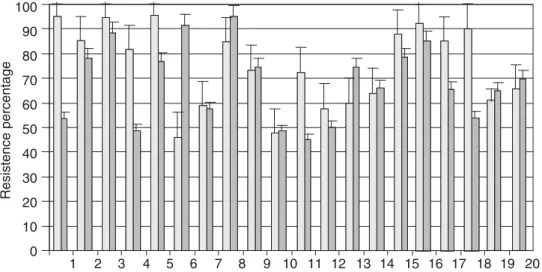
From all isolated strains, just 20 survived at pH 3.0 and CPBS conditions in 45% or more. Data of strains which did not survive are not shown. Survival at pH 3 is signifi-cant because ingestion of probiotic bacteria with food or dairy products raises the pH in stomach to 3.0 or higher. Resistant strains belonged to three identified species, Lb. salivarius being the most common with 15 strains (Figure 2). Others strains showed good survival to low pH (more than 50%), but they were discarded, because the resistance to CPBS was less than 0.1%. There were no statistical dif-ferences in the survival percentage (p>0.05), between the two main 1 species isolated (Table 1).
Autoaggregation and Hydrophobicity (microbial adhe-sion to hydrocarbons)
20 strains that survived to pH 3.0 and CPBS conditions were included to further characterization. They showed significant differences (p<0.05) in their autoaggregation and hydrophobicity properties. Strains 5, 6, 8, 9, 10, 11, 13, 18 and 20 showed an autoaggregation percentage su-perior to 40%, but strain 13 had less than 30% for hydro-phobicity, for which reason it was discarded as a potential probiotic. For hydrophobicity, strains 7, 15 and 19 showed less than 10% (Figure 3). Altogether, 8 strains showed au-toaggregation and hydrophobicity percentages superior to 40%, from these, 6 correspond to Lb. salivarius, 1 to Lb. reuteri and 1 to Lb. mucosae. This indicates that these strains posses autoaggregative and hydrophobic character-istics which are related to adhesion to epithelia.
Antagonism against Escherichia coli K88
8 strains that displayed superior autoaggregation and hydrophobicity properties were included in this experi-ment for antagonism. Whole cultures were used and halos of inhibition more than 20 mm against E. coli K88 were observed. There were no significant differences (p>0.05) between strains.
Accession numbers of sequences obtained from NCBI correspond to the fol-lowing species: AB289296.1 Lb. salivarius subsp. salivarius, AB289295.1 Lb. salivarius subsp. salicinus, DQ444477.1 Lb. salivarius, AB289270.1 Lb. reu-teri and AF126738.1 Lb. mucosae.
www.medigraphic.com
Lb. salivarius from 1 to 15, Lb. reuteri
from 16 to 19 and Lb. mucosae 20.
Figure 2. Resistance to pH 3.0 ( %) and conjugated porcine bile salts ( %) (CPBS) of Lactobacillus strains isolated from small intestine of piglets.
Table 1. Percentage of resistance to pH 3.0 and conjugated porcine bile salts (CPBS) of predominant species of Lactobacillus strains isolated from small intestine of piglets.
Species Resistance to pH 3.0 Resistance to CPBS
Lb. salivarius 66.5 ± 30 29 ± 35.5
Lb. reuteri 48.7 ± 39.2 16 ± 24.8
± indicates standard deviation
Quantitative antagonistic analysis, including all 8 strains showed pathogen growth decrease of 3 Log (Fig-ure 4). There were no differences between the effects of the
Lactobacillus strains tested (p>0.05). Antagonistic activi-ty of Lb. reuteri strains, which were not resistant to low pH or CPSB and did not show surface properties in the re-quired percentage, was quantified. They showed a de-crease of E. coli K88 growth of approximately 3 Log (data not shown).
DISCUSSION
Lactobacilli are established early in piglet intestine, and although succession occurs throughout lifetime of the pigs, they may remain as one of the predominant elements of the bacterial community (De Angelis et al., 2006).
In this work, we isolated one facultative heterofermen-tative and two obligate heterofermenheterofermen-tative Lactobacillus
species: Lb. salivarius, Lb. reuteri and Lb. mucosae, re-spectively. Similar results were reported by Robredo and Torres (2000) and Roos et al., (2000) in intestinal tract of
healthy adult pigs. Those results indicated that isolated species are maintained in the intestine throughout the whole life of pigs (Saarela et al., 2000; De Angelis et al., 2006).
Probiotic microorganisms need to resist the adverse fac-tors in the gastrointestinal tract when they pass through it, like the stomach acidity and bile salts, excreted in duode-num. For this investigation it was decided to select strains with a resistance more than 45%, to assure that bacteria ar-rive in suitable concentration (6 to 8 logarithms/g of con-sumed food) to the intestine, and exert their probiotic ef-fect (Shah et al., 1999).
In this work, all of three species were resistant to pH 3.0 and CPBS. Maxwell and Stewart (1995) found that Lb. aci-dophilus, Lb. fermentum and Lb. lactis were resistant to these adverse conditions in adult pigs. From those species, 20 strains survived the gastrointestinal transit more than 45%. Gómez-Zavaglia et al. (1998) and Kociubinski et al. (1999) obtained resistant strains to gastrointestinal transit over 23%. Those species were B. pseudolongum, B. infan-tis, B. animalis and B. breve. Aside, Ibrahim and Bezko-rovainy (1993) worked with strains of B. bifidum, B. breve,
B. infantis and B. longum, which were resistant to the ad-verse conditions of digestive tract. In general, variable re-sults have been documented in respect the resistance of low pH and bile salts of the Lactobacillus and Bifidobac-terium strains (Clark and Martin, 1994; Chung et al., 1999; Mishra and Prasad, 2005).
www.medigraphic.com
ESTE DOCUMENTO ES ELABORADO POR MEDIGRA-PHIC
depending on the activity of their H+-ATPase (Matsumoto
et al., 2004).
The greater adverse effect was observed for CPBS. Some authors have reported that the conjugated salts, mainly the glycodeoxicolic acid, are lethal for this bacterial genus and the mortality rate increases as pH diminishes. It has been found that biliary salts hydrolases produced by some Lacto-bacillus strains, are involved in the resistance (Grill et al., 2000; Tanaka et al., 2000; Kim et al., 2004).
Another desirable property of probiotic bacteria is the colonization in intestinal wall. This colonization is neces-sary in order to exert its beneficial effects (Tuomola et al., 2001). In probiosis, it is important to evaluate surface properties, like autoaggregation and hydrophobicity, be-cause they are used as a measurement directly related to adhesion ability to enterocytic cellular lines (Pérez et al., 1998; Del Re et al., 2000).
Autoaggregation besides determines the capacity of the bacterial strain to interact with itself, in a nonspecific way. Aside, when that hydrophobicity is high (more than 40%), it indicates the presence of hydrophobic molecules in the bacterial surface, like surface array proteins; wall interca-lated proteins, cytoplasmic membrane protein and lipids. (Ofek and Doyle, 1994; Pérez et al., 1998; Bibiloni et al., 1999; Bibiloni et al., 2001).
In this work, 20 strains were resistant to gastrointestinal transit, 8 had values of autoaggregation and hydrophobic-ity superior to 40%. According to Del Re et al. (1998) and Pérez et al. (1998), this percentage is the minimum neces-sary for considering a strain with adhesion abilities.
Pérez et al. (1998) found that B. breve strains isolated from humans were not autoaggregatives or hydrophobics.
Neither were adherent to cellular lines Caco-2. Time later, Bibiloni et al., (2001) related these non adherent strains with poor presence of protease-sensitive non polar like proteins molecules in their surface.
Del Re et al. (1998) concluded that the adhesion prop-erty is characteristic of each strain and cannot be general-ized to species. Ability to autoaggregate together with cell surface hydrophobicity could be used for preliminary screening to identify potentially adherent bacteria.
To improve the probiotic characterization, 8 strains that showed surface properties over 40%, were tested for their antagonism in vitro against E. coli K88. All strains showed inhibition zones and decreased the pathogen growth. Gusils et al. (2002) found that Lactobacillus and
Enteroccocus strains isolated from pig feces, do not inhib-it the growth of Yersinia enterocilitica, Salmonella chol-eraesuis, Salmonella typhimurium and Salmonella enterit-idis after 24 hours of plates incubation. The quantification of antagonism of the Lactobacillus strains in this work showed an E. coli K88 inhibition in 3 Log in 6 hours. Ac-cording to antagonistic values, resistance to gastrointesti-nal transit and adherence factors of isolated strains, it is possible to infer that these bacteria could be used as an al-ternative for the treatment of diarrhea in piglets. Neverthe-less, in vivo studies are necessary to confirm it. Recent work in other countries, using probiotic bacteria in pigs, that showed positive results in performance, decrease of intestinal infections and viral diseases (Casey et al., 2007, Schierack et al., 2007) encourage us to continue investi-gating our strains.
E. coli K88 has been identified as one of the main bac-terial producers of diarrhea in new born pigs. It has been
Lb. salivarius from 1 to 15, Lb. reuteri
from 16 to 19 and Lb. mucosae 20.
Figure 3. Percentages of autoaggre-gation ( %) and hydrophobicity ( %) of
www.medigraphic.com
suggested that probiotics can coaggregate pathogenic bteria and release antagonistic substances, like organic ac-ids (lactic mainly) and bacteriocins. Also there are studies that indicate that they can compete for adhesion sites with several microorganisms, but this is still not verified (Mul-der et al., 1997; Meng et al., 1998; Ouwehand et al., 1999; Doyle, 2001).
In conclusion, our results showed that 8 selected strains have potential probiotic value (Table 2). We found that the predominant species, Lb. salivarius, shows the best characteristics to fulfill the criteria of a probiotic strain. In addition, it is recommended that these strains be further analyzed according to the selection criteria like stimula-tion of the immunological system and adhesion to the pig mucosa and/or epithelium intestinal.
Table 2.Lactobacillus strains with the best values to be a probiotic potential.
Percentage of Percentage of resistance Percentage of Percentage of Percentage of Strain resistance to pH 3.0 to CPBS autoaggregation hydrophobicity inhibition against K88
5 95.4 76.6 59.1 76.1 97.2
6 46.0 91.4 52.9 57.8 98.4
8 84.6 95.0 54.0 73.5 98.0
9 73.4 74.4 64.9 65.5 98.2
10 47.7 48.5 58.4 42.1 98.3
11 65.4 69.1 69.8 49.7 98.3
18 72.5 45.0 61.2 81.5 97.9
20 90.2 53.8 67.6 62.1 97.8
Control = E. coli K88 culture without
Lactobacillus added
Lb. salivarius from 5 to 11, Lb. reuteri
18 and Lb. mucosae 20.
Figure 4. Antagonism against E. coli
K88 of Lactobacillus strains isolated from small intestine of piglets.
ACKNOWLEDGMENTS
We thank Alejandro Maldonado Kanzler, for critical re-viewing of the manuscript. This work was supported by the National Council for Science and Technology (CONA-CYT) who give a financial grant to Iñiguez-Palomares, to obtain her PhD degree.
REFERENCES
www.medigraphic.com
2. Bibiloni, R., P.F. Perez, & G.L. De Antoni. 1999. Factors en-volved in adhesion of bifidobacterial strains to epithelial cells in culture. Anaerobe 5:483-485.
3. Bibiloni, R., P.F. Pérez,, L.G. Garrote, E.A. Disalvo & G.L De Antoni . 2001. Surface characterization and adhesive properties of Bifidobacteria. Methods Enzymol 336:411-427.
4. Casey, P.G., G.E. Gardiner, G. Casey, B. Bradshaw, P.G. Lawlor, P.B. Lynch, F.C. Leonard, C. Stanton, R.P. Ross, G.F. Fitzgerald & C. Hill. 2007. A five-strain probiotic combination reduces pathogen shedding and alleviates disease signs in pigs challenged with Salmonella enterica Serovar Typhimurium. Appl Environ Microbiol 73:1858-1863.
5. Chung, H., Y.B. Kim, S.L. Chun, & G.E. Ji. 1999. Screening and selection of acid and bile resistant bifidobacteria. Int J Food Microbiol 47:25-32.
6. Clark, P.A. & J.H. Martin. 1994. Selection of bifidobacteria for use as dietary adjunts in cultured dairy foods: III – Tolerance to simulated bile concentrations of human small intestine. Cult Dairy Prod J 29:18-21.
7. Corona, V. 2001. Aislamiento e identificación de Bifidobacte-rium sp. a partir del contenido gástrico del cerdo. Thesis. Uni-versity of Sonora. Sonora, Mexico.
8. Corona, V. 2003. Evaluación probiótica de especies de Bifido-bacterium en cerdos lactantes. Thesis. Research Center in Food and Development. Sonora, Mexico.
9. De Angelis M., S. Siragusa, M. Berloco, L. Caputo, L. Settanni, G. Alfonsi, M. Amerio, A. Grandi, A. Ragni & M. Gobetti. 2006. Selection of potential probiotic lactobacilli from pig fe-ces to be used as additives in pelleted feeding. Research Micro-biol 157:792-801.
10. De los Reyes C., G. Limsowtin, P. Tailliez, L. Séchaud & J. Ac-colas J. 1992. A Lactobacillus helveticus-specific DNA probe detects restriction fragment length polymorphisms. Appl Envi-ronm Microbiol 58:3429-3432.
11. De Martinis, E., V. Alves & B. Franco. 2002. Fundamentals and perspectives for the use of bacteriocins produced by lactic acid bacteria in meat products. Food Reviews Int 18:191-208. 12. Del Re, B., A. Busetto, G. Vignola,, B. Sgorbati & D.L.
Palen-zona. 1998. Autoaggregation and adhesion ability in a Bifido-bacterium suis strain. Lett Appl Microbiol 27:307-310. 13. Del Re, B., B. Sgorbati, M. Miglioli & D. Palenzona. 2000.
Ad-hesion, autoaggregation and hydrophobicity of 13 strains of Bifidobacterium longum. Lett Appl Microbiol 31:438-442. 14. Doyle, M.E. 2001. Alternatives to antibiotic use for growth
pro-motion in animal husbrandy. Food Research Institute April 2001. 15. Fuller, R. 1999. Probiotics for farm animals. pp. 15-43. In
Tannok G.W. Probiotics: A Critical Review. Horizon Scientific Press. Wymondham, U.K.
16. García-Galaz, A., R. Perez-Morales, M.E. Díaz-Cinco & E. Acedo-Félix. 2004. Resistance of Enterococcus strains isolated from pigs to gastrointestinal tract and antagonistic effect against E. coli K88. Rev Latinoam Microbiol 46:5-11. 17. Gibson, G.R. & X. Wang. 1994. Regulatory effects of
bifido-bacteria on the growth of other colonic bifido-bacteria. J Appl Bacte-riol 77:412-420.
18. González, S.N., M.C. Apella, N.C. Romero, M.E. Nader de Macías & G. Oliver. 1993. Inhibition of enteropathogens by Lactobacilli strains used in fermented milk. J Food Prot 56:773-776.
19. Gómez-Gil, B., A. Roque, J. Turnbull & V. Inglis. 1998. A re-view on the use of microorganisms as probiotics. Rev Latinoam Microbiol 40:166-172.
20. Gómez-Zavaglia, A., G. Kociubinski, P. Pérez & G. De Antoni, 1998. Isolation and characterization of Bifidobacterium strains for probiotic formulation. J Food Prot 61:865-873.
21. Gómez-Zavaglia, A., G. Kociubinski, P. Pérez, E. Di Salvo & G. De Antoni. 2002. Effect of bile on the lipid composition and surface properties of bifidobacteria. J Appl Microbiol 93:794-799.
22. Gopal K.P., P. Prasad, J. Smart & H. Gill. 2001. In vitro adher-ence of Lactobacillus rhamnosus DR20 and Bifidobacterium lac-tis DR10 strains and their antagonistic activity against an entero-toxigenic Escherichia coli. Int J Food Microbiol 67:207-216. 23. Grill, J.P., S. Perrin & F. Schneider. 2000. Bile salt toxicity to
some bifidobacteria strains: role of conjugated bile salt hydro-lase and pH. Can J Microbiol 46:878-884.
24. Gusils, C., M. Bujazha & S. González. 2002. Preliminary stud-ies to design a probiotic for use in swine feed. Interciencia 27:409-413.
25. Hernández, F. 2003. Algunos aspectos de investigación básica y aplicada sobre probióticos. Memoria electrónica del 3er Sim-posio Mexicano de Probióticos. México D.F.
26. Hintze, J.L. 1997. NCSS 97 user’s guide-II [Computer software manual] Kaysville, UT: Number Cruncher Statistical Systems. 27. Ibrahim, S. & A. Bezkorovainy. 1993. Survival of
bifidobac-teria in the presence of bile salt. J Animal Sci 62:351-354. 28. Jonson, E. & P. Conway. 1992. Probiotics for pigs, pp. 84-93.
In Fuller R. (ed). Probiotics. The Scientific Basis. Chapman and Hall editorial. London, UK.
29. Kandler O. & N. Weiss. 1986. Genus Lactobacillus Beijerinck 1901, pp. 1209-1234. In P.H.A. Sneath, N.S. Mair, M.E. Sharpe & J.G. Holts (eds). Bergey´s Manual of Systematic Bac-teriology. vol. 2. Williams & Wilkins Co. Baltimore, Md. 30. Kim, G.B., S.H. Yi & B.H. 2004. Purification and
characteriza-tion of three different types of bile salt hydrolases from Bifido-bacterium strains. J Dairy Sci 87:258-266.
31. Kociubinski, G., P.F. Perez, M.C. Añon & G.L. De Antoni. 1999. A method for the screening of lactic acid bacteria with high inhibitory power. J Food Prot 59:739.
32. Lane D.J. 1996. 16S/23S rRNA sequencing, pp. 115-147 In Stackebrandt E. & M. Goodfellow (Eds). Nucleic Acid Tech-niques In Bacterial Systematics. John Wiley and Sons Chiches-ter, England.
33. Matsumoto, M., H. Oishi & Y. Benno. 2004. H+ ATPase activi-ty in Bifidobacterium with special reference to acid tolerance. Int J Food Microbiol 93:109-113.
34. Maxwell F.J. & C.S. Stewart 1995. The Microbiology of the Gut and the Role of Probiotics, pp. 155-177. In Varley M.A (ed). The neonatal Pig Development and Survival. Ed. Cab In-ternational. Leeds, UK.
35. Meng, Q., M.S. Kerley, T.J. Russel & G.L. Allee. 1998. Lectin-like activity of Escherichia coli K88, Salmonella choleraesuis and Bifidobacterium pseudolongum of porcine gastrointestinal origin. J Anim Sci 76:551-556.
36. Mishra V. & D. Prasad. 2005. Application of in vitro methods for selection of Lactobacillus casei strains as potential probiot-ics. Int J Food Microbiol 103:109-115.
37. Mulder, R.W.A.W., R. Havenaar & H. Huis in ´t Veld. 1997. In-tervention strategies: the use of probiotics and competitive ex-clusion microflora against contamination with pathogens in pigs and poultry, pp. 187-205. In Fuller R (ed). Probiotics 2: Appli-cation and Practical Aspects. Chapman & Hall. London, UK. 38. Ofek, I. & R.J. Doyle. 1994. Bacterial Adhesion to Cells and
Tissues, pp. 80-84. Chapman & Hall. New York.
39. Ouwehand, A.C., P.V. Kirjavainen, S. Colette & S. Salminen. 1999. Probiotics: Mechanisms and established effects. Int Dairy J 9:43-52.
www.medigraphic.com
41. Rinkinen, M., E. Westermarck, S. Salminen & A. Ouwehand. 2003. Absence of host specificity for in vitro adhesion of pro-biotic lactic acid bacteria to intestinal mucus. Vet Microbiol 97:55-61.
42. Robredo B. & C. Torres. 2000. Bacteriocin production by Lac-tobacillus salivarius of animal origin. J Clin Microbiol 38:3908-3909.
43. Rodríguez E., J. Arqués, M. Rodríguez & M. Medina. 2003. Reuterin production by lactobacilli isolated from pig faeces and evaluation of probiotic traits. Letters Appl Microbiol 37:259-263.
44. Roos S., F. Karner, L. Axelsson & H. Jonsson. 2000. Lactoba-cillus mucosae sp. nov., a new species with in vitro mucus-binding activity isolated from pig intestine. Int J System Evol Microbiol 50:251-258.
45. Saarela M., G. Mogensen, R. Fondén, J. Matto & T.M. Sand-holm. 2000. Probiotic bacteria : safety, funtional and techno-logical properties. J Biotech 84:197-215.
46. Schierack P., L.H. Wieler, D. Taras, V. Herwig, B. Tachu, A. Hlinak, M.F. Schmidt & Scharek L. 2007. Bacillus cereus var. toyoi enhanced systemic immune response in piglets. Vet Im-munol Immunopathol 118:1-11.
47. Shah, H., J. Lee, J. Pestka & Z. Unstund. 1999. Viability of bi-fidobacteria in commercial dairy products during refrigerated storage. J Food Prot 63:327-331.
48. Tanaka, H., H. Hashiba & J. Kok. 2000. Bile salt hydrolase of B. longum- biochemical and genetic characterization. Appl En-viron Microbiol 66:2502-2512.
49. Tuomola, E., R. Crittenden, M. Playne, E.I. Solauri & S. Salm-inen. 2001. Quality assurance criteria for probiotic bacteria. Am J Clin Nutr 73(Suppl):393S-398S.
50. Yusof, R.M., F. Haque, M. Ismail & Z. Hassan. 2000. Isolation of B. infantis and its antagonistic activity against ETEC O157 and Salmonella typhimurium S-285 in weaning foods. Asia Pa-cific J Clin Nutr 9:130-135.
Correspondence to:
Evelia Acedo Félix
Coordinación de Ciencia de los Alimentos Centro de Investigación en Alimentación y Desar-rollo A.C.
Apartado Postal 1735. Carretera a la Victoria Km. 0.6, Hermosillo, Sonora, México