Review
The Mycetoma Knowledge Gap: Identification of
Research Priorities
Wendy W. J. van de Sande1*, El Sheikh Maghoub2, Ahmed H. Fahal2, Michael Goodfellow3, Oliverio Welsh4, Ed Zijlstra5
1ErasmusMC, Department of Medical Microbiology and Infectious Diseases, Rotterdam, The Netherlands,2Mycetoma Research Centre, University of Khartoum, Khartoum, Sudan,3School of Biology, University of Newcastle, Newcastle upon Tyne, United Kingdom,4Dr. Jose E. Gonzalez University Hospital, Universidad Auto´noma de Nuevo Leo´n, Department of Dermatology, Ave Madero y Ave Gonzalitos, Colonia Mitras Centro, Monterrey, Nuevo Leon, Mexico,5Rotterdam Centre for Tropical Medicine, Rotterdam, The Netherlands
Introduction
Currently there are 17 infections listed by the World Health Organization as neglected tropical diseases and three others are cited as neglected conditions [1]. Until July 2013, mycetoma was absent from the WHO list, even though its estimated prevalence of two per 100,000 inhabitants was comparable to that of other recognized neglected diseases, such as Buruli ulcer and Human African trypanosomiasis [2,3].
Mycetoma is an implantation mycosis [4], characterized by large tumor-like swellings and located mainly in the extremities. It can be caused by taxonomically diverse microorganisms, both of bacterial (actinomycetoma) and fungal origin (eumycetoma) [5]. The most common causative agents include the fungusMadurella mycetomatisand the actinomycetesNocardia brasiliensis, Actinomadura madurae, Streptomyces somaliensis, andActinomadura pelletieri[3]. The disease is mainly found in tropical and subtropical regions of the world, and the majority of patients are reported from Mexico, Senegal, Sudan, and India, but its true prevalence and incidence are not well defined [3]. Furthermore, there are no rapid diagnostic tools, while treatment, especially for eumycetoma, is unsatisfactory, resulting in high morbidity, including amputation of limbs (submitted; see Acknowledgments).
On February 1, 2013, a landmark meeting was held in Geneva, Switzerland that was attended by experts on mycetoma from around the world. Its aim was to review all currently available information and to identify knowledge gaps and research priorities. It was concluded that basic epidemiological information is lacking: it is not known how many people are suffering from mycetoma or where the disease is most prevalent. Furthermore, early detection of mycetoma is difficult, while treatment is far from satisfactory for eumycetoma patients.
In this paper, we consider the knowledge gaps and research priorities identified at the Geneva meeting.
Epidemiology
The incidence, prevalence, and mapping of mycetoma Although mycetoma was first introduced to modern science in 1694 by Kaempfer in his dissertation [6], it is still not known how many people are affected by this disease. In order to get a rough estimation on the global burden of mycetoma, van de Sande performed a meta-analysis in which 8,763 cases were reviewed [3]. These cases were from various countries, including India, Mexico, Niger, and Sudan [7–16]. By dividing the number of reported cases by the country population in each year, an estimate of the prevalence per country was calculated. The estimated prevalence ranged from 3.49 cases per 100,000 inhabitants in Mauritania to ,0.01 cases per 100,000 inhabitants in many other countries [3]. The estimated prevalence for the endemic areas of mycetoma, Mexico and Sudan, were 0.15 and 1.81 cases per 100,000 inhabitants, respectively. Although this study gave insight into the prevalence of mycetoma, the estimates do not reflect the magnitude of the problem. The total number of patients is probably much higher, since the prevalence was mainly derived from single-center studies, and large epidemiological studies from a number of countries in which mycetoma is known to exist, such as South Africa, are lacking [3,17–19]. Furthermore, at the
Citation:van de Sande WWJ, Maghoub ES, Fahal AH, Goodfellow M, Welsh O, et al. (2014) The Mycetoma Knowledge Gap: Identification of Research Priorities. PLoS Negl Trop Dis 8(3): e2667. doi:10.1371/journal.pntd.0002667
Editor:Bodo Wanke, Fundac¸a˜o Oswaldo Cruz, Brazil, United States of America
PublishedMarch 27, 2014
Copyright:ß2014 van de Sande et al. This is an open-access article distributed under the terms of the Creative Commons Attribution License, which permits unrestricted use, distribution, and reproduction in any medium, provided the original author and source are credited.
Funding:The authors have indicated that no specific funding was received for this work.
Competing Interests:The authors have declared that no competing interests exist.
* E-mail: w.vandesande@erasmusmc.nl
Abstract
Mycetoma Research Centre in Khartoum, more than 6,400 patients have been seen and treated, but this has not been reported in the literature (Fahal, in preparation). Surveys performed in mycetoma-endemic villages in Sudan suggest a prevalence ranging from 0–8.5 per 1,000 inhabitants (Table 1) [20,21].
It can be concluded from the current situation that (inter)na-tional surveillance programs are needed to get a reliable indication of the prevalence of mycetoma. In the first instance, this could be achieved by using a surveillance form similar to the BU02 (http:// www.who.int/buruli/control/ENG_BU_02.pdf), which is used for Buruli ulcer [3]. The BU02 form is used to establish the prevalence of Buruli ulcer in different districts in Cameroon and the Democratic Republic of Congo (DRC). The Akonolinga district in Cameroon and the Songololo territory in DRC were found to have a high prevalence [22,23]. Subsequently, Buruli ulcer control projects launched in these two locations resulted in an increase in the number of Buruli cases detected [22,23]. As a result, there was a significant increase in the proportion of early lesions and simple ulcerative forms and a decrease in the proportion of relapse cases [23]. These results indicate that defining areas with a high prevalence and establishing informed local health institutes reduces the burden of disease. The application of a similar approach for mycetoma, including the use of standardized forms and the establishment of national reference centers, can be expected to generate more reliable data on the incidence and prevalence of mycetoma. In Sudan, part of the infrastructure and data needed are already available. A national Mycetoma Research Centre is situated in the capital, Khartoum. Furthermore, in 1956, Abbott already demonstrated that in Atbara, Ed Dueim, and Wad Medani the prevalence among hospital admissions was higher than in the capital (prevalence 9.2, 9.3, and 11.8 per 1,000 hospital admissions, respectively, versus 5.1 in Khartoum) [8]. The distance from these cities to Khartoum ranges from 190 km to 328 km, making the trip to the Mycetoma Research Centre difficult and expensive. The late presentation to the clinic is attributed to the nature of mycetoma, which is usually painless and slowly progressive, and the lack of health education. Furthermore, since most of the patients are of low socioeconomic status, financial constraints also play an important role. Therefore, more patients will be reached when regional health centers with healthcare personnel trained in the disease are established in highly endemic regions. The need was partially realized in October 2012 when a new Mycetoma center was opened in Wad Medani [24]. However, more satellite centers are needed in a country as large as Sudan. Better incidence and prevalence data are necessary to determine appropriate sites for new mycetoma institutes in the Sudan and other mycetoma regions, including India, Mexico, and Senegal.
The Mode of Transmission
Other information which is lacking is how people become infected with the causative agents of mycetoma. The primary reservoir of the causal agents is believed to be soil. Among the actinomycetes,A. madurae[25],A. pelletieri[25],N. asteroides[25– 27],N. brasiliensis[25,28],S. somalienis [25,26], and among the fungi, Falciformispora senegalensis (previously known as Lepto-sphaeria senegalensis) [25], M. mycetomatis[29], Neotestudina rosatii [25] and Scedosporium boydii [30–32] have been cultured from soil, though in many cases it is difficult to be certain that strains have been correctly identified, especially since they tend not to have been properly preserved. Several recent attempts to culture the fungusM. mycetomatisfrom soil have failed, although DNA of the organism was detected in 17 out of 74 soil samples and in one out of 22 thorn samples [33]. There is now evidence based on phylogenetic analyses that M. mycetomatis may be closely related to dung-inhabiting fungi, suggesting that its primary reservoir could overlap with the natural niche of these fungi [34]. Another mycetoma-causative agent,N. asteroides, has been isolated from cow dung in India [26]. More studies are needed to establish the environmental niches of the mycetoma causative agents and their mode of transmission to patients (Figure 1).
Irrespective of the natural niche of the causative agent, it has to reach the subcutaneous tissue in order to cause mycetoma. Historically, this disease has been associated with minor trauma caused by thorn pricks, stones, or snake and insect bites [35]. Also, how the grain itself forms has not been studied. Some studies have focused on the components of the grain but the formation itself is not understood [36–39]. The detection of remnants of thorns inside lesions of mycetoma patients has given credence to the importance of thorns [35], especially since thorns are abundant in mycetoma-endemic regions. F. senegalensis and Medicopsis romeroi (previously known asPyrenochaeta romeroi) have been isolated from thorns [35]. However, thorn pricks are also common in people who do not develop mycetoma, a result which suggests that other factors may be important and/or that the immune system has a role in the disease process.
The role of the immune system in mycetoma patients has been studied to some extent [40–43]. It would seem that there are no obvious defects in the immune system, but some single nucleotide polymorphisms have been linked with mycetoma development [41,42]. Furthermore, from animal experiments it appears that a Th2-response might be necessary to induce mycetoma [44–46]. Recently it was demonstrated that a co-infection with schistosomiasis, a strong Th2-inducer, was linked to mycetoma susceptibility [47]. The role of animals should be
Table 1.Prevalence of mycetoma as reported in Sudanese villages.*
Village Year Population Number of cases Prevalence per 1,000 Reference
Abu Gumri 1960 1,300 8 6.2 [20]
El Andalous 2011 2,835 24 8.5 [21]
Arkawit 1960 56 0 0 [20]
Beriab 1960 1,350 7 5.2 [20]
Denegila 1960 1,025 1 1.0 [20]
Rabua 1960 500 1 2.0 [20]
Seraam 1960 500 0 0 [20]
investigated in endemic areas where people live in close contact with animals (cattle, donkeys, dogs, sheep, chicken, etc.) that are kept in compounds and where the ground is covered with animal dung. The role of animal reservoirs has been shown to be important in the development of many other (subcutaneous) infections, as exemplified by plague [48], schistosomiasis [49], Lyme disease [50], and leishmaniaisis [51]. For mycetoma, intermediate hosts have not been detected, though, interestingly, N. asteroides, N. otidiscaviarum, andS. somaliensishave been isolated from the gut and casts of four different earthworm species [26]. Identifying the primary reservoir and the route of infection could help in developing control strategies to prevent transmis-sion of the causative agent to the subcutaneous tissue (Figure 1). Examples of such control measurements for other infectious diseases are numerous, ranging from mass drug administration to combat schistosomiasis [52] to distributing bed nets for the control of malaria [53]. The identification of risk factors associated with the development of mycetoma could provide clues to the natural route of infection. Epidemiological screening and culturing of causative agents from habitats in endemic regions is needed to answer these questions and plan effective
controls. Easy interventions such as the distribution of closed shoes, removal of cattle and other animals from the compound, or clearing thorny bushes from the compound’s immediate surroundings could possibly prevent mycetoma.
Development of Methods for Early Diagnosis
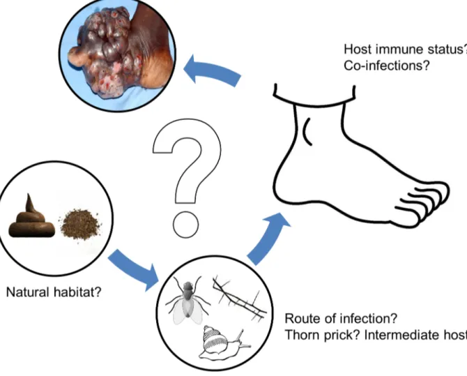
In order to be able to determine the true burden of mycetoma and to identify the primary reservoir(s) and potential intermediate host(s) of its causative agents, good, reliable, fast, and cheap diagnostic tools are needed. At present, it is very difficult to reliably discriminate between infected and noninfected individuals in the field (submitted; see Acknowledgments); early cases without lesions are almost impossible to identify since they don’t show classical signs of mycetoma and grains are still absent. The available diagnostic tools include imaging to determine the extent of lesions along different tissue planes, and cytological, histopathological examinations and culturing of grains to identify the causative agent (submitted; see Acknowledgments). Deep surgical biopsy material is needed for the latter two procedures. Identification of causal agents to the species level with histology is next to Figure 1. Possible hypothesis for mycetoma infection route.At the moment, many questions still remain about the various steps in the development of mycetoma. First, it is not known where the causative agents reside. What is the natural habitat of these agents? Both soil and dung have been implicated, but other niches could also be involved. Second, how is the causative agent introduced into the subcutaneous tissue? Is it via a thorn prick as implicated for years, or are vectors, such as insects, involved? When the causative agent is introduced in the subcutaneous tissue, will it always cause mycetoma, or does the host play a role as well? Do immune status, genetic background, or co-infections play a role in the development of mycetoma or even the extent of the infection?
impossible, while culturing them is difficult and time-consuming, especially for nonsporulating fungi (submitted; see Acknowledg-ments). Misidentification of causal agents in the past has caused poor therapeutic outcomes [54]. More reliable diagnostic tech-niques depending on DNA analysis have been developed for the detection of actinobacterial and fungal causal agents, but such approaches are too expensive for use in endemic areas at the present time [55,56]. Therefore, one of the first priorities is to develop fast and reliable diagnostic tools which can be used as point-of-care tests in regions where mycetoma is endemic. Since it has been demonstrated that 65% of all mycetoma cases can be attributed toM. mycetomatis, N. brasiliensis, A. madurae, A. pelletieri, N. asteroides, and S. somaliensis, the initial focus should be on the identification of these causative agents. Diagnostic methods which are most applicable are DNA-based identification tools such as loop-mediated isothermal amplification (LAMP), and serological assays.
LAMP has been successfully used in Africa and India [57–59] to diagnose Human African trypanosomiasis (HAT), malaria and visceral leishmaniasis. Sufficient DNA can be isolated from a dried blood spot on Whatman filter paper using simple extraction methods [58–60]. For the detection of Buruli ulcer, swabs or fine needle aspirates are used [61]. Isolated DNA can be amplified by using a heating bath and the resulting amplicons are usually visualized by adding (fluorescent) dyes [57–61]. This technique might be adapted for mycetoma, and various patient samples, including blood, serum, or fine needle aspirates, can be tested to determine the specificity and sensitivity of this assay in mycetoma patients.
Other diagnostic tools applicable in endemic settings are serological-based procedures, such as latex bead agglutination assays and dipsticks. By binding either an antigen [62,63] or a specific antibody [64,65] of the causative agent to a latex bead, a cheap and fast screening tool for serum can be developed that minimally requires the use of any equipment. Latex bead assays have been developed for other tropical infections, including visceral leismaniasis [62,64] and paracoccidioidomycosis [63]. In order to develop a specific agglutination assay, a discriminatory antigen needs to be selected. Whole genomes are available forN. brasiliensis [48] andS. somaliensis[47], while those of other causal agents such asM. mycetomatisandS. sudanensisare being generated. The genomes of these species will be ideal resources to select specific proteins for rapid serodiagnosis.
Treatment
Accurate diagnosis of the causal agents of mycetoma is a prerequisite for proper treatment. The current therapy includes medical treatment or a combination of medical treatment and surgery. Prevention by vaccination is not presently available. Mining the genomes of the most common causative agents could identify potential vaccine candidates. Currently, actinomycetoma is treated with antibacterials. The most common drug treatment for uncomplicated cases is trimethoprim-sulfamethoxazole (TMP-SMX) given for several months until cure is achieved [66,67]. A study in the 1970s by Mahgoub evaluated several antimicrobial combinations in 144 patients with actinomycetoma. In this study, the best clinical outcome was obtained with sulfamethoxazole plus streptomycin. This resulted in the following clinical outcomes: 63.2% cured, 21.5% greatly improved, and 11.1% showing some improvement [68]. In Sudan, the reported cure rate was 43.3%, excluding patients who did not complete the treatment [67]. At present, in Mexico, when there is no therapeutic response or improvement with TMP-SMX, or when the infection is severe and disseminated, or in a special location with a risk of spread to underlying vital tissues (head, neck, trunk,
abdomen, or inguinal region), a combination of amikacin (15 mg/kg/day) for 3 weeks plus trimethoprim-sulfamethoxazole (8/40 mg/kg/day) for 5 weeks (a cycle of treatment) is given for up to five cycles. This treatment has achieved a cure rate of .90% [66].
For eumycetoma, antifungal therapy consists mainly of ketoco-nazole or itracoketoco-nazole combined with surgical excision [67]. Unfortunately, this therapy is not quite successful. A recent study conducted at the Mycetoma Research Centre in Khartoum, Sudan showed that of the 1,242 eumycetoma patients studied, only 321 (25.9%) were cured, 35 (2.8%) had amputations, and 671 of the patients (54%) dropped out from the outpatient follow-up for various reasons. One of these reasons was the dissatisfaction with the therapy outcome [67]. In addition, in Sudan, most patients pay for their treatments. With a monthly income of$60, the costs of a ketoconazole ($30/month) or itraconazole treatment ($330/month) are a severe financial burden on an average household. Furthermore, it has been known to cause important side effects, such as hepatic toxicity, gynaecomastia, and skin discoloration [69,70]. Many patients stop therapy and are lost to follow-up in outpatient clinics, only to present later with a massive relapse, often requiring amputation.
For actinomycetoma, 168 out of 302 patients seen in the Mycetoma Research Centre in Khartoum did not complete their treatment. For eumycetoma, the corresponding figures were 671 out of 1,241 patients [67]. Therefore, there is an urgent need to develop better antifungal therapy, better treatment strategies, and a shorter treatment regime. A first step in improving therapy has been made by determining the in vitro susceptibilities of the most common fungal causative agentM. mycetomatisagainst a large panel of marketed antifungal agents [71–75].M. mycetomatisappeared to be most susceptible to the azole class of antifungal agents. Only poor inhibitory concentrations were found for other compounds. There is now a need to link in vitro susceptibility data with clinical outcome. There is also a need to generate minimal inhibitory concentration (MIC) breakpoints for in vitro susceptibility linked to therapeutic susceptibility or resistance.
In order to improve current therapy for M. mycetomatis infections, the best approaches are to select safer, newer azoles to which the organism is susceptible or use treatments that combine traditionally used azoles with (newly) available antifungal agents with different targets. Antifungal agents that have been used clinically in the treatment of eumycetoma include ketoconazole [76], itraconazole [77], posaconazole [78], voriconazole [79–82], and terbinafin [83]. The association of an azole with terbinafin is the most logical combination to be explored, as these two drugs have a different mode of action. Preliminary data on the use of liposomal amphotericin B are promising in terms of efficacy with good drug penetration (Fahal, personal communication). But since this drug is only available intravenously and is highly expensive, its feasibility in the long term for mycetoma treatment remains questionable. As 65% of patients have a secondary bacterial infection [84], the combination of antifungal and antibacterial treatment should be explored.
as well as bone density. A simple, reliable biomarker for cure is urgently needed to assess the response to treatment.
Conclusion
In order to be able to improve the current management of mycetoma, we have identified knowledge gaps for which research is urgently needed. These include the estimation of the burden of mycetoma in order to focus treatment and care on those locations with the highest prevalence. Furthermore, identification of the natural habitat of causal agents and establishing how they are introduced into subcutaneous tissues could lead to novel interven-tions, which ultimately will reduce its incidence. The development of methods for early diagnosis and therapeutic monitoring will result in better therapeutic outcomes. Improvement of therapeutic strategies in eumycetoma will also reduce the burden of disease. Now that the key research priorities have been highlighted, it is time to implement them. The WHO has taken an important first
step by including mycetoma on the list of NTDs. Further recognition by the international community is needed. Resources need to be made available to design control efforts and improve patient management.
Acknowledgments
The following work, cited in the text above, has been submitted for publication and is currently under review: van de Sande WWJ, Fahal AH, Goodfellow M, Mahgoub el S, Welsh O, et al. The merits and pitfalls of the currently used diagnostic tools in mycetoma. PLOS Negl Trop Dis.
References
1. WHO (2013) The 17 neglected tropical diseases. Geneva: World Health Organization.
2. Merritt RW, Walker ED, Small PL, Wallace JR, Johnson PD, et al. (2010) Ecology and transmission of Buruli ulcer disease: a systematic review. PLOS Negl Trop Dis 4: e911.
3. Van de Sande WWJ (2013) Global burden of human mycetoma: a systematic review and meta-analysis. PLOS Negl Trop Dis 7: e2550.
4. Queiroz-Telles F, Nucci M, Colombo AL, Tobon A, Restrepo A (2011) Mycoses of implantation in Latin America: an overview of epidemiology, clinical manifestations, diagnosis and treatment. Med Mycol 49: 225–236.
5. Ahmed AO, van Leeuwen W, Fahal A, van de Sande WWJ, Verbrugh H, et al. (2004) Mycetoma caused byMadurella mycetomatis: a neglected infectious burden. Lancet Infect Dis 4: 566–574.
6. Kaempfer E (1694) Disputatio physica medica inauguralis exhibens decadem observationem exoticarum [phD thesis]. Netherlands: Univeristy of Leiden. 7. Fahal AH, Sabaa AH (2010) Mycetoma in children in Sudan. Trans R Soc Trop
Med Hyg 104: 117–121.
8. Abbott P (1956) Mycetoma in the Sudan. Trans R Soc Trop Med Hyg 50: 11– 24; discussion, 24–30.
9. Lynch JB (1964) Mycetoma in the Sudan. Ann R Coll Surg Engl 35: 319– 340.
10. Develoux M, Audoin J, Treguer J, Vetter JM, Warter A, et al. (1988) Mycetoma in the Republic of Niger: clinical features and epidemiology. Am J Trop Med Hyg 38: 386–390.
11. Bakshi R, Mathur DR (2008) Incidence and changing pattern of mycetoma in western Rajasthan. Indian J Pathol Microbiol 51: 154–155.
12. Maiti PK, Ray A, Bandyopadhyay S (2002) Epidemiological aspects of mycetoma from a retrospective study of 264 cases in West Bengal. Trop Med Int Health 7: 788–792.
13. Chakraborti A, Singh K (1998) Mycetoma in Chandigarh and surrounding areas. Indian J Med Microbiol 16: 64–65.
14. Lopez Martinez R, Mendez Tovar LJ, Lavalle P, Welsh O, Saul A, et al. (1992) [Epidemiology of mycetoma in Mexico: study of 2105 cases]. Gac Med Mex 128: 477–481.
15. Joshi KR, Sanghvi A, Vyas MC, Sharma JC (1987) Etiology & distribution of mycetoma in Rajasthan, India. Indian J Med Res 85: 694–698.
16. Buot G, Lavalle P, Mariat F, Suchil P (1987) [Epidemiologic study of mycetomas in Mexico. Apropos of 502 cases]. Bull Soc Pathol Exot Filiales 80: 329–339. 17. Culligan GA, Grant C, Robbs GM, Crewe-Brown HH (1985) Actinomadura
pelletieri mycetoma from the Transvaal. A case report. S Afr Med J 68: 416– 418.
18. Freed CC, Bissett E, Martin PM, Ajello L (1975) Actinomycotic mycetoma due to Streptomyces somaliensis: report of a case in South Africa. Sabouraudia 13: 316–322.
19. Vismer HF, Morrison JG (1974) Mycetoma caused by Actinomadura (Streptomyces) madurae. The first South African case and the results of chemotherapy. S Afr Med J 48: 433–437.
20. Murray IG (1968) Laboratory aspects of mycetoma. In: Wolstenholme GEW, editor. Systemic mycosis. London, WI: J. & A. Churchill Ltd. pp. 78–93. 21. The Federal Ministry of Health: The National Mycetoma Control Programme
(2010) Report on the Epidemiological Study on Mycetoma at El Andalous Village, The White Nile State. Available: http://mycetoma.uofk.edu/news%26events/ pdf/AL%20Andalous%20final%20report.pdf. Accessed 19 February 2014. 22. Porten K, Sailor K, Comte E, Njikap A, Sobry A, et al. (2009) Prevalence of
Buruli ulcer in Akonolinga health district, Cameroon: results of a cross sectional survey. PLOS Negl Trop Dis 3: e466.
23. Phanzu DM, Suykerbuyk P, Imposo DB, Lukanu PN, Minuku JB, et al. (2011) Effect of a control project on clinical profiles and outcomes in buruli ulcer: a before/after study in Bas-Congo, Democratic Republic of Congo. PLOS Negl Trop Dis 5: e1402.
24. BNNICD (2012) Blue Nile Institute for Communicable diseases (BNNICD) newsletter. Wad Medani: Blue Nile Institute for Communicable Diseases. 3 p.
Top Five Papers
1. Abbott P (1956) Mycetoma in the Sudan. Trans R Soc Trop Med Hyg 50: 11–24; discussion, 24–30.
2. Ahmed AO, Mukhtar MM, Kools-Sijmons M, Fahal AH, de Hoog S, et al. (1999) Development of a species-specific PCR-restriction fragment length polymorphism analysis procedure for identification ofMadurella mycetomatis. J Clin Microbiol 37: 3175–3178.
3. van de Sande WWJ, Luijendijk A, Ahmed AO, Bakker-Woudenberg IA, van Belkum A (2005) Testing of the in vitro susceptibilities of Madurella mycetomatis to six antifungal agents by using the sensititre system in comparison with a viability-based 2,3-bis(2-methoxy-4-nitro-5-sulfophenyl)-5- [(phenylamino)carbonyl]-2H-tetra-zolium hydroxide (XTT) assay and a modified NCCLS method. Antimicrob Agents Chemother 49: 1364–1368. 4. Welsh O, Vera-Cabrera L, Welsh E, Salinas MC (2012)
Actinomycetoma and advances in its treatment. Clin Dermatol 30: 372–381.
5. Mahgoub ES, Gumaa SA (1984) Ketoconazole in the treatment of eumycetoma due toMadurella mycetomii. Trans R Soc Trop Med Hyg 78: 376–379.
Key Learning Points
N
Obtaining better incidence and prevalence data is necessary to determine appropriate sites for erecting mycetoma health care facilities.N
Gaining insight into the natural habitat and the mode of transmission of the mycetoma causative agents will open the door to preventive measures to reduce the number of mycetoma cases worldwide.N
Developing new, fast, and cheap diagnostic tools will enable early case detection, thereby enhancing the change of a better therapeutic outcome. Furthermore, these tools might also be able to monitor the therapeutic outcome.25. Segretain G, Mariat F (1968) [Research on the presence of agents of mycetoma in the soil and thorny plants of Senegal and Mauritania]. Bull Soc Pathol Exot Filiales 61: 194–202.
26. Parthasarathi K, Ranganathan LS, Anandi V, Zeyer J (2007) Diversity of microflora in the gut and casts of tropical composting earthworms reared on different substrates. J Environ Biol 28: 87–97.
27. Orchard VA, Goodfellow M (1980) Numerical classification of some named strains ofNocardia asteroidesand related isolates from soil. J Gen Microbiol 118: 295–312.
28. Gonzalez-Ochoa A, Sandoval MA (1960) [Isolation ofNocardia brasiliensisand
asteroidesfrom soils]. Rev Inst Salubr Enferm Trop 20: 147–151.
29. Thirumalachar MJ, Padhye AA (1968) Isolation ofMadurella mycetomifrom soil in India. Hindustan Antibiot Bull 10: 314–318.
30. Sotgiu G, Mazzoni A, Mantovani A, Ajello L, Palmer J (1966) Survey of soils for human pathogenic fungi from the Emilia-Romagna region of Italy. II. Isolation of Allescheria boydii, Cryptococcus neoformans and Histoplasma capsulatum. Am J Epidemiol 83: 329–337.
31. Ajello L (1952) The isolation ofAliescheria boydiishear, an etiologic agent of mycetomas, from soil. Am J Trop Med Hyg 1: 227–238.
32. Ajello L, Zeidberg LD (1951) Isolation ofHistoplasma capsulatumandAllescheria boydiifrom soil. Science 113: 662–663.
33. Ahmed A, Adelmann D, Fahal A, Verbrugh H, van Belkum A, et al. (2002) Environmental occurrence ofMadurella mycetomatis, the major agent of human eumycetoma in Sudan. J Clin Microbiol 40: 1031–1036.
34. de Hoog GS, Abdalla Ahmed S, Najafzadeh MJ, Sutton DA, Saradeghi Keisari M, et al. (2013) Phylogenetic findings suggest possible new habitat and routes of infection of human eumycetoma. PLOS Negl Trop Dis 7: e2229.
35. Fahal AH (2006) Mycetoma, Clinicopathological Monograph. Khartoum: Khartoum University Press.
36. Findlay GH, Vismer HF (1974) Black grain mycetoma. A study of the chemistry, formation and significance of the tissue grain in Madurella mycetomi infection. Br J Dermatol 91: 297–303.
37. Findlay GH, Vismer HF (1977) Black grain mycetoma. Atomic absorption and spark source mass spectrophotometry of the tissue grain in Madurella mycetomi infection. Br J Dermatol 97: 497–499.
38. Findlay GH, Vismer HF, Kiebenberg NW (1979) Black grain mycetoma: the ultrastructure of Madurella mycetomi. Mycopathologia 67: 51–54.
39. Ibrahim AI, El Hassan AM, Fahal A, van de Sande WW (2013) A histopathological exploration of the Madurella mycetomatis grain. PLOS One 8: e57774.
40. Mahgoub ES, Gumaa SA, El Hassan AM (1977) Immunological status of mycetoma patients. Bull Soc Pathol Exot Filiales 70: 48–54.
41. van de Sande WW, Fahal A, Verbrugh H, van Belkum A (2007) Polymorphisms in genes involved in innate immunity predispose toward mycetoma susceptibil-ity. J Immunol 179: 3065–3074.
42. Mhmoud N, Fahal A, Van de Sande WWJ (2013) Polymorphisms of interleukin-10 and CCL5 genes in mycetoma. Med Mycol 51: 527–533.
43. Mendez-Tovar LJ, Mondragon-Gonzalez R, Vega-Lopez F, Dockrell HM, Hay R, et al. (2004) Cytokine production and lymphocyte proliferation in patients withNocardia brasiliensisactinomycetoma. Mycopathologia 158: 407–414. 44. Cavanagh LL (1974) Attempts to induce mycetoma in monkeys and mice using
Madurella mycetomi. Sabouraudia 12: 258–262.
45. Ahmed AO, van Vianen W, ten Kate MT, van de Sande WW, van Belkum A, et al. (2003) A murine model ofMadurella mycetomatiseumycetoma. FEMS Immunol Med Microbiol 37: 29–36.
46. Mahgoub ES (1978) Experimental infection of athymic nude New Zealand mice, nu nu strain with mycetoma agents. Sabouraudia 16: 211–216.
47. van Hellemond JJ, Vonk AG, de Vogel C, Koelewijn R, Vaessen N, et al. (2013) Association of eumycetoma and schistosomiasis. PLOS Negl Trop Dis 7: e2241. 48. Prentice MB, Rahalison L (2007) Plague. Lancet 369: 1196–1207.
49. Gryseels B, Polman K, Clerinx J, Kestens L (2006) Human schistosomiasis. Lancet 368: 1106–1118.
50. Radolf JD, Caimano MJ, Stevenson B, Hu LT (2012) Of ticks, mice and men: understanding the dual-host lifestyle of Lyme disease spirochaetes. Nat Rev Microbiol 10: 87–99.
51. Kweku MA, Odoom S, Puplampu N, Desewu K, Nuako GK, et al. (2011) An outbreak of suspected cutaneous leishmaniasis in Ghana: lessons learnt and preparation for future outbreaks. Glob Health Action 4. doi:10.3402/ gha.v4i0.5527
52. Zhang Y, MacArthur C, Mubila L, Baker S (2010) Control of neglected tropical diseases needs a long-term commitment. BMC Med 8: 67.
53. Curtis CF, Maxwell CA, Magesa SM, Rwegoshora RT, Wilkes TJ (2006) Insecticide-treated bed-nets for malaria mosquito control. J Am Mosq Control Assoc 22: 501–506.
54. Mhmoud NA, Ahmed SA, Fahal AH, de Hoog GS, Gerrits van den Ende AH, et al. (2012)Pleurostomophora ochracea, a novel agent of human eumycetoma with yellow grains. J Clin Microbiol 50: 2987–2994.
55. Ahmed AO, Mukhtar MM, Kools-Sijmons M, Fahal AH, de Hoog S, et al. (1999) Development of a species-specific PCR-restriction fragment length polymorphism analysis procedure for identification of Madurella mycetomatis. J Clin Microbiol 37: 3175–3178.
56. Quintana ET, Wierzbicka K, Mackiewicz P, Osman A, Fahal AH, et al. (2008)
Streptomyces sudanensis sp. nov., a new pathogen isolated from patients with actinomycetoma. Antonie Van Leeuwenhoek 93: 305–313.
57. Namangala B, Hachaambwa L, Kajino K, Mweene AS, Hayashida K, et al. (2012) The use of Loop-mediated Isothermal Amplification (LAMP) to detect the re-emerging Human African Trypanosomiasis (HAT) in the Luangwa and Zambezi valleys. Parasit Vectors 5: 282.
58. Matovu E, Kuepfer I, Boobo A, Kibona S, Burri C (2010) Comparative detection of trypanosomal DNA by loop-mediated isothermal amplification and PCR from flinders technology associates cards spotted with patient blood. J Clin Microbiol 48: 2087–2090.
59. Surabattula R, Vejandla MP, Mallepaddi PC, Faulstich K, Polavarapu R (2013) Simple, rapid, inexpensive platform for the diagnosis of malaria by loop mediated isothermal amplification (LAMP). Exp Parasitol 134: 333– 340.
60. Verma S, Avishek K, Sharma V, Negi NS, Ramesh V, et al. (2013) Application of loop-mediated isothermal amplification assay for the sensitive and rapid diagnosis of visceral leishmaniasis and post-kala-azar dermal leishmaniasis. Diagn Microbiol Infect Dis 75: 390–395.
61. Ablordey A, Amissah DA, Aboagye IF, Hatano B, Yamazaki T, et al. (2012) Detection ofMycobacterium ulceransby the loop mediated isothermal amplification method. PLOS Negl Trop Dis 6: e1590.
62. Akhoundi B, Mohebali M, Shojaee S, Jalali M, Kazemi B, et al. (2013) Rapid detection of human and canine visceral leishmaniasis: assessment of a latex agglutination test based on the A2 antigen from amastigote forms ofLeishmania infantum. Exp Parasitol 133: 307–313.
63. Silveira-Gomes F, Sarmento DN, Pinto TM, Pimentel RF, Nepomuceno LB, et al. (2011) Development and evaluation of a latex agglutination test for the serodiagnosis of paracoccidioidomycosis. Clin Vaccine Immunol 18: 604– 608.
64. Attar ZJ, Chance ML, el-Safi S, Carney J, Azazy A, et al. (2001) Latex agglutination test for the detection of urinary antigens in visceral leishmaniasis. Acta Trop 78: 11–16.
65. Darani HY, Ahmadi F, Zabardast N, Yousefi HA, Shirzad H (2010) Development of a Latex Agglutination Test as a Simple and Rapid Method for Diagnosis ofTrichomonas vaginalisInfection. Avicenna J Med Biotechnol 2: 63–66.
66. Welsh O, Vera-Cabrera L, Welsh E, Salinas MC (2012) Actinomycetoma and advances in its treatment. Clin Dermatol 30: 372–381.
67. Zein HA, Fahal AH, Mahgoub el S, El Hassan TA, Abdel-Rahman ME (2012) Predictors of cure, amputation and follow-up dropout among patients with mycetoma seen at the Mycetoma Research Centre, University of Khartoum, Sudan. Trans R Soc Trop Med Hyg 106: 639–644.
68. Mahgoub ES (1976) Medical management of mycetoma. Bull World Health Organ 54: 303–310.
69. Findor JA, Sorda JA, Igartua EB, Avagnina A (1998) Ketoconazole-induced liver damage. Medicina (B Aires) 58: 277–281.
70. Bernuau J, Durand F, Pessayre D (1997) Ketoconazole-induced hepatotoxicity. Hepatology 26: 802.
71. van de Sande WWJ, Fahal AH, Bakker-Woudenberg IAJM, van Belkum A (2010) Madurella mycetomatis is not susceptible to the echinocandin class of antifungal agents. Antimicrob Agents Chemother 54: 2738–2740.
72. van de Sande WWJ, Luijendijk A, Ahmed AO, Bakker-Woudenberg IA, van Belkum A (2005) Testing of the in vitro susceptibilities ofMadurella mycetomatisto six antifungal agents by using the sensititre system in comparison with a viability-based 2,3-bis(2-methoxy-4-nitro-5-sulfophenyl)-5- [(phenylamino)carbonyl]-2H-tetrazolium hydroxide (XTT) assay and a modified NCCLS method. Antimicrob Agents Chemother 49: 1364–1368.
73. Kloezen W, Meis JF, Curfs-Breuker I, Fahal AH, Van de Sande WWJ (2012) In vitro antifungal activity of isavuconazole toMadurella mycetomatis. Antimicrob Agents Chemother 56: 6054–6056.
74. van Belkum A, Fahal AH, van de Sande WW (2011) In vitro susceptibility of
Madurella mycetomatis to posaconazole and terbinafine. Antimicrob Agents Chemother 55: 1771–1773.
75. Ahmed AO, van de Sande WWJ, van Vianen W, van Belkum A, Fahal AH, et al. (2004) In vitro susceptibilities ofMadurella mycetomatis to itraconazole and amphotericin B assessed by a modified NCCLS method and a viability-based 2,3-Bis(2-methoxy-4-nitro-5- sulfophenyl)-5-[(phenylamino)carbonyl]-2H-tetra-zolium hydroxide (XTT) assay. Antimicrob Agents Chemother 48: 2742–2746. 76. Mahgoub ES, Gumaa SA (1984) Ketoconazole in the treatment of eumycetoma
due toMadurella mycetomii. Trans R Soc Trop Med Hyg 78: 376–379. 77. Fahal AH, Rahman IA, El-Hassan AM, Rahman ME, Zijlstra EE (2011) The
safety and efficacy of itraconazole for the treatment of patients with eumycetoma due toMadurella mycetomatis. Trans R Soc Trop Med Hyg 105: 127–132. 78. Negroni R, Tobon A, Bustamante B, Shikanai-Yasuda MA, Patino H, et al.
(2005) Posaconazole treatment of refractory eumycetoma and chromoblasto-mycosis. Rev Inst Med Trop Sao Paulo 47: 339–346.
79. Lacroix C, de Kerviler E, Morel P, Derouin F, Feuilhade de Chavin M (2005)
Madurella mycetomatis mycetoma treated successfully with oral voriconazole. Br J Dermatol 152: 1067–1068.
80. Loulergue P, Hot A, Dannaoui E, Dallot A, Poiree S, et al. (2006) Successful treatment of black-grain mycetoma with voriconazole. Am J Trop Med Hyg 75: 1106–1107.
82. Schaenman JM, Digiulio DB, Mirels LF, McClenny NM, Berry GJ, et al. (2005)
Scedosporium apiospermumsoft tissue infection successfully treated with voricona-zole: potential pitfalls in the transition from intravenous to oral therapy. J Clin Microbiol 43: 973–977.
83. N’Diaye B, Dieng MT, Perez A, Stockmeyer M, Bakshi R (2006) Clinical efficacy and safety of oral terbinafine in fungal mycetoma. Int J Dermatol 45: 154–157. 84. Ahmed AO, Abugroun ES (1998) Unexpected high prevalence of secondary