Subclinical abnormal glucose tolerance is a predictor of
death in liver cirrhosis
Diego García-Compeán, Joel Omar Jáquez-Quintana, Fernando Javier Lavalle-González, José Alberto González-González, Linda Elsa Muñoz-Espinosa, Jesús Zacarías Villarreal-Pérez, Héctor J Maldonado-Garza
Diego García-Compeán, Joel Omar Jáquez-Quintana, José Alberto González-González, Héctor J Maldonado-Garza, Gas-troenterology Service and Department of Internal Medicine, versity Hospital “Dr. José E. Gonzalez” and Medical School, Uni-versidad Autónoma de Nuevo León, Monterrey 64320, México
Fernando Javier Lavalle-González, Jesús Zacarías Villarreal-Pérez, Endocrinology Service and Department of Internal Medicine, University Hospital “Dr. José E. González” and Medi-cal School, Universidad Autónoma de Nuevo León, Monterrey 64320, México
Linda Elsa Muñoz-Espinosa, Liver Unit, University Hospi-tal “Dr. José E. González” and Medical School, Universidad Autónoma de Nuevo León, Monterrey 64320, México
Author contributions: García-Compeán D designed and coor-dinated the research and wrote the manuscript; Jáquez-Quintana JO and González-González JA evaluated patients at inclusion and follow-up; Lavalle-González FJ and Villarreal-Pérez JZ par-ticipated in laboratory testings and revision of the manuscript; Muñoz-Espinosa LE and Maldonado-Garza HJ reviewed the manuscript.
Supported by Gastroenterology and Endocrinology Services of the University Hospital, Universidad Autónoma de Nuevo León, Monterrey, México
Correspondence to: Diego García-Compeán, MD, Gastroen-terology Service and Department of Internal Medicine, Univer-sity Hospital “Dr. José E. Gonzalez” and Medical School, Uni-versidad Autónoma de Nuevo León, Madero y Gonzalitos S/N, Monterrey 64320, México. digarciacompean@prodigy.net.mx
Telephone: +52-81-83487315 Fax:+52-81-89891381
Received: November 10, 2013 Revised: February 5, 2014
Accepted: March 19, 2014
Published online: June 14, 2014
Abstract
AIM: To determine if subclinical abnormal glucose tol-erance (SAGT) has influence on survival of non-diabetic patients with liver cirrhosis.
METHODS: In total, 100 patients with compensated
PROSPECTIVE STUDY DOI:10.3748/wjg.v20.i22.7011 © 2014 Baishideng Publishing Group Inc. All rights reserved.
liver cirrhosis and normal fasting plasma glucose were included. Fasting plasma insulin (FPI) levels were mea-sured, and oral glucose tolerance test (OGTT) was performed. According to OGTT results two groups of patients were formed: those with normal glucose toler-ance (NGT) and those with SAGT. Patients were fol-lowed every three months. The mean follow-up was 932 d (range of 180-1925). Survival was analyzed by the Kaplan-Meyer method, and predictive factors of death were analyzed using the Cox proportional hazard regression model.
RESULTS: Of the included patients, 30 showed NGT and 70 SAGT. Groups were significantly different only in age, INR, FPI and HOMA2-IR. Patients with SAGT showed lower 5-year cumulated survival than NGT patients (31.7% vs 71.6%, P = 0.02). Differences in survival were significant only after 3 years of follow-up. SAGT, Child-Pugh B, and high Child-Pugh and Model for End-Stage Liver Disease (MELD) scores were independent predictors of death. The causes of death in 90.3% of cases were due to complications related to liver disease.
CONCLUSION: SAGT was associated with lower sur-vival. SAGT, Child-Pugh B, and high Child-Pugh and MELD scores were independent negative predictors of survival.
© 2014 Baishideng Publishing Group Inc. All rights reserved.
Key words: Diabetes mellitus; Liver cirrhosis; Oral glu-cose tolerance test; Survival; Gluglu-cose metabolism dis-orders
MATERIALS AND METHODS
Patients
Patients with LC, who were seen in our hospital from Au-gust 2007 to AuAu-gust 2012, were prospectively evaluated. Only those with normal FPG (< 100 mg/dL), no history of overt DM, with clinical or histological diagnosis of LC, aged over 18 years old and without clinically evident
complications of liver disease (e.g., esophageal or gastric
variceal bleeding, hepatic encephalopathy, hepatorenal syndrome, infection, spontaneous bacterial peritonitis, and moderate to severe ascites), were selected for the study (patients with complications were excluded in order to avoid the effect of medications used for treating them, on glucose metabolism of patients). Clinical diagnosis of LC was made by using a combination of clinical symp-toms, laboratory tests and imaging studies (abdominal
ultrasonography or CT scan)[21,22]. The etiology of
cirrho-sis was determined as follows: Alcohol-related cirrhocirrho-sis was determined when daily alcohol consumption was > 80 g in men and > 40 g in women for at least 10 years with negative viral, metabolic and autoimmune markers. Diagnosis of hepatitis C virus and B virus related
cir-rhosis was determined with specific viral markers (HBsAg or anti-HCV). Autoimmune liver disease was diagnosed with specific autoimmune markers (anti-nuclear antibod -ies, smooth muscle antibodies or liver-kidney anti-microsomal antibodies). Meanwhile, cryptogenic cirrhosis was established in the absence of any of the causes above described[23].
Informed consent was obtained from each patient. The protocol was approved by The Research and Ethics Committee of the Faculty of Medicine of the Autono-mous University of Nuevo Leon in Monterrey.
Fasting plasma insulin and OGTT
Fasting plasma insulin (FPI) levels were measured and
OGTT was performed according to the World Health
Organization criteria[24]. Patients were fasted for at least
12 h, and baseline plasma glucose was measured; then, they were given an oral glucose load (75 g). Additionally, plasma glucose (PG) levels were measured every 30 min for 2 h. The results were interpreted as follows: a) normal glucose tolerance (NGT) if 2-h PG was < 140 mg/dL; IGT if 2-h PG between 140 mg/dL and 200 mg/dL;
and DM if 2-h PG > 200 mg/dL. Plasma HbA1c was
not used for diagnosing DM due to its low sensitivity re-ported in cirrhotic patients[19].
FPI was measured using the
electrochemilumines-cence method (normal values: 5-20 μU/mL).
HOMA2-IR index (Homeostasis Model Assessment 2-Insulin Re
-sistance Index) was calculated using the following online software (http://www.dtu.ox.ac.uk/homacalculator/in-dex.php).
Assessments
The following data were recorded in a database specially
DM is associated with low survival of cirrhotic patients. This issue is important since a high proportion of cir-rhotic patients (about 70%) without history of DM and with normal fasting plasma glucose have subclinical abnormal glucose tolerance. The use of oral glucose tolerance test for early recognition and treatment of DM may improve prognosis of these patients.
García-Compeán D, Jáquez- Quintana JO,Lavalle-González FJ, González-González JA,Muñoz-Espinosa LE,Villarreal-Pérez JZ, Maldonado-Garza HJ. Subclinical abnormal glucose tolerance is a predictor of death in liver cirrhosis. World J Gastroenterol
2014; 20(22): 7011-7018 Available from: URL: http://www. wjgnet.com/1007-9327/full/v20/i22/7011.htm DOI: http://dx.doi. org/10.3748/wjg.v20.i22.7011
INTRODUCTION
Overt diabetes mellitus (DM) has been reported in
21%-30% of patients with liver cirrhosis (LC)[1]. DM may
arise from a progressive disorder of insulin secretion in
the presence of liver and muscle resistance to insulin[2,3].
DM is related to LC in two ways: (1) type 2 DM (T2DM) (often associated with metabolic syndrome) causes nonal-coholic fatty liver disease (steatosis, steatohepatitis, LC, and
hepatocellular carcinoma)[4-7]; and (2) DM may develop as
a complication of LC, which is known as “hepatogenous
diabetes” (HD)[8,9]. As liver disease advances, DM becomes
clinically evident[10,11]. T2DM and HD are associated with
an increased risk of complications of chronic liver diseases and mortality[7,9,12-17].
On the other hand, subclinical abnormal glucose tolerance (SAGT) disorders, such as impaired glucose tol-erance or DM (called subclinical since they are detected only by means of an oral glucose tolerance test), may be observed in 45% and 22%, respectively, of patients with
LC and no history of DM[18,19]. Therefore, the prevalence
of DM and impaired glucose tolerance (IGT) may be underestimated when only fasting plasma glucose (FPG) levels are taken into account. Our group has recently published a study determining the prevalence of T2DM,
DH and IGT in patients with LC who underwent oral
glucose tolerance test (OGTT). In total, 86% of the cases had either overt or subclinical IGT and DM, with or without insulin resistance (IR)[18].
The negative influence of SAGT on the survival of
cirrhotic patients was demonstrated in one study car-ried out in Japan, which found that patients with IGT or
DM diagnosed by OGTT had a significantly lower 5-year
survival rate[20]. However, similar findings have not been
reported by others elsewhere.
The aims of this study were: (1) to evaluate if SAGT
designed for the study: gender, age, and family history of DM as well as laboratory blood test results (serum hemo-globin, leukocytes, platelets, BUN, creatinine, albumin,
bilirubin, aspartate aminotransferase, γ-glutamyltranspe
ptidase, alkaline phosphatase, cholesterol, triglycerides, and INR). The body mass index (BMI) was calculated and construed as follows: normal if < 24.9, overweight if
between 25 and 29.9, and obesity if > 30[25]. Liver
func-tion was estimated using Child-Pugh and Model for
End-Stage Liver Disease (MELD) classifications[26,27].
Follow-up
All patients were followed every 3 mo in the outpatient department. A full clinical examination was made in each interview. Laboratory blood testing (serum hemoglobin, hematocrit, blood cell count, glucose, creatinine, BUN, liver function tests and coagulation tests) was also per-formed. Moreover, plasma alpha-fetoprotein and liver ul-trasonography were performed every 6 mo. Additionally, hospital admissions were recorded during which clinical course of the patient was determined. Patients or their relatives were contacted by telephone when he or she did not attend to appointments. Callings were done in order to determine the clinical status of the patient, or to in-quire whether their death occurred.
During follow-up patients were treated with life style
and diet modifications, and in particular cases with oral
hypoglycemic agents or insulin administration. No an-tidiabetic medication was given to those with impaired glucose tolerance.
Statistical analysis
Continuous variables are expressed as means and stan-dard deviations, while non-continuous variables as
me-dians and ranges. Categorical variables are expressed as relative proportions. Intergroup comparisons were made
by Student’s t, χ2 and Mann-Whitney tests. Variables were
analyzed using the Cox proportional hazard regression model in order to determine independent predictive
fac-tors of mortality[28]. Only the variables that were
statisti-cally significant in univariate analysis were analyzed in
multivariate analysis. The results were expressed as HR
with 95%CI, and the P value was calculated.
The cumulative survival was analyzed using the Ka-plan-Meier method, and the curves were compared using
log-rank test. A P value less than 0.05 was considered
statistically significant. All statistical analyses were done
using the statistical package SPSS v17.0 (Chicago, Illinois, United States).
RESULTS
Patient population
Of 293 cirrhotic patients evaluated, 124 (42.3%) met the
inclusion criteria. However, 24 (8.1%) of them were not finally included due to the following reasons: 13 refused
to participate in the study, 8 found difficult to attend consultation because they lived in another state, and 3 did not attend their appointments for performing OGTT (Figure 1).
Finally, 100 (34.1%) patients were included in the study. Their clinical and biochemical characteristics are shown in Table 1. Although BMI is not the best param-eter for assessing nutritional status in cirrhotic patients (particularly in those with ascites), this method showed that most of them (69%) were overweight or obese. The etiology of cirrhosis was predominantly alcoholic and cryptogenic (47% and 26%, respectively). The diagnosis
Evaluated patients with LC n = 293
Fulfilled selection criteria
n = 124 (42.3%)
13 Refused to participate 8 Found difficult to attend
consultations 3 Did not returned for OGTT
Included patients
n = 100 (34.1%)
OGTT
Normal
n = 30 (20.0%)
IGT
n = 44 (29.3%)
DM
n = 26 (17.3%)
times of follow-up, oral antidiabetic medications were prescribed in 6, and subcutaneous insulin was prescribed in 2 out of 12 patients with overt DM.
In total, 49% of alcoholic patients consistently stopped alcohol consumption, 47% of patients with hep-atitis C virus infection were treated with antiviral therapy, 50% and 90% of patients with hepatitis B virus infection or autoimmune liver disease were treated. There were no dropouts.
Cumulative survival
The mean survival time was 1448 d (range: 1256-1925) in the patients with NGT, while it was 1116 d (range
990-1790) in the patients with SAGT (P = 0.045).
Cumu-lative survival curve of patients with NGT and SAGT is
shown in Figure 2. SAGT patients had significantly lower
survival (P = 0.02).
One to 5-year survival rates are shown in Table 2.
Sta-tistically significant differences were observed only after
three years of follow-up. Increasing differences were ob-served over time until the end of follow-up.
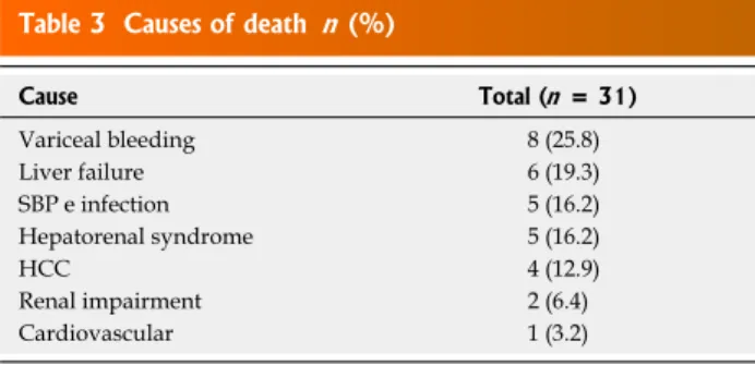
Causes of death
In total 31 patients died (5 from the NGT group and 26 from the SAGT group). The causes of death are shown in Table 3. Twenty eight (90.3%) patients died due to the following complications related to liver disease: 8 due to esophageal variceal bleeding (25.8%), 6 due to chronic liver failure (19.3%), 5 due to spontaneous bacterial peri-tonitis and other infections (16.2%), 5 due to hepatorenal syndrome (16.2%) and 4 due to hepatocellular carcinoma (12.9%). Additionally, 2 of the remaining patients died due to renal impairment (6.4%), and 1 due to
cardiovas-of LC was confirmed by liver biopsy in 44% cardiovas-of cases. In
total, 47% and 53% of patients were assigned into the
groups A and B of the Child-Pugh classification, respec -tively. The mean Child-Pugh score was 6.9 ± 1.8 points and the mean MELD score was 8.9 ± 4.8. The mean
values of FPI levels and HOMA2-IR index were 15.9 ±
10.2 μU/mL and 2.67 ± 1.6, respectively.
Clinical characteristics and survival rate of patients
Two groups were formed according to the results of OGTT: those with NGT (30 patients) and those with SAGT (70 patients, 44 with IGT and 26 with DM). Clini-cal and biochemiClini-cal characteristics are shown in Table 1.
SAGT patients were significantly older and had higher val
-ues of INR, FPI and HOMA2-IR. Although Child-Pugh
and MELD scores were mildly higher in patients with
SAGT, these differences were not statistically significant.
Follow-up
The mean follow-up was 932 d (range 180-1925). At the end of follow-up, 19 (19%) patients (5 NGT patients and 14 SAGT patients) developed overt IGT (7 cases) or overt DM (12 cases). All patients with SAGT were pre-scribed dietary and life style modifications. At different
Table 1 Clinical and biochemical characteristics of cirrhotic patients
Variable Total (n = 100) NGT (n = 30) SAGT (n = 70)
Age, yr 53.9 ± 11.8 49.0 ± 12.0 56.0 ± 11.11
BMI, kg/m2 26.9 ± 4.2 26.4 ± 4.40 27.21 ± 4.15
Males 60 (60) 18 (60) 42 (60) Etiology: Alcohol 47 (47) 14 (46.6) 33 (47.1) HBV 4 (4) 1 (3.3) 3 (4.2) HCV 13 (13) 2 (6.6) 11 (15.7) Autoimmunity 10 (10) 3 (10) 7 (10) Cryptogenic 26 (26) 10 (33.3) 16 (22.8) Liver biopsy 44 (44) 13 (43.3) 31 (44.2) Length of LC, mo 21.6 + 27.6 21.4 ± 30.6 21.7 ± 33.2 Hemoglobin, g/dL 11.8 ± 2.3 11.6 ± 2.1 11.9 ± 2.3 Platelets, × mm3 128140 ± 89501 159186 ± 112108 114834 ± 73269
INR 1.3 ± 0.3 1.2 ± 0.3 1.36 ± 0.32
Creatinine, mg/dL 0.8 ± 0.2 0.90 ± 0.33 0.83 ± 0.2 Albumin, g/dL 3.2 ± 0.73 3.33 ± 0.71 3.21 ± 0.74 ALT, UI/L 46.5 ± 40.8 48.9 ± 53.9 45.4 ± 34.0 Total bilirubin,
mg/dL
1.71 ± 0.90 1.55 ± 0.88 1.78 ± 0.91
Fasting plasma insulin, μU/mL
15.9 ± 10.2 11.9 ± 7.6 18.2 ± 113
HOMA2 score 2.67 ± 1.6 2.36 ± 1.46 4.6 ± 3.34
Child-Pugh A 47 (47) 15 (50) 32 (45.7) B 53 (53) 15 (50) 38 (54.2) Child Pugh score 6.8 ± 1.8 6.5 ± 1.61 6.82 ± 1.67 MELD score 8.94 ± 4.8 8.3 ± 5.2 9.21 ± 4.63 Survival, d 1237 1448 1116
(990-1925) (1256-1925) (990-1790)5
Data are expressed as absolute numbers (percentage) or mean ± SD. 1P
= 0.003; 2P = 0.025; 3P = 0.03; 4P = 0.009; 5P = 0.045. NGT: Normal glucose
tolerance; SAGT: Subclinical abnormal glucose tolerance; HOMA: Ho-meostatic model assessment; LC: Liver cirrhosis. BMI: Body mass index; MELD: Model for End-Stage Liver Disease; HBV: Hepatitis B virus; HCV: Hepatitis C virus; INR: International normalized ratio; ALT: Alanine trans-aminase.
Cumulated surviv
al
1.0
0.8
0.6
0.4
0.2
0.0
NGT
SAGT
P = 0.02
0.00 500.00 1000.00 1500.00 2000.00 Follow-uo period (d)
Figure 2 Cumulated survival of cirrhotic patients with normal glucose tolerance or subclinical abnormal glucose tolerance. Note that significant
cular disease (3.2%). There were no significant
differ-ences in the causes of death among patients with NGT vs
those with SAGT.
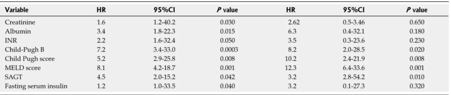
Predictors of death
Serum creatinine, albumin, INR, Pugh B, Child-Pugh and MELD scores, SAGT as well as FPI were statistically significant according to univariate analysis.
However, only Child-Pugh B (HR = 8.2, 95%CI: 2.0-28.5, P = 0.02), Child-Pugh score (HR = 10.2, 95%CI: 2.4-21.9,
P = 0.008), MELD score (HR = 12.3, 95%CI: 6.4-33.6,
P = 0.001) and SAGT (HR = 3.2, 95%CI: 2.8-54.2, P = 0.01) were independent predictors of survival according to multivariate analysis (Table 4).
DISCUSSION
Our study suggests that SAGT may have negative im-pact on the survival of patients with LC, as those who showed this abnormality had lower 5-year survival than those who had NGT. Significant differences in survival were observed after 3 years of follow-up (Table 2). These results are consistent with another study carried out in Japan, with 58 cirrhotic patients without overt DM who
underwent OGTT[20]. In that study, 5-year survival rate
was lower in patients with IGT and DM compared to those with NGT. Albumin and abnormal OGTT were
independent negative predictors of survival[20]. Despite
the similarities in results of both studies, ours differs in the following points: (1) 74% of our patients had alco-holic and cryptogenic cirrhosis, while 78.5% of patients
in the Japanese study had viral etiology (HBV and HCV).
The causes of chronic liver disease in our patients were identical to those observed in most Western countries, where alcohol and metabolic syndrome are the predomi-nant causes (especially nowadays, the frequency of obe-sity and nonalcoholic fatty liver disease have dramatically increased), while viral etiology is predominantly observed
in Asian countries[29-31]; (2) At time of inclusion, our
pa-tients did not have liver-related complications, whereas in the Japanese study, up to 30% had ascites and hepatic encephalopathy; and (3) The number of patients in our study was almost twice.
The mechanism by which SAGT may be involved in
the occurrence of death of cirrhotic patients is difficult to explain. DM may induce progression of liver fibrosis, inflammation and liver failure, or may increase cardiovas
-cular complications and atherosclerosis. In a study with virus C-infected patients, IR and DM were significantly
associated with liver fibrosis[32]. Furthermore, in other
reports, it has been suggested that DM can increase fibro -sis, incidence of hepatocellular carcinoma, and resistance
to antiviral therapy in patients with hepatitis C[33]. DM
may be involved in the progression of liver fibrosis and inflammation through diverse mechanisms: it is likely that
adipokine production (such as leptin and Tumor necrosis
factor-alpha, which activate inflammatory pathways exac -erbating liver injury) is increased by insulin resistance[34-36]. Leptin and oxidative stress associated with liver inflam-mation may activate transforming growth factor beta 1
(TGF-β1), which is one of the most potent profibrogenic
cytokines produced in the liver. TGF-β1 activates hepatic
stellate cells which are the major source of collagen Ⅰ and
Ⅲ and extracellular matrix proteins[37-39]. In our study,
SAGT as well as high Child-Pugh and MELD scores were independent predictors of death. This suggests that SAGT combined with other etiologic agents of liver cir-rhosis might have induced liver failure and death.
On the other hand, as it has been pointed out in other publications, most of our patients with SAGT died from
liver disease related complications[9,20,40] (Table 3). DM
increases the incidence of severe infections by inducing immunosuppression. Cirrhotic patients with DM have a higher prevalence of infections compared to non-diabetic ones. In a recent study, spontaneous bacterial peritonitis was more frequent in patients with cryptogenic cirrhosis (which is associated with DM) compared to those with
cirrhosis of other causes[41]. These infected patients had
higher in-hospital mortality due to sepsis, liver failure, and
hepatorenal syndrome[42]. In addition, DM may increase
the risk of variceal bleeding, as postprandial hyperglyce-mia that occurs in diabetic patients produces splanchnic
vasodilatation and increases the flow and pressure of the
porto systemic venous system[43]. Also, esophageal
vari-ceal bleeding increases the risk of infection and death by
inducing bacterial intestinal translocation[44]. DM has been
also associated with an increased risk of hepatic
encepha-lopathy (HE). A significant association between malnutri
-tion and diabetes with HE was observed in a study with
cirrhotic patients[45]. In multivariate analysis, Child-Pugh
classification, malnutrition and DM showed independent correlations with HE. Plasma ammonium ion levels were
related to insulin resistance and muscle mass. Authors
Table 2 Annual cumulated survival of cirrhotic patients n (%)
Year(s) NGT (n = 30) SAGT (n = 70) P value (χ2 test)
1 2 (93) 9 (87) 0.36 2 4 (83.2) 14 (79) 0.34 3 4 (83.2) 23 (51.4) 0.04 4 4 (83.2) 24 (47.5) 0.03 5 5 (71.6) 26 (31.7) 0.02
NGT: Normal glucose tolerance; SAGT: Subclinical abnormal glucose tolerance.
Table 3 Causes of death n (%)
Cause Total (n = 31)
Variceal bleeding 8 (25.8) Liver failure 6 (19.3) SBP e infection 5 (16.2) Hepatorenal syndrome 5 (16.2) HCC 4 (12.9) Renal impairment 2 (6.4) Cardiovascular 1 (3.2)
concluded that nutritional status and insulin resistance
might be implicated in the pathogenesis of HE[45]. On the other hand, some authors reported low
fre-quency of diabetic complications in cirrhotic patients[46].
However, these observations have been recently chal
-lenged: in two published studies, patients with chronic hepatitis C compared to controls, showed significant higher carotid intima media thickness, greater number of
carotid plaques and carotid atherosclerosis affection[47,48].
In one of these studies[47], older age and severe hepatic
fibrosis were independently associated with early carotid
atherosclerosis. In the other one[48], diabetes and
meta-bolic syndrome were associated with carotid plaques. The precise prevalence of cardiovascular disease or athero-sclerosis and their role in the induction of death in
cir-rhotic patients with DM need to be defined in future well
conducted studies.
In our study, we did not observe significant differ-ences in the causes of death among patients with NGT compared to those with SAGT (Table 3). This may be due to the small number of individuals in each group. Some differences might be clearly evident using a larger sample of patients.
Although the negative impact of DM on survival of cirrhotic patients is well known a long time ago, the ben-eficial effects of early detection and treatment of DM and IGT for reducing complications and mortality rates have not been clearly determined. In our study, analysis of this issue was not performed due to the low number of patients. This important point has to be determined in future studies.
In conclusion, SAGT disorders were associated with reduced long term survival in patients with compensated LC and normal FPG. Additionally, SAGT, Child-Pugh B, and high Child-Pugh and MELD scores were
indepen-dent negative predictors of survival. These findings sug -gest that SAGT may give rise to morbid conditions that increase mortality of patients. According to this, OGTT is useful for accurate assessment of mortality in non-diabetic cirrhotic patients.
COMMENTS
Background
Overt diabetes mellitus (DM) may be found in 30% of patients with liver
cir-rhosis of diverse etiology. Since a long time ago, it has been known that overt DM is linked to a reduction of survival of patients with liver cirrhosis. Causes of death of these patients are mostly due to liver disease complications. Nonethe-less, 60% of non-diabetic patients with liver cirrhosis may have abnormal glu-cose tolerance disorders, either impaired gluglu-cose tolerance (IGT) or DM, after an oral glucose tolerance test.
Research frontiers
The impact of subclinical forms of IGT and DM on survival of cirrhotic patients has not been extensively described. In this paper, the authors demonstrate that patients with these forms of abnormal glucose tolerance have reduced long term survival than those with normal glucose tolerance. Nevertheless, this study does not allow demonstrating the mechanisms by which abnormal glucose tol-erance may reduce survival of patients
Related publications
The impact of subclinical forms of abnormal glucose tolerance on survival of cir-rhotic patients has been described in only one study performed in Japan. In this study, patients with IGT and DM detected by oral glucose tolerance test (OGTT) had reduced 5-year survival than those with normal glucose tolerance. Innovations and breakthroughs
Most of the patients in the previously published study had cirrhosis of viral etiol-ogy and 30% had liver complications, while in this study, patients were clinically compensated and most of them had alcoholic and cryptogenic etiology. In addi-tion the sample size of this study was double.
Applications
The practical applications of the findings of this study are clear: early detection
of abnormal glucose tolerance conditions through OGTT allows early treatment of DM and consequently a reduction of complications and mortality of patients. The use of OGTT is suggested in all patients with liver cirrhosis without previ-ous DM, in order to assess prognosis of survival. Those with DM may be included in a close monitoring and an early treatment program. Future studies concerning the mechanisms by which DM induces death of cirrhotic patients may allow focusing prevention and therapeutic measures.
Terminology
Subclinical abnormal glucose tolerance refers to impaired glucose tolerance and diabetes mellitus detected by an oral glucose tolerance test.
Peer review
The investigators correlated the subclinical abnormal glucose tolerance and reduced survival in cirrhosis patients, and found that the subclinical abnormal glucose tolerance was associated with lower survival.
REFERENCES
1 Hickman IJ, Macdonald GA. Impact of diabetes on the se-verity of liver disease. Am J Med 2007; 120: 829-834 [PMID: 17904449 DOI: 10.1016/j.amjmed.2007.03.025]
2 Petrides AS, Vogt C, Schulze-Berge D, Matthews D, Stroh-meyer G. Pathogenesis of glucose intolerance and diabetes mellitus in cirrhosis. Hepatology 1994; 19: 616-627 [PMID: 8119686 DOI: 10.1002/hep.1840190312]
3 Petrides AS, Stanley T, Matthews DE, Vogt C, Bush AJ, Lambeth H. Insulin resistance in cirrhosis: prolonged re-duction of hyperinsulinemia normalizes insulin sensitivity.
Table 4 Univariate and multivariate analyses: Independent predictors of death
Variable HR 95%CI P value HR 95%CI P value
Creatinine 1.6 1.2-40.2 0.030 2.62 0.5-3.46 0.650 Albumin 3.4 1.8-22.3 0.015 6.3 0.4-32.1 0.180 INR 2.2 1.6-32.4 0.050 3.5 0.3-23.6 0.230 Child-Pugh B 7.2 3.4-33.0 0.0003 8.2 2.0-28.5 0.020 Child Pugh score 5.2 2.9-25.8 0.008 10.2 2.4-21.9 0.008 MELD score 8.1 4.2-18.7 0.001 12.3 6.4-33.6 0.001 SAGT 4.5 2.0-15.2 0.042 3.2 2.8-54.2 0.010 Fasting serum insulin 1.2 1.0-33.5 0.040 3.2 0.1-27.3 0.320
MELD: Model for end stage liver disease; INR: International normalized ratio; SAGT: Subclinical abnormal glucose tolerance.
Hepatology 1998; 28: 141-149 [PMID: 9657106 DOI: 10.1002/ hep.510280119]
4 Lewis JR, Mohanty SR. Nonalcoholic fatty liver disease: a review and update. Dig Dis Sci 2010; 55: 560-578 [PMID: 20101463 DOI: 10.1007/s10620-009-1081-0]
5 Blendea MC, Thompson MJ, Malkani S. Diabetes and Chron-ic liver disease: Etiology and pitfalls in monitoring. Clin Dia-betes 2010; 28: 139-144 [DOI: 10.2337/diaclin.28.4.139] 6 Porepa L, Ray JG, Sanchez-Romeu P, Booth GL. Newly
di-agnosed diabetes mellitus as a risk factor for serious liver disease. CMAJ 2010; 182: E526-E531 [PMID: 20566726 DOI: 10.1503/cmaj.092144]
7 Loria P, Lonardo A, Anania F. Liver and diabetes. A vicious circle. Hepatol Res 2013; 43: 51-64 [PMID: 23332087 DOI: 10.1111/j.1872-034X.2012.01031.x]
8 García-Compean D, Jaquez-Quintana JO, Maldonado-Gar-za H. Hepatogenous diabetes. Current views of an ancient problem. Ann Hepatol 2009; 8: 13-20 [PMID: 19221528] 9 Holstein A, Hinze S, Thiessen E, Plaschke A, Egberts EH.
Clinical implications of hepatogenous diabetes in liver cirrhosis. J Gastroenterol Hepatol 2002; 17: 677-681 [PMID: 12100613 DOI: 10.1046/j.1440-1746.2002.02755.x]
10 Del Vecchio Blanco C, Gentile S, Marmo R, Carbone L, Coltorti M. Alterations of glucose metabolism in chronic liver disease. Diabetes Res Clin Pract 1990; 8: 29-36 [PMID: 2153513 DOI: 10.1016/0168-8227(90)90093-9]
11 Tolman KG, Fonseca V, Dalpiaz A, Tan MH. Spectrum of liver disease in type 2 diabetes and management of pa-tients with diabetes and liver disease. Diabetes Care 2007; 30: 734-743 [PMID: 17327353 DOI: 10.2337/dc06-1539]
12 Bianchi G, Marchesini G, Zoli M, Bugianesi E, Fabbri A, Pisi E. Prognostic significance of diabetes in patients with cirrhosis. Hepatology 1994; 20: 119-125 [PMID: 8020880] 13 Lonardo A, Carulli N, Loria P. HCV and diabetes. A
two-question-based reappraisal. Dig Liver Dis 2007; 39: 753-761 [PMID: 17611176]
14 Quintana JO, García-Compean D, González JA, Pérez JZ, González FJ, Espinosa LE, Hernández PL, Cabello ER, Vil-larreal ER, Rendón RF, Garza HM. The impact of diabetes mellitus in mortality of patients with compensated liver cirrhosis-a prospective study. Ann Hepatol 2011; 10: 56-62 [PMID: 21301011]
15 Harrison SA. Liver disease in patients with diabetes melli-tus. J Clin Gastroenterol 2006; 40: 68-76 [PMID: 16340637 DOI: 10.1097/01.mcg.0000190774.91875.d2]
16 de Marco R, Locatelli F, Zoppini G, Verlato G, Bonora E, Muggeo M. Cause-specific mortality in type 2 diabetes. The Verona Diabetes Study. Diabetes Care 1999; 22: 756-761 [PMID: 10332677 DOI: 10.2337/diacare.22.5.756]
17 El-Serag HB, Everhart JE. Diabetes increases the risk of acute hepatic failure. Gastroenterology 2002; 122: 1822-1828 [PMID: 12055590 DOI: 10.1053/gast.2002.33650]
18 García-Compeán D, Jáquez-Quintana JO, Lavalle-González FJ, Reyes-Cabello E, González-González JA, Muñoz-Espino-sa LE, Vázquez-Elizondo G, Villarreal-Pérez JZ, Maldonado-Garza HJ. The prevalence and clinical characteristics of glu-cose metabolism disorders in patients with liver cirrhosis. A prospective study. Ann Hepatol 2012; 11: 240-248 [PMID: 22345342]
19 Cacciatore L, Cozzolino G, Giardina MG, De Marco F, Sacca L, Esposito P, Francica G, Lonardo A, Matarazzo M, Varria-le A. Abnormalities of glucose metabolism induced by liver cirrhosis and glycosylated hemoglobin levels in chronic liver disease. Diabetes Res 1988; 7: 185-188 [PMID: 3402168] 20 Nishida T, Tsuji S, Tsujii M, Arimitsu S, Haruna Y, Imano
E, Suzuki M, Kanda T, Kawano S, Hiramatsu N, Hayashi N, Hori M. Oral glucose tolerance test predicts prognosis of pa-tients with liver cirrhosis. Am J Gastroenterol 2006; 101: 70-75 [PMID: 16405536 DOI: 10.1111/j.1572-0241.2005.00307.x] 21 Cozzolino G, Lonardo A, Francica G, Amendola F,
Cac-ciatore L. Differential diagnosis between hepatic cirrhosis and chronic active hepatitis: specificity and sensitivity of physical and laboratory findings in a series from the Medi -terranean area. Am J Gastroenterol 1983; 78: 442-445 [PMID: 6869354]
22 Udell JA, Wang CS, Tinmouth J, FitzGerald JM, Ayas NT, Simel DL, Schulzer M, Mak E, Yoshida EM. Does this pa-tient with liver disease have cirrhosis? JAMA 2012; 307: 832-842 [PMID: 22357834 DOI: 10.1001/jama.2012.186] 23 Caldwell SH, Oelsner DH, Iezzoni JC, Hespenheide EE, Battle
EH, Driscoll CJ. Cryptogenic cirrhosis: clinical characteriza-tion and risk factors for underlying disease. Hepatology 1999;
29: 664-669 [PMID: 10051466 DOI: 10.1002/hep.510290347] 24 Alberti KG, Zimmet PZ. Definition, diagnosis and
clas-sification of diabetes mellitus and its complications. Part 1: diagnosis and classification of diabetes mellitus provisional report of a WHO consultation. Diabet Med 1998; 15: 539-553 [PMID: 9686693]
25 Nguyen DM, El-Serag HB. The epidemiology of obesity. Gastroenterol Clin North Am 2010; 39: 1-7 [PMID: 20202574 DOI: 10.1016/j.gtc.2009.12.014]
26 Pugh RN, Murray-Lyon IM, Dawson JL, Pietroni MC, Wil-liams R. Transection of the oesophagus for bleeding oesoph-ageal varices. Br J Surg 1973; 60: 646-649 [PMID: 4541913 DOI: 10.1002/bjs.1800600817]
27 Kamath PS, Kim WR. The model for end-stage liver disease (MELD). Hepatology 2007; 45: 797-805 [PMID: 17326206 DOI: 10.1002/hep.21563]
28 Cox D. Regression models and life tables. J Roy Statist Soc 1972; 34: 187-220
29 Petrasch S, Stein H, Kosco MH, Brittinger G. Follicular dendritic cells in non-Hodgkin lymphomas: localisation, characterisation and pathophysiological aspects. Eur J Cancer 1991; 27: 1052-1056 [PMID: 1832893 DOI: 10.1016/ S0140-6736(08)60383-9]
30 Angulo P. GI epidemiology: nonalcoholic fatty liver disease. Aliment Pharmacol Ther 2007; 25: 883-889 [PMID: 17402991 DOI: 10.1111/j.1365-2036.2007.03246.x]
31 Tellez-Avila FI, Sanchez-Avila F, García-Saenz-de-Sicilia M, Chavez-Tapia NC, Franco-Guzman AM, Lopez-Arce G, Cerda-Contreras E, Uribe M. Prevalence of metabolic syn-drome, obesity and diabetes type 2 in cryptogenic cirrhosis. World J Gastroenterol 2008; 14: 4771-4775 [PMID: 18720537 DOI: 10.3748/wjg.14.4771]
32 Taura N, Ichikawa T, Hamasaki K, Nakao K, Nishimura D, Goto T, Fukuta M, Kawashimo H, Fujimoto M, Kusu-moto K, Motoyoshi Y, Shibata H, Abiru N, Yamasaki H, Eguchi K. Association between liver fibrosis and insulin sensitivity in chronic hepatitis C patients. Am J Gastroen-terol 2006; 101: 2752-2759 [PMID: 17026566 DOI: 10.1111/ j.1572-0241.2006.00835.x]
33 Pattullo V, Heathcote J. Hepatitis C and diabetes: one treat-ment for two diseases? Liver Int 2010; 30: 356-364 [PMID: 20040049 DOI: 10.1111/j.1478-3231.2009.02185.x]
34 Roden M. Mechanisms of Disease: hepatic steatosis in type 2 diabetes--pathogenesis and clinical relevance. Nat Clin Pract Endocrinol Metab 2006; 2: 335-348 [PMID: 16932311 DOI: 10.1038/ncpendmet0190]
35 Flisiak R, Pytel-Krolczuk B, Prokopowicz D. Circulating transforming growth factor beta(1) as an indicator of hepatic function impairment in liver cirrhosis. Cytokine 2000; 12: 677-681 [PMID: 10843744 DOI: 10.1006/cyto.1999.0660] 36 Whitehead JP, Richards AA, Hickman IJ, Macdonald GA,
Prins JB. Adiponectin--a key adipokine in the metabolic syndrome. Diabetes Obes Metab 2006; 8: 264-280 [PMID: 16634986 DOI: 10.1111/j.1463-1326.2005.00510.x]
37 Bertolani C, Marra F. The role of adipokines in liver fibro -sis. Pathophysiology 2008; 15: 91-101 [PMID: 18602801 DOI: 10.1016/j.pathophys.2008.05.001]
Kaser S, Clouston AD, Powell EE, Tilg H. Adiponectin and its receptors in patients with chronic hepatitis C. J Hepatol 2005;
43: 929-936 [PMID: 16139921 DOI: 10.1016/j.jhep.2005.05.030] 39 Svegliati-Baroni G, Ridolfi F, Di Sario A, Casini A, Marucci
L, Gaggiotti G, Orlandoni P, Macarri G, Perego L, Benedetti A, Folli F. Insulin and insulin-like growth factor-1 stimulate proliferation and type I collagen accumulation by human hepatic stellate cells: differential effects on signal trans-duction pathways. Hepatology 1999; 29: 1743-1751 [PMID: 10347117 DOI: 10.1002/hep.510290632]
40 Hagel S, Bruns T, Herrmann A, Stallmach A, Schmidt C. Abnormal glucose tolerance: a predictor of 30-day mortality in patients with decompensated liver cirrhosis. Z Gastro-enterol 2011; 49: 331-334 [PMID: 21391163 DOI: 10.1055/ s-0029-1245933]
41 Sorrentino P, Tarantino G, Conca P, Perrella A, Perrella O. Clinical presentation and prevalence of spontaneous bacte-rial peritonitis in patients with cryptogenic cirrhosis and features of metabolic syndrome. Can J Gastroenterol 2004; 18: 381-386 [PMID: 15190393]
42 Cheruvattath R, Balan V. Infections in Patients With End-stage Liver Disease. J Clin Gastroenterol 2007; 41: 403-411 [PMID: 17413611 DOI: 10.1097/01.mcg.0000248018.08515.f9] 43 Moreau R, Chagneau C, Heller J, Chevenne D, Langlet
P, Deltenre P, Hillaire S, Lefilliatre P, Pateron D, Sogni P, Valla D, Lebrec D. Hemodynamic, metabolic and hormonal
responses to oral glibenclamide in patients with cirrhosis re-ceiving glucose. Scand J Gastroenterol 2001; 36: 303-308 [PMID: 11305519 DOI: 10.1080/003655201750074654]
44 Thalheimer U, Triantos CK, Samonakis DN, Patch D, Bur-roughs AK. Infection, coagulation, and variceal bleeding in cirrhosis. Gut 2005; 54: 556-563 [PMID: 15753544 DOI: 10.1136/gut.2004.048181]
45 Kalaitzakis E, Olsson R, Henfridsson P, Hugosson I, Bengts-son M, Jalan R, BjörnsBengts-son E. Malnutrition and diabetes mel-litus are related to hepatic encephalopathy in patients with liver cirrhosis. Liver Int 2007; 27: 1194-1201 [PMID: 17919230] 46 Marchesini G, Ronchi M, Forlani G, Bugianesi E, Bianchi
G, Fabbri A, Zoli M, Melchionda N. Cardiovascular disease in cirrhosis--a point-prevalence study in relation to glu-cose tolerance. Am J Gastroenterol 1999; 94: 655-662 [PMID: 10086647]
47 Petta S, Torres D, Fazio G, Cammà C, Cabibi D, Di Marco V, Licata A, Marchesini G, Mazzola A, Parrinello G, Novo S, Li-cata G, Craxì A. Carotid atherosclerosis and chronic hepatitis C: a prospective study of risk associations. Hepatology 2012;
55: 1317-1323 [PMID: 22135089 DOI: 10.1002/hep.25508] 48 Adinolfi LE, Restivo L, Zampino R, Guerrera B, Lonardo
A, Ruggiero L, Riello F, Loria P, Florio A. Chronic HCV infection is a risk of atherosclerosis. Role of HCV and HCV-related steatosis. Atherosclerosis 2012; 221: 496-502 [PMID: 22385985 DOI: 10.1016/j.atherosclerosis.2012.01.051]
8226 Regency Drive, Pleasanton, CA 94588, USA
Telephone: +1-925-223-8242
Fax: +1-925-223-8243
E-mail: bpgoffice@wjgnet.com
Help Desk: http://www.wjgnet.com/esps/helpdesk.aspx
http://www.wjgnet.com
I S S N 1 0 0 7 - 9 3 2 7