FGF2 plays a key role in embryonic cerebrospinal fluid trophic properties
over chick embryo neuroepithelial stem cells
C. Martín
a, D. Bueno
b, M.I. Alonso
a,c, J.A. Moro
a,c, S. Callejo
a, C. Parada
b, P. Martín
a,
E. Carnicero
a, A. Gato
a,c,⁎
aDepartamento de Anatomía y Radiología, Facultad de Medicina, Universidad de Valladolid, C/Ramón y Cajal 7, 47005 Valladolid, Spain bDepartament de Genètica, Facultat de Biologia, Universitat de Barcelona, Av. Diagonal 645, 08028 Barcelona, Catalonia, Spain
cLaboratorio de Desarrollo y Teratología del Sistema Nervioso, Instituto de Neurociencias de Castilla y León (INCYL), Universidad de Valladolid, Valladolid, Spain
Received for publication 16 March 2006; revised 28 April 2006; accepted 8 May 2006 Available online 19 May 2006
Abstract
During early stages of brain development, neuroepithelial stem cells undergo intense proliferation as neurogenesis begins. Fibroblast growth
factor 2 (FGF2) has been involved in the regulation of these processes, and although it has been suggested that they work in an autocrine
–
paracrine
mode, there is no general agreement on this because the behavior of neuroepithelial cells is not self-sufficient in explants cultured in vitro.
In this work, we show that during early stages of development in chick embryos there is another source of FGF2, besides that of the
neuroepithelium, which affects the brain primordium, since the cerebrospinal fluid (E-CSF) contains several isoforms of this factor. We also
demonstrate, both in vitro and in vivo, that the FGF2 from the E-CSF has an effect on the regulation of neuroepithelial cell behavior, including cell
proliferation and neurogenesis.
In order to clarify putative sources of FGF2 in embryonic tissues, we detected by in situ hybridization high levels of mRNA expression in
notochord, mesonephros and hepatic primordia, and low levels in brain neuroectoderm, corroborated by semiquantitative PCR analysis.
Furthermore, we show that the notochord segregates several FGF2 isoforms which modify the behavior of the neuroepithelial cells in vitro. In
addition, we show that the FGF2 ligand is present in the embryonic serum; and, by means of labeled FGF2, we prove that this factor passes via the
neuroepithelium from the embryonic serum to the E-CSF in vivo.
Considering all these results, we propose that, in chick embryos, the behavior of brain neuroepithelial stem cells at the earliest stages of
development is influenced by the action of the FGF2 contained within the E-CSF which could have an extraneural origin, thus suggesting a new
and complementary way of regulating brain development.
© 2006 Elsevier Inc. All rights reserved.
Keywords:FGF2; Cerebrospinal fluid; Neuroepithelial precursor behavior; Neurogenesis; Brain development; Stem cells; Neural tube; Chick embryos
Introduction
In vertebrates, early brain development takes place at the
expanded anterior end of the neural tube. At these early
developmental stages, the brain wall is formed by a
pseudo-stratified neuroepithelium, mainly built by pluripotent stem
cells. This neuroepithelial wall also encloses a large cavity
containing the embryonic cerebrospinal fluid (E-CSF).
The highly dynamic cellular behavior of neuroepithelial stem
cells at these stages of development includes both an intense
proliferation phase in which these cells increase in number to
generate a large number of progenitor cells in the central
nervous system (CNS) and the initiation of neurogenesis. The
mechanisms involved in controlling in vivo replication and
differentiation of neuroepithelial stem cells are not fully known.
It has been suggested that proliferation and neurogenesis are
sequentially driven during the ontogeny of neural cells, and that
morphogenetic factors involved in the inducing the proliferation
of neuroepithelial cells also promote their transition to the
⁎ Corresponding author.
E-mail address:[email protected](A. Gato).
differentiation phase, thus initiating neurogenesis (
Panchision
and McKay, 2002
). Although it has been proposed that in
different species, several growth factors could be involved in
the regulation of these processes through different sorts of
interactions, several studies conducted in many different
experimental conditions have shown that FGF2 and,
subse-quently, EGF are involved in regulating the proliferation and
differentiation of neural precursors at early stages of
develop-ments (
Tropepe et al., 1999; Vaccarino et al., 1999a; Junier,
2000; Ford-Perriss et al., 2001; Panchision and McKay, 2002;
Dono, 2003
). However, so as to understand what influences
embryonal brain development, it is important to know of the
origin and modus operandi of its regulating factors.
Regarding the role of FGF2 in neuroepithelial
develop-ment, it has been suggested that it acts in an intracrin (
Bikfalvi
et al., 1995; Davis et al., 1997
) and autocrine
–
paracrine
manner (
Vaccarino et al., 1999a,b; Panchision and McKay,
2002
, among others). In this regard, it has been shown that
FGF2 immunoreactivity (
Kalcheim and Neufeld, 1990; Dono
and Zeller, 1994; Raballo et al., 2000; Dono, 2003
) or mRNA
(
Zúñiga Mejia Borja et al., 1993; Savage and Fallon, 1995;
Ozawa et al., 1996; Han, 1997 Vaccarino et al., 1999a
) is
expressed from early stages of development in a
developmen-tally regulated manner at numerous distinct sites, including the
proliferative neuroepithelium in several species, specially in
rodents. In addition, FGF2 signaling involves high affinity
transmembrane tyrosine-kinase receptors (FGFR1 to FGFR4),
which are also expressed at distinct sites during
embryogen-esis in a developmentally regulated manner, including the
CNS (
Fayein et al., 1992; Ozawa et al., 1996; Wilke et al.,
1997; Okada-Ban et al., 2000; Reuss and von Bohlen und
Halbach, 2003
).
In the last few years, it has been shown that the intra-cavity
E-CSF has an important effect on brain development. For
instance, we have recently reported that E-CSF, which has a
complex protein composition (
Gato et al., 2004; Parada et al.,
2005a
), exerts a trophic action on the neuroepithelial cells,
promoting their survival, proliferation and neurogenesis, and
has a role in regulating the expression of some genes involved in
the patterning of the CNS, i.e.,
otx2
(
Gato et al., 2005; Parada et
al., 2005b
). In this regard, it has been suggested that CSF is
involved in the regulation of cortical histogenesis during fetal
and early postnatal stages (
Miyan et al., 2003
). Moreover, the
theory that FGF2 has an autocrine
–
paracrine action on
neuroepithelial cells implies a tissue self-sufficiency in
regulating cell behavior; we have also demonstrated that
neuroepithelial explants cultured in vitro with a chemically
defined medium are unable to self-induce a normal level of
proliferation and neurogenesis of neuroepithelial cells, and that
they require the action of specific E-CSF components on such
cells (
Gato et al., 2005
).
However, there are many reports that enable us to presume
that the FGF2 could act on the neuroepithelium by means other
than those autocrine
–
paracrine previously proposed. FGF2 is
known to be one of the prototypic heparin binding growth
factors (HBGF) and may act over longer distances, as it has
been reported that members of the HBGF family may be
secreted at a certain distance from their target cells. It is also
known that FGF2 normally travels via extracellular routes
bound to heparan-sulphate proteoglycans (HSPG), facilitating
the displacement of the growth factor, and protecting them
from enzymatic degradation, as well as, in some cases,
concentrating them near the target cells or working in
collaboration with the FGF2-receptor union (
Yayon et al.,
1991, Nugent and Edelman, 1992, Joseph et al., 1996,
Brickman et al., 1998
). These findings indicate that during
development FGF2 is a factor which can act at a distance
between cells of the same or different structure. In this regard, it
has been shown that proteoglycans are present inside the brain
cavity of chick embryos during the period of maximum
cerebral expansion. (
Gato et al., 1993, Alonso et al., 1999
).
Considering all this data, we hypothesize that a source of
FGF2 other than the neuroepithelium itself could be necessary
to maintain normal in vivo behavior of neuroepithelial cells, and
that the E-CSF may provides the neuroepithelium with this
function during early development.
In these work, we show that in chick embryos at early
stages of CNS development, E-CSF contains FGF2, that this
FGF2 is involved in regulating the behavior of
neuroectoder-mal cells, including cell proliferation and neurogenesis, and
our results suggest that the FGF2 of the E-CSF might come
from the embryonic serum and may have, at least in part, an
extraneural origin.
Material and methods
Obtaining E-CSF and embryonic serum
Fertile chicken eggs were incubated at 38°C in a humidified atmosphere to obtain chick embryos at developmental stage HH25 (Hamburger and Hamilton, 1951). To obtain E-CSF, after incubation, the extraembryonic membranes were removed, the embryos were rinsed twice in sterile saline solution and placed in a dry Petri dish, and surrounding fluid was aspirated to avoid CSF contamination. A glass microneedle (30-μm inner diameter at the tip) connected to a microinjector (Medical System PLI 100) was carefully placed on the middle of the mesencephalic cavity under dissecting microscope control. CSF was slowly aspirated, avoiding contact with the neuroepithelial wall so as to obtain samples not contaminated by neuroepithelial cells. A sufficient number of embryos were used to obtain an adequate amount of CSF. To minimize protein degradation, E-CSF samples were kept at 4°C during this procedure, and immediately afterwards, they were aliquoted, lyophilized, and frozen at−40°C until used.
Although the organotypic cultures in which this E-CSF was used were made with mesencephalic neuroectodermal tissue from embryos at HH19–20, to be cultured 24 h in vitro (up to the equivalent of stage HH23), the E-CSF was obtained from embryos at stage HH25 in order to obtain a reasonable amount of this fluid (approximately 5μl/embryo). It has been reported that the protein composition of the E-CSF monitored by SDS-polyacrylamide gel electropho-resis and silver staining is not qualitatively modified between HH19 to 28 (Gato et al., 2004).
Organotypic cultures of mesencephalic neuroectoderm
Organotypic cultures of mesencephalic neuroectoderm were performed as described inGato et al. (2005). Briefly, this took place as follows: chick embryos at HH20 were dissected out of extraembryonic membranes, and the ectoderm and mesoderm covering the mesencephalic vesicle was removed using a tungsten needle. Following this, the mesencephalic roof of the neuroepithelium was cut with microscissors. The explants, after extensive washing in a serum-free medium, were placed on small pieces of Millipore filters (0.8-μm pore size) previously boiled in distilled and de-ionized water. After equilibrating the filters in a serum-free medium for 15 min, the explants were placed on the filter with the apical surface in close contact with the filter. To avoid the detachment of the explants, these were peripherically fixed to the filter with the tungsten needle. They were then transferred to a culture well containing a serum-free medium (DMEM F12, Sigma) supplemented with 1% ascorbic acid and cultured as a Trowell's tissue culture, as described inBrunet et al. (1993)for 24 h at 37°C and 5% CO2 (which chronologically corresponds to stage HH23). Finally, the explants were fixed in Carnoy, embedded in paraffin and processed for histological sectioning.
For the different sets of experiments, cultures were made only in a serum-free medium (400μl per dish) as negative controls, or alternatively they were supplemented with the following: (1) E-CSF from H.H. 25 chick embryos at 1/ 7 v/v, as a positive control; (2) E-CSF + 2μl of FGF2 antibody, diluted 1/100 (Sigma F-3393), to gauge the effect of the specific immunodeprivation of FGF2 in the E-CSF; (3) E-CSF + 2μl of FGF2 antibody diluted 1/100 (Sigma F3393), previously blocked with FGF2 at a 1/10 ratio (human recombinant Sigma F0291-25UG), to assess the blocking capacity of FGF2's biological activity by the FGF2 antibody employed; (4) 60 ng of FGF2 (human recombinant, Sigma F0291-25UG) in 400μl culture medium in order to confirm the effect of this isolated factor; and (5) E-CSF + 2μl of NGF antibody (Sigma N-6655) at 1/100 dilution, to discard any unspecific effects of the antibody added to the culture medium. It is important to note that according to the supplier, both antibodies block the biological activity of the recognized molecule. Occasionally we carried out neuroepithelial explants cultured with a medium previously conditioned by notochord. This took place as follows: fragments of cephalic notochord from 25 H.H. stage chick embryo were dissected with tungsten needles under microscope. Culture plates with 400 μl of specific medium (DMEM F12, supplemented with 1% ascorbic acid), plus 3 fragments of notochord per dish, were cultured for 24 h under the conditions explained above; after the notochord fragments were removed, use was made of the specific medium for neuroepithelium culture already described. Occasionally we did cultures with a notochord-conditioned medium + FGF2 antibody (2μl of FGF2 antibody, diluted 1/100) to verify that the effect of the conditioned medium is due to the presence of this factor.
In ovo mesencephalic cavity microinjection
Chick embryos at HH20 were microinjected in ovo into the brain cavity, with 400 nl of the same antibodies to FGF2, or alternatively to NGF, as those employed in the in vitro study, both at 1/100 dilution, to compare the effects of the immunodeprivation with respect to the in vitro experiments (see above). Other embryos were microinjected with 400 nl of a solution of 50 ng of FGF2 ligand in 400μl of Hank sterile, to compare with the experimental conditions cited above. Controls were performed by microinjecting the solvent of the antibodies, namely, PBS. Microinjection was performed with a glass microneedle (10-μm inner diameter at the tip) connected to a microinjector (Medical System PLI 100) into the mesencephalic cavity through a small opening made in the extraembryonic membranes with a tungsten needle. The microinjected embryos were reincubated in ovo for 24 h, and they were removed from the egg at HH23.
BrdU incorporation and tubulin immunostaining
In order to test the effect of the different experimental conditions on the behavior of the neuroepithelial cells, we selected the main parameters, cell replication and neurogenesis. Determination of BrdU incorporation into cell nuclei was performed by adding BrdU to the culture medium at just 1 h from the
end of the culture, at a final concentration of 5μM (for the organotypic culture), or by microinjecting 190 nl of BrdU in the outflow of the heart, once again at 1 h from the end of the culture (for in ovo experiments). Immediately after that, the explants or embryos were fixed in Carnoy for 20 min, dehydrated in an alcohol series, passed through xylene and embedded in paraffin. After transversal sectioning of the tissues, they were deparaffinated, and BrdU was detected following standard procedures. The sections were incubated in a solution containing a monoclonal antibody to BrdU (Dako) at 1/100 for 30 min. To detect the primary antibody, the avidin–extravidin system conjugated to peroxidase (mouse anti-rabbit 1/20 for 30 min and extravidin 1/20 for 10 min; Sigma) was used and staining was undertaken with DAB. We visualized and photographed the preparations using a Nikon microphot-FXA photomacroscope. A quantita-tive analysis of nuclear BrdU incorporation was performed by counting the number of BrdU-positive nuclei in 34 microscopic fields of 1400μm2with 5 samples from 5 different explants. The average of each condition and the standard error were plotted, and their significance was tested by a two-tailed Student'sttest.
We detected neuronal differentiation by β3-tubulin expression. After treatment, the explants or the embryos maintained in ovo from different experimental conditions were fixed in Carnoy and processed as described below for histological study. Sections were blocked in 1% BSA in PBS and subsequently incubated in a solution containing a monoclonal anti-tubulin antibody at 1/500 (BAbCO), and, after extensive washes, they were incubated with a solution containing an anti-mouse antibody conjugated to FITC at 1/64 (Sigma) for 1 h at room temperature. In order to facilitate identification of the β3-tubulin-positive cells, we used single plane images obtained by confocal laser microscopy in which cells showing a negative nucleus surrounded by a labeled cytoplasm were deemed positive (seeFig. 2G).
A quantitative analysis ofβ3-tubulin expressing cells in vitro and in ovo was performed by counting the number of neuroepithelial cells with immunostained cytoplasm in 20 microscopic fields of 1900 μm2 from at least 4 different samples. In the whole of the analysis, the average of each condition and the standard error were plotted, and their significance was tested by a two-tailed Student'sttest.
TUNEL assay
In order to rule out the possibility of the influence of the E-CSF FGF2 on replication and neurogenesis in the neuroepithelial explants being the result of a non-specific effect on cell survival, we assessed apoptosis by means of the TUNEL technique on paraffin sections from formalin-fixed explants. Apoptotic cells were detected using the Apoptosis Detection System Fluorescein Kit (Promega) following the manufacture's instructions. Visualization was made with a confocal microscope (Zeiss LSM-310). We performed quantitative analysis by counting the number of stained nuclei of neuroepithelial cells in 20 microscopic fields of 1900μm2from at least 4 different samples. In the whole of the analysis, the average of each condition and the standard error were plotted and their significance was tested by a two-tailed Student'sttest.
PC12 cells in vitro culture
In order to evaluate the capacity of our chosen anti-FGF2 antibody to block the biological activity of FGF2, we conducted a biological assay on in vitro cultured PC12 cells.
SDS-polyacrylamide gel electrophoresis, 2D electrophoresis and
Western blot analysis
SDS-polyacrylamide gel electrophoresis (SDS-PAGE) was performed under denaturing conditions according to the method of Laemmli (1979), with a Miniprotean II electrophoresis system (BioRad). Molecular mass standards of high and low range (BioRad) were also used. SDS-PAGE was performed in a discontinuous buffer system for 30 min at 200 V.
For 2D gel electrophoresis. The first dimension was run in a strip of 24 cm, pH 7–11 (Amersham Bioscience), in the IPGPhor system, within a gradient from 50 to 8000 V (accumulated voltage: 96,000 V/h). The second dimension was run in a 24 × 21 cm, 12,5% acrylamide (50% de Duracryl) gel electro-phoresis by using the Ettan Dalt II system (Amersham Bioscience), at 3 w/gel for 30 min, followed by 19 w/gel for 4 h.
For Western blot analysis, proteins were electrotransferred from the electrophoresis gel to nitrocellulose membrane (Trans-Blot Transfer Medium, BioRad), for 1 h at 100 V (for SDS-PAGE) or overnight at 17 V (for 2D gel electrophoresis), using a basic transfer buffer following standard protocols. The immunoblotting was developed either with Opti-4CN (Sigma) or chemilumi-nescence (Biological Industries) on an X-ray film.
With a view to determining whether FGF2 is capable of passing from the embryonic fluid to the E-CSF, we conducted a Western blot SDS-PAGE of aliquots of 50μl E-CSF from embryos at developmental stage HH25, 5μl of a solution of 100 ng/μl of commercial FGF2 (Sigma F5542) linked to FITC, as we describe later or 5μl of a solution of 100 ng/μl of glutathione-S-transferase– alcohol–dehydrogenase (GST-Adh) linked to FITC, as described below. Detection was made with antibody rabbit anti-FGF2 (Sigma F3393, 1/2000), mouse anti-GST (kindly provided by N. Cols, Dept. of Genetics, U. of Barcelona, Spain) 1/2000 or mouse anti-FITC (Sigma F5636) 1/2000 and subsequently with a secondary antibody anti-rabbit or anti-mouse IgG (Sigma) conjugated to peroxidase.
In order to rule out crossed reactions with other ligands, we performed a Western blot SDS-PAGE of aliquots of 0.5μl of different solutions (100μg/μl): EGF (Sigma, E6135), FGF-1 (Sigma, F5542), FGF2 (Sigma, F0291-25UG) and FGF8 (Sigma, F1802), which were exposed to the anti-FGF2 antibody (Sigma, F3393).
The presence of different isoforms of FGF2 in the E-CSF, embryonic serum and notochord-conditioned medium, were detected by a Western blot of bidimensional electrophoresis of E-CSF (250μl) from 25 H.H. stage chick embryos, 150μl of embryonic serum from 25 H.H. stage chick embryos and 250μl notochord conditioned culture medium (see below). Immunostaining was with anti-FGF2 antibody (Sigma F3393) or anti-FGF2 antibody previously incubated with FGF2 factor (Sigma F0291-25UG) at 1/10 ratio.
FITC coupling, microinjection and detection
Commercial FGF2 stabilized with albumin (human recombinant, Sigma F0291-25UG) and a glutathione-S-transferase–alcohol–dehydrogenase (GST-Adh) fusion protein were coupled to fluorescein isothiocyanate Isomer I (FITC, Sigma). The GST-Adh fusion protein (kindly provided by N. Cols, Dept. of Genetics, U. of Barcelona, Spain) was a recombinant protein with a molecular mass of 38 kDa made from the recombination of theSchistosoma japonicum GSTand theDrosophila lebanonensis Adhgenes. This recombinant protein was used as a negative control as its amino acidic sequence did not show similarity with any known chick protein.
The coupling with FITC was performed as described inHarlow and Lane (1988)for antibodies. The unbounded dye was separated by gel filtration (Sephacryl S-200, Pharmacia). The final ratio of absorbance at 495/280 nm for the conjugated proteins was 0.81 for FGF2-FITC and 0.93 for GST-Adh-FITC. 500 nl of both conjugates at 0.2 mg/ml in NaCl physiological solution (9 g/l) were microinjected into the outflow of the heart of H.H.22 chick embryos with a glass microneedle (10-μm inner diameter at the tip) connected to a microinjector (Medical System PLI 100), and the E-CSF from these embryos was recovered after 24 h of in ovo development (i.e., HH25), as described above. To identify the conjugates, FGF2-FITC and GST-Adh-FITC in the E-CSF of the microinjected embryos, this fluid was run in a 12% SDS-PAGE and transferred to a nitrocellulose membrane; FGF-2, GSH and FITC were detected by Western blot immunolabeling as described in the previous section.
RNA in situ hybridization and semiquantitative PCR analysis
RNA in situ hybridization was performed as described byBueno et al. (1996) for one probe using single-stranded digoxigenin–UTP-labeled (Boehringer Mannheim) antisense riboprobes. Fgf2 riboprobe (fgf2-288; Savage and Fallon, 1995; EcoRI digestion and T3 transcription), recognizing all knownfgf2isoforms, was used. Hybridization was developed with NBT/ BCIP according to standard protocols for 6 to 36 h, depending on the embryo developmental stage. To avoid the trapping of reagents in the cephalic cavities, embryos older than HH18 were dissected in two symmetrical halves prior to whole-mount hybridization.
After probe detection, embryos were embedded in paraffin, sectioned at 10μm and mounted on gelatine-coated slides. Photomicrographs were taken using a Stemi V6 stereomicroscope (Zeiss) and an Axiophot microscope (Zeiss). Images were digitalized by capturing them with a CCD Coopix 995 camera (Nikon), and they were assembled by using Photoshop software.
PCR analysis was made on several different embryonic tissues, dissected under the control of a dissecting microscope. The total mRNA was purified by using the RNeasy Mini Kit for RNA extraction (Qiagen). Primers used for FGF2 PCR amplification were as follows: forward, 5′ctgcagcttcaagcagaag3′, which amplifies all known isoforms; forward, 5′gcttccagccagccgaggg3′, which amplifies the Low Molecular Isoforms (LMW) isoform; forward, 5′ tggacggccgcggcaggg3′and 5′gagcatcaccacgctgcc3′, which amplify different High Molecular Isoforms (HMW); and reverse, 5′, for all amplification reactions. Primers used for actin controls were the following: forward, 5′ tggatgatgatattgctgc3′; and reverse, 5′atcttctccatatcatccc3′. A regime consisting of 94°C for 2 min (1 cycle), 94°C for 1 min–45°C for 30 s–72°C for 45 s (35 cycles), followed by a single terminal extension step (72 C for 5 min) on an Eppendorf Mastercycler was used for all primer sets. Actin was used as an internal control for semiquantitative comparison. Sample dissection was independently performed twice, and each PCR experiment was performed with 4–5 separate RNA-cDNA collections.
Results
FGF2 is present in the chick E-CSF
FGF2 contained within chick E-CSF is involved in regulating
the behavior of neuroepithelial stem cells
To check whether E-CSF is an alternative source of FGF2
capable of influencing neuroectodermal cell proliferation and
differentiation, and thus whether it is one of the active
compo-nents of this fluid, responsible at least in part for its previously
described trophic role, we performed two experiments.
Firstly, mesencephalic organotypic cultures were made in
several different conditions (
Fig. 2
). As a positive control, we
used explants cultured with whole E-CSF supplemented
medium (
Fig. 2
A), and, as a negative control, we used explants
cultured only in a chemically defined medium (
Fig. 2
B). As we
described in the introduction, the presence of E-CSF in the
culture, medium is capable of activating replication and
neurogenesis in neuroepithelial cells. To test whether the
FGF2 contained within the E-CSF was involved in regulating
the behavior of neuroepithelial stem cells, mesencephalic
explants were cultured with E-CSF in which this ligand had
been specifically immunodeprived by the previous adding of
the monoclonal antibody to FGF2. In these organotypic
cultures, both BrdU incorporation and
β
3-tubulin expression
Fig. 1. Western blot analysis from a 2D gel electrophoresis (A, B and C) developed with chemiluminescence to increase sensibility. (A) E-CSF from chick embryos at stage HH25, labeled with monoclonal antiFGF2 antibody showing the presence of diverse isoforms of the FGF2 in E-CSF. (B) E-CSF from chick embryos at stage H.H. 25, labeled with monoclonal antiFGF2 antibody preabsorbed with FGF2 ligand showing the absence of spots labeled in panel A, corroborating the specificity of the label. (C) Embryonic serum from chick embryos at stage HH25, labeled with monoclonal antiFGF2 antibody showing the presence of diverse isoforms of the FGF2 in embryonic serum. (D) Western blot analysis from a SDS-PAGE electrophoresis of several growth factors (indicated at the bottom of the image) labeled with monoclonal antiFGF2 antibody showing no cross-reaction except with FGF2. Molecular weight is indicated on the left side and the isoelectric point (pI) appears at the bottom of the images. The red squares in panel A delimit the positive spots for FGF2 of the E-CSF in the different ranges of molecular weight.were drastically reduced, to approximately 50% and 25%,
respectively (
Figs. 2
C, H and I), with respect to the positive
control. Interestingly, the number of proliferating and
differen-tiating cells in explants treated with E-CSF plus the anti-FGF2
antibody was similar to that in negative controls, suggesting that
the FGF2 present in E-CSF plays a key role in E-CSF biological
activity. In order to verify that anti-FGF2 antibody is capable of
selectively blocking the biological activity of the FGF2 ligand,
in vitro cultured PC12 cells, as shown in
Fig. 2
J; the PC12 cells
underwent differentiation in the presence of FGF2, triggering
the emission of neurites, a process almost totally inhibited when
the anti-FGF2 antibody was added to the FGF2-supplemented
culture medium (
Fig. 2
K). These data allow us to affirm that the
action of the anti-FGF2 antibody is attributable to its capacity to
block the biological activity of the FGF2 ligand.
Subsequently, and to corroborate these results,
mesence-phalic neuroepithelial explants were cultured in serum-free
medium supplemented with commercial FGF2 (
Fig. 2
E). The
rate of BrdU incorporation and neuronal differentiation reached
elevated values (
Figs. 2
H and I), albeit not so high as those of
the positive controls; what is more, although the differences
observed were statistically significant, the number of BrdU and
β
3-tubulin-positive cells was nearly double that of both
explants cultured with the medium supplemented with E-CSF
and immunodeprived with anti-FGF2 antibody, and negative
controls (
Figs. 2
B and C).
To discard any unspecific effect due to the addition of the
antibody to the culture medium, mesencephalic neuroepithelial
explants were cultured in E-CSF supplemented medium to
which a commercial antibody to NGF was added (
Fig. 2
F). We
used this antibody as a negative control since it has been
reported that NGF does not act on brain development at these
developmental stages (
Ford-Perriss et al., 2001; Panchision and
McKay, 2002
). In these organotypic cultures, the rate of BrdU
incorporation and neuronal differentiation reached the same
level as in positive controls (
Figs. 2
H and I).
Finally, to rule out the possibility of the influence of FGF2
on replication and differentiation of the neuroepithelial cells
being due to an indirect action on cell survival, we conducted
a study of the presence of apoptotic cells on the in vitro
cultured neuroepithelial explants. The results demonstrate
(
Figs. 2
L and M) that the addition of FGF2 to the culture
medium does not significantly reduce the number of apoptotic
cells in respect of the explants cultured in a defined medium;
however, the presence of apoptotic cells was scarce in the
explants with E-CSF added to the culture medium. These data
indicate that FGF2 does not have a direct influence on cell
survival, and that this might be controlled by other factors
present in the E-CSF.
Secondly, to check whether FGF2 from the E-CSF is able
to influence neuroectoderm proliferation and differentiation in
vivo, 20 H.H. chick embryos were microinjected in the brain
cavity with the monoclonal antibody to FGF2 to selectively
block this ligand in vivo, with the FGF2 ligand and
alternatively with a commercial antibody to NGF as a control
for immunodeprivation (see above); following 24 h culture,
no macroscopic malformations were appreciated in the
embryonic brain under any experimental condition. Embryos
microinjected with the antibody to FGF2 showed a reduction
in the number of BrdU-positive nuclei to approximately 40%
with respect to the controls, a statistically significant finding
(
Figs. 3
A, B and J); however, the embryos microinjected with
the FGF2 ligand and antibody to NGF did not show any
significant variation in the number of BrdU-positive nuclei
(
Figs. 3
C, D and J).
With respect to neurogenesis, embryos microinjected with
the antibody to FGF2 showed a reduction in the number of
β
3-tubulin-immunoreactive cells to approximately 50% with
respect to the controls, again statistically significant (
Figs. 3
E,
F and K); yet embryos microinjected with FGF2 ligand
presented a significant reduction with respect to the controls,
but their values were higher tan those of the embryos
microinjected with the anti-FGF2 antibody (
Figs. 3
G and K).
The antibody to NGF did not show any significant reduction
in the number of
β
3-tubulin-immunoreactive cells (
Figs. 3
H
and K).
Finally, to test whether FGF2 immunodeprivation affects
only the ligand contained within the E-CSF or also the ligand
contained within the neuroepithelial tissue, the distribution of
the antiFGF2 antibody microinjected into the brain cavity was
analyzed 24 h after injection. As shown in
Fig. 3
I, the
microinjected antibody to FGF2 was confined within the brain
cavity, suggesting that FGF2 immunodeprivation only affects
the ligand contained within the E-CSF.
These data indicate that, in chick embryos, the FGF2
contained within E-CSF exerts a mitogenic and differentiating
role on the neuroectodermal cells both in vitro and in vivo.
Origin of E-CSF FGF2
To throw light on the embryological origin of the FGF2
detected within the chick E-CSF, i.e., to determine whether the
neuroectoderm is the source of the FGF2 contained within
E-CSF, or whether, instead, it comes at least in part from any other
embryonic tissue or organ, we carried out a study of
fgf2 mRNA
expression by in situ hybridization at developmental stages
HH13 to HH27, and semiquantitative PCR analyses of different
embryonic tissues of H.H.25 chick embryos were performed.
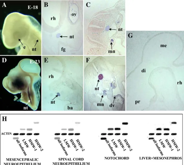
Fgf2
in situ hybridization analysis showed that none of the
known isoforms of this gene could be clearly detected at any site
on the neuroectoderm in all developmental stages analyzed, i.e.,
from HH13 to 27 (
Figs. 4
A, D, G and data not shown). It is
important to note that in some in situ hybridizations, a diffuse
NBT/BCIP precipitate in the neuroepithelium was observed
(not shown), although this was hard to interpret as a positive
hybridization.
Despite this apparent low level of
fgf2
expression in the
neuroectoderm, this gene was clearly expressed at other distinct
sites of interest to us. At HH18,
fgf2
started to be expressed in
the notochord, the rope-shaped structure that it is known to be
responsible for the secretion of distinct signaling molecules
(
Figs. 4
A, B and C).
Fgf2
expression in the notochord was
detected from H.H.18 to H.H.27. The intensity of this
structure's staining increased notably with growth, and at
stage 23 H.H. it was much greater than that of other structures
(
Figs. 4
D, E and F).
Fgf2
expression was also detected in the
mesonephros from H.H.13 to H.H. 27 (
Figs. 4
C, F, and data not
shown), and in a portion of the liver primordium, an organ that
is known to supply different molecules and cells to the
embryonic serum, in a location close to the ductus venosus,
from HH22 to 27 (
Fig. 4
F, and data not shown). In addition,
shown): i.e., in the mesoderm close to the lens during eye
development, from HH18 to 27, in the developing heart, in the
foregut, and in the limb bud.
Due to the presence of very faint NBT/BCIP precipitate
in the neuroectoderm in some hybridizations, and as other
authors had reported the expression of
fgf2
in some
neuroectodermal tissues, such as the mesencephalon and
the spinal cord, when radiolabeled riboprobes were used, a
semiquantitative PCR analysis of different portions of the
neuroectoderm (i.e., mesencephalon and spinal cord) was
performed for different
fgf2
isoforms. The result was then
compared with a semiquantitative PCR analysis of the
tissues where
fgf2
expression was clearly detected in our in
situ hybridizations (i.e., the notochord and the liver and
mesonephros area). This analysis clearly showed that
different LMW and HMW
fgf2
isoforms were expressed
in the notochord and in the liver and mesonephros area
(
Fig. 4
H), as well as in the neuroectodermal tissue of the
mesencephalon and the spinal cord. The relative level of
fgf2
expression in the tissues analyzed with respect to actin
control detected by this semiquantitative PCR amplification
allows us to suggest that, in chick embryos,
fgf2
is
expressed at much higher levels in the notochord and in the
liver and mesonephros area than in the neuroectoderm,
which is in agreement with the results obtained by in situ
hybridization.
The results obtained with neuroepithelial explants cultured
with a chemically defined medium, together with those of
immunodeprivation of FGF2 in the E-CSF, both in vitro and in
vivo, and also the low
fgf2
expression detected in the
neuroepithelium as compared with other embryonic tissues
and organs, indicate that neuroepithelial FGF2 production is not
able to self-induce a normal level of neuroepithelial cell
proliferation and neurogenesis; in addition, these findings
suggest that the FGF2 contained within the E-CSF may be
produced at least in part by extraneural tissues and then shifted
into the E-CSF via the embryonic serum. To support this
hypothesis, we contemplate three conditions. (1) The
extra-neural tissues which express
fgf2
should be capable of liberating
soluble forms of FGF2. (2) FGF2 should be present in the
embryonal serum. (3) FGF2 should be able to pass from the
serum to the E-CSF via the neuroepithelium.
five times superior to the value for the explants cultured in a
non-conditioned medium (
Figs. 5
C, E and I). Both effects
drastically diminished when the FGF2 antibody was added to
the conditioned medium (
Figs. 5
F, G, H and I), as a result of
which we conclude that this is due to the presence of FGF2.
As regards the second point, we performed a Western blot
analysis of embryonic serum and E-CSF with an antibody to
FGF2 after a bidimensional electrophoresis. This analysis
showed the presence of 34, 24 and 17 kDa
antiFGF2-im-munoreactive spots in both the E-CSF and the embryonic serum
(
Figs. 1
A and C).
To test the third point, commercial FGF2 was coupled to
FITC and microinjected into the outflow of heart of chick
embryos at HH24. The E-CSF from these embryos was
recovered at HH27 and run in a SDS-PAGE.
Commercial FGF2 coupled to FITC before injection appears
as a 34-kDa band on the Western blot recognized by both the
anti-FGF2 antibody (
Fig. 6
A) and the anti-FITC antibody (
Fig.
6
B), which indicates that the FGF2-FITC union is stable.
In
Fig. 6
B appears another band of 66 kDa which
corresponds to the bovine seroalbumin bound to FITC, used
by the suppliers to stabilize the FGF2 ligand.
Twenty-four hours following intravascular microinjection of
FGF2-FITC, the Western blot of the E-CSF revealed a band of
34 kDa labeled with both the anti-FGF2 antibody (
Fig. 6
C) and
the anti-FITC antibody (
Fig. 6
D). This indicates that the
FGF2-FITC injected in the vascular system of the embryo passes to the
E-CSF, while a protein of higher MW (albumin) does not,
suggesting that certain proteins could pass from the embryonic
serum to the E-CSF in accordance with their molecular weight.
To analyze the effect of protein size on this favored uptake, a
fusion protein not existent in chick but with a similar molecular
mass to FGF2 was also coupled to FITC and microinjected into
the outflow of heart of chick embryos at H.H.24 (GST-Adh,
predicted MW = 38 kDa). Preinjection solution of
GST/Adh-FITC was detected by Western blot as a band of 38 kDa both
with the anti-GST antibody (
Fig. 6
E), and with the anti-FITC
antibody (
Fig. 6
F). This indicates that the GST/Adh-FITC
union was stable. Twenty-four hours after intravascular
microinjection of the GST/Adh-FITC protein, the Western
blot of the E-CSF showed no band with any of the two
antibodies we employed (
Figs. 6
G and H), suggesting that
E-CSF uptake of this fusion protein from the embryonic serum
was not favored.
These findings suggest that the FGF2 is capable of selective
incorporation from the embryonic serum to the E-CSF in chick
embryos, whereas other proteins of a similar molecular weight
are incapable of behaving in the same way.
Discussion
The results of our study demonstrate the following: (1)
different isoforms of FGF2 are present in the CSF and serum of
chick embryos in early stages of development; (2) in this species
the FGF2 in E-CSF is directly involved in both the in vivo and
in vitro proliferation and neural differentiation of
neuroepithe-lial cells; (3) neuroepitheneuroepithe-lial cells express low levels of
fgf2
mRNA
and the FGF2 from serum is incorporated into the
E-CSF, suggesting that some FGF2 contained within E-CSF may
have an extraneural origin.
FGF2 is an important active component of E-CSF
Embryonal CSF has a complex protein composition which
differs from that of the adult both in terms of quality and
quantity; also, it has been suggested that it might play diverse
biological roles during brain development (
Birge et al., 1974;
Fielitz et al., 1984; Checiu et al., 1984; Dziegielewska et al.,
2000; Miyan et al., 2003; Gato et al., 2004; Parada et al.,
2005b
). In this regard, we have recently demonstrated that
E-CSF exercises an important trophic effect in vitro on
neuroepithelial cells during early chick embryo development
(
Gato et al., 2005
), despite the fact that the molecules involved
are still to be identified. In this paper, we show that chick
embryo E-CSF contains different isoforms of FGF2 which
could influence the behavior of the neuroepithelial cells; this
factor is considered the main instrument involved in activating
replication and neurogenesis from neural stem cells (
Vaccarino
et al., 1999b; Panchision and McKay, 2002
).
Flamme et al.
mechanisms in which other molecules such as proteoglycans
play a role.
These results suggest that an extraneuroepithelial source of
FGF2 might participate in regulating the behavior of neural
stem cells during the development of the embryo.
The FGF2 in E-CSF is involved in controlling neuroepithelial
replication and neurogenesis
In this study, we show that selective in vitro and in vivo
immunoblocking of the biological activity of the FGF2 in
E-CSF at early stages of development, notably reduces the trophic
effect of this fluid on the neuroepithelium which we recently
described (
Gato et al., 2005
), thereby significantly diminishing
mitotic activity and neurogenesis. However, our neuroepithelial
cell apoptotic study show that FGF2 does not play a key role in
cell survival, which could be controlled by other E-CSF factors.
In order to demonstrate that these effects are not due to an
immunoblocking of FGF2 biological activity in the
neuroe-pithelium itself, we have verified that the anti-FGF2 antibody
injected into the embryonal brain cavity does not penetrate the
neuroepithelial tissue but remains confined to the cavity of the
brain vesicles; this ensures the specificity of the antibody acting
on the FGF2 of E-CSF. In this way, we show that, in vitro,
disruption of replication and neurogenesis in the
neuroepithe-lium cultured in a defined medium is to a large extent
compensated for by the exogenous addition of FGF2. These
data suggest, therefore, that the FGF2 is a key component in the
trophic action of E-CSF on neuroepithelial stem cells.
In fact, FGF2 is considered a trophic factor involved in the
behavior of CNS stem cells, on which it exerts a strong
mitogenic and neurogenetic influence both in embryonal and
adult stages. It has been demonstrated that during the
development of the embryo (above all, in in vitro studies)
FGF2 has an intense mitogenic effect on neuroepithelial stem
cells and is capable of triggering neurogenesis (
Murphy et al.,
1990; Tropepe et al., 1999; Vaccarino et al., 1999b; Raballo et
al., 2000; Panchision and McKay, 2002; Reuss and von
Bohlen und Halbach, 2003
). In this regard, our findings
provide experimental evidence of these actions of FGF2 in
vivo on the neuroepithelial cells of chick embryos in early
stages of development, this accord with the intense reduction
in replication and neurogenesis described in the
neuroepithe-lium of FGF2 knockout mice (
Raballo et al., 2000
). Despite
the references in these processes to other factors such as EGF
and IGF1 (
Drago et al., 1991; Tropepe et al., 1999
), the
general opinion is that during early developmental stages
neuroepithelial stem cell proliferation and differentiation
mainly depend on FGF2. Similar effects have been described
for FGF2 on neural progenitor cells in postnatal and adult
stages (
Tao et al., 1996, 1997; Wagner et al., 1999; Cheng et
al., 2001
), although it has also been attributed a role as a
neuroprotector, in the repair of CNS lesions (
Patterson et al.,
1993; Yoshimura et al., 2003; Reuss and von Bohlen und
Halbach, 2003
), and disruptions have been described of FGF2
levels in the cerebrospinal fluid among patients with
neurodegenerative disorders (
Stopa et al., 2001
).
The existence of a specific spatio-temporal pattern of fgf2
expression in the neuroepithelium of rodent embryos has been
shown in various studies (
Vaccarino et al., 1999a
,
b
;
Dono et
al., 1998; Dono, 2003
). Our findings show that fgf2 mRNA
expression in the CNS of chick embryos detected by means of
in situ hybridization at the analyzed stages is much more
lower than that detected in other embryonic structures, such as
the notochord and the mesonephros. Such differences have
been confirmed by semiquantitative PCR, which allows us to
make a comparative estimation of the fgf2 expression levels.
In fact, fgf2 expression by both in situ hybridization and
immunochemistry on the neuroepithelium of avian embryos is
controversial because, despite most authors have shown a very
low fgf2 expression or not expression at all in the CNS (
Han,
1997
;
Savage and Fallon, 1995
, on chick embryos; and
Kalcheim and Neufeld, 1990
, on quail embryos), other authors
have described the presence of FGF2 in the telencephalon and
mesencephalon of chick embryos at HH17 (
Dono, 2003
).
However, the lack of histological section in this latter report
makes very difficult to evaluate the tissular location and the
intensity of FGF2 immunochemistry. In this way, our data
support the idea that in chick embryos fgf2 exhibits a low
level of expression in the neuroepithelium, and thus, this
difference with respect to rodents suggests the existence of
phylogenetic variations.
The different members of the family of FGFs act via different
types of receptors, although it is reported (
Tropepe et al., 1999
)
that the action of FGF2 on the early development of the CNS is
mediated mainly by FGFr1 and 2. These receptors are expressed
differentially in the neuroepithelium of chick and rodent
embryos, with FGFr1 presenting a more ubiquitous pattern
than FGFr2 (
Wilke et al., 1997; Walshe and Mason, 2000;
Trokovic et al., 2005
); however, certain authors have described a
greater concentration of FGFr for both avian and rodent embryos
in those regions of the neuroepithelium which are close to the
ventricular cavities in contact with E-CSF (
Heuer et al., 1990;
Raballo et al., 2000
).
The coincidence in FGF2 expression and that of its
receptors in the neuroepithelium has led to a theory of
autocrine
–
paracrine behavior on the part of FGF2 during the
development of the CNS. However, previous studies we
carried out (
Gato et al., 2005
) have demonstrated that
mesencephalic neuroepithelia of chick embryos cultured in a
chemically defined medium without external supplements are
incapable of maintaining normal levels of proliferation and
neurogenesis during a short 24-h period. This indicates that
intrinsic factors in the developing brain are insufficient to
maintain normal levels of proliferation and neurogenesis,
suggesting that factors outside the neuroepithelium are
involved in regulating these processes; this coincides with
the findings of
Drago et al. (1991)
and
Raballo et al. (2000)
,
in that an exogenous contribution of FGF2 is required in the
regulation of neuroepithelial cell behavior.
replication and neuroepithelial neurogenesis, showing that the
FGF2 of E-CSF has a direct influence on the behavior of
neuroepithelial cells at early stages of development. These
findings are coherent with the apical positioning of the FGF2
and its receptors described by
Raballo et al. (2000)
, who
suggest that these cells could incorporate FGF2 from the
cerebrospinal fluid itself.
The abovementioned data suggest the existence of two
simultaneous routes by which FGF2 acts on the
neuroepithe-lium during chick brain development: one of an autocrine
–
paracrine mechanism within the neuroepithelium itself and the
other an exogenous source in E-CSF. One possible
explana-tion of this is given in the studies carried out by
Qian et al.
(1997)
and
Raballo et al. (2000)
, who suggest that
neurogenesis from neuroepithelial progenitor cells requires
low concentrations of FGF2; as a result, those cells influenced
only by the low levels of FGF2 from inside the
neuroepithe-lium, namely, those situated at its basal portion, would be
subject to neural differentiation, while the cells capable of
proliferation are situated near to the apical side, that is, next
to the cerebral cavity, where they can receive the influence of
the FGF2 from the E-CSF.
Finally, the presence of FGF2 in the cerebrospinal fluid is
constant both during brain development and in the postnatal
period, in which it plays an important physiological role and is
involved in the physiopathology of certain neurodegenerative
disorders. In this paper, we show that the FGF2 of embryonal
cerebrospinal fluid also play a key role in the development of
the CNS.
The extraneural origin of E-CSF in the embryonal
cerebrospinal fluid
Taking into consideration both the fact that our results reveal
low levels of
fgf2
expression in the neuroepithelium of the brain
primordium of chick embryos, when compared with other
extraneural structures, and, as we established previously, that
the neuroepithelium seems insufficient to maintain normal
levels of proliferation and neurogenesis, we suggest that the
FGF2 from E-CSF might have an extraneural origin.
In order to test this hypothesis, we have attempted to check
whether extraneural primordia (notochord) are capable of
liberating diffusible and active forms of FGF2, if this factor is
present in the serum of embryos during the stages we studied
and if it is able to pass from the serum to E-CSF.
In this paper, we demonstrate that during the initial
phases of chick embryo development, there are different
structures, such as the hepatic primordium, the mesonephros
and, above all, the notochord, which present much higher
levels of FGF2 expression than the neuroepithelium itself.
Moreover, we have seen that the notochord liberates different
soluble isoforms of FGF2, which are capable of activating
the replication and neurogenesis in vitro of neuroepithelial
cells. Such findings are consistent with previous studies
demonstrating the capacity of chick embryo notochord to
produce FGF2 (
Hebrok et al., 1998
); what is more,
ectopically implanted notochord induces in vivo an increase
in the mitotic activity of the neuroblasts in chick spinal cord
(
van Straaten et al., 1989
).
Secondly, we have observed that HMW and LMW isoforms
of FGF2 are present in the serum of chick embryos at very early
stages of development (E 25 H.H.), which demonstrates the
existence of diffusible forms of FGF2 circulating, the origin of
which could be in the structures with high levels of mRNA
expression of
fgf2
, permitting it to act remotely on target cells.
Finally, we demonstrate that the FGF2 stained with
exogenous FITC injected into the embryonic vascular system
passes to the E-CSF in a short space of time, while other
proteins of the same or greater molecular weight are incapable
of this process. Considering that at these stages of development
the cavity of the embryonic brain is a physiologically sealed
system, surrounded exclusively by neuroepithelial cells in
which the choroid plexuses are yet to develop, we may affirm
that the FGF2-FITC has selectively traversed the
neuroepithe-lium in a basal
–
apical direction. These data suggest that there
might exist within the neuroepithelium some type of specific
transportation mechanism for this molecule, a matter which
requires further investigation. In adults, it has been proven that
the choroid plexuses are responsible for regulating the
concentration of FGF2 levels in cerebrospinal fluid (
Stopa et
al., 2001
). However, in early phases of embryonic development,
when the choroid plexus are undeveloped or not yet functional,
the neuroepithelium itself might assume this function. In this
regard, the existence has been shown of a mechanism that
selectively internalizes specific proteins in the E-CSF of chick
embryos (
Moro and Uriel, 1981
), which testifies to the
transportation capacity of this epithelium.
This paper contributes new ideas regarding the regulation
and differentiation of neuroepithelial cells in early stages of
chick embryo development, confirming the important trophic
role, previously described by us, of E-CSF in this species,
and providing evidence that FGF2 plays a key role in this
action.
Acknowledgments
The authors thank M. Ros from Cantabria University for fgf2
mRNA riboprobe, to N. Cols from University of Barcelona for
the GST-Adh fusion protein, to Dr. David Rixham for language
translation assistance, and to Pilar Martín and Isabel Garcia for
technical support.
References
Alonso, M.I., Gato, A., Moro, J.A., Barbosa, E., 1998. Disruption of proteoglycans in neural tube fluid byβ-D-xyloside alters brain enlargement in chick embryos. Anat. Rec. 252, 499–508.
Alonso, M.I., Gato, A., Moro, J.A., Martín, P., Barbosa, E., 1999. Involvement of sulfated proteoglycans in embryonic brain expansion at earliest stages of development in rat embryos. Cells Tissues Organs 165, 1–9.
Birge, W.J., Rose, A.D., Haywood, J.R., Doolin, P.F., 1974. Development of the blood-cerebrospinal fluid barrier to proteins and differentiation of cerebrospinal fluid in the chick embryo. Dev. Biol. 41, 245–254. Brickman, Y.G., Ford, M.D., Gallagher, J.T., Nurcombe, V., Bartlett, P.F.,
Turnbull, J.E., 1998. Structural modification of fibroblast growth factor-binding heparan sulfate at a determinative stage of neural development. J. Biol. Chem. 273 (8), 4350–4359.
Brunet, C.L., Sharpe, P.M., Ferguson, M.W., 1993. The distribution of epidermal growth factor binding sites in the developing mouse palate. Int. J. Dev. Biol. 37 (3), 451–458.
Bruni, J.E., 1998. Ependymal development, proliferation, and functions: a review. Microsc. Res. Tech. 41 (1), 2–13.
Bueno, D., Skinner, J., Abud, H., Heath, J.K., 1996. Double in situ hybridization on mouse embryos for detection of overlapping regions of gene expression. Trends. Genet. 12, 385–387.
Checiu, I., Prelipceanu, O., Popescu, O., 1984. The role of cerebrospinal fluid during embryonic development. A biochemical study. Morphol. Embryol. (Bucur) 30 (4), 243–250.
Cheng, Y., Tao, Y., Black, I.B., DiCicco-Bloom, E., 2001. A single peripheral injection of basic fibroblast growth factor (bFGF) stimulates granule cell production and increases cerebellar growth in newborn rats. J. Neurobiol. 46 (3), 220–229.
Davis, M.G., Zhou, M., Ali, S., Coffin, J.D., Doetschman, T., Dorn II, G.W., 1997. Intracrine and autocrine effects of basic fibroblast growth factor in vascular smooth muscle cells. J. Mol. Cell. Cardiol. 29 (4), 1061–1072.
Dono, R., 2003. Fibroblast growth factor as regulators of central nervous system development and function. Am. J. Physiol. 284, R867–R881.
Dono, R., Zeller, R., 1994. Cell-type-specific nuclear translocation of fibroblast growth factor-2 isoforms during chicken kidney and limb morphogenesis. Dev. Biol. 163 (2), 316–330.
Dono, R., Texido, G., Dussel, R., Ehmke, H., Zeller, R., 1998. Impaired cerebral cortex development and blood pressure regulation in FGF-2-deficient mice. Embo J. 17 (15), 4213–4225.
Drago, J., Murphy, M., Carroll, S.M., Harvey, R.P., Bartlett, P.F., 1991. Fibroblast growth factor-mediated proliferation of central nervous system precursors depends on endogenous production of insulin-like growth factor I. Proc. Natl. Acad. Sci. 88 (6), 2199–2203.
Dziegielewska, K.M., Knott, G.W., Saunders, N.R., 2000. The nature and composition of the internal environment of developing brain. Cell. Mol. Neurobiol. 20 (1), 41–56.
Fayein, N.A., Courtois, Y., Jeanny, J.C., 1992. Basic fibroblast growth factor high and low affinity binding sites in developing mouse brain, hippocampus and cerebellum. Biol. Cell. 76 (1), 1–13.
Fielitz, W., Esteves, A., Moro, R., 1984. Protein composition of cerebrospinal fluid in the developing chick embryo. Brain Res. 315 (1), 111–115. Flamme, I., Schulze-Osthoff, K., Jacob, H.J., 1919. Mitogenic activity of
chicken chorioallantoic fluid is temporally correlated to vascular growth in the chorioallantoic membrane and related to fibroblast growth factors. Development 111 (3), 683–690.
Ford-Perriss, M., Abud, H., Murphy, M., 2001. Fibroblast growth factor in the developing central nervous system. Clin. Exp. Pharmacol. Physiol. 28 (7), 493–503.
Gato, A., Moro, J.A., Alonso, M.I., Pastor, J.F., Represa, J., Barbosa, E., 1993. Chondroitin sulphate proteoglycan and embryonic brain enlargement in the chick. Anat. Embryol. 188, 101–106.
Gato, A., Martín, P., Alonso, M.I., Martín, C., Pulgar, M.A., Moro, J.A., 2004. Analysis of cerebrospinal fluid protein composition in early developmental stages in chick embryo. J. Exp. Zool. 301, 280–289.
Gato, A., Moro, J.A., Alonso, M.I., Bueno, D., De La Mano, A., Martin, C., 2005. Embryonic cerebrospinal fluid regulates neuroepithelial survival, proliferation, and neurogenesis in chick embryos. Anat. Rec. 284A (1), 475–484.
Guimond, S.E., Turnbull, J.E., 1999. Fibroblast growth factor receptor signalling is dictated by specific heparán sulphate saccharides. Curr. Biol. 9 (22), 1343–1346.
Hamburger, V., Hamilton, H.L., 1951. A series of normal stages in the development of the chick embryo. J. Morphol. 88, 49–92.
Han, J.K., 1997. Expression of the fibroblast growth factor-2 gene during chick development. Mol. Cells 7 (2), 208–213.
Hanneken, A., Frautschy, S., Galasko, D., Baird, A., 1995. A fibroblast growth factor binding protein in human cerebral spinal fluid. NeuroReport. 6 (6), 886–888.
Harlow, E., Lane, D., 1988. Antibodies. A laboratory manual, Cold Spring Harbour Laboratory. Cold Spring Harbour, New York.
Hebrok, M., Kim, S.K., Melton, D.A., 1998. Notochord repression of endodermal Sonic hedgehog permits pancreas development. Genes. Dev. 12 (11), 1705–1713 (Jun. 1).
Heuer, J., Von Bartheld, Ch.S., Kinoshit, A.Y., Evers, P.C., Bothwell, M., 1990. Alternating phases of FGF receptor and NGF receptor expression in the developing chicken nervous system. Neuron 5, 283–296.
Joseph, S.J., Ford, M.D., Barth, C., Portbury, S., Bartlett, P.F., Nurcombe, V., Greferath, U., 1996. A proteoglycan that activates fibroblast growth factors during early neuronal development is a perlecan variant. Development 122 (11), 3443–3452.
Junier, M.P., 2000. What role(s) for TGFalpha in the central nervous system? Prog. Neurobiol. 62 (5), 443–473.
Kalcheim, Ch., Neufeld, G., 1990. Expression of basic fibroblast growth factor in the nervous system of early avian embryos. Development 109, 203–215.
Milev, P., Monnerie, H., Popp, S., Margolis, R.K., Margolis, R.U., 1998. The core protein of the chondroitin sulfate proteoglycans phosphocan is a high-affinity ligand of fibroblast growth factor-2 and potentiates its mitogenic activity. J. Biol. Chem. 273 (34), 21439–21442.
Miyan, J.A., Nabiyouni, M., Zendah, M., 2003. Development of the brain: a vital role for cerebrospinal fluid. Can. J. Physiol. Pharmacol. 81, 317–328. Moro, R., Uriel, J., 1981. Early localization of alpha-fetoprotein in the developing nervous system of the chicken. Oncodev. Biol. Med. 2 (6), 391–398.
Murphy, M., Drago, J., Bartlett, P.F., 1990. Fibroblast growth factor stimulates the proliferation and differentiation of neural precursor cells in vitro. J. Neurosci. Res. 25, 463–475.
Nicholson, Ch., 1999. Signals that go with the flow. Trends Neurosci. 22 (4), 143–145.
Nugent, M.A., Edelman, E.R., 1992. Kinetics of basic fibroblast growth factor binding to its receptor and heparan sulfate proteoglycan: a mechanism for cooperativity. Biochemistry 31, 8876–8883.
Nurcombe, V., Ford, M.D., Wildschut, J.A., Bartlett, P.F., 1993. Developmental regulation of neural response to FGF-1 and FGF-2 by heparán sulfate proteoglycan. Science 260 (5104), 103–106.
Okada-Ban, M., Thiery, J.P., Jouanneau, J., 2000. Fibroblast growth factor-2. Int. J. Biochem. Cell Biol. 32 (3), 263–267.
Ozawa, K., Uruno, T., Miyakawa, K., Seo, M., Imamura, T., 1996. Expression of the fibroblast growth factor family and their receptor family genes during mouse brain development. Mol. Brain Res. 41, 279–288.
Panchision, D.M., McKay, R.D.G., 2002. The control of neural stem cells by morphogenic signals. Curr. Opin. Genet. Dev. 12, 478–487.
Parada, C., Gato, A., Aparicio, M., Bueno, D., 2005a. Proteome analysis of chick embryonic cerebro-spinal fluid. Proteomics (accepted to publication).
Parada, C., Martín, C., Alonso, M.I., Moro, JA., Bueno, D., Gato, A., 2005b. Embryonic cerebrospinal fluid collaborates with the isthmic organizer to regulate mesencephalic gene expression. J. Neurosci. Res. 82 (3), 333–345. Patterson, S.L., Grady, M.S., Bothwell, M., 1993. Nerve growth factor and a fibroblast growth factor-like neurotrophic activity in cerebrospinal fluid of brain injured human patients. Brain Res. 605 (1), 43–49.
Qian, X., Davis, A.A., Goderie, S.K., Temple, S., 1997. FGF2 concentration regulates the generation of neurons and glia from multipotent cortical stem cells. Neuron 18 (1), 81–93.
Raballo, R., Rhee, J., Lyn-Cook, R., Leckman, J.F., Schwartz, M.L., Vaccarino, F.M., 2000. Basic fibroblast growth factor (Fgf2) is necessary for cell proliferation and neurogenesis in the developing cerebral cortex. J. Neurosci. 20 (13), 5012–5023.
Ribatti, D., Urbinati, C., Nico, B., Rusnati, M., Roncali, L., Presta, M., 1995. Endogenous basic fibroblast growth factor is implicated in the vasculari-zation of the chick embryo chorioallantoic membrane. Dev. Biol. 170 (1), 39–49.
Ruoslahti, E., Yamaguchi, Y., 1991. Proteoglycans as modulators of growth factor activities. Cell 64 (5), 867–869.
Savage, M.P., Fallon, J.F., 1995. FGF2 mRNA and its antisense message are expressed in a developmentally specific manner in the chick limb bud and mesonephros. Dev. Dyn. 202, 343–353.
Sperinde, G.V., Nugent, M.D., 2000. Mechanisms of fibroblast growth factor 2 intracellular processing: a kinetic analysis of the role of heparan sulfate proteoglycans. Biochemistry 39 (13), 3788–3796.
Stopa, E.G., Berzin, T.M., Kim, S., Song, P., Kuo-LeBlanc, V., Rodriguez-Wolf, M., Baird, A., Johanson, C.E., 2001. Human choroid plexus growth factors: what are the implications for CSF dynamics in Alzheimer's disease? Exp. Neurol. 167 (1), 40–47.
Tao, Y., Black, I.B., DiCicco-Bloom, E., 1996. Neurogenesis in neonatal rat brain is regulated by peripheral injection of basic fibroblast growth factor (bFGF). J. Comp. Neurol. 376 (4), 653–663.
Tao, Y., Black, I.B., DiCicco-Bloom, E., 1997. In vivo neurogenesis is inhibited by neutralizing antibodies to basic fibroblast growth factor. J. Neurobiol. 33, 289–296.
Tooyama, I., 1993. [Fibroblast growth factors (FGFs) in neurodegenerative disorders] Rinsho. Shinkeigaku (12), 1270–1274.
Trokovic, R., Jukkola, T., Saarimaki, J., Peltopuro, P., Naserke, T., Weisenhorn, D.M., Trokovic, N., Wurst, W., Partanen, J., 2005. Fgfr1-dependent boundary cells between developing mid- and hindbrain. Dev. Biol. 278 (2), 428–439. Tropepe, V., Sibilia, M., Ciruna, B.G., Rossant, J., Wagner, E.F., Van der Kooy, D., 1999. Distinct neural stem cells proliferate in response to EGF and FGF in the developing mouse telencephalon. Dev. Biol. 208, 166–188. Vaccarino, F.M., Schwartz, M.L., Raballo, R., Nilsen, J., Rhee, J., Zhou, M.,
Doetschman, T., Coffin, J.D., Wyland, J.J., Hung, Y.T., 1999a. Changes in
cerebral cortex size are governed by fibroblast growth factor during embryogenesis. Nat. Neurosci. 2 (3), 246–253.
Vaccarino, F.M., Schwartz, M.L., Raballo, R., Rhee, J., Lyn-CooK, R., 1999b. Fibroblast growth factor signaling regulates growth and morpho-genesis at multiple steps during brain development. Curr. Top. Dev. Biol. 46, 179–201.
van Straaten, H.W., Hekking, J.W., Beursgens, J.P., Terwindt-Rouwenhorst, E., Drukker, J., 1989. 0Effect of the notochord on proliferation and differentiation in the neural tube of the chick embryo. Development 107 (4), 793–803.
Walshe, J., Mason, I., 2000. Expression of FGFR1, FGFR2 and FGFR3 during early neural development in the chick embryo. Mech. Dev. 90 (1), 103–110.
Wagner, J.P., Black, I.B., DiCicco-Bloom, E., 1999. Stimulation of neonatal and adult brain neurogenesis by subcutaneous injection of basic fibroblast growth factor. J. Neurosci. 19 (14), 6006–6016.
Wilke, T.A., Gubbels, S., Schwartz, J., Richman, J.M., 1997. Expression of fibroblast growth factor receptors (FGFR1, FGFR2, FGFR3) in the developing head and face. Dev. Dyn. 210 (1), 41–52.
Yayon, A., Klagsbrun, M., Esko, J.D., Leder, P., Ornitz, J.D., 1991. Cell surface, heparin-like molecules are required for binding of basic fibroblast growth factor to its high affinity receptor. Cell 64, 841–848.
Yosimoto, T., Houkin, K., Takahashi, A., Abe, H., 1997. Evaluation of ces in cerebrospinal from patients with moyamoya disease. Clin. Neurol. Neurosurg. 99 (Suppl. 2), S218–S220.
Yoshimura, S., Teramoto, T., Whalen, M.J., Irizarry, M.C., Takagi, Y., Qiu, J., Harada, J., Waeber, C., Breakefield, X.O., Moskowitz, M.A., 2003. FGF-2 regulates neurogenesis and degeneration in the dentate gyrus after traumatic brain injury in mice. J. Clin. Invest. 112 (8), 1202–1210.