Bases cerebrales de procesos cognitivos y motivacionales en la adicción a la cocaína
165
0
0
Texto completo
(2) Departamento de Psicología Básica, Clínica y Psicobiología Facultad de Ciencias de la salud. Bases cerebrales de procesos cognitivos y motivacionales en la adicción a la cocaína Tesis doctoral presentada por: Juan Carlos Bustamante Fernandiz Para obtener el grado de doctor por la Universitat Jaume I de Castellón. Directores: Dr. César Ávila Rivera Dr. Alfonso Barrós Loscertales Programa de doctorado Psicopatología, Salud y Neuropsicología Castellón, Julio 2012.
(3)
(4) Departamento de Psicología Básica, Clínica y Psicobiología Facultad de Ciencias de la salud. Neural bases of cognitive and motivational processes in cocaine addiction Doctoral thesis presented by: Juan Carlos Bustamante Fernandiz To obtain the grade of doctor by the University Jaume I of Castellón. Supervisors: Dr. César Ávila Rivera Dr. Alfonso Barrós Loscertales Ph.D. program Psicopatología, Salud y Neuropsicología Castellón, Julio 2012.
(5)
(6) Neural bases of cognitive and motivational processes in cocaine addiction 2012. For my family….
(7)
(8) Neural bases of cognitive and motivational processes in cocaine addiction 2012. “Research is to see what everybody else has seen, and to think what nobody else has thought” Albert Szent-Gyorgyi “Research is formalized curiosity. It is poking and prying with a purpose” Zora Neale Hurston. “La ciencia es aquello sobre lo cual cabe siempre discusión” José Ortega y Gasset “La ciencia más útil es aquella cuyo fruto es el más comunicable” Leonardo Da Vinci.
(9)
(10) Neural bases of cognitive and motivational processes in cocaine addiction 2012. Table of contents. Acknowledgments....................................................................................... vi Abbreviations list........................................................................................ viii Abstract....................................................................................................... xi Resumen..................................................................................................... xiii PREFACE................................................................................................... 1 Justification..................................................................................... 1 CHAPTER 1. GENERAL INTRODUCTION........................................... 5 1.1. The neurobiology of cocaine addiction.................................. 7 1.1.1. Dopamine circuitry implications in cocaine addiction................................................................ 7 1.1.2. Structural and functional brain implications of cocaine addiction............................................... 11 1.1.2.1.. Structural alterations in cocaine addiction......................................... 12. 1.1.2.2.. Functional alterations in cocaine addiction........................................ 13. 1.1.2.2.1. Cognitive control functioning in cocaine addiction................ 14 Inhibitory control............................ 14 Working memory............................. 16 Decision making............................... 17. ii.
(11) Neural bases of cognitive and motivational processes in cocaine addiction 2012. 1.1.2.2.2. Motivational salience attribution and cocaine addiction............... 19. Reward processing of drug-related stimuli............................................. 20 Reward processing of non-drug-related stimuli............................................. 21 1.1.3. Drug-free state and neurobiological implications in cocaine addiction.................................................. 22 1.2 Magnetic Resonance Imaging (MRI): the technique............... 25 1.2.1. Magnetic Resonance Imaging (MRI)...................... 25 1.2.2. Functional Magnetic Resonance Imaging (fMRI)........................................................ 26 1.2.3. Methodological toolboxes for MRI and fMRI: implications in cocaine addiction......... 28 1.2.3.1.. Voxel-Based Morphometry (VBM).............................................. 28. 1.2.3.2.. Biological Parametric Mapping (BPM), statistical toolbox for multimodal analysis in neuroimaging................ 29. CHAPTER 2. EXPERIMENTAL SECTION.............................................. 31 2.1.. Overview of the studies...................................................... 31 2.1.1 Objective and hypotheses....................................... 35. 2.2.. Barrós-Locertales A., Garavan H., Bustamante J.C., et al. (2011).. Reduced striatal volume in cocaine dependent patients. Neuroimage, 56, 1021-1026......................................................................... 37 2.3.. Bustamante J.C., Barrós-Loscertales A., Ventura-Campos N., et al. (2011). Right parietal hypoactivation in a cocaine-dependent. group during a verbal working memory task. Brain research, 1375, 111-119……………………………………………………. 2.4.. 39. Barrós-Loscertales A., Bustamante J.C., Ventura-Campos N., et al. (2011). Lower activation in the right frontoparietal network iii.
(12) Neural bases of cognitive and motivational processes in cocaine addiction 2012. during a counting Stroop task in a cocaine-dependent group. Psychiatry research, 194, 111-118……………..………….. 42 2.5.. Bustamante J.C., Barrós-Loscertales A., Costumero V., et al. (submitted).. Modulation. of. abstinence. duration. on. striatal. functioning during monetary reward processing in cocaine patients………………………………..…………………….. 44 CHAPTER 3. GENERAL DISCUSSION…………...………………….. 47 3.1 General conclusions……………………………...………….. 52 CHAPTER 4. FUTURE RESEARCH LINES……………...…………... 53 REFERENCES. OF. GENERAL. INTRODUCTION. AND. GENERAL. DISCUSSION……………………………………………...…………… 57 RESUMEN GENERAL EN CASTELLANO……………...…………... 83 R.1. Planteamiento y metodología utilizada………….………… 84 R.2. Objetivos e hipótesis………………………………………. 89 R.3. Principales aportaciones y conclusiones…………………... 91 R.4. Líneas de investigación futuras……………………………. 94. iv.
(13) Neural bases of cognitive and motivational processes in cocaine addiction 2012. v.
(14) Neural bases of cognitive and motivational processes in cocaine addiction 2012. Acknowledgements. I wish to thank all the people who have been with me while preparing this work, and who have given me their support and helped motivate me every day. Your role throughout this process has been essential for the preparation of a complex, good quality doctoral thesis. Thanks to Dr. César Ávila Rivera for giving me the opportunity to join a research group that undertakes its work in one of my research interests, functional Magnetic Resonance Imaging (fMRI), and its application in Neuropsychology. Thank you for being the motor of a lab that grows “crescendoing” and which is developing an interesting project with much clinical and experimental applicability. I appreciate all your help with and all your advice on my PhD training. I also wish to thank Dr. Barrós Loscertales; Alfonso, thank you for everything that you have taught me and provided me with. I feel very lucky for having the opportunity of working with a person like him. He has allowed me to obtain all the professional and personal values that I believe are essential for a good scientific and academic career. Thanks too to Dr. Maria Antònia Parcet for her clinical contributions, and for offering me the possibility of making good use of her expertise and pragmatic vision in the Neuropsychology field. I also thank Dr. Cristina Forn for giving me lots of advice which have made my work over these 4-5 years much more fruitful and satisfactory. Thanks too to Dr. Ana Sanjuán for being an example of attitude at work. I am also grateful to Noelia for being patient and for providing me methodological help and support, which have been basic in all the work that I am going to present. Thanks also to Víctor for his useful attitude and vi.
(15) Neural bases of cognitive and motivational processes in cocaine addiction 2012. for asking questions all the time, which produce interesting ideas that have improved the quality of the methodology in our papers. Thanks to Paola, Marian and Patri for providing freshness to the lab. Thanks to Maya for giving another perspective of research work, based on an open-minded style of work, which implies travelling abroad and working in different research groups. Thanks to the “new Javi” and Elisenda for showing me their motivation in doing our fieldwork, and I appreciate them motivating me more, at the same time, to continue in my work. And last but not least, thanks to Aina and the “old Javi” for giving me something, which in my opinion, is essential; their friendship and support at all times. Thanks to my family, you are incredible! I do not know what to say, just thanks with all my heart and soul. Without you I would not have reached this point. I need you and I will always need you. Thanks to everyone!!. The research reported in this thesis was supported by the grant “Beca de Formación de Personal Investigador UJI, PREDOC/2007/13”.. vii.
(16) Neural bases of cognitive and motivational processes in cocaine addiction 2012. Abbreviations list. ACC, Anterior Cingulate Cortex ACG, Anterior Cingulate Gyrus AC-PC, Anterior Commisure-Posterior Commisure ADHD, Attention Deficit Hyperactivity Disorder AH, Allostatic Hypothesis BA, Brodmann Area BOLD, Blood Oxygen Level-Dependent BPM, Biological Parametric Mapping DA, Dopamine DAT, Dopamine Transporter DLPFC, Dorsolateral Prefrontal Cortex DS, Dorsal Striatum DSM, Diagnostic and Statistical Manual of Mental Disorders DTI, Diffusion Tensor Imaging ERP, Event-Related Potentials FDR, False Discovery Rate fMRI, functional Magnetic Resonance Imaging FOV, Field of View FWE, Family Wise Error FWHM, Full Width at Half Maximum GM, Gray Matter GLM, General Lineal Model HDR, Haemodynamic Response HMRF, Hidden Markov Random Fields IFC, Inferior Frontal Cortex IFG, Inferior Frontal Gyrus viii.
(17) Neural bases of cognitive and motivational processes in cocaine addiction 2012. IGT, Iowa Gambling Task IPC, Inferior Parietal Cortex IPG, Inferior Parietal Gyrus IPL, Inferior Parietal Lobule ISI, Intestimuli Interval LPFC, Lateral Prefrontal Cortex LPPs, Late Positive Potentials MFG, Medial Frontal Gyrus MidFG, Middle Frontal Gyrus MIDT, Monetary Incentive Delay Task MNI, Montreal Neurologic Institute MPFC, Medial Prefrontal Cortex MR, Magnetic Resonance MRI, Magnetice Resonance Imaging Nacc, Nucleus Accumbens OCD, Obsesive-Compulsive Disorder OFC, Orbitofrontal Cortex PET, Positron Emmision Tomography PFC, Prefrontal Cortex RDS, Reward Deficiency Syndrome RFT, Random Field Theory ROI, Region of Interest RT, Reaction Time SD, Standard Deviation SMA, Supplementary Motor Area SPG, Superior Parietal Gyrus SPM, Statistical Parametric Mapping SSRT, Stop-Signal Reaction Time STC, Superior Temporal Cortex T, Tesla TE, Echo Time TR, Repetition Time VBM, Voxel-Based Morphometry ix.
(18) Neural bases of cognitive and motivational processes in cocaine addiction 2012. VMPFC, Ventromedial Prefrontal Cortex VS, Ventral Striatum VTA, Ventral Tegmental Area WAIS, Wechsler Adults Intelligence Scale WFU, Wake Forest University WCST, Wisconsin Card Sorting Test WM, White Matter. x.
(19) Neural bases of cognitive and motivational processes in cocaine addiction 2012. Abstract. Cocaine. addiction. is. related. with. neurological. and. neuropsychiatric. complications, which need to be considered in treatment. Use of Magnetic Resonance Imaging (MRI), such as structural MRI and functional MRI, improves the neuroanatomical and neurofunctional analysis of addiction-related changes. Several studies have shown that addiction alters the dopaminergic mesocorticolimbic circuitry of self-control and incentive salience to subserve the transition from voluntary drug use to habitual, compulsive drug abuse. Some have analysed if cocaine alterations are associated with consumption patterns, effect of abstinence and treatment maintenance. This thesis aims to give more evidence regarding the brain changes subserving alterations in cognitive and motivational processing in cocaine addiction. Four studies have been conducted in this thesis. The morphometric study shows that cocaine addicts present reduced grey matter volume in the dorsal striatum, and that the regional grey matter volume of the amygdala inversely correlates with years of cocaine use. The first functional study reveals that cocaine-dependent patients display hypoactivation in the inferior parietal gyrus during a verbal working memory n-back task. The second functional study reports that cocaine-dependent patients present hypoactivation in the inferior frontal cortex, the inferior parietal cortex and the superior temporal cortex during an interference-control Counting Stroop task. Finally, the third functional study indicates that patients present hypoactivation in the caudate during a monetary reward anticipation task. Moreover, the functional regulation of the striatum during reward processing seems to be associated with abstinence and time on treatment. In conclusion, cocaine addiction has been associated with structural and functional alterations in the mesocorticolimbic circuitry. Attentional-cognitive changes relate to frontal and parietal hypoactivations, while striatum function and volume reduce xi.
(20) Neural bases of cognitive and motivational processes in cocaine addiction 2012. in cocaine addicts. Moreover, the cocaine-free time may determine the regulation of the altered striatal pattern activation during monetary reward processing. Future endeavors could focus on analysing the neural bases of the interaction between cognitive and motivational processes, and the connectivity between cortical and subcortical dopamine regions.. xii.
(21) Neural bases of cognitive and motivational processes in cocaine addiction 2012. Resumen. La adicción a la cocaína se asocia con complicaciones neurológicas y neuropsiquiátricas que deben ser consideradas en los tratamientos. El uso de la Resonancia Magnética (RM), específicamente la RM estructural y la RM funcional, mejora el análisis neuroestructural y neurofuncional de los cambios relacionados con la adicción. La literatura previa ha mostrado que la adicción a la cocaína altera el circuito cerebral dopaminérgico mesocorticolímbico de control cognitivo y saliencia de incentivo; una alteración que está a la base del proceso de transición de un uso voluntario de la droga a un abuso compulsivo de la misma. Por otra parte, son pocos los estudios que han analizado si las alteraciones se relacionan con patrones de consumo y efectos de la abstinencia o el mantenimiento del tratamiento. El objetivo de esta tesis es intentar dar más evidencia empírica a los cambios cerebrales asociados a las alteraciones cognitivas y motivacionales en adicción a la cocaína. Cuatro estudios se desarrollaron para la confección de la misma. El estudio de morfometría mostró que los adictos a la cocaína presentaban una reducción del volumen de sustancia gris en el estriado dorsal y que el volumen de sustancia gris en la amígdala correlacionaba inversamente con los años de consumo. El primer estudio funcional mostró que los pacientes presentaban una hipoactivación a nivel del giro parietal inferior durante una tarea n-back de memoria de trabajo verbal. El segundo estudio funcional mostró que los pacientes tenían una hipoactivación en el córtex frontal inferior, córtex parietal inferior y córtex temporal superior durante una tarea Counting Stroop de control de interferencia. El último estudio mostró que los pacientes presentaban una hipoactivación en el caudado durante la anticipación de recompensas monetarias. Además, se observó que la regulación de la funcionalidad del estriado durante el procesamiento de recompensas está relacionada con la abstinencia y el tiempo en tratamiento. xiii.
(22) Neural bases of cognitive and motivational processes in cocaine addiction 2012. En conclusión, la adicción a la cocaína se asocia con alteraciones estructurales y funcionales en el circuito mesocorticolímbico. Cambios cognitivo-atencionales se relacionan con hipoactivaciones a nivel frontal y parietal y la función estriatal y su volumen se ven reducidos en la adicción a la cocaína. Además, el período en el que el dependiente está libre del consumo puede asociarse con una regulación de los patrones estriatales de activaciones alteradas durante el procesamiento de recompensas monetarias. Líneas de investigación futuras se pueden relacionar con el análisis de las bases neurales de la interacción entre procesos cognitivos y motivacionales, y la conectividad. entre. regiones. dopaminérgicas. xiv. corticales. y. subcorticales..
(23)
(24) Neural bases of cognitive and motivational processes in cocaine addiction 2012. Preface Justification In the last few years, the number of cases of cocaine dependence has considerably increased to become the second most prevalent illegal psychoactive drug in Spain (National Drugs Plan, the Spanish Observatory on Drugs Report, 2009). Therefore, it is an important subject for the public health system and for professionals working on drug dependence. Responsibility is not for those who work directly with patients only; it is also necessary to study the cocaine addiction process in research environments to allow methodological controls to be able to test hypotheses and to put forward theories. Along these lines, the effort made in this research field focuses on comprehending the complex profile of a cocaine-addictive person at three levels: biological, behavioural and social. Chronic cocaine use is related with medical, neurological and neuropsychiatric complications, which have to be considered in the treatment (Bolla et al., 1998), and it is important that treatment programmes take into account a neurobiological model that has to accurately explain the patient’s clinical reality. Neuropsychology, which studies the relationship between behaviour and the brain, establishes an interesting point of view to study cocaine addiction because its efforts focus on the study of cognitive/motivational brain-behavioural processes. The technological revolution that has taken place in recent years allows the use of new technology in research. The use of techniques such as structural Magnetic Resonance Imaging (MRI) and functional Magnetic Resonance (fMRI) Imaging improves neuroanatomical and neurofunctional approaches in distinct clinical populations, such as addictive behaviours. The great spatial and good time resolutions of these techniques and their non-invasive properties provide the scientific community with interesting and. 1.
(25) Neural bases of cognitive and motivational processes in cocaine addiction 2012. complementary data to those obtained by other techniques, such as Possitron Emission Tomography (PET), Event-Related Potentials (ERP), among others. Nowadays, addiction emphasises uncontrolled drug use rather than tolerance and physiological dependence (O’Brien, 2008). Several studies report alteration in executive control functions in cocaine addiction (Garavan and Hester, 2007; Kubler et al., 2005, Tomasi et al., 2007a; Bolla et al., 2003). Moreover, some studies indicate that addiction alters the reward function (Garavan et al., 2000; Jia et al., 2011). The interaction of motivational and cognitive neurobehavioural components in cocaine dependence is basic in comprehending the addiction process and the different degrees of severity implicated. Compulsive drug use develops due to an inflexible behaviour, and it persists despite the considerable cost that cocaine addiction involves (Everitt et al., 2001). Addiction alters the neural bases of motivation and self-control, and affects processes like cognitive control, decision making and reward processing (Baler and Volkow, 2006; Goldstein and Volkow, 2002¸Volkow et al., 2011a, 2011b). The isolate consumption of a drug does not produce addiction, but maintaining the frequency and time of consumptions produces structural and functional brain changes (Kalivas and O’Brien, 2008). Uncertainty about the relationship between neural bases of addiction and manifestation of addictive behaviours offers this aspect, and the clinical implications that it may has, a clear value of research interest for several scientists in this particular field. The neurobehavioural study of both cognitive aspects (e.g., cognitive control) and motivational aspects (e.g., reward processing), and also the possible structural changes associated with cocaine addiction, will offer possibilities to produce empirical evidence to develop theoretical models. These models not only implicate one “psychopathology of the patient” dimension, but also a “biological” dimension (a neural substrate of addiction processes), which could be modulated by genetic and environmental variables. The neuroimaging studies methodology provides an understanding of the cocaine patients profile by considering aspects like neurotransmission systems, brain structural and functional bases, cognitive state, personality traits, personal-social situation and clinical situation. This amount of 2.
(26) Neural bases of cognitive and motivational processes in cocaine addiction 2012. knowledge will relate with the establishment of more complete, effective and efficient treatments (Kampman, 2010; Volkow et al., 2011a, 2011b, 2011c). The general aim of this doctoral thesis is to provide the addictions field with more empirical evidence for the possible structural and functional implications in the cognitive. and. motivational. alterations. relating. with. maintenance. of. the. compulsive/impulsive substance use behavioural pattern in cocaine addiction by generalising them to therapeutics approaches to confer clinical significance to the results.. 3.
(27) Neural bases of cognitive and motivational processes in cocaine addiction 2012. 4.
(28) Neural bases of cognitive and motivational processes in cocaine addiction 2012. Chapter 1.General Introduction Behavioural. disinhibition. associated. with. cognitive. and. motivational. components, which are related with failures in the top-down control of fronto-striatal circuits and overactivity within striatal circuitry, may explain both the impulsive and compulsive patterns in some disorders (Fineberg et al., 2010). These psychopathological disorders are often highly heritable and include obsessive-compulsive disorder (OCD), body dysmorphic disorder, Tourette’s syndrome, trichotillomania, attention deficit hyperactivity disorder (ADHD), pathological gambling and substance addictions (Fineberg et al., 2010). Impulsivity is defined as a predisposition towards rapid, unplanned reactions to internal or external stimuli with diminished regard to negative consequences (Chamberlain and Sahakian, 2007; Potenza, 2007). Compulsivity represents a tendency to unpleasantly perform repetitive acts habitually, leading to functional impairment (Hollander and Cohen, 1996; Chamberlain et al, 2006). Longitudinal studies have demonstrated that individuals who demonstrate poor behavioural self-control or high novelty seeking as young children are substantially more likely to initiate substance use in adolescence (King et al., 2004; Màsse and Tremblay, 1997). On the other hand, prolonged consumption can lead to drug-taking compulsion, and addicted losses control habitual behaviour and are unable to reverse repetitive response patterns no longer driven by drugs (Koob and Volkow 2010). Some neurobiological models establish that psychostimulant addiction could be a transition between impulsive and compulsive behaviours (Dalley et al. 2011; Koob and Volkow, 2010; Everitt et al., 2008; Pierce and Vanderschuren, 2010). These neurobiological models establish the existence of separate, yet intercommunicating, ‘compulsive’ and ‘impulsive’ cortico-striatal circuits that are differentially modulated by neurotransmission, and which play different roles in the various stages of the transition process of addiction from social drug use to habitual and compulsive drug use (Robbins, 2007; Brewer and Potenza, 2008; Everitt et al., 2008; Pierce and Vanderschuren, 2010). In the impulsive circuit, a ventral striatal 5.
(29) Neural bases of cognitive and motivational processes in cocaine addiction 2012. component (VS/nucleus accumbens) may drive the impulsive behaviours underlying cocaine seeking and intake over extended periods of time (Everitt et al., 2008); in the compulsive circuit, a dorsal striatal component (DS/caudate nucleus) may drive compulsive behaviours. Compulsive behaviours result from maladaptative stimulusresponse habits in which the ultimate goal of behaviour has been devalued and responding is governed by drug-related stimuli, which also function when presented as a result of instrumental responses. The persisting quality of these habits has been likened to a pathological wanting of drugs and also relates simultaneously with lack of control of habitual behaviours. Goldstein and Volkow (2002) and Volkow et al. (2011b) proposed that at the core of drug addiction, we find the processes of loss of self-directed behaviours to become “automatic sensory-driven formulas”, as well as the attribution of primary salience to the drug of abuse at the expense of natural reinforcers, which are related with the impulsive behavioural pattern. Then, when these processes become chronic, behavioural compulsion patterns appear as relapse and mental compulsion patterns like withdrawal or craving. The proposed syndrome of impaired response inhibition and salience attribution (I-RISA syndrome) consists in four behavioural dimensions that are interrelated in a positive loop (Goldstein and Volkow, 2002): drug intoxication, drug craving, compulsive drug administration and drug withdrawal. Drug intoxication is related with the experience of strong effects of the drug based on positive and negative reinforcement, and the repeated associations between the drug and these effects allow the attribution of primary salience to drugs. Craving is associated with the learned response that links drug-related stimuli to a pleasurable experience, producing drug expectation and its desire. Moreover, compulsive drug taking is maintained to avoid unpleasant withdrawal symptoms, or even when the drug is no longer perceived as pleasurable and adverse physical reactions to it appear. In short, addictive drugs like cocaine are initially taken simply to achieve their pleasant drug effects, and after addiction, they are consumed to escape withdrawal symptoms due to only the sensitisation to the drug of the incentive salience brain system, but also to impaired response inhibition (Robinson and Berridge, 2003; Volkow et al., 2011b). 6.
(30) Neural bases of cognitive and motivational processes in cocaine addiction 2012. 1.1.. The neurobiology of cocaine addiction. 1.1.1. Dopamine circuitry implications in cocaine addiction Prefrontal components (e.g., orbitofrontal cortex, OFC; inferior frontal cortex, IFC; Anterior Cingulate Cortex, ACC) may exert inhibitory control over the formerly cited impulsive and compulsive dopaminergic circuitries (Robbins, 2007; Brewer and Potenza, 2008). Hyperactivity or hypoactivity within the striatal components and abnormalities (presumably hypoactivity) in the prefrontal components may, thus, result in increased impulsive or compulsive behaviours (Fineberg et al., 2010). In addiction impairment typically begins in the more primitive subcortical areas of the brain that process reward to then move on to other neocortical areas relating with more complex cognitive functions (Volkow et al., 2010). Thus, in addition to reward (see Figure 1), addiction can produce severe disruptions in learning (memory, conditioning, habituation), executive function (impulse inhibition, working memory, decision making, delayed gratification), cognitive awareness (interoception), and even emotions (mood and stress reactivity) (Volkow et al., 2010).. Figure 1. During addiction, the enhanced value of the drug in the reward, motivation, and memory circuits overcomes the inhibitory control exerted by the prefrontal cortex, thereby favoring a positivefeedback loop initiated by the consumption of the drug and perpetuated by the enhanced activation of the motivation/drive and memory circuits. Extracted from Volkow et al. (2011d).. 7.
(31) Neural bases of cognitive and motivational processes in cocaine addiction 2012. The mesocortical dopamine circuit, which includes the prefrontal cortex (PFC), the OFC and the ACC, seems to be involved in the conscious experience of drug intoxication, drug incentive salience, craving and compulsive drug administration. The mesolimbic dopamine circuit, which includes the nucleus accumbens (VS component), the caudate (DS component), the amygdala and the hippocampus, has been associated with acute reinforcing drug effects, and also with memory and conditioned responses, which have also been linked to craving and to compulsive drug processes. This circuit is also likely to be involved in emotional and motivational changes during withdrawal (Goldstein and Volkow, 2002). Cocaine produces its reinforcing effects by potentiating these dopamine circuits (Hurd and Pontén, 2000). Indeed, enhancement of cocaineevoked dopamine (DA) levels in a terminal projection area of mesocorticolimbic neurons like the nucleus accumbens, which is related with behavioural sensitisation (Zapata et al. 2003; Kalivas et al., 1998), is thought to be responsible for the development of the impulsive and compulsive drug-taking behaviour related with addictive disorders (Robinson & Berridge, 1993). Scientific literature has shown that addiction implicates alterations in the neural circuitry normally involved in pleasure, incentive motivation and learning (Wise 1989, Robbins and Everitt 1996, Berridge and Robinson 1998, Di Chiara 1999, Kelley 1999, Hyman and Malenka 2001, Kelley & Berridge 2002). DA pathways in the mesolimbic system perform an important function in reward and reinforcement (Wise, 2002). Addiction is related with a sensitisation of the reward system due to repeated stimulation of mesolimbic DA pathways, increasing reward-seeking behaviours (Robinson and Berridge, 1993). This fact relates with inhibitory control (associated with poor prefrontal functioning) and may facilitate DA impulsive-motivated behaviours. Excessive DA release and stimulation may deplete DA stores and lead to anhedonia and depression (Koob and Le Moal, 1997), producing compulsion to seek stronger rewards to avoid DA deficiency. The demonstration of decreased DA receptors in chronic cocaine users (Volkow et al, 1999a) suggests a down-regulation in response to persistently elevated postsynaptic DA concentrations, which is consistent with the hypothesis of a dysregulated DA system after repeated stimulation of DA release. As Volkow et al. (2002a) reported, in drug abusers, lower striatal dopamine receptors 8.
(32) Neural bases of cognitive and motivational processes in cocaine addiction 2012. correlate significantly with lower brain glucose metabolism in the OFC (involved with salience attribution, whose disruption results in compulsive behaviours) and in ACC (involved with inhibitory control and error monitoring, whose disruption results in impulsivity). The dopamine transporter (DAT) removes dopamine from the extracellular space, thus terminating signalling, and it is also critically involved in the rewarding and stimulating effects of psychostimulants. DAT blockers, like cocaine, inhibit the uptake of DA (Ferris et al., 2011), increasing DA levels and extending the magnitude and duration of DA signalling (see Figure 2). Psychostimulant-induced DA elevations are related with regulation of motor, mood, motivation, fronto-attentional functioning, learning and addiction processes (Wanat et al., 2009; Schultz, 2007). What starts as an increased DA release, leading to increased ventral ACC activity and increased reward seeking (Wise, 2002), may end up as a compulsive drive towards increased levels of reward stimulation to restore resultant DA deficiency. This compulsive drive may be exacerbated by deficient impulse control and decision making, linked to the OFC, ventromedial prefrontal cortex (VMPFC) and ACC (Adinoff, 2003).. Figure 2. Cocaine blocks the dopamine transporters and dopamine molecules are accumulated in the intercellular space striking the receiving cell’s receptors and causing an intensified response in the receiving cell. Also a downregulation of the dopamine receptors in the receiving cell may take place. Extracted from Fowler et al. (2007).. 9.
(33) Neural bases of cognitive and motivational processes in cocaine addiction 2012. Low dopamine D2 receptor (DRD2) availability is associated with drug liking for stimulants (Volkow et al., 1999b) and the reinforcing effect of cocaine has also been related to its ability to block the DA transporter (Ritz et al., 1987). As animal studies report, drug intoxication is associated with higher extracellular dopamine concentrations in striatal, limbic and frontal brain regions (Ritz et al., 1987; Hurd et al., 1989; Goeders and Smith, 1986), which relate with positive reinforcing effects. On the other hand, although increased DA has been frequently associated with positive affective states, it is possible that, under certain conditions, DA levels increasingly stimulate the neuronal systems associated with aversion, thus causing an animal to increase cocaine intake to block these negative experiences (Hurd and Pontén, 2000). Human studies have shown that increasing extracellular DA concentrations in the nucleus accumbens is thought to be a final common pathway for cocaine (Everitt and Robbins, 2005). In non-dependent drug abusers, cocaine self-administration increases the extracellular DA levels within the ventral limbic striatum (Cox et al., 2009). DA release is also related with craving mechanisms. DA agonists produce an enhancement in cocaine cue reactivity (Robbins et al., 1992). Re-exposure to the environmental cues previously associated with cocaine use produces a strong conditioned response, which increases subjective measures of cocaine craving, while DA release may also mediate some conditioned responses to cocaine cues (Berger et al., 1996). High concentrations of DA metabolites (plasma homovanillic acid), which represents high concentrations of DA, correlate with cocaine craving in newly abstinent patients (Berger et al., 1996). When non-treatment seeking cocaine-dependent volunteers listen to cocaine-related scripts or view cocaine-related videos, a DA release is selectively induced within the associative and sensorimotor regions of the DS (Volkow et al., 2006a; Wong et al., 2006). Sensitisation mechanisms in animals, such as locomotor activating and dopamine-releasing effect (Fontana et al., 1993), occur after repeated use of cocaine, and are believed to enhance the incentive salience of drugrelated stimuli related to craving and drug seeking (Robinson and Berridge, 2003). DA deficiency during withdrawal evidences the fact that DA agonists effectively reverse post-cocaine deficits in brain stimulation reward in rats (Markou and Koob, 1992), and confirms a link between withdrawal and DA neurotransmission in 10.
(34) Neural bases of cognitive and motivational processes in cocaine addiction 2012. mesolimbic areas and attenuated brain stimulation reward (Weiss et al., 2001). By taking into account that dopamine partial agonists can reverse psychostimulant withdrawal, it has been suggested that DA tone dysregulation relates with the motivational. effects. of. withdrawal. (Koob,. 2009).. Regarding. relapse,. DA. neurotransmission mediates cocaine-induced reinstatement (Self and Nestler, 1998; Shalev et al., 2002; Spealman et al., 1999). Increasing the DA release in the nucleus accumbens (Nacc) can reinstate cocaine-seeking behaviour (Stewart et al., 1984). Moreover, a lot of dopaminergic agonists are greater inducers of relapse in cocaine and heroin addiction (De Wit and Stewart, 1983; Wise et al., 1990; Self et al., 1996a; 1996b), and DA antagonists can block the priming effects of heroin, amphetamine and cocaine (Ettenberg, 1990; Shaham and Stewart, 1996; Weissenborn et al., 1996). In short, DA mesocorticolimbic circuitry plays an essential role in the cocaine addiction process, and is related with alterations in this neurotransmission system based on the dysregulation of DA levels in the synaptic space to produce cognitive and motivational changes in distinct phases of addiction, which allow a transition from voluntary drug use to habitual and compulsive drug abuse (Everitt et al., 2008). 1.1.2. Structural and functional brain implications of cocaine addiction Cocaine addiction is associated with neural changes in the frontostriatal brain system, which may be related with impulsivity and drug-related compulsivity (Jentsch and Taylor 1999; Everitt and Robbins, 2005; Porrino et al., 2007; Schoenbaum and Shaham, 2008). Imaging studies have shown functional and structural alterations in PFC regions that play an important role in executive functions such as cognitive control, decision. making,. emotional. regulation,. motivation. and. salience. attribution,. demonstrating the catastrophic consequences of PFC disruption in cocaine addiction (Volkow and Fowler, 2000; Volkow et al.,2006b). Impaired cognitive control plays a fundamental role in drug-seeking behaviours in abusers, and successful functionality requires top-down control of the PFC to the striatum and limbic regions involved in reward. and. emotion. processing. (Heatherton. and. Wagner,. 2011).. Motivational/emotional alterations in cocaine addiction are related with structural and functional alterations in several regions, including the ACC, the OFC, the dorsolateral 11.
(35) Neural bases of cognitive and motivational processes in cocaine addiction 2012. PFC (DLPFC), the amygdala, the striatum, and other limbic brain regions (see Figure 3). Neurobiology of cocaine addiction results in not only an enhanced motivational value of the drug at the expense of other natural reinforcers, but in an impaired ability to inhibit the intentional actions associated with strong desires to take the drug, resulting in impulsive and compulsive drug taking in cocaine addiction (Volkow et al., 2003).. Figure 3. Major brain regions with structural and functional alterations, including the Prefrontal cortex regions (as the OFC, DLPFC), ACC, the OFC, the amygdala, hippocampus, the striatum (Nacc, caudate, putamen) with a clear role in cocaine addiction. Extracted from Fowler et al. (2007).. 1.1.2.1. Structural alterations in cocaine addiction Cocaine dependence is associated with the structural neuroadaptations related to behavioural and brain functional changes in cocaine users. Many studies based on these neuroadaptations have focused on DA circuitry, and particularly on the striatum (Koob et al., 1994; White and Kalivas, 1998). Nonetheless, cocaine addiction is also related to spread reductions in grey matter (GM) density and volume in cortical brain regions (Matochik et al., 2003; Tanabe et al., 2009; Franklin et al., 2002; Bartzokis et al., 2000a; Sim et al., 2007; Ersche et al., 2011), as well as in frontal (Lim et al., 2002, 2008), insular (Lyoo et al., 2004) and callosal (Moeller et al., 2005) white matter (WM). Previous studies applying Voxel-Based Morphometry (VBM), based on morphometric changes (Ashburner and Friston, 2000), have reported GM reductions in 12.
(36) Neural bases of cognitive and motivational processes in cocaine addiction 2012. the volume of the OFC, the ACC, the insula, the superior temporal cortex (Matochik et al., 2003; Franklin et al., 2002; Lim et al., 2008; Tanabe et al., 2009) and the cerebellum (Sim et al., 2007) in cocaine addiction. Yet others have failed to find these differences (Connolly et al., 2009; Narayana et al. 2010). On the other hand, and by means of manual volume segmentation, Jacobsen et al. (2001) showed an increased volume of the striatal structure, like the caudate head and putamen, based on brain volume differences. Martínez et al. (2004) do not find these differences. The last structural findings have shown loss of GM in the following: OFC, IFC, medial frontal cortex (MPFC), DLPFC, insula, ACC, temporoparietal cortex, amygdala, parahippocampus, caudate and cerebellum (Ersche et al., 2011; Weller et al., 2011; Moreno-López et al., 2012), as well as increased GM in the putamen, the caudate, globus pallidum and the cerebellum in cocaine-dependent men (Ersche et al., 2011). Moreno-López et al. (2012) also found lower WM volumes in the inferior and medial frontal cortices, the superior temporal cortex, the ACC, the insula and the caudate. Lack of consensus among studies could be related with methodological and sample differences. All these structural changes associated with cocaine addiction process affect the striato-cortico-limbic circuitry linked to the cognitive and motivational/emotional component of the process. These effects could be related with long-term drug use based on maintenance of drug-seeking and drug-consuming behaviours due to impulsive and compulsive behaviours. 1.1.2.2. Functional alterations in cocaine addiction Volume differences in a given brain area related to drug use may affect functional activation, as measured by brain Blood Oxygen Level Dependent (BOLD) patterns (Aron and Paulus, 2007). Thus, the cell numbers in a structure, as reflected by its volume measure, could be important for capillary recruitment associated with brain activity (Makris et al., 2004), suggesting reduced functionality in an area of reduced volume. Cocaine dependence has been related to a wide range of cognitive and motivational/affective dysfunctions, and fMRI has proven an invaluable technique to study these drug-related changes (Aron and Paulus, 2007). Different fMRI studies have shown cognitive (Garavan et al., 2008), motivational (Goldstein et al., 2007a; Garavan 13.
(37) Neural bases of cognitive and motivational processes in cocaine addiction 2012. et al., 2000) and motor (Hanlon et al., 2009) deficits in cocaine-dependent patients, which directly relates with the functionality of dopamine-mediated regions. 1.1.2.2.1. Cognitive control functioning in cocaine addiction Stimulant abusers present significant deficits in executive control processes (Bechara, 2005). Mental flexibility or cognitive monitoring, and decision-making processes become altered executive functions in cocaine addiction (Bolla et al., 1998). Moreover, there are differences between cocaine abstinent and controls in selective (Verdejo-García et al., 2005) and sustained (Morgan et al., 2006) attention, visuospatial functions, memory and concentration (Berry et al., 1993). It has been suggested that executive control implicates not only one simple network, but two different connected networks: a frontoparietal network comprising DLPFC, inferior parietal lobe (IPL), dorsal frontal cortex, intraparietal sulcus, precuneus and middle cingulate cortex; and a cingulo-opercular. network. composed. of. anterior. prefrontal. cortex,. anterior. insula/medial frontal operculum, dorsal anterior cingulate/medial superior frontal cortex and thalamus (Dosenbach et al., 2008). These two networks may be affected by cocaine addiction to an unknown extent. -. Inhibitory control Some studies have demonstrated that cocaine-dependent individuals exhibit an attentional system that is biased to detect and process cocaine-related stimuli; in drug Stroop tasks, substance abusers have been seen to take longer to name the colour of drug-words than neutral words (Cox et al., 1999; Mogg and Bradley, 2002; Copersino et al., 2004; Hester et al., 2006; Vadhan et al., 2007). Moreover, other research works have indicated an association between long-term cocaine use and impairments of inhibitory processes (Ardila et al., 1991; Horner et al., 1996; Volkow et al., 1996; Biggins et al., 1997; Fillmore and Rush, 2002; Colzato et al., 2007). Cocaine abuse is related with patterns of premature responding (Bauer, 2001), persevering behaviour, and the inability to adapt behaviour to environmental changes (Lane et al., 1998).. 14.
(38) Neural bases of cognitive and motivational processes in cocaine addiction 2012. Previous research, which compared patients and controls using the Stroop task, has found differences in several frontal and cingulate areas. Bolla et al. (2004) revealed how the ACC and the right lateral prefrontal cortex (LPFC) were less active in a group of cocaine-dependent individuals during the execution of a manual version of the Stroop task. Although Goldstein et al. (2001) did not find brain functional differences between cocaine-dependent patients and a group of matched controls while performing an eventrelated colour-word Stroop task, their results reveal increased activation in the OFC, which is associated with lesser and greater conflict in controls and patients, respectively, while groups did not differ in terms of task performance. Cocaine-using subjects have shown impaired behavioural performance in GO– NOGO tasks (Fillmore and Rush, 2002). Kaufman et al. (2003) demonstrated that the ACC, which is critical for cognitive control, is less responsive in chronic cocaine users during a GO-NOGO task. They showed STOP-related hypoactivity in the right insula and a rostral region of the ACC, two regions that have been identified with emotional processes (Whalen et al., 1998). The ACC has also been seen to be implicated in inhibitory control (Casey et al., 1996; Ponesse et al., 1998). In one of our studies (Bustamante et al., 2008), we showed lower activation in the MPFC of cocainedependent patients during a GO-NOGO task with reward contingencies, as in previous studies (Hester and Garavan, 2004; Kaufman et al., 2003). Moreover, compensatory hyperactivation in the right IFC has shown in cocaine patients during a GO-NOGO task (Kaufman et al., 2003). In a stop-signal task, Fillmore and Rush (2002) established that cocaine users display less ability to inhibit a response, less likelihood of inhibiting responses, and that they required more time to inhibit responses as estimated by their higher stop-signal reaction time (SSRT) in comparison with controls. Li et al. (2008) showed hypoactivation in the rostral ACC as a specific deficit in controlling the inhibitory response in men with chronic cocaine abuse. Moreover, they found greater activation in the visual and posterior parietal regions in controls when compared with cocaine users. These results are also consistent with an earlier work demonstrating frontal and temporo-parietal hypoperfusion associated with chronic cocaine use (Strickland et al, 15.
(39) Neural bases of cognitive and motivational processes in cocaine addiction 2012. 1993), while greater activation of the posterior parietal and visual cortical regions suggests that healthy individuals pay more attention to the task (Li et al., 2008). These alterations in the frontal brain regions relating with inhibitory control functioning may be associated with the cocaine-related cognitive deficits linked to the relationship between drug cues and compulsive drug self-administration in drug dependence (Goldstein et al., 2001; Carpenter et al., 2006). These dysexecutive consequences of drug abuse indicate that cocaine users may be compromised in the endogenous and volitional control of their behaviour. Consequently, their behaviour may be disproportionately determined by environmental cues (e.g., drug-craving cues) and by habitual and compulsive behavioural patterns. The effect of this would be to compound drug abuse maintenance: if chronic cocaine users are influenced especially by environmental contingencies and cues, then an inhibitory dysfunction may reduce their capacity to inhibit these external influences (Kaufman et al., 2003). -. Working memory In general, cocaine patients poorly perform a verbal working memory task, such as the n-back task (Beatty et al., 1995; Bolla et al., 2000; Goldstein et al., 2004; Verdejo-García et al., 2006; Tomasi et al., 2007a). Some studies have reported a functional alteration in the PFC in cocaine dependence (Adinoff et al., 2001; 2003; Goldstein and Volkow, 2002; Goldstein et al., 2004; Kosten et al., 2004), which could be related with patients’ limited working memory capacity (Tomasi et al., 2007a). However, there is also evidence to establish that cocaine patients, in comparison to controls, hypoactivate the anterior cingulate gyrus (ACG) (Tomasi et al., 2007a; Kubler et al., 2005), the medial frontal gyrus (MFG), the middle frontal gyrus (MidFG) and the parietal cortex (Kubler et al., 2005). For control subjects, parametric increases of working memory load enhance brain activation bilaterally in the prefrontal and the parietal cortices (Tomasi et al., 2007a). In patients, an increased working memory load produces lower BOLD signal increases in these brain regions. These dysfunctional patterns in a fronto-parietal network could be related with limitations in working memory capacity among cocaine patients. Another study (Tomasi et al., 2007b) has revealed greater activations in the frontal and parietal brain regions in cocaine16.
(40) Neural bases of cognitive and motivational processes in cocaine addiction 2012. dependent users in comparison to a control group, such as engagement of attentional resources for supporting working memory functions. Thus, the dysfunctional activation frontoparietal pattern in cocaine users (Adinoff & cols., 2001, 2003; Goldstein et al., 2004; Goldstein & Volkow, 2002; Kosten et al., 2004; Volkow et al., 1988, Bolla et al., 2004) could be associated with limitations in the capacity of the working memory frontoparietal network (Tomasi et al., 2007a; Nyberg et al., 2009). Specifically, the parietal cortex is related with attentional response-orienting processes (Corbetta y Shulman, 2002) and limitations in the working memory load (Nyberg et al., 2009). In short, the literature establishes differential patterns of activation between cocaine addicts and controls in a frontoparietal network related with the working memory function and attentional-related processes. There is evidence for an impaired integrative function of those regions involved with executive control and attention in cocaine abusers, and it is likely to underlie the cognitive disruption in cocaine patients. Diverse studies have established the prefrontal cortex to be the main neural substrate holding the neurocognitive pattern and the addiction process in cocaine-dependent subjects, but other cortical brain regions are relevant in cognitive control functioning in cocaine addiction. -. Decision making Cocaine addicts’ inability to consider the consequences of both their behaviour with a view to modifying it and their poor risk assessment is similar to the impairment noted in patients with VMPFC lesions (Damasio, 1985; Bechara et al., 1994; 1998; Grant et al., 1997; Bartzokis et al., 2000b). The Iowa gambling task (IGT) proves a valid decision-making measure (Bechara et al., 1998). A study reported that polydrug abusers perform worse in the Gambling Task when compared to controls (Grant et al. 1997). Cocaine use impairs risk assessment and advantageous decision making (Bartzokis et al., 2000b; Bolla et al., 2003). Cocaine-dependent subjects show impaired performance in decision-making tasks such as the Gambling task (Reavis and Overman, 2001; Adinoff et al., 2003).. 17.
(41) Neural bases of cognitive and motivational processes in cocaine addiction 2012. Tanabe et al. (2007) reported that substance-dependent individuals display lower ventral medial frontal activity during decision-making processes, as well as less right prefrontal activity. Cocaine abusers exhibit greater activation during the IGT in the right OFC, but less activation in the right DLPFC, which are involved in planning and working memory required for IGT, and in the left MFC involved in planning and performance in the IGT (Bolla et al., 2003). For compensation processes, cocaine abusers may overactivate the right OFC. OFC hyperactivity in cocaine abusers during the IGT may reflect an abnormally intense focus because they are thinking about the winning/rewarding emotional aspects of the task, and this thought process could exaggerate the value of the high reward and suppress the negative value of high loss. OFC dysfunction could be a predisposing factor which may lead to poor decision making that relates with certain “myopia” for the future consequences of their choices (Cunha et al., 2011) and to the social dysfunction among cocaine patients. Thus in cocaine subjects, Adinoff et al. (2003) reported less regional cerebral blood flow in the OFC, the ACC and the DLPFC during gambling task execution, which could be related with impaired performance. In conclusion, cocaine abusers display functional abnormalities in the prefrontal neural networks involved in decision-making. This compromised decision-making is likely to contribute to maintaining addiction, thus making abstinence difficult (Bolla et al., 2003). Specifically, the important social behaviour deficits, risk-taking tendencies and personality alterations seen in cocaine addiction are related with the link between the OFC and the decision-making dysfunction (Cunha et al., 2011). The neuroscientific literature has concentrated on the involvement of DA in the drug addiction process because the ability of drugs of abuse to increase brain DA concentrations in the limbic and striatal brain regions is considered crucial for their reinforcing effects (Di Chiara, 1999). Otherwise in drug addiction, we can observe functional and structural changes in dopamine circuits, including not only limbic areas, but also the frontal cortex (Goldstein and Volkow, 2002). As Tomasi et al. (2007a) remarked, multiple neuroimaging studies have been done to characterise the neurocircuitry involved in cocaine addiction, mostly focussing on reward processing (related with the motivational and emotional aspects implicating addiction), but also on 18.
(42) Neural bases of cognitive and motivational processes in cocaine addiction 2012. complex cognitive functions (related with executive functions; e.g., cognitive control). The literature clearly places importance on the structural and functional alterations in the striato-cortico-limbic system as a neural substrate of altered processes of the topdown control and incentive sensitisation presented in cocaine addiction. 1.1.2.2.2. Motivational salience attribution and cocaine addiction Environmental stimuli associated with effects of self-administered drugs gain incentive salience through the Pavlovian conditioning process. Drugs produce sensitisation of autonomic activity or distortions in sensory processing, and exaggeration of incentive salience of stimuli that already predict important environmental events such as drug-related cues (Everitt and Robbins, 2005). There are theories to explain the role of the brain reward pathways in mediating relapse, which is the core motivational symptom of compulsive drug taking and intense drug craving. Impulsivity theories have determined that addicted users are characterised by prominent traits of impulsivity resulting from some combinations of overactive mesolimbic reward approach circuitry and deficient frontocortical punishment avoidance circuitry to produce a greater functional engagement of the dopaminergicmotivational brain circuitry during reward processing (Bechara, 2005; Bickel et al., 2007; Newman and Wallace, 1993). On the other hand, the reward deficiency syndrome (RDS) hypothesis (Blum et al., 2000) has established that addicted individuals display deficit to recruit the dopaminergic-motivational circuitry by non-drug rewards, but abused drugs are able to normalise DA levels, thus motivating drug-taking behaviour (Bjork et al., 2008). The allostatic hypothesis (AH) (Koob and Le Moal, 1997; Koob et al., 2004) has posited that neurochemical effects of chronic drug use also cause an under-responsiveness in the mesolimbic incentive neurocircuitry to non-drug rewards and cues (Koob and Le Moal, 2005). These effects produce anhedonia and a generalised dysphoric mood (Bjork et al., 2008), and relapse risk implicates the alleviation of that discomfort and negative affect (Weiss et al., 2001). However, this theory has its limitations; as regards the positive incentive processes caused directly by drugs themselves, withdrawal states are not especially powerful in motivating drug-seeking behaviour (Stewart & Wise 1992; Robinson and Berridge, 2003). Besides, this theory 19.
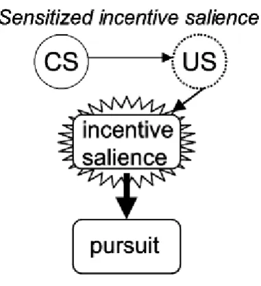
(43) Neural bases of cognitive and motivational processes in cocaine addiction 2012. cannot explain why addicts so often relapse into drug-taking, even after being free from withdrawal (Robinson and Berridge, 2003). There is another theory, the Incentive-Sensitisation Theory, which suggests that relapse is explained by drug-like processes that activate reward pathways, such as the acute effect of the drug itself (Stewart et al., 1984; Wise and Bozarth, 1987; Robinson and Berridge, 1993; Self and Nestler, 1995). Drugs, like cocaine, can alter the reward brain circuits mediating the attribution of incentive salience (see Figure 4), a basic incentive-motivational function (Robinson and Berridge, 2003). These neural circuits become hypersensitive (neural sensitisation) to specific drug effects and to drug-related stimuli at both the micro (neurotransmission) and macro (brain systems) levels. Thus, excessive attribution of incentive salience to drug-related representations could cause “pathological wanting” (Goldstein and Volkow, 2002; Robinson and Berridge, 2003; Volkow et al., 2011a, 2011b). Thus, the next two points go on to present the main neuroimaging findings in cocaine addiction relating with processing drug and non-drugrelated stimuli to compare evidence for highly responsiveness to drug-associated cues in brain reward regions, and for altered responsiveness of brain reward regions to non-drug rewards. Figura 4. According to the incentivesensitisation theory the critical change is in the ability of drug cues (the dashed US evoked by a drug cue) to engage a sensitized motivational response of incentive salience (as indicated by the starbust). This enhanced motivational response is primarily responsible for compulsive drug pursuit in addiction. Extracted from Robinson and Berridge (2003).. -. Reward processing of drug-related stimuli If compared with neutral stimuli, drug-related stimuli elicit enhance physiological reactivity in addicted individuals (Carter & Tiffany, 1999). Some authors 20.
(44) Neural bases of cognitive and motivational processes in cocaine addiction 2012. have demonstrated that larger late positive potentials (LPPs) are elicited by drug-related pictures if compared with neutral pictures in cocaine, heroin and alcohol addiction (Franken et al., 2003; 2004; van de Laar et al., 2004; Namkoong et al., 2004). These results have been interpreted as an increased centralisation of neural resources in drugrelated stimuli if compared with neutral stimuli (Dunning et al., 2011). Goldstein et al. (2009) demonstrated that drug words increase brain activations in the mesencephalon, a major source of dopamine release during motivationally salient situations or conditioned stimuli appearances (Robinson and Berridge, 1993; McClure et al., 2003). When cocaine-related stimuli are presented, and if we compare this scenario with non-drug-rewarding stimuli presentation (i.e., erotic stimuli), various brain regions become activated in cocaine abusers: ACG, OFC, DLPFC, retrosplenial cortex, peristriate cortex, IFL and temporal pole (Maas et al., 1998; Childress et al., 1999; Garavan et al., 2000; Kilts et al., 2001; Wexler et al., 2001; Bonson et al., 2002; Grant et al., 1996; Wang et al., 1999). In cocaine addicts, even a brief exposure to drug cues evokes limbic system activation (Childress et al., 2008) as with exposure to natural-sexual cues. In this sense, cocaine users’ brain response to “unseen” drug and sexual cues reflects Pavlovian learning, but has evolutionary implications; rapid response to reward signals for food and sex would have a survival advantage. Cocaine confers no such advantage, but strongly activates the reward circuitry. The limbic brain treats cocaine cues “as if” they were signals for highly desirable natural rewards (Childress et al., 2008). -. Reward processing of non-drug-related stimuli A deficient electrophysiological response to pleasant and unpleasant images has been shown to be evident during current use of cocaine (Dunning et al., 2011), as occurs in other drug addiction and dependence cohorts (Lubman et al., 2009; Zijlstra et al., 2009), and is related with impairments in sustaining non-drug-related goal-oriented motivation and predisposition of addicted patients to drug use as a compensation mechanism (Dunning et al., 2011).. 21.
(45) Neural bases of cognitive and motivational processes in cocaine addiction 2012. Goldstein et al. (2007) showed altered sensitivity to gradients in monetary value in the OFC in cocaine-dependent men. Asensio et al. (2010) showed that, if compared with controls, a cocaine group displayed hypoactivated DS and VS, thalamus, parietal cortex and dorsomedial prefrontal cortex while processing erotic-related pictures. On the other hand, some studies have suggested that some cocaine-addicted individuals are hypersexual (Washton and Stone-Washton, 1993) and that some substance-dependent individuals may be hyperresponsive to monetary rewards (Bechara et al., 2002; Jia et al., 2011). These results could indicate impairments that may reflect inefficient processing, reward hypersensitivity, greater salience, or other possibilities or combinations (Knutson et al., 2000; Bjork et al., 2008; Jia et al., 2011). It can be postulated that reactivity to monetary rewards in cocaine patients increases in comparison with control subjects as money acts as a secondary reinforcer towards substance use, thus increasing anticipation and outcome responses (Jia et al., 2011). Briefly, the neuroimaging literature shows a functional alteration in reward processing neural systems in cocaine addicts, which is characterised by hyperactivity to drug-related cues, and by either hyperactivity or hypoactivity regarding other rewarding stimulus processing. Alterations have been found in the emotional processing related with cocaine addict patients’ reduced ability to experience pleasure by natural reinforcers, thus attributing excessive incentive salience to drugs and stimuli, which remain associated with drugs after repeated drug use. 1.1.3. Drug-free state and neurobiological implications in cocaine addiction As shown in this introduction, in different neuroimaging studies into cocaine addiction, no consensus between studies has been reached as regards results. Perhaps methodological issues could represent the main reason for this lack. Thus, characteristics of samples in relation to illness severity or treatment-seeking status are one of the most common methodological differences (Moeller et al., 2012). In this sense, most studies into cocaine addiction have not reached an agreement on the clinical situation of cocaine patients that have participated in their works using short-term or long-term abstinents, or even using active cocaine users. Indeed, the marked heterogeneity of treatment-seeking samples (determined by length of abstinence, in22.
(46) Neural bases of cognitive and motivational processes in cocaine addiction 2012. patient status, remission status, etc.) in the same study could lead to an increase in data variability and to effects on the power of the neuroimaging results (Moeller et al., 2012). The literature has shown differences in brain structure and brain functioning in those who have attained relatively long periods of abstinence, and also in those who have recently abstained. Regarding the acute effects of cocaine, they could affect the brain structure and function in a wide range of dopaminergic brain regions including, but not limited to, regions involved in reward and executive control functions (Breiter el al., 1997; Breiter and Rosen, 1999; Bartzokis et al., 1999a, 1999b; Hanlon et al., 2009; Kaufman et al., 2003; Lyoo et al., 2004; Hester and Garavan, 2004; Kubler et al., 2005; Goldstein et al., 2007, 2009; Garavan et al., 2008; Li et al., 2008; Sim et al., 2007; Franklin et al., 2002; Ersche et al., 2011). Specifically, if we take into account the neuroimaging data on abstinence duration, abstinent patients were those who showed reduced GM volume in the PFC (Fein et al., 2002), the medial OFC (Tanabe et al., 2009; Matochik et al., 2003; Moreno-López et al., 2012), the lateral OFC (Matochik et al., 2003), the right inferior frontal gyrus (IFG) (Moreno-López et al., 2012), the right ACG (Matochik et al., 2003), the right insula (Moreno-López et al., 2012), the left amygdala and the bilateral caudate (Moreno-López et al., 2012) in comparison to controls. Bell et al. (2011) showed that a short-term abstinent group, a mid-term abstinent group and a long-term abstinent group differed in terms of the WM integrity of the bilateral inferior longitudinal fasciculus, the right anterior thalamic radiation, the right ventral posterolateral nucleus of the thalamus, the left superior corona radiata, superior longitudinal fasciculus bilaterally, the right cingulum and the right precentral gyrus. Moreover, all abstinent patients, if compared with controls, exhibited WM differences in left anterior callosal fibres, the left genu and the splenium of the corpus callosum, right superior longitudinal fasciculus, right callosal fibres and the superior corona radiata bilaterally (Bell et al., 2011). In a PET study during a Stroop task (Bolla et al., 2004) recently abstinent cocaine patients displayed less activation in the left ACC and the right lateral PFC, but greater activation in the right ACC when compared to controls. Recently abstinent cocaine patients also showed greater activation in the right OFC while performing the IGT, but less activation in the right DLPFC and the left MFC (Bolla et al., 2003). On the other hand, in an fMRI study during a verbal working 23.
(47) Neural bases of cognitive and motivational processes in cocaine addiction 2012. memory task, longer abstinent patients presented attention-related functional alterations in the putamen, the ACC, the MidFG, the MFG, the superior parietal gyrus (SPG), the parahippocampal gyrus, the amygdala, the mesencephalon and the thalamus (Tomasi et al., 2007). Another sample of longer abstinent patients (at least two months) showed an altered activation in the right IFG and the right MFG during an inhibitory control task (Bustamante et al., 2008). Otherwise, there are very few data available that elucidate structural and functional recovery with abstinence. Thus, a PET study has shown that striatal dopaminergic dysregulation may partly recover with abstinence (Volkow et al. 2001). A diffusion tensor imaging (DTI) study (Xu et al., 2010) found that self-reported abstinence. correlated positively with white matter integrity in the right superior. longitudinal fasciculus, the right body of the corpus callosum, the right posterior limb of the internal capsule and the left cerebellum. One fMRI study found positive correlations between self-reported duration of abstinence and activation during a cognitive control task in the left posterior ACC, the left ventral MPFC and the right putamen (Brewer et al., 2008). Additionally, Moeller et al. (2012) demonstrated a partial recovery of brain response during an interference control drug Stroop task in the dopaminergic midbrain and the thalamus in relation to maintained abstinence. Moreover, Connoly et al (2012) showed that short-term abstinence is related with increased inhibition-related dorsolateral and inferior frontal activity, indicative of the need for increased inhibitory control while long-term abstinence relates with increased error-related ACC activity, indicative of heightened behavioural monitoring. On the other hand, little is known about the effects of abstinence on functional effects during motivational reward processing in cocaine addiction. Specifically, only one fMRI study has shown that activation in the thalamus, the culmen and the Nacc during a monetary reward processing task is correlated with self-reported abstinence (Jia et al., 2011) In short, we observe that a drug-free state in cocaine patients brings about changes in the brain’s structure and function, conferring protracted abstinence therapeutic implications. However, very little is known about the neurobiology of those who successfully avoid relapse, maintain the guidance of treatment and remain abstinent over long periods of time (Bell et al., 2011; Connoly et al., 2012). 24.
(48) Neural bases of cognitive and motivational processes in cocaine addiction 2012. 1.2. Magnetic Resonance Imaging (MRI): the technique. 1.2.1. Magnetic Resonance Imaging (MRI) MRI is based on the application of intense magnetic fields for creating biological tissues. These magnetic fields use the Tesla (T, 1T are 1000 Gauss) as a unit measure and are generated by an electromagnet in the MRI scanner. In human studies, the intensity of magnetic fields is between 0.5 and 9.4 T. In the scanner, MRI sequences, which are series of oscillatory magnetic gradients and electromagnetic fields, are applied to detect properties and types of tissues. These tissues are, principally, GM, WM and cerebrospinal fluid. Pulses sequences with a specific frequency are absorbed by the atomic nuclei of the tissue of interest. These frequencies are specific for each atomic nucleus. Hydrogen is the most common nucleus in the human body and the frequencies of pulses are adjusted for that nucleus with a unique proton. After absorption, protons emit the electromagnetic energy that they have captured, and this energy is the “signal”. Signal intensity depends on the concentration of these nuclei in the tissue and their magnetic properties. Therefore, if diverse tissues are differentiated in the concentration of hydrogen nuclei, the electromagnetic energy emitted by each tissue will differ. However, another important aspect is the spatial localisation of the tissues in MRI images. This spatial localisation is established in units called “voxels”, like spatial localisation in a digital image in pixels. Thus, a voxel is a unit of volume with three dimensions (x, y, z). The spatial resolution of MRI images is determined by the size of the voxel in each dimension; if the voxel is smaller, then we will have more possibilities to delimit small brain structures. The signal obtained from each voxel has an intrinsic variability that is independent of the tissue, but dependent on the measuring technique used, called “noise”. Then, the MRI image is not an absolute measure of the signal emitted by a tissue, but is a measure of the contrast between the signal emitted by a tissue and the noise inherent to the technique used, called “signal to noise ratio”. The “signal to noise ratio” is the magnitude of the difference in intensity between diverse amounts of signal 25.
(49) Neural bases of cognitive and motivational processes in cocaine addiction 2012. divided by the variability of the measures. The “signal to noise ratio” is bigger the larger the voxel size. In summary, to obtain brain structural images in an MRI scanner: 1. An electromagnet generates a magnetic field for the alignment of hydrogen protons in a specific direction. 2. Sequences of pulses are applied at a specific frequency (radiofrequency pulses) to change the orientation of the hydrogen protons at a specific angle. 3. The interruption of the pulse causes hydrogen protons to return to their original position, as established by the electromagnet. 4. When protons return to their original position, they emit energy, that is captured by a receptor and transformed into images by considering the position of each voxel and the signal emitted by each one. 1.2.2. Functional Magnetic Resonance Imaging (fMRI) MRI allows us to study the brain structure as a static representation. With this technique, specifically Functional Magnetic Resonance Imaging (fMRI), we can study short-term physiologic changes related with brain functionality; we can also visualise functional dynamic changes. The main characteristic of the images obtained for the brain structure study by MRI is their “spatial resolution”. The study of brain functioning by MRI (fMRI) introduces an important variable: “temporal resolution”. This variable establishes the velocity of registering the physiologic changes related with brain functioning. This temporal resolution depends on the “sampling rate”, which is the frequency of the fMRI signal measure over time, and is between 2 and 4 seconds. It means that physiologic changes are measured every 2-4 seconds in the acquisition process. These physiologic changes, which represent variations or changes in the quantity of deoxyhaemoglobin in the blood (Huettel et al., 2004), are related with functional changes in a given brain region. Functional changes are defined as the intensity and variation of the oxyhaemoglobin changes in a brain region related with the neural 26.
Figure


+7
Documento similar
When examining the relationship between language and general cognitive functioning, some studies suggest that children with prenatal alcohol exposure are likely to show difficulties
Government policy varies between nations and this guidance sets out the need for balanced decision-making about ways of working, and the ongoing safety considerations
In recent years, photometric redshift galaxy surveys have arisen as powerful probes of the Large Scale Structure (LSS) of the Universe and dark energy.. The Dark Energy Survey (DES)
The objective of this study was to identify factors related to dysfunctional family functioning that may be associated with the severity of symptoms among adolescent patients with
The success of computational drug design depends on accurate predictions of the binding free energies of small molecules to the target protein. ligand-based drug discovery
The fluctuations of the complex amplitude, measured for a given spatial frequency as a function of wavelength (top panel), can be related to the different values of the corresponding
in in S0s the accretion events may be more rare with respect S0s the accretion events may be more rare with respect to the spirals so the accreted gas is not enough to fill to
Instrument Stability is a Key Challenge to measuring the Largest Angular Scales. White noise of photon limited detector
