E Mbongo-Kama et al.MDR3 mutations in cholesterol cholelithiasis 143
medigraphic.com
Annals of Hepatology 2007; 6(3): July-September: 143-149
Annals of Hepatology
Concise Review
MDR3 mutations associated with intrahepatic and
gallbladder cholesterol cholelithiasis: an update
Elvire Mbongo-Kama;1 Florence Harnois;2 Didier Mennecier;2 Eleonore Leclercq;1 Pascal Burnat;1 Franck Ceppa1
Abstract
Background: The recurrent microlithiasis represents one of the most frequent clinical forms of lithiasis of the bile ducts. This affection is characterized by the presence of cholesterolic microgallstones on hepatic canaliculars, and belongs to a heterogeneous group of autosomal recessive liver disorders. Radiological di-agnosis can be confirmed by analysis of MDR3 gene, coding a protein involved in physiologic translocation of phospholipids in bile. Discovery of MDR3 mutations is of particular interest, since normally associated with good effectiveness of medication by ursodesoxycholic acid. AIM: To review MDR3 mutations in humans as-sociated with recurrent cholesterol microlithiasis and to suggest a practical approach for MDR3 gene analy-sis. Results: 48 mutations of MDR3 gene have been re-ported in humans to date, from which 43 (89.5%) in the coding region, and 5 splice site mutations have been as-sociated to cholesterol cholelithiasis. 21 (43.8%) of the 43 precited mutations are located in only 8 exons on 28, near transmembrane or nucleotide binding do-mains of the protein. From the 22 remaining described mutations, 9 (18.8%) are restricted to exon 14. We sug-gest therefore to start analysis of MDR3 gene by screening exons 6, 7, 9, 10, 12, 14, 17, 23 and 24 with an appropriate protocol in this diagnosis associated with effective treatment. In conclusion such therapeutic ori-entation is valuable, since recurrent cholesterolic mi-crolithiasis occurs relatively early in life, and by the fact that recurrence of symptoms may occur despite cholecystectomy, or shock-wave therapy.
Key words: ABC transporters, gallbladder diseases, In-trahepatic cholestasis, Multidrug resistance protein 3.
1BEGIN Military Hospital, Department of Biochemistry. 2Department of Hepatology and Gastroenterology.
Address for correspondence: Franck Ceppa
BEGIN Military Hospital, Department of Biochemistry. 69 avenue de Paris, 94160 Saint-Mandé, France. Telephone number: 33(0)1 4398 4844
E-mail: [email protected]
Manuscript received and accepted: 26 May and 6 July 2007
The recurrent microlithiasis represents one of the most frequent clinical forms (25% of the cases) of lithiasis of the bile ducts. This affection is characterized by the presence of cholesterolic microgallstones on hepatic canaliculars, and belongs to a heterogeneous group of autosomale recessive liver disorders. It occurs especially in young adults, before the age of forty and appears to be strongly related to meta-bolic syndrome.1 Prevalence is greater in women and
in-creases considerably with age. In some cases, several genet-ic abnormalities of phospholipids canalgenet-icular gene MDR3 are responsible for occurrence of this pathology.
Diagnosis of recurrent microlithiasis is very often ra-diological (70 to 80% of cases). Intrahepatic hypere-chogenic foci along the biliary tree may be evidenced by ultrasonography, and hepatic bile composition may be determined by duodenoscopy. In a second time, detection of mutations in MDR3 is of particular interest, since asso-ciation with good effectiveness of the medical treatment by ursodesoxycholic acid has been demonstrated.2
1. Transport carriers of cholesterol in bile
Bile is an aqueous solution that contains various lip-ids: bile salts, phospholipids and cholesterol. At low con-centrations, hydrosoluble bile salts are dispersed as monomers. Bile phospholipids are hydrophobic lecithins that organize themselves in vesicles in water. In bile, coex-istence of bile salts and phospholipids allows solubiliza-tion of cholesterol as micelles.
If a supersaturation of cholesterol occurs, cholesterol in excess is incorporated in vesicles containing two layers of phospholipids. This condition is necessary but does not lead to lithiasis development per se. Nucleation (or cristallization) occurs when vesicles and micelles concen-trate in mucus and agglomerate in liposomes. After solu-bilisation of phospholipids, cholesterol monohydrate crystals constitute first sediment macroscopically visible (sludge) for calculi formation and growth that is favoured by bile stagnation or impaired motility of gallbladder.
2. Clinical and epidemiological aspects
Cholelithiasis is characterized by cholesterol or calcium bilirubinate calculi. Cholesterol cholelithiasis is the most common form of cholelithiasis, associated with calculi of cholesterol in the gallbladder, due to an excess of bile
Artemisa
medigraphic.com
cholesterol compared to solubilizing molecules, namely bile salts and phospholipids.
Taking all ages into account, prevalence of cholesterol cholelithiasis is estimated to be 10 to 20% of the popula-tion in industrialized countries. It is more prevalent in women and increases considerably with age. This hetero-geneous disease can present as gallstones more than 2 mm in diameter or as microlithiasis, often referred to as bil-iary sludge. Symptoms are found in only 10% of patients, complications such as acute cholecystitis may be ob-served in approximately 1-2% of patients, and acute pan-creatitis is even rare (less than 2% of patients with gall-stones on a period of 20 or 30 years). Other complications are represented by cancer of the gallbladder or migration of calculi in the common bile duct. Treatment for this dis-ease is mainly based on surgery with cholecystectomy or biliary drainage with preoperative or perendoscopic ex-traction of the common bile duct calculi. Recurrence of symptoms may occur despite cholecystectomy, or shock-wave therapy. Low bilirubin contents are observed in about 20% of cases of hepatic lithiasis.3
Various presentations of MDR3 mutations are ob-served:
• Homozygous nonsense mutations in the MDR3 gene are thought to be responsible for progressive familial intrahepatic cholestasis-3 (PFIC type 3), characterized by a chronic cholestasis with high serum GGT levels, a characteristic histologic picture (nonsuppurative in-flammatory cholangitis with portal inflammation and ductular proliferation), and liver failure before adult-hood.10
• Intrahepatic Cholestasis of Pregnancy (ICP) is a liver disorder associated with increased risk of intrauterine fetal death and prematurity. There is an increasing evi-dence that genetically determined dysfunction in multi-drug resistance protein 3 might be a risk factor for ICP development.14
• Low phospholipid-associated cholelithiasis is charac-terized by the association of MDR3 mutations and low biliary phospholipid concentration with symptomatic and recurring cholelithiasis. This syndrome is infre-quent and corresponds to a peculiar small subgroup of patients with symptomatic gallstone disease.4
Therefore, at least three human liver diseases are due to a single gene deficiency.
3. Pathogenesis of intrahepatic and gallbladder cholelithiasis
Some previous reports suggested that there are hepat-ic transport and secretion defects of phospholipids (phosphatidylcholine) in the setting of increased choles-terogenesis and decreased bile acid synthesis.2
Phospho-lipid concentrations are significantly decreased in the
he-patic bile issuing from both affected and unaffected bile ducts of hepatic lithiasis patients. The loss of phospho-lipid secretion appears to be the main cause of the non suppurative destruction cholangitis that is observed in patients with progressive familial intrahepatic cholestasis. This provides an important linkage between a transport defect of phospholipids and the development of cholangi-opathies and cholesterol lithiasis. Because phospholipids are main carrier and solvent of cholesterol in hepatic bile, genetic variations in biliary phospholipid metabolism might increase the risk of gallstone formation.3
Many studies have documented ethnic and familial clusters of cholesterol gallstones, indicating that gallstone susceptibility has important genetic components and that cholesterol cholelithiasis might be caused by complex in-teractions between multiple genes and environment.2
However the pathogenesis of recurrent cholesterolic mi-crolithiasis has not been yet fully elucidated. Reports of familial gallstone disease and the high incidence in the in-dustrialized countries point to genetic factors.5
Rosmorduc et al identified mutations in the ABCB4 (MRD3) gene in 6 symptomatic adult patients with a pecu-liar form of bipecu-liary gallstone disease characterized by intra-hepatic sludge and gallbladder cholesterol gallstones asso-ciated with mild chronic cholestasis, recurrence of symp-toms after cholecystectomy, and prevention of recurrence by ursodeoxycholate.2 In this study, several lines of
evi-dence suggest that a MDR3 gene defect is involved in this form of cholesterol cholelithiasis. First, the MDR3 protein is the physiologic translocator of phospholipids in bile. Sec-ond, phospholipids (mainly phosphatidylcholine) are the main carrier and solvent of biliary cholesterol. Third, all the patients showed evidence of both intrahepatic and gall-bladder cholesterol cholelithiasis. Fourth, the bile from 2 patients was supersatured with cholesterol, associated with a low phospholipid concentration. However, bile salt concen-trations were not determined in the hepatic biles in this study. Wang and Carey have demonstrated that five crystal-lization pathways are present in pathophysiologically rele-vant model biles as functions of increasing lecithin frac-tion.6 The five crystallization pathways, detection times of
medigraphic.com
ESTE DOCUMENTO ES ELABORADO POR MEDI-GRAPHIC
Ursodesoxycholic acid was shown to up-regulate the expression of the protein at the canalicular membrane, minimizing the toxicity of the endogenous hydrophobic bile acids and increase the pool of protective hydrophilic bile acids.4
4. Involvement of MDR3 gene in cholelithiasis
The function of the MDR3 gene has been further elu-cidated through disruption of its murine homologue mdr2. de Vree et al comparing histopathological profile of two patients and that observed in mdr2 (-/-) mice re-ported a homozygous 7 bp deletion beginning at amino acid 132, and a nonsense mutation in codon 957 (C/T), both introducing a stop codon.10
In addition to an absence of biliary phospholipid, mdr2(-/-) mice had strongly impaired cholesterol secre-tion.10 Both the decreased expression levels and
alter-ation of subcellular localizalter-ation of MDR3 in the liver may correlate with impaired biliary phospholipids secre-tion. These findings taken together with those in mdr2(-/-) mice indicate that the MDR3 gene is a monogenic risk factor for cholesterol microlithiasis.3
Heterozygosity for a mutated MDR3 seems to predis-pose individuals to cholestasis and/or cholesterol lithia-sis when other pathogenetic mechanisms alter transcrip-tional regulation, intracellular trafficking, or function within the canalicular membrane and other canalicular ABC transporters.3,8
The early occurrence of gallstones in two patients of Rosmorduc’s study might be consistent with a MDR3 dose effect similar to those observed for JAG1 gene expression in some patients with Alagille syndrome.2 There is a
non-sense mutation (1327insT) located in exon 12 in the first nucleotide-binding domain (NBD1) resulting in a frame-shift and introducing a stop codon 4 codons downstream, leading to a truncated protein of 446 amino acids. A mis-sense mutation changed an amino acid threonine into a valine in another patient (T175V). This amino acid has re-cently been shown to be included in a very conserved cluster of 4 amino acids at position 169-172 (TRLT) in the central portion of intracellular loop of the protein required for adenosine triphosphatase activity. Two patients had the S320F mutation, located in exon 9 at the extremity of transmembrane domain (TM5). The substitution changed an amino acid serine, which is very conserved between the human MDR3 gene and its rodent homologues, into an ar-omatic phenylalanine. Furthermore, this study has shown that the ABC transporters (which include the P-glycopro-tein) are formed by two homologous and symmetric halves, the transmembrane domains 5-6 and 11-12, which participate in drug binding. Indeed most of mutations lo-cated within these specific transmembrane domains affect the substrate specificity of P-glycoprotein by modulating the transport, initial binding, or release of some of its sub-strates. The last mutation was located in exon 26 close to
the second adenosine triphosphate-binding domain in the patient. All of these mutations, which differ from those pre-viously published, may alter the biological function of the MDR3 protein and the biliary phospholipid secretion, leading to a high cholesterol-to-phospholipid ratio in bile and thus a high cholesterol saturation index.
The fact that patients of Rosmorduc’s study did not develop severe hepatobiliary disease during childhood may be explained by the residual MDR3 activity that pre-vents them until biliary phospholipid secretion falls below a critical threshold under the influence of some additional host and/or environmental factors.
5. MDR3 mutations
Diagnosis of cholesterol microlithiasis is done fortu-itously during radiological exams for others indications in more than 90% of patients. Ultrasonography is actually the diagnosis exam. More than 40 MDR3 mutations have been identified and described in humans according to literature.
MDR3 mutations in intrahepatic cholestasis of pregnancy
Dixon et al as Jacquemin et al identified heterozygous mutations of the MDR3 gene in patients exhibiting cholestasis only during pregnancy.9,11 Dixon et al
de-scribed a missense mutation in exon 14 resulting in a dis-rupted trafficking to the cell surface and subsequent loss of function of the MDR3 protein.9
However, 1712delT mutation identified by Jacquemin was unlikely to play any significant role in obstetric cholestasis in affected Finnish women.12 A third mutation
(R144X), again at the heterozygous state, was further iden-tified in this disorder unique to pregnancy.13 Analyzing
genetic variability in BSEP (Bile Salt Export Pump) and MDR3 in intrahepatic cholestasis of pregnancy, Pauli-Magnus et al identified two mutations affecting evolu-tionarily conserved amino acids (S320F described below and further again identified by Keitel et al and G762E) and four splicing mutations.14,15 An analysis of exon 14
report-ed three mutations among 80 ICP italian patients.16 Very
recently, a new intronic MDR3 mutation c.3486+5G>A re-sulting in a 54 bp (3465-3518) inframe deletion via cryp-tic splicing site activation was proved to be an ICP caus-ative locus. Indeed, linkage analysis of the ICP trait versus this intragenic MDR3 variant yielded a LOD score of 2.48, and either stillbirths and symptomatic gallstone disease were more prevalent in heterozygous relatives than in rela-tives without the mutation (P = 0.00341).17
MDR3 mutations in PFIC3 and low phospholipid as-sociated cholelithiasis
medigraphic.com
mutations.18 Three of those 16 mutations have been
re-ported previously.2 In 12 out of 17 patients, the MDR3
de-fect was characterized on both alleles and on one allele in 5 patients. Six mutations led to a premature truncation of the protein. Five of these 6 mutations were homozygous in 7 patients, and 1 mutation was heterozygous in 1 patient. This last patient was compound heterozygous and had a heterozygous missense mutation on the second allele. The remaining 10 mutations were missense mutations, predict-ing an amino acid change in presumed important domains of the protein. Out of the 10 missense mutations, 4 were homozygous and 6 heterozygous. In these heterozygous, the mutation on the second allele has not been identified by PCR-SSCP. One mutation was found in transmembrane domain 2, 6, 7, and 12. Two mutations were located in the first Walker A motif and 3 in the first Walker B motif. One mutation was found between the transmembrane domain 6 and the first Walker A motif. In patients who had a ho-mozygous or heterozygous mutations, heterozygosity was confirmed in their parents. These parents were heterozy-gous for a mutation leading to premature truncation of the protein in 2 cases and for the R652G missense mutation (considered to be a polymorphism) in one case. According to Jacquemin, MDR3 should be associated with FIC1 and BSEP genes in genetic investigation of PFIC.19
In Rosmorduc’s study, sequence analysis of the RT-PCR products from mononuclear cells showed the 4 fol-lowing mutations described above:2
• Three missense mutations:
- 523 A > G (ACG > GCG; Thr175 > Val) in exon 6, was present as a heterozygous mutation in one patient. This mutation was not detected in 102 chromosomes (corresponding to 51 independent control patients), showing that it did not correspond to a simple poly-morphism in the MDR3 gene. However, it was present in the 2 youngest children of this patient. - 959C > T (TCC > TTC; Ser320 > Phe) in exon 9, was
present at homozygous state in two patients. This mutation was not detected in 126 chromosomes (corresponding to 63 dependent control patients). - 3481 C > T (CCC > TCC; Pro1161 > Ser) in exon 26,
was present as a homozygous mutation in one pa-tient. This mutation was not detected in 114 chro-mosomes (corresponding to 57 independent control patients).
• A 1-bp insertion at nucleotide 1327 in exon 12 (as de-scribed above) was found as heterozygous mutation in two patients. This mutation was not found in another member of the family who was asymptomatic and pre-sented with normal liver enzyme levels. However it was present in the youngest brother of one the two patients. This mutation was not detected in 110 chromosomes (corresponding to 55 independent control patients). In parents of each patient, heterozygous or homozygous mutations were found at least on one of them.
An epidemiological study concerning infantile onset chronic cholestasis in Taiwan showed that only one pa-tient out of 47 had mutation in MDR3 gene, namely a ho-mozygous 719-bp deletion encompassing exon 5 to 9 and leading to protein truncation.20
It is interesting to note that deletions of the MDR3 gene were also found in transmembrane domains of the protein in primary hepatolithiasis, which contrasts with ordinary cholelithiasis, in which stones are usually locat-ed in the gallbladder and/or extrahepatic bile ducts:3
• A 77-bp deletion at nucleotides 537-613 in exon 7 in TM3, which results in a frameshift at codon 179 and an early stop codon predicting a truncated protein of only 215 amino acids, found as a heterozygous mutation in two of 16 patients.
• A 1-bp deletion at nucleotide 1015 in exon 10 in TM 6 was found as a heterozygous mutation in one of those two patients.
• A 242-bp deletion at nucleotides 2683-2924 in exons 22-23 in TM 11 was found as a heterozygous mutation in the same patient. No other mutations were found in the other 14 patients.
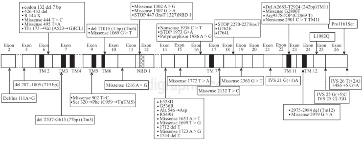
All mutations described above are represented along MDR3 encoding cDNA on Figure 1.
Protocol for MDR3 analysis
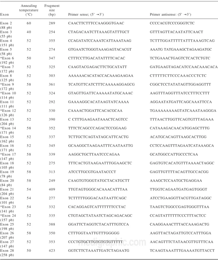
To our knowledge, 18 out of 28 exons are affected by mutations associated with recurrent cholesterol microlithi-asis. More precisely 48 mutations of MDR3 gene have been reported to date, from which 43 (89.5%) in the cod-ing region. Twenty one (43.8%) are located in only 8 exons near transmembrane or nucleotide binding domains of the protein. From the 22 remaining described mutations, 9 (18.8%) are restricted to exon 14. Lastly, five mutations at splice site have been associated to cholesterol cholelithi-asis. In our laboratory we start therefore analysis of MDR3 gene by scanning exons 6, 7, 9, 10, 12, 14, 17, 23 and 24 (corresponding primers are marked with an asterisk in Table I). Focus on this coding region increases chance of mutation discovering, although for any of mutation de-scribed above no study was initiated to look for either a founder effect (except for T175V),4 or a hot spot status.
Briefly, genomic DNA is isolated from peripheralblood lymphocytes of patient by automated nucleic acids ex-tractor MagnapureTM system (Roche diagnostics, Meylan,
amplifica-medigraphic.com
tionis carried out for 36 cycles (30 sec at 95° C, 12 sec at temperature mentioned in Table I and 20 sec at 72°C) and ends by 1 cycle for 7 min at 72° C . PCR productsare separated on 1% agarose gel and visualized with ethidi-um bromide before purification using High Pure PCR Product Purification Kit (Roche Diagnostics, Meylan, France). Sequencing in both sense is carried out in 20-µl mixture containing 1 µL of either 5 µM primer sense or antisens, 8 µl of master Mix (GenomeLab DTCS Quick-start Kit, Beckman Coulter, Villepinte, France) and 1.5 µl of purified DNA. Sequence reaction protocol consists of 30 cycles (20 sec at 96°C, 20 sec at 50°C and 4 min at 60°C), afterwards a precipitation of samples with ethanol is necessary before injection in CEQ 8000TM analyser.
Sequences are analyzed by InvestigatorTM software
(Beckman Coulter, Villepinte, France).
5. Discussion
Defects of the phospholipid export pump MDR3 result in impaired biliary excretion of phosphatidylcholine and a variety of cholestatic syndromes ranging from progres-sive familial intrahepatic cholestasis in neonates to biliary cirrhosis in adults.21 Interestingly, great majority of
report-ed mutations affects functional domains of the protein leading to start the analysis near these regions. More spe-cifically, mutations are more frequently localized in sym-metrical positions in the protein, in transmembrane do-mains TM2, TM3, TM4, TM5 and TM6 (around exons 6 to 9) in one side, and TM 11 and TM12 (around exons 23-24) in the other side of the protein. The molecular de-fects either result in truncation of the open reading frame (frame shift and non-sense) or amino acid substitutions
(missense). If frame-shift mutations are associated with severe RNA depletion, most missense mutations are ac-companied by faint or absent MDR3 canalicular stain-ing.18 Reduced MDR3 expression can result from
mes-senger RNA destabilization or degradation of a misfold-ed protein. Functional consequences and resulting phenotype of missense mutations, such as those present in the NBD or Walker motifs, can be predicted with some level of confidence, and their linkage to disease state is uncomplicated. However, mutations in other regions of the coding sequence that do not affect protein expression are more difficult to interpret. In absence of more exten-sive genetic analysis, it is difficult to conclude that these defects are the sole cause of the disease.
Different mutations in a single gene can generate dis-parate phenotypes. Loss-of-function mutations that abol-ish protein expression lead to severe cholestasis in infan-cy and do not respond to treatment with ursodesoxycho-late. Conversely, patients carrying missense mutations, which may permit limited protein expression and func-tion, manifest disease later in life and respond to ursodes-oxycholate.8
Main mutations in the hepatic phospholipid transport-er that have been found in ICP patients are missense or splicing mutations at the heterozygous state. Some au-thors warn against the possibility that other rare underly-ing hepatic disorders may be unmasked durunderly-ing pregnancy with cholestasis as its first manifestation.22
In a similar way it is noteworthy that I764L and L1082Q mutations that have been mentioned in Figure 1 were in-volved in specific situations of drug-induced cholestasis and drug-induced hepatocellular injury, respectively. Therefore one should consider these genetically
medigraphic.com
Table I. Primers used for amplification of MDR3 coding region (exons with an asterisk are those to analyze in priority).
Annealing Fragment
temperature size
Exon (°C) (bp) Primer sense; (5’ →3’) Primer antisense: (5’ →3’)
Exon 2 6 0 289 CAACTTCTTTCCAAGGGTGAAC CCCCACGTCCCGGGTCTC
(88 pb)
Exon 3 4 0 254 CTAGACAATCTTAAAGTATTTGCT GTTTAGTTACAATATTCAACT
(55 pb)
Exon 4 5 2 355 CCAGATATCCAAATCATTAAATAAG TCTTTGGATTTTTATTTAAAGTCAG
(151 pb)
Exon 5 5 2 274 GTGAATCTGGGTAAAGAGTACACGT AAATG TATGAAAGCTAGAAGATGC
(58 pb)
*Exon 6 5 0 347 CTTTCCTTGACATATTTTCACAC TCTGAAACTGAGTCTCACTCTGTC
(192 pb)
*Exon 7 5 2 325 CAAGTACGAGACTTCTGCATATT GATGAAGTAGACATCCAACAAACACA
(172 pb)
Exon 8 5 2 303 AAAAAACACATACCACAAAGAAGAA CTTTTTCTTCCCAAACCCTCTC
(125 pb)
*Exon 9 5 8 361 TCATGTTCATCTTTCAAAAAGGAGCG CGGCTCCTATATAGTTGGAGGTTT
(172 pb)
*Exon 10 5 2 324 GTAGTTGATTCAAAAATATGCAAAC AAGTTTAGGTTTATCCTTTCCTTT
(114 pb)
Exon 11 5 2 292 GAAAAGGCACATAAGTATCAAAA AGGAATATGATTCAGCAAATTCCA
(111 pb)
*Exon 12 5 2 330 CAAAACTGGATTCACACGCAA TGAAAAAAAAGTATCAAATAAGGGA
(126 pb)
Exon 13 5 2 390 C CTTTGAAGAATAAACTCAGTCC TTTAACTTGGTTCAGTGTTTAGAAA
(204 pb)
*Exon 14 5 8 352 TTTCTCAGCCCAGACTCCGGAAG CATAAAGACAACATGGAGCTTTG
(171 pb)
Exon 15 5 2 337 TCTTGCTCAGTATAGCATTCACTG ACATGCACAGTTAAGCACTTGG
(162 pb)
Exon 16 5 2 345 GCAAGGCTAAGAATTTCAATAATTG CCTCCAAGTTTAGAATCATAAAGCA
(171 pb)
*Exon 17 5 8 339 AAGGCTGCTTAATCCCAGAA GCATGGCCATTGCCCTCAA
(147 pb)
Exon 18 5 2 275 TTTCACTGTAAGAATTTGGAAGCTC GAGTGTCACATGTTTAAAACTAGGC
(105 pb)
Exon 19 5 8 313 ATCCTTGCGTGAATACCCT GAGTTGTTTTACAGTTGCCACGG
(78 pb)
Exon 20 5 8 249 CAAGTGTGGGTATGCTACATGCTT AAAGCTCCAATGCTGAGGAA
(84 pb)
Exon 21 5 4 409 TTGTAGTGGGCACAAACATTTAA TTGGTCAGAATGATGAGTGGGT
(204 pb)
Exon 22 5 4 277 TCTTTTTGGGACAATAATTCAGC ATCCTGAAGGTTACGTTGATAGGC
(101 pb)
*Exon 23 5 4 332 CACAGGAGTCATTTTTTTCCTAC TAAGTCTGGCCGAGTGGGTTTAA
(141 pb)
*Exon 24 5 2 335 CTGTAGCTATAATCTAGCAGACAGC CCAGTATTTTTTCCCTTTACTCC
(157 pb)
Exon 25 5 2 388 GGATTCTAGGTCTACATTTGTCCA CAAGGAAACTTTACCAAAGACTG
(198 pb)
Exon 26 5 8 350 CTTTGGTAATTGTTTGGGGG AAGTTACTAGATTGTCCATTTGGA
(207 pb)
Exon 27 5 2 353 CCCTGTGCTTGTGTGTGTTTTT AACAGTTTCTATAACGTTGTTTCAA
(147 pb)
Exon 28 5 0 423 GGTCTTCTAAATTGATCTAGAATG TCAAGTAAATTTGAAAATGTTACCT
(258 pb)
mined functional impairments as predisposition factors rather than causal mutations for intrahepatic cholesta-sis.23 The mutation screening method used in our
labora-tory is unable to detect major DNA rearrangements, and does not include the promoter or other potential
medigraphic.com
described, CYP7A1 seems to be a second important actor, since it is the rate-limiting enzyme in the synthesis of bile salts from cholesterol in the liver. Recently, several common DNA polymorphisms in the ABCG8 gene were discovered that are associated with variations in plasma sterols, which could also influence biliary cholesterol se-cretion, but there is still a paucity of human studies.23
Lastly, it is tempting to speculate that MDR3 defects could also play an important role in cholangiopathies in humans. Indeed, MDR3 variants could play a role as modifi-er gene in primary biliary cirrhosis and primary sclmodifi-erosing cholangitis, but their exact role needs further clarification.24
6. Conclusion
Multidrug resistance protein 3, as an ABC transport-er, participates to hepatic transport and secretion of phospholipid (phosphatidylcholine) and at this ac-count, may be at origin of common biliary cholesterol microlithiasis. Progressive familial intrahepatic cholestasis-3 is a heterogeneous group of autosomal re-cessive liver disorders characterized by early onset of cholestasis that progresses to hepatic fibrosis, cirrhosis, end-stage liver disease before adulthood. High gamma-glutamyltranspeptidase levels are an additional criteri-on characteristic to PFIC3 that is necessary to ccriteri-onsider analysis of MDR3 gene. In that case, we showed that a preliminary scanning of 8 exons which represents less than the third of the coding region allows detection of main deleterious mutations. Such work is valuable since an effective treatment is now identified. At the same time several drugs for the treatment of cholestatic liver diseases that target MDR3 expression and func-tion are tested, further underscoring the clinical signifi-cance of this transport system.
E. Mbongo-Kama and F. Ceppa contributed equally to this work.
References
1. Boland LL, Folsom AR, Rosamond WD. Hyperinsulinemia,
dyslipidemia, and obesity as risk factors for hospitalized gallbladder disease: a prospective study. Ann Epidemiol 2002; 12: 131-40.
2. Rosmorduc O, Hermelin B, Poupon R. MDR3 gene defect in
adults with symptomatic intrahepatic and gallbladder cholesterol cholelithiasis. Gastroenterology 2001; 120: 1459-67.
3. Kano M, Shoda J, Sumakazi R, Oda K, Nimura Y, Tanaka N, et al. Mutations identified in the human multidrug resistance P-glyco-protein (ABCB4) gene in patients with primary hepatolithiasis.
Hepatol Res 2004; 29: 160-6.
4. Rosmorduc O, Poupon R. Low phospholipid associated cholelithi-asis: association with mutation in the MDR3/ABCB4 gene.
Orphanet J Rare Dis 2007; 2: 29 [Epub ahead of print].
5. Weiss KM, Ferrell RE, Hanis CL, Styne PN. Genetics and epide-miology of gallbladder disease in New World native peoples. Am J
Hum Genet 1984; 36: 1259-78.
6. Wang DO, Carey MC. Complete mapping of crystallization path-ways during cholesterol precipitation from model bile: influence of physical-chemical variables of pathophysiologic relevance and
iden-tification of a stable liquid crystalline state in cold, dilute and hydro-philic bile salt-containing systems. J Lipid Res 1996; 37: 606-30. 7. Carey MC, Lamont JT. Cholesterol gallstone formation.
Physi-cal-chemistry of bile and biliary lipid secretion. Prog Liver Dis 1992; 10: 139-63.
8. Ortiz D, Arias IM. MDR3 Mutations: a Glimpse into Pandora’s box and the future of canalicular pathophysiology.
Gastroenter-ology 2001; 120: 1549-51.
9. Dixon PH, Weerasekera N, Linton KJ, Donaldson O, Chambers J, Egginton E, Weaver J, et al. Heterozygous MDR3 missense mu-tation associated with intrahepatic cholestasis of pregnancy: evi-dence for a defect in protein trafficking. Hum Mol Genet 2000; 9: 1209-17.
10. De Vree JML, Jacquemin E, Sturm E, Cresteil D, Bosma PJ, Aten J, Deleuze JF, et al. Mutations in the MDR3 gene cause progres-sive familial intrahepatic cholestasis. Proc Natl Acad Sci USA 1998; 95: 282-7.
11. Jacquemin E, Cresteil D, Manouvrier S, Boute O, Hadchouel M. Heterozygous non-sense mutation of the MDR3 gene in familial intrahepatic cholestasis of pregnancy. Lancet 1999; 353: 210-1. 12. Eloranta ML, Heiskanen JT, Hiltunen MJ, Mannermaa AJ, Punnonen KR, Heinonen ST. Multidrug resistance 3 gene muta-tion 1712delT and estrogen receptor alpha gene polymorphisms in Finnish women with obstetric cholestasis. Eur J Obstet Gynecol
Reprod Biol 2002; 104: 109-12.
13. Gendrot C, Bacq Y, Brechot MC, Lansac J, Andres C. A second heterozygous MDR3 nonsense mutation associated with intrahe-patic cholestasis of pregnancy. J Med Genet 2003; 40: e32. 14. Pauli-Magnus C, Lang T, Meier Y, Zodan-Marin T, Jung D,
Breymann C, Zimmermann R, et al. Sequence analysis of bile salt export pump (ABCB11) and multidrug resistance p-glycopro-tein 3 (ABCB4, MDR3) in patients with intrahepatic cholestasis of pregnancy. Pharmacogenetics 2004; 14: 91-102.
1 5 . Keitel V, Vogt C, Haussinger D, Kubitz R. Combined muta-tions of canalicular transporter proteins cause severe intrahe-patic cholestasis of pregnancy. Gastroenterology 2006; 131: 6 2 4 - 9 .
16. Floreani A, Carderi I, Paternoster D, Soardo G, Azzaroli F, Esposito W, Variola A, et al. Intrahepatic cholestasis of pregnancy: three novel MDR3 gene mutations. Aliment Pharmacol Ther 2006; 23: 1649-53.
17. Schneider G, Paus TC, Kullak-Ublick GA, Meier PJ, Wienker TF, Lang T, Van de Vondel P, et al. Linkage between a new splicing site mutation in the MDR3 alias ABCB4 gene and intrahepatic cholestasis of pregnancy. Pharmacogenet Genomics 2007; 17: 47-60.
18. Jacquemin E, de Vree JML, Cresteil D, Sokal E, Sturm E, Dumont M, Scheffer GL, et al. The wide spectrum of multidrug resistance 3 deficiency: from neonatal cholestasis to cirrhosis of adulthood.
Gastroenterology 2001; 120: 1448-58.
19. Jacquemin E. Progressive familial intrahepatic cholestasis: ge-netic basis and treatment. Clin Liver Dis 2000; 4: 753-63. 20. Chen HL, Chang PS, Hsu HC, Lee JH, Ni YH, Hsu HY, Jeng YM,
et al. Progressive familial intrahepatic cholestasis with high gamma-glutamyltranspeptidase levels in Taiwanese infants: role of MDR3 gene defect? Pediatr Res 2001; 50: 50-5.
21. Trauner M, Fickert P, Wagner M. MDR3 (ABCB4) defects: a paradigm for the genetics of adult cholestatic syndromes. Semin
Liver Dis 2007; 27: 77-98.
22. Arrese M. Cholestasis during pregnancy: rare hepatic diseases unmasked by pregnancy. Ann Hepatol 2006; 5: 216-8. 23. Lang C, Meier Y, Stieger B, Beuers U, Lang T, Kerb R,
Kullak-Ublick GA, et al. Mutations and polymorphisms in the bile salt export pump and the multidrug resistance protein 3 associated with drug-induced liver injury. Hepatology 2007; 45: 150-8. 24. VanBerge-Henegouwen GP, Venneman NG, Portincasa P,