Liver fibrosis in bile duct-ligated rats correlates with
increased hepatic IL-17 and TGF-
βββββ
2 expression
Adelaida Sara M. Zepeda-Morales,* Susana Del Toro-Arreola,** Leonel García-Benavides,*** Blanca E. Bastidas-Ramírez,* Mary Fafutis-Morris,** Ana L. Pereira-Suárez,** Miriam R. Bueno-Topete*
* Instituto de Investigación en Enfermedades Crónico-Degenerativas, Departamento de Biología Molecular y Genómica. ** Laboratorio de Inmunología, Departamento de Fisiología. *** Instituto de Terapéutica Experimental y Clínica, Departamento de Fisiología. Centro Universitario de Ciencias de la Salud , Universidad de Guadalajara, Guadalajara, Jal., México.
A B S T R A C T A B S T R A C TA B S T R A C T A B S T R A C T A B S T R A C T
Background and rationale for the study. Background and rationale for the study.Background and rationale for the study. Background and rationale for the study.
Background and rationale for the study. IL-17, TGF-β1/2 are cytokines involved in the development of kidney, pulmonary and liver fibrosis. However, their expression kinetics in the pathogenesis of cholestatic liver fibrosis have not yet been fully explored. The aim of the study was to analyze the expression of IL-17, RORγt, NKp46, TGF-β1, and TGF-β2 in the liver of rats with bile duct ligation (BDL). Results.Results.Results.Results.Results. Hepatic IL-17A gene expression analyzed by qRT-PCR showed a dramatic increase of 350 and 10 fold, at 8 and 30 days post BDL, respectively. TGFβ1 and TGFβ2 gene expression significantly increased throughout the whole fibrotic process. At the protein level in liver homogenates, IL-17, TGF-β1, and RORγt significantly increased at 8 and 30 days after BDL. Interestingly, a significant increase in the protein levels of TGF-β2 and decrease of NKp46 was observed only 30 days after BDL. Unexpectedly, TGF-β2 exhibited stronger signals than TGF-β1 at the gene expression and protein levels. Histological analysis showed bile duct proliferation and collagen deposition. Conclusions.Conclusions.Conclusions.Conclusions. Our results suggest that pro-fibrogenic cytokinesConclusions. IL-17, TGF-β1 and, strikingly, TGF-β2 might be important players of liver damage in the pathogenesis of early and advanced expe-rimental cholestatic fibrosis. Th17 cells might represent an important source of IL-17, while NK cell depletion may account for the perpetuation of liver damage in the BDL model.
Key words. Key words.Key words. Key words.
Key words. ROR γ. TGF-β. NK cells bile duct ligation.
May-June, Vol. 15 No. 3, 2016: 418-426
INTRODUCTION
Liver fibrosis may result from a variety of chronic liver diseases, including hepatitis B, hepatitis C, and alcoholic liver disease. It is characterized by the accumulation of ex-tracellular matrix (ECM) and scar formation. Several inju-ry-triggered events playing critical roles in the pathogenesis of liver fibrosis, such as recruitment of inflammatory cells to the damaged area and profibrogenic cytokines release, have been described. However, transforming growth factor beta (TGF-β) and resident hepatic stellate cells (HSC) constitute the major mediators of liver injury.1,2
TGF-β is a member of a large family of multifunctional cytokines that exist in at least three isoforms denominated
β1, β2, and β3, presenting 80% homology in their amino acid sequence. TGF-β1 binds to specific receptors on the
cell surface, transducing signals to the nucleus by means of Smad proteins, upregulating collagen gene expression and resulting in collagen deposition within the liver.3 TGF-β1
isoforms play distinct roles in wound healing with
TGF-β1/2 having predominantly pro-scarring roles and TGF-β3 having mainly anti-scarring effects.4-6 TGF-β2 transcripts
have been identified in the bile duct epitelial cells of pa-tients with biliary atresia, alcoholic cirrhosis, and cirrho-sis secondary to hepatitis B.7,8 The participation of this
cytokine in remodeling the ECM that stimulates the ca-nonical Smad signaling pathway through Smad 2/3 in the pathogenesis of extraocular muscles fibrosis has been re-ported by Wordinger, et al.9 To date, reports dealing with
TGF-β2 kinetics in the progression of chronic hepatic in-jury involved in cholestatic processes in humans and ex-perimental models are limited.
The Official Journal of the Mexican Association of Hepatology, the Latin-American Association for Study of the Liver and
the Canadian Association for the Study of the Liver
Manuscript received: Manuscript received: Manuscript received: Manuscript received:
Manuscript received: August 01, 2015. Manuscript accepted:Manuscript accepted:Manuscript accepted:Manuscript accepted:Manuscript accepted: September 09, 2015.
Recently, the profibrogenic role of interleukin 17 (IL-17), a new cytokine, has been proposed in skin, lung, and liver fibrosis.10-12 IL-17A is the founding member of the
IL-17 cytokine family, which now includes six major iso-forms, IL-17A, B, C, D, E, and F. IL-17F is most closely related to IL-17A. The two molecules share 50% amino acid sequence homology. Different reports have demon-strated that IL-17A and IL-17F share some biological activ-ities including the induction of cytokines and chemokines involved in inflammatory responses.13,14 However,
addi-tional activities specifically concerning to each cytokine have not been fully elucidated.
Association studies in patients with chronic liver injury by alcohol, hepatitis B, and hepatitis C show overexpres-sion of IL-17, which interestingly correlates with the de-gree of fibrosis.15,16 Experimentally, dramatic decrease of
liver injury and fibrosis in IL-17A or IL-17R(-/-) knock-out mice with induced cholestatic or hepatotoxic fibrosis has been reported.17,18 On the other hand, IL-17 represents
the most important cytokine produced by Th17 cells. Dif-ferentation of Th17 cells requieres the combined action of IL-6, IL-1β, TGF-β, and IL-23. These cytokines promote the expression of the lineage-specific transcription factor orphan nuclear receptor γt (ROR γt: in mice) or RORc (in human), wich is necessary and sufficient for develop-ment of Th17 cells.18,19 Therefore, RORγt would suggest
the presence of Th17 cells.
The production of IL-17 could not be restricted only to Th17 cells, but other cell populations, such as CD8+T cells, γδT cells, and natural killer (NK) cells, may be a source for this cytokine. Considering the fact that up to 30% of the lymphocytes in hepatic parenchyma are repre-sented by NK cells, it is factible to speculate that activated NK cells might be additional producers of IL-17. Several activating receptors, such as NKp46 and NKG2D, charac-terize to these cells in rodent models; however only the first one is mostly restricted to NK cells.20
Notwithstanding this, IL-17 expression in the different stages of the bile duct ligation (BDL)-induced cholestatic fibrosis, as well as its source, is unknown.
Based on this background, it is important to elucidate the IL-17, TGF-β1, and TGF-β2 expression kinetic pattern in the progress of experimental fibrosis, as well as the par-ticipation of Th17 and NK cells as major IL-17 sources.
MATERIAL AND METHODS
Cholestasis-induced by bile duct ligation
Male Wistar rats weighing 200-250 g, obtained from Charles Rivers (Boston, MA, USA) used in this study were kept under 12:12 h light/dark cycles, room tempera-ture of 22 °C, constant humidity, and free access to food
and water. Cholestatic fibrosis was induced by bile duct ligation (BDL) according to the method of Lee, et al., 1986.21 Animals were anesthetized with 30 mg/kg i.p. of
Zoletil 50 (Virbac). Laparotomy was performed under an-tiseptic conditions. A mid-line incision in the abdomen exposing the muscle layers and the linea alba was made. The edge of the liver was then raised and the duodenum pulled down to expose the common bile duct. The com-mon bile duct was doubly ligated with 4-0 silk thread and sectioned between the ligatures. Incisions were closed with silk thread. Control rats (sham) underwent the same surgical procedure except for ligation and section of the bile duct. Ligated animals (n = 15) were divided into two experimental groups (BDL), one corresponding to 8 days and the other one to 30 days evolution BDL, respectively. Finally, 5 survivor animals remained in each BDL group; meanwhile all sham animals (control) survived (n = 10). Experimental and control animals were sacrificed on day 8 or on day 30 post-surgery and representative liver samples were obtained.
All animal procedures were performed according to the guidelines of the Ethics Committee of University of Guad-alajara based on technical specifications for the production, care, and use of laboratory animals NOM-062-ZOO-1999 and the International Guiding Principles for Biomedical Research Involving Animals established in 1985.22,23
RNA extraction and cDNA synthesis
Isolation of total RNA from rat livers was carried out according to the method described by Chomczynski and Sacchi.24 Briefly, liver tissue was obtained from three
dif-ferent lobes and homogenized utilizing a Polytron system in the presence of Trizol (Invitrogen, Carlsbad, CA, USA). Two μg of RNA were reverse-transcribed in 0.05 M Tris-HCl, pH 8.3, 40 mM KCl, 7 mM MgCl2 buffer containing 0.05 μg/μL random hexamers, 1 mM dNTP mix, 0.05 U/mL RNAse inhibitor, and 200 U/mL M-MLV reverse transcriptase. Samples were incubated for 10 min at 70 °C, and prior to incubation for 60 min at 37 °C. Re-verse transcriptase was inactivated by heating the sample tubes to 95 °C for 10 min, and the complementary DNA (cDNA) obtained were used immediately or stored at -20 °C until use.
Quantitative-reverse
transcriptase-polymerase chain reaction
GenBank Nucleotide database sequences. Primers were synthesized by integrated DNA Technologies (USA). Primer sequences, amplicon length, and aligning tempera-ture are shown in table 1. All qRT-PCR reactions were performed in a final volume of 15 μL consisting of 3 μL of 1X PCR buffer (Light cycler fast start DNA master plus reaction mix SYBRgreen I, Roche Applied Science, Basel, Switzerland), 0.5 μM of sense and antisense primers, and 100 ng of cDNA. Reaction conditions were the following: 50 °C for 2 min and 95 °C for 10 min, followed by 40 cy-cles at 95 °C for 15 s and at 60 °C for 30 s. Additionally, melting analysis was performed, setting reactions at 95 °C for 10 sec, at 60 °C for 1 min, and by slowly heating the samples to 95 °C at a temperature rate of 0.1 °C/s. TaqMan probes (Applied Biosystems, Foster City, CA, USA) to amplify TGF-β2 were employed. Gene amplification was normalized against 18S rRNA expression. Complementa-ry DNA (cDNA) was analyzed by triplicate and mean val-ues were calculated. Relative quantification by the 2-ΔΔCT method was carried out, considering control groups as an internal calibrator.
Western blotting
Detection of TGF-β1, TGF-β2, IL-17, ROR-γ, and NKp46 was performed by Western blotting. Proteins were extracted from liver homogenates using RIPA buffer (SIGMA R 0278; Sigma, NJ, USA). Proteases inhibitor cocktail (Santa Cruz Biotechnology SC-29030, Santa Cruz, CA, USA) was added and clarified by centrifugation at 4 °C 12,000 rpm for 15 min. Protein concentration was deter-mined by the Bradford method (Coomassie brilliant blue G-250, 95% ethanol, concentrated phosphoric acid and wa-ter). The total protein (40 g) was mixed with loading buffer undbuffer reducing conditions, electrophoresed on 7.5 -12% SDS-polyacrylamide gels and transferred to a polyvi-nylidene difluoride membrane (Bio-Rad, CA, USA). Non-specific binding was blocked with 3% milk. Subse-quently, membranes were incubated with primary anti-bodies overnight: TGF-β1 (1:500) (sc - 52893), TGF-β2 (1:500) (sc - 374659), IL-17 (1:500) (sc - 374218), ROR-γt
(1:500) (sc - 28559), and NKp46 (1:500) (sc - 292746). All antibodies were from Santa Cruz Biotechnology. HRP-conjugated anti-mouse or anti-rabbit secondary antibodies were used to reveal the blots: anti-mouse IgG (1:5,000) (SC - 2005) and anti-rabbit IgG (1:5000) (SC - 2004) were from Santa Cruz Biotechnology. Immune detection was performed using a chemiluminescence imaging system (Millipore, Billerica, MA, USA). Densitometric analysis was carried out with the Kodak MI 5.0 Image Analyzer (Kodak, Rochester, NY, USA). As an internal control to confirm that similar amounts of protein were loaded into each lane, GAPDH levels were determined using a mono-clonal anti-gapdh IgG (1:5,000) (sc - 32233) and were re-vealed with anti-mouse IgG peroxidase (sc-2005).
Histological analysis
Liver sections were randomly taken from the right, me-dian, and left lobes of rat livers in each experimental group and immediately fixed by immersion in 4% paraformalde-hyde diluted in phosphate saline buffer. The tissue were dehydrated in graded ethylic alcohol, and embedded in paraffin. Sections 5 μm thick were stained with Masson’s trichrome and analysed by light microscopy for determi-nation of pathological changes.
Statistical methods
Statistical analysis was performed with SPSS Statistics software ver. 20.0 (SPSS, Inc., Chicago, IL, USA). Nor-mally distributed variables were analyzed using one-way Analysis of variance (ANOVA) or the Student t test. Bon-ferroni and Dunnett post-hoc tests were employed for de-termination of statistical significance. Data are shown as the mean ± standard deviation (SD) of the mean. Only p values ≤ 0.05 were considered significant.
RESULTS
Cytokine expression in the liver tissue was determined by qRT-PCR. A highly significant increase in IL-17A
Table 1. Primer set sequences for quantitative-Reverse transcriptase-polymerase chain reaction (qRT-PCR) assays.
Gene Bases (n) Sequence Product length Tm ºC
TGF-β1 Rat F 21 TCCTTGCCCTCTACAACCAAC 115 60
R 20 TCCACCTTGGGCTTGCGACC
IL-17A Rat F 21 CCGAGATAACTTTGAGGCATA 292 62
R 20 AACGAGGTTTGACTTTCACA
TGF-β2 Rat Gene Bank Acces NM_031131 I.D 95 60
Figure 1. Figure 1.Figure 1. Figure 1.
Figure 1. Gene expression of IL-17A, TGF-β1, and TGF-β2 by qRT-PCR. The relative quantification was carried out utili-zing the 2 –ΔΔCt± standard deviation (SD)
method and ribosomal 18s gene expres-sion as housekeeping gene. Results are expressed as Expression relative units (ERU). Each experiment was performed in triplicate. A.A.A.A.A. IL-17A expression, * p ≤ 0.05 control vs. BDL day 30, ** p ≤ 0.001 control vs. BDL day 8. B.B.B.B.B. TGF-β2 expre-ssion, * p ≤ 0.05 control vs. BDL day 8, ** p ≤ 0.001 control vs. BDL day 30. C.
C.C. C.
C. TGF-β1 expression, * p ≤ 0.05 control
vs. BDL day 8; ** p ≤ 0.001 control vs. BDL day 30, and (DDDD) TGF-D β1/TGF-β2 ratio EUA.
expression of 350 (p < 0.001) and 10 times (p < 0.05) more induction than the control after 8 and 30 days of BDL respectively, was observed (Figure 1A).
A very interesting behavior was exhibited by TGF-β2, which showed a significant increase up to 10 times more than that exhibited by the control group (p < 0.05), during early stages of fibrosis. More importantly, TGF-β2 was strikingly overexpressed (up to 93 times more) at 30 days after BDL when compared to the control group (p < 0.001), as seen in figure 1B.
Regarding TGF-β1 expression, it was induced 5 times (p < 0.05) and 7.5 times (p < 0.001) after 8 and 30 days BDL, respectively, when compared with control (Figure 1C). It is noteworthy to mention that a prevailing expres-sion pattern of TGF-β2 over TGF-β1 of 2:1 after 8 and 30 days BDL was appreciated (Figure 1D).
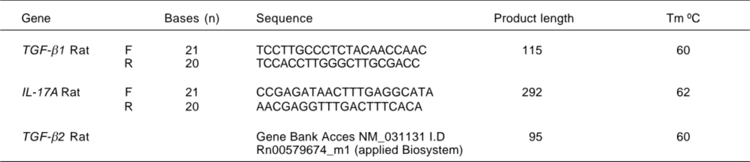
At the protein level in the liver tissue, significant IL-17 elevation of 2 and 3.5 times more than the control after 8 (p < 0.05) and 30 days (p < 0.001) BDL was exhibited (Figure 2A). However, the strongest effect was appreciat-ed at the gene expression level.
A significant increase in TGF-β1 concentration after 8 (257 ± 5 ARU vs. 154 ± 10 ARU; p < 0.05) and 30 days
(555 ± 15 ARU vs. 154 ± 10 ARU; p < 0.001) BDL in comparison with control was observed (Figure 2B). Like-wise, TGF-β2 showed a tendency toward increasing after 8 days BDL, and a significant increase (877 ± 35 ARU vs. 334 ± 50 ARU; p < 0.001) in the late stages of fibrosis when compared to control (Figure 2C). A prevailing pat-tern of TGF-β2 over TGF-β1 after 8 (p < 0.05) and 30 days (p < 0.001) BDL at the protein level similar to that already described at the expression level, was exhibited (Figure 2D).
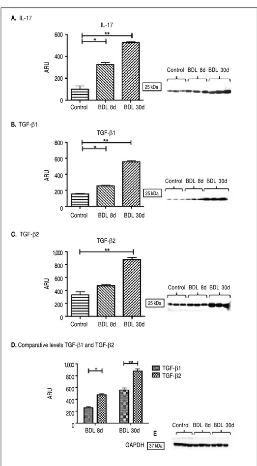
Interestingly, there was an increase of three times more than the control of ROR-γt (p < 0.001), a Th17 cell-specific transcriptional factor, throughout the whole fibrotic process (Figure 3A). In contrast, liver NKp46 level was significantly (p < 0.001) reduced after 30 days BDL when compared with the control (Figure 3B). Additionally, histopathological analysis exhibited normal morphology of the portal triad and hepatic lobules in control animals (Figure 4A); meanwhile, duct proliferation and periductal fibrosis after 8 days BDL, could be appreciated. This pattern became more apparent and substituting for nearly all of the hepatic parenchyma after 30 days BDL (Figures 4B and 4C).
A. A. A. A.
A. mRNA IL-17A/18s B.B.B.B.B. mRNA TGF-β2/18s
C. C. C. C.
C. mRNA TGF-β1/18s D. D. mRNA D. D. D. TGF-β1/TGF-β2 400
300
15
5 1 0
EUA
Control BDL 8d BDL 30d Control BDL 8d BDL 30d
Control BDL 8d BDL 30d Control BDL 8d BDL 30d 140
120 100 80
15
5 1 0
EUA
10
8
6
4
1 0
EUA
14
12
10
8
1
0
EUA
TGF-β1/TGF-β2
Figure 2. Figure 2. Figure 2. Figure 2.
Figure 2. IL-17, TGF-β1, and TGF-β2 proteins determined by Western blot. GA-PDH was used as an internal control. Each bar represents the mean value ± standard deviation (SD) of five rats. Re-sults are expressed as area relative units (ARU). A.A.A.A.A. IL-17 protein, 25 kDa, * p ≤ 0.05 control vs. BDL day 8; ** p ≤ 0.001 control vs. BDL day 30. B.B.B.B.B. TGF-β1 pro-tein, 25 kDa, * p ≤ 0.05 control vs. BDL day 8; ** p ≤ 0.001 control vs. BDL day 30. C.C.C.C.C. TGF-β2 protein, 25 kDa, ** p ≤ 0.001 control vs. BDL day 30. D.D.D.D.D. Com-parative levels TGF-β1 and TGF-β2 pro-teins, * p ≤ 0.05 BDL day 8, * p ≤ 0.001 BDL day 30. E.E.E.E.E. GAPDH protein, 37 kDa.
A. A. A. A. A. IL-17
Control BDL 8d BDL 30d 600
400
200
0
ARU
B. B. B. B. B. TGF-β1
Control BDL 8d BDL 30d 800
600
400
200
0
ARU
Control BDL 8d BDL 30d 1,000
800
600
400
200
0
ARU
BDL 8d BDL 30d 1,000
800
600
400
200
0
ARU
C. C. C. C. C. TGF-β2
D. D. D. D.
D. Comparative levels TGF-β1 and TGF-β2
E E E E E IL-17
TGF-β1
TGF-β2
TGF-β1 TGF-β2
GAPDH 37 kDa 25 kDa 25 kDa 25 kDa
Control BDL 8d BDL 30d
Control BDL 8d BDL 30d
Control BDL 8d BDL 30d
DISCUSSION
In this study, the participation of IL-17 and TGF-β1/2 in the pathogenesis of experimental cholestasis was as-sessed. Various reports dealing with the role of IL-17 in lung, skin, and liver fibrosis have been published.10-12
Heightened gene expression and increase of IL-17 pro-tein level in early and advanced experimental cholestatic fibrosis was observed in our study. These findings have not been previously described in the literature. Accumu-lation of IL-17 positive cells around the damaged bile ducts in primary biliary cirrhosis (PBC), as well as the
Figure 4. Figure 4.Figure 4.
Figure 4.Figure 4. Histology findings induced by experimental cholestasis. Representative images of liver sections from bile duct ligation (BDL) and control rats. A.A.A.A.A. Control groups. B.B.B.B.B. BDL rats day 8. C.C.C.C. BDL rats day 30. Extrahepatic cholestasis due to prolonged obstruction of bile flow in BDL rats resulted in extensiveC. proliferation of bile ducts in enlarged portal tracts and periductal fibrosis (scale bars: 200 μm).
Figure 3. Figure 3. Figure 3. Figure 3.
Figure 3. ROR-γt and NKp46 proteins determined by Western blot. GAPDH was used to normalize protein concentration as an internal control. Each bar represents the mean value ± standard deviation (SD) of five rats. Results are expressed as area relative units (ARU). A.A.A.A.A. ROR-γt protein, 63 kDa; ** p ≤ 0.001 control vs. BDL day 8, ** p ≤ 0.001 control vs. BDL day 30. B.
B. B. B.
B. NKp46 protein, 47 kDa, ** p ≤ 0.001 control vs. BDL day 30, ** p ≤ 0.001 BDL day 8 vs. BDL day 30, and CCCCC. GAPDH protein, 37 kDa.
A A A A A
B B B B B
600
400
200
0
ARU
63 kDa
Control BDL 8d BDL 30d
Control BDL 8d BDL 30d 400
300
200
100
0
ARU
47 kDa
Control BDL 8d BDL 30d
Control BDL 8d BDL 30d
37 kDa
ability of biliary epithelial cell cultures stimulated with IL-17 to produce Th17-inducible cytokines (IL-6 and IL-1β) and Th17-maintaining cytokine (IL-23) have been reported by Nakanuma, et al.25 These results suggest that
periductal IL-17 secreting cells might facilitate the migra-tion of inflammatory cells around the bile ducts, aggravat-ing the chronic cholangitis in the BDL model. In addition, serum TNF-α, TGF-β1 levels and fibrosis score were significantly decreased in knockout mice with induced cholestatic fibrosis, when compared with the WT mice.18
Moreover, induction of collagen type 1 expression by IL-17 in HSC cultures promoting their activation into fibrogenic myofibroblasts via STAT-3, have been observed in vitro.17
Thus, the above observations indicate that in the BDL model, IL-17 might not only exacerbate the local inflam-matory process, but might also contribute to the deposi-tion of extracellular matrix components through collagen production. It is noteworthy to mention that, in our study the increase of IL-17 was more evident at the level of gene expression than at the protein level. This finding might be explained due to the fact that the anti-IL-17 antibody used in our study is designed to recognize total IL-17, and the gene expression was specific for IL-17A.
This cytokine represents the most studied isoform in chronic liver disease. Additionally, a dramatic overexpres-sion of IL-17F mRNAwas also observed in our study (data not shown), evidencing the importance of understanding the biological participation of distinct IL-17 isoforms (A-F) in liver damage.
IL-17 constitutes the major cytokine produced by Th17 cells; however, T cells, CD8+ T cells, γδ T cells, NKT cells, innate lymphoid cells, eosinophils, neutrophils, and monocytes can also produce IL-17. The presence of tran-scription factors and cytokines, such as STAT3, 6, IL-1β, and TGF-β, are required for Th17 differentiation, leading to the induction of the expression of RORγt, a master gene regulator. Likewise, IL-23 expression is nec-essary for maintaining Th17 phenotype and function.18,19
We analyzed the expression of RORγt as an indicator of the presence of Th17 cells and statistically significant in-creased expression of RORγt in the liver of cholestatic rats during the chronic stages of liver injury, suggesting that Th17 could be an important source of IL-17 in the BDL model was demonstrated in our study. These results are also supported by the TGF-β1/2 overexpression observed both in the early as well as in the late stages of fibrosis, where TGF-β is essential to Th17 cells differentiation in rodents. A methodological flaw in our study has been the fact that we did not identify the exact cell population producing IL-17; therefore, it will be necessary to plan future studies, which include immunohistochemestry assays in hepatic biopsy or multicolor flow cytometric analysis in isolated
hepatic cells, in order to discriminate inflammatory infil-trate from hepatic parenchyma (biliary epithelial cells, Kupffer cells or HSC cells) as a probable source for IL-17.
Parallel to these findings, the decreased expression of NKp46 exhibited in our study suggests that in the BDL model, it might not be a considerable source of IL-17; however, it is likely to be contributing to the exacerbation of the liver damage process.
An interesting antifibrogenic mechanism in the model of CCl4 chronic intoxication, orchestrated by the cytotox-ic capacity of NK cells, has been described. In rodents, ac-tivation of HSC releases retinoic acid leading to RAE-1 synthesis as a metabolic by-product, sensitizing NK cells through their receptor NKG2D to produce INFγ and TRAIL, inducing the arrest of the HSC cell cycle and death, blocking the perpetuation of hepatic injury.26,27
Future multicolor flow cytometry staining by using a het-erogeneus gamma of markers, besides NKp46 (CD16, CD94, CD158, NKG2D, for example), will lead us to identify and quantify intrahepatic NK cells, which will clarify in turn, the participation of NK cells during the whole process of hepatic damage by cholestasis.
On the other hand, it is important to elucidate the co-operative role established between IL-17 and TGF-β1/2 in the development of liver experimental fibrosis in BDL model, since a regulatory role of TGF-β1 in allograft fibrosis has been attributed to IL-17 recently.
TGF-β is a key molecule in diverse fibrotic disorders, it can promote fibroblast proliferation, differentiation, migration, adhesion, and survival. TGF-β can induce cytokine secretion and, most importantly, upregulation of collagen and ECM synthesis, playing a critical role in pathological fibrogenesis.28 The role of TGF-β1 has been
well documented in diverse models of acute and chronic hepatic injury. Recently, however, TGF-β2 has been rec-ognized as an important cytokine implicated in the patho-physiology of fibrosis induced by different etiologies. In early and advanced models of renal fibrosis, TGF-β2 expression was markedly increased and specific targeting of TGF-β2 attenuated the renal fibrosis process in experi-mental diabetes.29,30 Likewise, in ocular fibrosis, TGF-β2
participates in ECM remodeling through the stimulation of the canonical Smad signaling pathway via Smad2/3.9
Kouroumalis, et al. have described intrahepatic bile ductules as a novel source of TGF-β isoforms (1/2/3) in patients with PBC and HCV cirrhosis.31 This finding is
relevant due to the displaying features of epitelial-to-mes-enchymal transition (EMT) of cholangyiocytes during he-patic fibrosis.31-34
Studies in vitro and in vivo have demonstrated the partici-pation of TGF-β1 in the induction of collagen production as well as in EMT-associated changes in several liver cell types, including cholangiocytes.33,35-37
In the future, the elucidation of TGF-β2 role in EMT will contribute to further understand the hepatic fibrosis process. In the same context, obliteration of TGF-β2 in BDL model would provide useful informa-tion regarding the participainforma-tion of this cytokine in fi-brosis progression.
Finally, it is important to point out that inflammation and fibrosis are preponderant mechanisms during the de-velopment of the cholestasis. In this context, we might conclude that, based on our present results, TGF-β and IL-17 are important protagonists in the pathophysiology of this disease.
ABBREVIATIONS
• BDL: bile duct ligation. • ECM: extracellular matrix. • HSC: hepatic stellate cells. • IL-17: interleukin 17. • NK: natural killer cells. • PBC: primary biliary cirrhosis.
• TGF-βββββ: transforming growth factor beta.
REFERENCES
1. Bataller R, Brenner DA. Liver fibrosis. J Clin Invest 2005; 115: 209-18.
2. Seki E, De Minicis S, Österreicher CH, Kluwe J, Osawa Y, Brenner DA, Schwabe RF, et al. TLR4 enhances TGF-β signaling and hepatic fibrosis. Nat Med 2007; 13: 1324-32.
3. Inagaki Y, Okazaki I. Emerging insights into Transforming growth factor beta Smad signal in hepatic fibrogenesis. Gut
2007; 56: 284-92.
4. Ferguson MWJ, O’Kane S. Scar-free healing: from embryon-ic mechanisms to adult therapeutembryon-ic intervention. Philos Trans R Soc Lond B Biol Sci 2004; 359: 839-50.
5. Ferguson MWJ, Foreman, David M, Shah, Mamta. Neutralisa-tion of TGF-β1 and TGF-β2 or exogenous addition of TGF-β3 to cutaneous rat wounds reduces scarring. J Cell Sci
1995; 108: 985-102.
6. Durani P, Occleston N, O’Kane S, Ferguson MWJ. Avotermin: a novel antiscarring agent. Int J Low Extrem Wounds 2008; 7: 160-8.
7. Lee S-Y, Chuang J-H, Huang C-C, Chou M-H, Wu C-L, Chen C-M, Hdich, et al. Identification of transforming growth fac-tors actively transcribed during the progress of liver fibrosis in biliary atresia. J Pediatr Surg 2004; 39: 702-8.
8. Santos RM, Norton P, Degli Esposti S, Zern MA. TGF-beta isoforms in alcoholic liver disease. J Gastroenterol 1998; 33: 383-9.
9. Zode GS, Sethi A, Brun-Zinkernagel A-M, Chang I-F, Clark AF, Wordinger RJ. Transforming growth factor-β2 increases extracellular matrix proteins in optic nerve head cells via ac-tivation of the Smad signaling pathway. Mol Vis 2011; 17: 1745-58.
10. Gasse P, Riteau N, Vacher R, Michel M-L, Fautrel A, di Pado-va F, Fick L, et al. IL-1 and IL-23 mediate early IL-17A pro-duction in pulmonary inflammation leading to late fibrosis.
PloS One 2011; 6: e23185.
11. Tan Z, Qian X, Jiang R, Liu Q, Wang Y, Chen C, Wang X, et al. IL-17A plays a critical role in the pathogenesis of liver fi-brosis through hepatic stellate cell activation. J Immunol
2013; 191: 1835-44.
12. Nakashima T, Jinnin M, Yamane K, Honda N, Kajihara I, Maki-no T, Masuguchi S, et al. Impaired IL-17 signaling pathway contributes to the increased collagen expression in sclero-derma fibroblasts. J Immunol 2012; 188: 3573-83.
13. Toy D, Kugler D, Wolfson M, Vanden Bos T, Gurgel J, Derry J, Tocker J, et al. Cutting edge: interleukin 17 signals through a heteromeric receptor complex. J Immunol 2006; 177: 36-9. 14. Iwakura Y, Nakae S, Saijo S, Ishigame H. The roles of IL-17A in inflammatory immune responses and host defense against pathogens. Immunol Rev 2008; 226: 57-79.
15. Ge J, Wang K, Meng Q-H, Qi Z-X, Meng F-L, Fan Y-C. Impli-cation of Th17 and Th1 cells in patients with chronic active hepatitis B. J Clin Immunol 2010; 30: 60-7.
16. Lemmers A, Moreno C, Gustot T, Maréchal R, Degré D, Demetter P, de Nadai P, et al. The interleukin-17 pathway is involved in human alcoholic liver disease. Hepatol 2009; 49: 646-57.
17. Meng F, Wang K, Aoyama T, Grivennikov SI, Paik Y, Scholten D, Cong M, et al. IL-17 signaling in inflammatory cells, Kupffer cells and Hepatic Stellate cells exacerbates liver fibrosis. Gastroenterology 2012; 143: 765-76.
18. Hara M, Kono H, Furuya S, Hirayama K, Tsuchiya M, Fujii H. Interleukin-17A plays a pivotal role in cholestatic liver fibro-sis in mice. J Surg Res 2013; 183(2): 574-82.
19. Veldhoen M, Hocking RJ, Atkins CJ, Locksley RM, Stockinger B. TGFbeta in the context of an inflammatory cytokine milieu supports de novo differentiation of IL-17-producing T cells.
Immunity 2006; 24: 179-89.
20. Moretta A, Bottino C, Vitale M, Pende D, Cantoni C, Mingari MC, Biassoni R, et al. Activating Receptors and Coreceptors Involved in Human Natural Killer Cell-Mediated Cytolysis.
Annu Rev Immunol 2001; 19: 197-223.
21. Lee SS, Girod C, Braillon A, Hadengue A, Lebrec D. Hemo-dynamic characterization of chronic bile duct-ligated rats: ef-fect of pentobarbital sodium. Am J Physiol 1986; 251: 176-80.
22. De Aluja AS. Laboratory animals and official Mexican norms (NOM-062-ZOO-1999). Gac Médica México 2002; 138: 295-8. 23. Mappes TA DD. Biomedical ethics. 6th ed. Boston:
McGraw-Hill; 2006.
24. Chomczynski P, Sacchi N. Single-step method of RNA isola-tion by acid guanidinium thiocyanate-phenol-chloroform ex-traction. Anal Biochem 1987; 162: 156-9.
25. Harada K, Shimoda S, Sato Y, Isse K, Ikeda H, Nakanuma Y. Periductal interleukin-17 production in association with biliary innate immunity contributes to the pathogenesis of cholangio-pathy in primary biliary cirrhosis. Clin Exp Immunol 2009; 157: 261-70.
stim-ulating NK cell cytotoxicity. Hepatol Baltim Md 2006;44: 1441-51.
27. Tian Z, Chen Y, Gao B. Natural killer cells in liver disease.
Hepatol 2013; 57: 1654-62.
28. Blobe GC, Schiemann WP, Lodish HF. Role of transforming growth factor beta in human disease. N Engl J Med 2000; 342: 1350-8.
29. Yu L, Border WA, Huang Y, Noble NA. TGF-beta isoforms in renal fibrogenesis. Kidney Int 2003; 64: 844-56.
30. Hill C, Flyvbjerg A, Rasch R, Bak M, Logan A. Transforming growth factor-beta2 antibody attenuates fibrosis in the experimental diabetic rat kidney. J Endocrinol 2001; 170: 647-51.
31. Wells RG. Cellular sources of extracellular matrix in hepatic fibrosis. Clin Liver Dis 2008; 12: 759-68.
32. Omenetti A, Porrello A, Jung Y, Yang L, Popov Y, Choi SS, Witck R, et al. Hedgehog signaling regulates epithelial-mes-enchymal transition during biliary fibrosis in rodents and hu-mans. J Clin Invest 2008; 118: 3331-42.
33. Rygiel KA, Robertson H, Marshall HL, Pekalski M, Zhao L, Booth TA, Jones DEJ, et al. Epithelial-mesenchymal transition contributes to portal tract fibrogenesis during human chronic liver disease. Lab Investig J Tech Methods Pathol 2008; 88: 112-23.
34. Sato Y, Harada K, Ozaki S, Furubo S, Kizawa K, Sanzen T, Yasoshima M, et al. Cholangiocytes with mesenchymal
fea-tures contribute to progressive hepatic fibrosis of the poly-cystic kidney rat. Am J Pathol 2007; 171: 1859-71.
35. Kaimori A, Potter J, Kaimori J-Y, Wang C, Mezey E, Koteish A. Transforming growth factor-beta1 induces an epithelial-to-mesenchymal transition state in mouse hepatocytes in vit-ro. J Biol Chem 2007; 282: 22089-101.
36. Xia J-L, Dai C, Michalopoulos GK, Liu Y. Hepatocyte growth factor attenuates liver fibrosis induced by bile duct ligation.
Am J Pathol 2006; 168: 1500-12.
37. Zeisberg M, Yang C, Martino M, Duncan MB, Rieder F, Tan-jore H, Lalluri R,et al. Fibroblasts derive from hepatocytes in liver fibrosis via epithelial to mesenchymal transition. J Biol Chem 2007; 282: 23337-47.
Correspondence and reprint requests: Miriam Ruth Bueno-Topete, M.D.
Instituto de Investigación en Enfermedades Crónico – Degenerativas, Departamento de Biología Molecular y Genómica, Centro Universitario de Ciencias de la Salud,
Universidad de Guadalajara.
Sierra Mojada, Núm. 950, Col. Independencia, Guadalajara, Jalisco. C.P. 44340, México.