Hepatic Angiosarcoma with
Kasabach-Merritt Phenomenon:
A Case Report and Review of the Literature
Fumika Fujii,*,** Takefumi Kimura,*,** Naoki Tanaka,*** Daisuke Kubota,** Ayumi Sugiura,** Takeji Umemura,** Shuichi Wada,* Eiji Tanaka**
* Department of Gastroenterology, Nagano Red Cross Hospital, Nagano, Japan. ** Department of Internal Medicine, Division of Gastroenterology, Shinshu University School of Medicine, Matsumoto, Japan. *** Department of Metabolic Regulation, Shinshu University Graduate School of Medicine, Matsumoto, Japan.
July-August, Vol. 17 No. 4, 2018: 655-660
INTRODUCTION
Disseminated intravascular coagulopathy (DIC) is characterized by massive microthrombi appearing through various mechanisms.1 Enhanced thrombolysis and con-sumption of coagulation factors in DIC lead to bleeding tendency, thrombocytopenia, hypofibrinogenemia, and in-creased circulating fibrinogen degradation product (FDP).1 Although DIC is frequently accompanied by se-vere infection, leukemia, and cancer cell invasion of the bone marrow, some cases are caused by tumors of vascular origin, such as cavernous hemangioendothelioma.1 Hepat-ic angiosarcoma is an uncommon malignant vascular tu-mor of the liver that is very rarely accompanied with DIC.2 We herein report a case of hepatic angiosarcoma showing Kasabach-Merritt phenomenon (KMP), review
its clinical features, and compare it with existing cases in the literature.
CASE REPORT
A 76-year-old woman was referred to our outpatient clinic due to bouts of persistent or massive gingival bleed-ing followbleed-ing teeth extraction two weeks prior. On admis-sion, she had no history of drinking, blood transfuadmis-sion, medicine or supplement regimen, or occupation handling vinyl chloride or heavy metals. She did not suffer from weight loss, general fatigue, anorexia, or abdominal full-ness or pain. Physical examination showed no jaundice, purpura, lymphadenopathy, or splenomegaly, but a soft liver was palpable in the epigastric region. Laboratory findings revealed decreased platelet count (56,000/μL) and
The Official Journal of the Mexican Association of Hepatology, the Latin-American Association for Study of the Liver and
the Canadian Association for the Study of the Liver
Manuscript received: Manuscript received:Manuscript received:
Manuscript received:Manuscript received: January 03, 2017. Manuscript accepted:Manuscript accepted:Manuscript accepted:Manuscript accepted:Manuscript accepted: May 24, 2017.
DOI:10.5604/01.3001.0012.0949
A B S T R A C T A B S T R A C T A B S T R A C T A B S T R A C T A B S T R A C T
A 76-year-old woman was referred to our hospital due to massive gingival bleeding following teeth extraction. Laboratory findings sug-gested disseminated intravascular coagulopathy (DIC). Enhanced computed tomography and magnetic resonance imaging disclosed multiple hypervascular liver masses of 2-6 cm in diameter, the largest of which displaying an irregular enhancement pattern. We con-sidered that her DIC was caused by the multiple liver masses and commenced repeated erythrocyte/fresh frozen plasma infusion and gabexate mesilate administration. However, the DIC proved uncontrollable and trans-arterial embolization could not be attempted. The patient eventually died 4 months after admission due to spontaneous hepatic tumor rupture and hepatic failure. Post-mortem he-patic tumor biopsy led to a final diagnosis of hehe-patic angiosarcoma with Kasabach-Merritt phenomenon (KMP). Among the 7 cases of hepatic angiosarcoma representing KMP found in the literature, mortality occurred within 4 months of the appearance of bleeding tendency primarily due to abdominal bleeding and hepatic failure. The possibility of hepatic angiosarcoma should be considered in pa-tients with DIC and hypervascular liver tumors. Since treatment is uncertain and prognosis is poor, novel diagnostic and therapeutic advances are needed for angiosarcoma.
Key words. Key words.Key words. Key words.
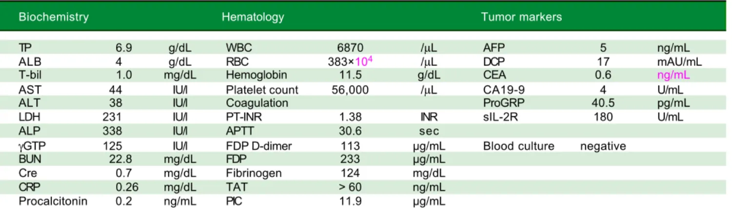
circulating fibrinogen level (124 μg/mL) with elevated FDP level (233 μg/mL), which were suggestive of DIC (Table 1). Microscopic examination of peripheral blood cells detected no atypical leukocytes or fragmented eryth-rocytes. Serum levels of lactate dehydrogenase and the tu-mor markers alpha fetoprotein, des-gamma-carboxy prothrombin, carcinoembryonic antigens, carbohydrate antigen, pro-gastrin-releasing peptide, and soluble inter-leukin 2 receptor were all within normal ranges (Table 1). Negative blood culture results and normal levels of serum C-reactive protein and procalcitonin indicated a low pos-sibility of underlying severe infection or inflammation (Table 1).
Enhanced computed tomography (CT) disclosed mul-tiple hypervascular liver masses of 2-6 cm in diameter (Figure 1A). On magnetic resonance imaging (MRI), the largest tumor was T1 hypointense and T2 hyperintense (Figure 1B). Dynamic contrast enhanced MRI revealed ir-regular areas of enhancement at the periphery (Figure 1C) that were hypoechoic on routine ultrasound (Figure 1D).
Based on the above findings, we considered the possi-bility of a tumor of vascular origin, such as hemangioen-dothelioma or angiosarcoma. In spite of repeated packed red blood cell and fresh frozen plasma transfusion along with gabexate mesilate administration, the patient’s gingi-val bleeding did not stop and alveolar hemorrhage ap-peared 43 days after admission. A relationship between the multiple liver tumors and DIC was suspected, but prima-ry liver tumor treatment, such as trans-arterial emboliza-tion (TAE), could not be attempted due to the patient’s poor general condition and uncontrollable systemic bleed-ing tendency; bleedbleed-ing would persist for several days fol-lowing routine blood sampling from the peripheral vein.
Spontaneous hepatic tumor rupture and hepatic failure eventually occurred and she died 120 days after admission.
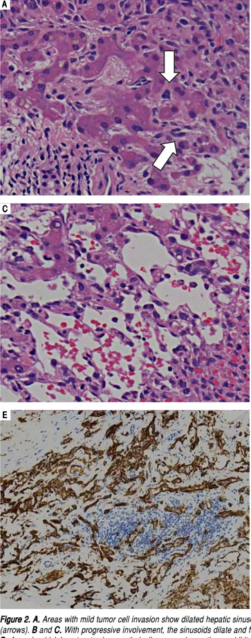
Post-mortem hepatic tumor biopsy revealed various pathological findings. Liver parenchyma with mild inva-sion of tumor cells exhibited dilated hepatic sinusoids lined by hypertrophied endothelial cells displaying atypi-cal hyperchromatic nuclei (Figure 2A). In severely in-volved areas, these bizarre cells had proliferated in dilated sinusoids, causing liver cell plate atrophy (Figure 2B and C) and forming clusters in areas which the hepatocytes had disappeared entirely (Figure 2D). Immunohisto-chemical analysis revealed CD31 and CD34 positivity in tumor cells, leading to the final diagnosis of hepatic angi-osarcoma (Figure 2E and F).
DISCUSSION
Hepatic angiosarcoma is a malignant mesenchymal tu-mor with a very low incidence of 0.14-0.25 per million in-habitants that represents 1.8% of all primary liver tumors.3 Several etiological factors have been implicated in this dis-ease, including exposure to thorotrast, vinyl chloride, cop-per, radium, sex steroids, and arsenic compounds.4 Although the present case contained none of these ele-ments, numerous other reports support the existence of hepatic angiosarcoma without such factors.2
The symptoms of hepatic angiosarcoma are largely nonspecific. Abdominal pain is the most common com-plaint, followed next by weakness, fatigue, and weight loss.2 Moreover, there are no physical or laboratory find-ings characteristic of this disease. The lack of specific findings makes accurate diagnosis of hepatic angiosarcoma difficult.
Table 1. Laboratory data on admission.
Biochemistry Hematology Tumor markers
TP 6.9 g/dL WBC 6870 /μL AFP 5 ng/mL
ALB 4 g/dL RBC 383×104 /μL DCP 17 mAU/mL
T-bil 1.0 mg/dL Hemoglobin 11.5 g/dL CEA 0.6 ng/mL
AST 44 IU/l Platelet count 56,000 /μL CA19-9 4 U/mL
ALT 38 IU/l Coagulation ProGRP 40.5 pg/mL
LDH 231 IU/l PT-INR 1.38 INR sIL-2R 180 U/mL
ALP 338 IU/l APTT 30.6 sec
γGTP 125 IU/l FDP D-dimer 113 μg/mL Blood culture negative
BUN 22.8 mg/dL FDP 233 μg/mL
Cre 0.7 mg/dL Fibrinogen 124 mg/dL
CRP 0.26 mg/dL TAT > 60 ng/mL
Procalcitonin 0.2 ng/mL PIC 11.9 μg/mL
A AA
AA BBBBB
D DD DD
Figure 1. A. Figure 1. A.Figure 1. A. Figure 1. A.
Figure 1. A. Enhanced CT scan shows multiple enhanced liver masses measuring 2-6 cm in diameter. The largest tumor in S4 measures 6 cm and exhibits an irregular enhancement pattern (arrow). B.B.B.B.B. The largest tu-mor in S4 shows T1 hypointensity and T2 hyperintensity on MRI (arrows). C
C C C
C and D.D.D.D.D. The largest tumor in S4 is seen with an irregular enhancement pattern on enhanced MRI (CCCCC, arrows) and as hypoechoic on ultrasonography (DDDDD, arrows).
C CC CC
Unexplainable DIC may be a clue in identifying hepat-ic tumors of vascular origin. In 1940, Kasabach and Merritt described a new syndrome in a boy with kaposiform he-mangioendothelioma, severe thrombocytopenia, anemia,
and consumption coagulopathy.3 KMP with angiosarcoma was later detected in the skin,4 breast,5 and bone.6 Recent-ly, ultrastructural examination of kaposiform hemangioen-dothelioma samples with KMP revealed trapping of
p r e p r ep r e
p r ep r e 30s30s30s30s30s 60s60s60s60s60s 120s120s120s120s120s 180s180s180s180s180s T 1 W I
T 1 W I T 1 W I T 1 W I
Figure 2. A. Figure 2. A. Figure 2. A. Figure 2. A.
Figure 2. A. Areas with mild tumor cell invasion show dilated hepatic sinusoids lined by hypertrophied endothelial cells with atypical hyperchromatic nuclei (arrows). BBBB and C.B C.C. With progressive involvement, the sinusoids dilate and fill with malignant endothelial cells (BC.C. BBBB, arrows) with liver cell plate atrophy (C)(C)(C)(C)(C). D.
D. D. D.
D. Areas in which hepatocytes have entirely disappeared sometimes exhibit solid malignant cell growth (arrows). E E E E E and F.F.F.F. Immunohistochemical analysis. TheF. tumor cells are positive for the endothelial markers CD31 (E) and CD34 (F)(F)(F)(F)(F).
A A A A
A BBBBB
C CC
CC DDDDD
E EE
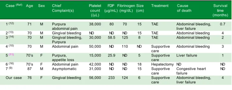
Table 2. Case reports of hepatic angiosarcoma with Kasabach-Merritt phenomenon.
Case (Ref) Age Sex Chief Platelet FDP Fibrinogen Size Treatment Cause Survival
Complaint(s) count (μg/mL) (mg/dL) (cm) of death time
(/μL) (months)
1 (12) 71 M Purpura 38,000 80 70 15 TAE Abdominal bleeding, 0.7
abdominal pain liver failure
2 (13) 70 M Gingival bleeding ND ND ND 15 TAE Abdominal bleeding 4
3 (14) 70 M Gingival bleeding, 30,000 58.5 125 8 TAE Abdominal bleeding 2
Purpura
4 (10) 70 M Abdominal pain 50,000 ND 110 ND Supportive Abdominal bleeding 3
care
5 (11) 70’s F Purpura, 15,000 25.9 ND 5 Supportive Liver failure 1
appetite loss care
6 (16) 70’s F Abdominal pain 42,000 ND ND 18 Hepatectomy ND ND
7 (9) 87 M Asymptomatic 31,000 ND ND 15 Supportive Congestive heart ND
care failure
Our case 76 F Gingival bleeding 56,000 233 124 6 Supportive Abdominal bleeding, 4
care liver failure
F: female. FDP: fibrinogen degradation product. M: male. ND: not described. Ref: reference. TAE: transcatheter arterial embolization.
platelets, erythrocytes, lymphocytes, and macrophages in tumor cells and intra-tumoral channels.7 Many thrombi were generated and a large amount of platelets and coagu-lation factors were consumed in the tumor, leading to DIC.7 Judging from the clinical course and pathological biopsy findings in the present case, we considered that the DIC caused by multiple hepatic angiosarcoma was KMP.
Due to the nonspecific physical/laboratory findings of hepatic angiosarcoma, imaging modalities play a critical role in its diagnosis. Angiosarcoma has various hallmark features in CT/MRI that reflect its heterogeneous histo-logic composition. When angiosarcoma appears as multi-ple nodular lesions, most include enhancement foci to enable distinction from benign hemangiomas showing nodular enhancement.8 Hemorrhagic lesions may appear in cases of very large angiosarcoma.9 Our patient exhibited irregular areas of enhancement at the periphery of the larg-est lesion. Although we could not obtain a specimen, we surmised that it corresponded to cavernous areas lined by malignant cells and ensuing hepatocyte atrophy.
There is uncertainty on how patients with KMP should be treated. The clinical features, treatment, and outcomes of 7 case reports of hepatic angiosarcoma with KMP are presented in table 2. Patient age ranged from 70 to 87 years and initial symptoms primarily included gingival bleed-ing, purpura, and abdominal pain. All cases possessed thrombocytopenia and large tumors of more than 5 cm in diameter. Tumor resection and TAE were considered risky because of uncontrollable bleeding tendency. More-over, TAE was insufficient in most cases and required ad-ditional treatments. Survival was less than 4 months due to rapid tumor progression, with intraperitoneal bleeding and liver failure representing the main causes of death.
Al-though there are no established therapeutic strategies for angiocarcinoma at present, several reports have demon-strated the potential of angiogenic agents, such as anti-VEGF drugs, tyrosine kinase inhibitors, such as sunitinib, and combinations of such molecular targeted agents as sor-afenib and sunitinib to be at least partially effective.15 The establishment of novel therapeutic interventions is needed.
In conclusion, we report a rare case of hepatic angiosar-coma that had manifested as bleeding tendency. The possi-bility of hepatic angiosarcoma should be considered for patients with DIC and liver tumor.
ABBREVIATIONS
• CT: computed tomography.
• DIC: disseminated intravascular coagulopathy. • FDP: fibrinogen degradation product.
• KMP: Kasabach-Merritt phenomenon. • MRI: magnetic resonance imaging.
FUNDING
No funding source to declare.
CONFLICT OF INTEREST
No conflict of interest exists.
REFERENCES
2. Locker GY, Doroshow JH, Zwelling LA, Chabner BA. The clinical features of hepatic angiosarcoma: a report of four cases and a review of the English literature. Medicine 1979; 58: 48-64.
3. Kasabach HH, Merritt KK. Capillary hemangioma with exten-sive purpura. Am J Dis Child 1940; 59: 1063-70.
4. Imafuku S, Hosokawa C, Moroi Y, Furue M. Kasabach-Merritt syndrome associated with angiosarcoma of the scalp suc-cessfully treated with chemoradiotherapy. Acta Dermato-ve-nereologica 2008; 88: 193-4.
5. Bernathova M, Jaschke W, Pechlahner C, Zelger B, Bodner G. Primary angiosarcoma of the breast associated Kasa-bach-Merritt syndrome during pregnancy. Breast 2006; 15: 255-8.
6. Choi JJ, Murphey MD. Angiomatous skeletal lesions. Semi-nars in Musculoskeletal Radiology 2000; 4: 103-12.
7. Yuan SM, Hong ZJ, Chen HN, Shen WM, Zhou XJ. Kaposi-form hemangioendothelioma complicated by Kasabach-Mer-ritt phenomenon: ultrastructural observation and immunohistochemistry staining reveal the trapping of blood components. Ultrastructural Pathology 2013; 37: 452-5.
8. Koyama T, Fletcher JG, Johnson CD, Kuo MS, Notohara K, Burgart LJ. Primary hepatic angiosarcoma: findings at CT and MR imaging. Radiology 2002; 222: 667-73.
9. Habringer S, Boekstegers A, Weiss L, Hopfinger G, Meiss-nitzer T, Melchardt T, Egle A, et al. Kasabach-Merritt
phe-nomenon in hepatic angiosarcoma. British Journal of
Haematology 2014; 167: 716-8.
10. Alliot C, Tribout B, Barrios M, Gontier MF. Angiosarcoma
vari-ant of Kasabach-Merritt syndrome. European Journal of
Gastroenterology & Hepatology 2001; 13: 731-4.
11. Homma M, Kushima M, Saito K. An autopsy case of angiosa-rcoma of the liver with aggressive course. Division of Diag-nostic Pathology 2010; 27: 110-4.
12. Kudo M, Hirasa M, Takakuwa H. A case of hepatic hemangi-osarcoma associated with kasabach-merritt syndrome and intraperitoneal bleeding. Kanzo 2016; 25: 1605-11.
13. Saito M, Watanabe Y, Fujita M. An autopsy case of hepatic angiosarcoma associated with Kasabach-Merritt syndrome and tumor rupture. Shindanbyouri 2000; 17: 369-71. 14. Tsuji K, Yoshida H, Sakurai Y. A case of angiosarcoma of the
liver with Kasabach-Merritt syndorome and investigated on the tumor growth, retrospectively. Kanzo 2001; 42: 210-16. 15. Park MS, Ravi V, Araujo DM. Inhibiting the VEGF-VEGFR
pathway in angiosarcoma, epithelioid hemangioendothelioma,
and hemangiopericytoma/solitary fibrous tumor. Current
Opinion in Oncology 2010; 22: 351-5.
16. Gonzalez Rodriguez FJ, Dominguez Comesana E, Portela Serra JL, Lede Fernandez A, Pinon Cimadevila MA. Urgent surgery in a Kasabach-Merrit syndrome associated with a giant hepatic angiosarcoma. Cirugia Espanola 2014; 92: 370-2.
Correspondence and reprint request: Takefumi Kimura, M.D., Ph.D.
Department of Internal Medicine, Division of Gastroenterology, Shinshu University School of Medicine, 3-1-1 Asahi, Matsumoto,
Japan