R Igetei et al. Detection of p53 CODON 249 mutation in Nigerian patients 339
www.medigraphic.com
Annals of Hepatology 2008; 7(4): October-December: 339-344
Annals of Hepatology
Original Article
Detection of p53 codon 249 mutation
in Nigerian patients with hepatocellular carcinoma
using a novel evaluation of cell-free DNA
Rufina Igetei;1 Jesse A. Otegbayo;1 Dennis A. Ndububa;2 Olufunmilayo A. Lesi;3 Chiaka I. Anumudu;4 Pierre
Hainaut;5 Emmanuelle Gormally6
1Department of Medicine, University College Hospital, Ibadan. 2Department of Medicine, Obafemi Awolowo University
Teaching Hospitals Complex, Ile-Ife, Nigeria.
3Department of Medicine, Lagos University Teaching Hospital,
Lagos.
4Zoology Department, University of Ibadan, Ibadan. 5Molecular Carcinogenesis Unit.
6International Agency For Research On Cancer, Lyons, France.
Address for correspondence:
Dr. R Igetei. E-mail: [email protected]
Manuscript received and accepted: 8 July and 1 September 2008
Abstract
Objectives: This case-control study was done to deter-mine the association and prevalence of p53 codon 249 mutation using cell-free DNA in the plasma of patients with hepatocellular carcinoma (HCC) in South-West-ern Nigeria. Method: Eighty-five adults with HCC and seventy-seven age and gender matched controls with-out evidence of liver disease or malignancy involving any part of the body, were recruited. Plasma DNA was analyzed for p53 codon 249 by restriction fragment length polymorphism. Patient evaluation was done by means questionnaire interview, clinical examination, laboratory and radiological tests. The prevalence of the p53 codon 249 mutation was expressed as a per-centage amplifiable DNA samples analyzed from HCC patients while that of controls was expressed in the same way. Fisher’s exact test or the student t-test where appropriate were used to assess statistical significance of prevalence between both groups as well as compari-son of some characteristics in the HCC cases between those who had codon 249 mutation and those who did not. Associations between the various parameters as-sessed were determined by odds ratio and significant difference was specified at p < 0.05. Results: p53 codon 249 mutation was present in 6 (7.6%) of the 79 samples from the HCC patients with amplifiable plasma DNA while none (i.e. 0%) of the 73 samples with amplifiable plasma DNA from the controls had this mutation. This prevalence is significantly higher among HCC patients than controls (0.029). The mutation was also found to
be significantly associated with HCC (odds ratio = 2.00; 95% C I: 1.70 – 2.35). Conclusion: The prevalence of the p53 codon 249 mutation from plasma DNA of hepa-tocellular carcinoma patients is significantly higher than among controls in South-Western Nigeria and the presence of this mutation is significantly associated with HCC in this region.
Key words: p53 codon 249 mutation, cell-free/plasma DNA, hepatocellular carcinoma, Nigeria.
Introduction
Hepatocellular carcinoma which is a malignant growth of the hepatocytes commonly affects middle-aged men in South-East Asia and sub-Saharan Africa.1-6 The common aetiologic agents associated with this malignan-cy are hepatitis B virus (HBV), hepatitis C virus (HCV), alcohol and aflatoxin B1 (AFB1). The out-come of this disease is poor as a result of late presentation. To im-prove the prognosis of this disease, several techniques have been advocated for screening and surveillance of high risk subjects. This is to facilitate early diagnosis at a stage when the disease is potentially curable.2
The only single test that is diagnostic of HCC is his-tology on liver biopsy specimens or cyhis-tology on fine needle aspiration samples from the liver, which are inva-sive in nature. These procedures are sometimes risky and better avoided in some patients who are not fit to under-go these procedures especially when they present late. Moreover, it may be inconvenient to perform liver biop-sy on the same subject at a periodicity of three months which is the time interval advocated presently for screen-ing and surveillance programmes.7,8
Various genetic aberrations have been associated with HCC.9 Of these, the p53 gene mutation is the most stud-ied. A specific mutation in codon 249 of the p53 gene, induced by AFB1 toxicity in which the third base gua-nine is substituted by thymine (AGG → AGT) is associat-ed with HCC.3,10,11 This leads to the substitution of argin-ine by serargin-ine in the p53 protein causing folding abnor-mality of the DNA binding domain on the protein. The worldwide prevalence of this genetic abnormality is
Artemisa
www.medigraphic.com
11%,9 and has also been found in 13 to 36% of aflatoxinrelated tumours from South-east Asia.9,12 In sub-Saharan Africa, 36 to 66% of HCC patients had this mutation.13-15 In Nigeria, where the rate of food contamination by afla-toxin B1 and the prevalence of HBV viral markers among HCC patients are high, Ndububa et al found this mutation in 1 (5.5%) of the 18 tumour samples taken from HCC patients in South-western Nigeria.16
Most of the genetic analyses done initially were on DNA extracted from tumour tissues because genetic mate-rial is stored in the nucleus of nucleated cells. Recent re-search however, has shown that it is now possible to iso-late DNA from body fluids like plasma/serum and urine which carries the same genetic material as the original tu-mour. Thus, cell-free DNA in the plasma and in the above-mentioned body fluids can be used as a surrogate material to detect genetic alterations present in the original tu-mour.10,11,17 The source of this cell-free DNA is not certain. It is however, thought to be from tumour cells which have un-dergone necrosis or apoptosis. The objective of this study was to determine the association and prevalence of the p53 codon 249 mutation in plasma DNA of HCC patients.
Materials and methods
Eight-five consecutive and consenting adults with HCC who fulfilled the diagnostic criteria for HCC as stat-ed by the European Association for the Study of the Liv-er (EASL) in Barcelona in the year 2000 wLiv-ere recruited into the study. Seventy-seven age and gender matched adults being managed for ailments other than liver dis-ease or malignancy were also recruited from the Medical Outpatient Department or the Medical wards of the Uni-versity College Hospital (UCH), Ibadan, Obafemi Awolowo University Teaching Hospital (OAUTH), Ile-Ife and the Lagos University Teaching Hospital (LUTH) La-gos, all in South-western Nigeria.
After obtaining an informed consent from each subject, a questionnaire was administered to obtain socio-demograph-ic data and other relevant clinsocio-demograph-ical history. Clinsocio-demograph-ical examina-tion and relevant laboratory investigaexamina-tions were done.
Genomic DNA was extracted from the plasma using QiAmp DNA blood mini kit according to the manufactur-er’s blood and body fluid spin protocol (Qiagen, Hilden, Germany). Purified DNA was eluted from the QiAmp sili-ca column with 200 μL of water (PCR-grade, Sigma, St Louis, MO, USA).
The eluted DNA (8 μL) was amplified by polymerase chain reaction (PCR) for exon 7 of the p53 gene using spe-cific primers: forward primer 5’-CTTGCCACAGGTCTC-CCCAA-3’ and reverse primer 5’ –AGGGGTCACCG-GCAAGCAGA-3’. The protocol for the reaction was 15 minutes denaturation at 95 °C, then 50 cycles of denatur-ation at 94 °C, primer annealing at 60 °C, and extension at 72 °C at 30 seconds each, followed by a 10-minute final extension step at 72 °C. The amplification product
con-taining 254 base-pairs (bp) was visualized on 3% agarose gel with ethidium bromide. 10 uL of PCR products were then digested with restriction endonuclease Hae III (Boe-hringer Mannheim GmbH, Mannheim, Germany) which cleaves GG/CC sequence between codon 249 and 250 to generate 92 bp and 66 bp fragments. The presence of codon 249 mutation yields an undigested 158 bp frag-ment which is identified on 3% agarose gel. Gel bands containing the 249 mutation were excised from samples that had the mutation and following elution of DNA from the gel, nested PCR was done with the primers; (sense) 5’ AGGCGCACTGGCCTCATCTT 3’ and (antisense) 5’ TGTGCAGGGTGGCAAGTGGC 3’. Products of the nested PCR were visualized on 2% agarose gel with ethidium bromide, followed by sequencing of the purified nested PCR product. Sequencing was done by automated, dideoxy sequencing (sequencer AbiPrism 3100, Perkin-Elmer). All PCR and enzyme digestion of PCR products were done twice along with control samples consisting wild-type p53 gene, 249-mutated p53 gene and PCR water as negative control. A third PCR and enzyme digestion was done when discordant results were obtained from the analysis for further clarification.
Statistical analyses were done using SPSS software. The prevalence of p53 codon 249 mutation in HCC Pa-tients was expressed as a percentage of amplifiable DNA samples analysed from HCC patients and that of controls was also expressed in the same way. Association between this genetic mutation and HCC was determined by odds ratio. Fisher’s exact test was used to assess statistical sig-nificance of prevalence between both groups. Compari-son of some characteristics in the HCC cases between those who had codon 249 mutation and those who did not have this mutation and statistical significance was done using Pearson chi-square, Fisher’s exact test or the student t test where appropriate (p < 0.05).
The University of Ibadan/University College Hospital Institutional Review Committee (UI/UCH IRC) approved the study.
Results
A total of 162 Nigerian adults were studied. Eighty-five (53.5%) were HCC cases while 77 (47.5) were age and gender matched controls. The mean age of the HCC subjects was 44.82 ± 14.22 years while that of controls was 44.29 ± 14.07 years. Both were statistically similar (p = 0.812). The male: female ratio was similar in both groups (p = 0.693). The peak age incidence was in the 36–45 years age range, with 27.1% of the total HCC subjects belonging to this age range.
Prevalence of p53 codon 249 mutation
R Igetei et al. Detection of p53 CODON 249 mutation in Nigerian patients
www.medigraphic.com
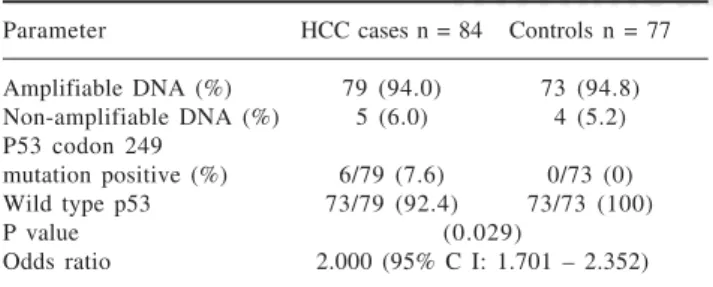
plasma samples analyzed in both groups, exon 7 of thep53 gene could not be amplified in 9 (5.6%) samples; 5 (6.0%) samples in the HCC cases and 4 (5.2%) samples in the control group. More details are shown in table I.
Codon 249-mutation positive samples
Among those with amplifiable plasma DNA (i.e.79 HCC cases and 73 controls), 6 HCC subjects had codon 249 mutation (i.e. G → T transversion) in the p53 gene while none of the subjects in the control group had this mutation. The prevalence of p53 codon 249 mutation among HCC subjects was 7.6% while it was 0% among controls and the difference in prevalence between the two groups was statistically significant (p = 0.029). The presence of this mutation was significantly associated with HCC. Those with the p53 codon 249 mutation were twice at risk for having HCC than those who did not have this mutation (odds ratio: 2.000; 95% confidence inter-val: 1.701–2.352). Direct DNA sequencing of the puri-fied nested PCR product of the codon 249 mutated sam-ples did not show any other mutation in this gene. The electrophoretic pattern of enzyme digestion of PCR prod-uct seen on agarose gel is shown in figure 1.
The characteristics of hepatocellular carcinoma patients based on p53 codon 249 status
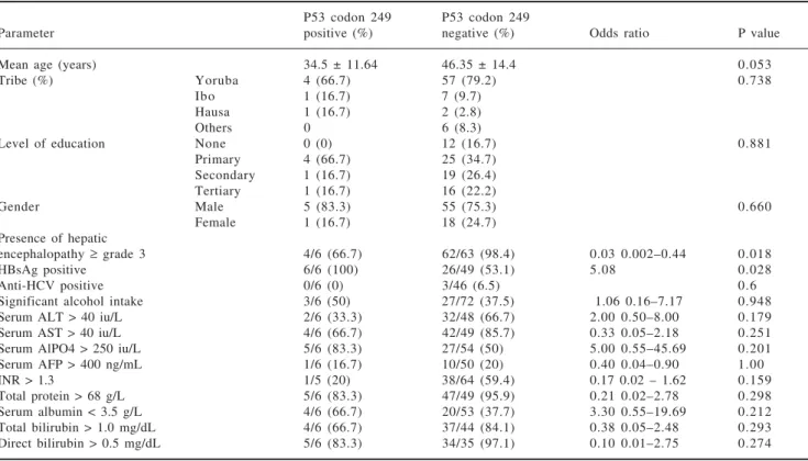
In an attempt to further characterize the HCC patients who had p53 codon 249 mutation, comparison was made between this group of subjects and the rest of the HCC patients who were negative for this mutation based on some clinical and biochemical parameters. The patients with this genetic mutation had a mean age of 34.5 ± 11.64 years and seemed to be younger than the HCC pa-tients who were negative for this mutation (p = 0. 053). Five of the 6 HCC patients who had the p53 codon 249 mutation were males while 1 was female. The gender dis-tribution between this group and the rest of the HCC pa-tients was similar (p = 0.660). Both groups of papa-tients were similar in tribal distribution and the level of educa-tion attained (p = 0.738 and 0.881 respectively). All the six subjects who had p53 codon 249 mutation were posi-tive for hepatitis B surface antigen (HBsAg) and this pro-portion of HCC subjects was significantly higher than
the rest of the HCC subjects who were HBsAg-positive but did not have this mutation (p = 0.035). It was not pos-sible to evaluate the risk associated with the develop-ment of this mutation in HBsAg-positive patients for lo-gistic reasons. The codon 249 mutation-positive patients who drank significant amount of alcohol and those who were anti-HCV positive were similar to the rest of the HCC patients (p = 0.948 and 0.6 respectively). None of these two factors was associated with the development of this genetic mutation. Though, a high proportion of the codon 249 mutation-positive HCC patients had elevated liver enzymes and reversal of the albumin: globulin ratio, they were similar to the rest of the HCC patients who were negative for this mutation. More HCC patients with codon 249 mutation had hepatic encephalopathy ≥ grade 3 on presentation than the rest of the HCC patients (p = 0.018) (Table II).
Discussion
The prevalence of the p53 codon 249 mutation is sig-nificantly higher among hepatocellular carcinoma pa-tients and its presence is associated with a two fold risk for HCC among this group of patients in South-western Nigeria. The observed prevalence in this study is howev-er, lower than those reported earlier in some countries in South-east Asia and sub-Sahara Africa9,12-15 though, con-sistent with a previous observation among HCC subjects from the same region.16
Table I. Prevalence of p53 codon 249 mutation.
Parameter HCC cases n = 84 Controls n = 77
Amplifiable DNA (%) 79 (94.0) 73 (94.8) Non-amplifiable DNA (%) 5 (6.0) 4 (5.2) P53 codon 249
mutation positive (%) 6/79 (7.6) 0/73 (0) Wild type p53 73/79 (92.4) 73/73 (100)
P value (0.029)
Odds ratio 2.000 (95% C I: 1.701 – 2.352)
Legend:
Number 1 = DNA molecular weight ladder
Number 2 = Negative control containing PCR water with no DNA Number 3 = wild-type p53 control
Number 4 = Control sample of positive p53 codon 249 mutation Number 5 = Patient’s sample positive for p53 codon 249 mutation
Figure 1. Electrophoretic pattern of enzyme digestion of PCR product seen on agarose gel.
www.medigraphic.com
The reason for this relatively lower prevalence ratecompared with previous studies is not likely to be due to the age and gender characteristics of the subjects recruit-ed into this study which is similar to those observrecruit-ed in these studies.9,12-15 The sample size in the index study is not small compared with the number of subjects recruited into those studies and is therefore unlikely to be respon-sible for the prevalence rate observed in this study. The prevalence of this genetic mutation in the index study may not be related to the fact that genomic analysis was done on plasma DNA. Other studies where the prevalence of this genetic mutation has been determined using plas-ma DNA have reported higher values than that observed in this study.3,10,11,15 Moreso, previous studies on p53 codon 249 mutation have shown a concordance rate of 88–90% between paired tumour and plasma samples.10,11 A more sensitive technique than that utilized in this study such as short oligonucleotide mass analysis (SOMA) may have detected a higher rate than what is currently observed in this study. Such observation was made by Lleonart et al.18 This however, is unlikely to ex-plain the dissimilarity in prevalence rates between the in-dex study and those earlier studies3,11,15 as the observed prevalence rates were obtained by similar technique as that used in this study.
The high prevalence of this genetic mutation among HCC patients which has been implicated as a molecular pathogenetic mechanism for HCC in countries in the above-mentioned regions has also been associated with consumption of food heavily contaminated by
Aflatox-in B1 as well as HBV infection endemicity.19,20 The source of aflatoxin is food items which are contaminat-ed by aflatoxin. The relatively lower prevalence value obtained in this study is similar to the prevalence rate of toxic levels of serum aflatoxin of 8.2 and 9% of rural and urban dwellers respectively in this same region (South-western Nigeria).21 It is however, incompatible with the degree of food contamination by aflatoxin re-ported earlier in South-western Nigeria where most of the food items such as yam, cassava and other processed food derived from these tuber crops as well as palm-oil and smoked-fish which are the staple food consumed have been reported to be heavily contaminated by afla-toxin.22 The food items which are most likely to be con-taminated are grains and other items which are pro-cessed by drying and stored under poor conditions which favour the growth of Aspergillus flavus, the fun-gus which produces aflatoxin.
Groundnut has also been reported to be the most heavily contaminated food item by aflatoxin.22,23 Some authors have associated the amount of groundnut ingest-ed daily with significant levels of serum aflatoxin and the presence of this genetic mutation.24 A higher preva-lence rate of this genetic mutation may have been ob-served if this study was done in Northern Nigeria where grains and groundnut are the staple food. One limitation in this study is insufficient data to quantify the daily consumption of groundnuts by the subjects. Based on the tribal distribution of the subjects, those from Northern Nigeria in this study who had this genetic abnormality
Table II. Characteristics of hepatocellular carcinoma patients based on p53 codon 249 status.
P53 codon 249 P53 codon 249
Parameter positive (%) negative (%) Odds ratio P value
Mean age (years) 34.5 ± 11.64 46.35 ± 14.4 0.053
Tribe (%) Yoruba 4 (66.7) 57 (79.2) 0.738
Ibo 1 (16.7) 7 (9.7)
Hausa 1 (16.7) 2 (2.8)
Others 0 6 (8.3)
Level of education None 0 (0) 12 (16.7) 0.881
Primary 4 (66.7) 25 (34.7) Secondary 1 (16.7) 19 (26.4) Tertiary 1 (16.7) 16 (22.2)
Gender Male 5 (83.3) 55 (75.3) 0.660
Female 1 (16.7) 18 (24.7) Presence of hepatic
encephalopathy ≥ grade 3 4/6 (66.7) 62/63 (98.4) 0.03 0.002–0.44 0.018
HBsAg positive 6/6 (100) 26/49 (53.1) 5.08 0.028
Anti-HCV positive 0/6 (0) 3/46 (6.5) 0.6
R Igetei et al. Detection of p53 CODON 249 mutation in Nigerian patients
www.medigraphic.com
ESTE DOCUMENTO ES ELABORADO PORMEDI-GRAPHIC
were similar to those of the same region who had wild type p53 gene. Though the inference from this study may not be reliable as a result of the few subjects who were positive for this mutation, the role of tribal origin and di-etary staple in the acquisition of this genetic mutation need to be further evaluated.
Current evidence suggests that following exposure to aflatoxin, the susceptibility to having this genetic muta-tion and subsequently, HCC is determined by the detoxi-fying effect of the enzymes responsible for its metabo-lism; epoxide hydrolase (EPHX) and gluthathione S-transferase M1 (GSTM1) and the efficacy of the DNA repairing process.25 Mutations in the genes coding for these enzymes have been found to be overrepresented in individuals with high serum levels of AFB1-albumin ad-ducts and HCC subjects.26,27 The EPHX and GSTM1 gen-otypes of the HCC subjects in this environment are not known. The presence or absence of mutations in the genes coding for these enzymes and its contribution to the low prevalence of p53 codon 249 in this region also require further evaluation.
All the HCC patients with p53 codon 249 mutation were HBsAg positive and this was significantly higher than the HCC patients who were negative for this genetic mutation. Significant association between HBsAg posi-tivity and this genetic mutation could not be evaluated statistically for logistic reasons. Previous studies have demonstrated a combined effect of these factors on HCC, where a multiplicative effect of both factors was ob-served.24 The infection by HCV does not seem to play a role in the development of the p53 codon 249 mutation as none of those who had this mutation was positive for anti-HCV. No study has so far reported an association be-tween this genetic mutation and HCV infection.15,28 There was also no association between significant alcohol in-gestion and the p53 codon 249 mutation. This observa-tion is consistent with that in earlier studies where low prevalence of this genetic mutation have been reported in places where significant alcohol ingestion is highly prevalent among HCC patients.15,28
This is the first report of p53 mutation using cell-free DNA in Nigeria. The observed prevalence rate of this ge-netic mutation among HCC subjects in this study is how-ever, quite similar to that from analysis of 18 tumour samples obtained by liver biopsy, where the same tech-nique of RFLP was employed.16 This study was done in South-western Nigeria, the same geographical location where UCH is also located. The similarity in findings from both studies (i.e. prevalence rates of 5.516 and 7.6% in this study) are not unexpected, as both sample popula-tions were derived from similar ethnic backgrounds and share a common staple diet rich in tuber.
From the observed prevalence rate of the p53 codon 249 mutation in this study, it seems unlikely to be the major molecular hepatocarcinogenetic pathway for HCC in South-western Nigeria.
Despite the low observed prevalence rate of this ge-netic mutation, it could still be useful as early molecular screening diagnostic tool for HCC because of its signifi-cant association with this disease. Moreso, this irrevers-ible genetic mutation has been observed in HCC patients 1 year prior to histological diagnosis.10,24 The use of sero-logical studies such as the antibody to the p53 protein may not be very useful as similar proportions of HCC pa-tients and controls at the UCH, Ibadan have been found to have detectable titre levels of this antibody. It is not surprising because of a possible high rate of false posi-tive antibody reaction. This may be due to the high ex-posure rate to frequent infections such as malaria which is holoendemic and other parasitic infections with subse-quent high circulating antibody titre. A similar explana-tion has been given for the high rate of false-positive anti-HCV status among sickle cell disease patients by Mutimer et al.30 Otegbayo et al reported that though re-duced transferrin and increased alpha 2 macroglobulin in HBV carriers might suggest active liver disease, these two in addition to haptoglobulin lack predictive value for the development of HCC in HBV carriers.31
Since circulating cell-free DNA could be found in the plasma of a large number of HCC patients and controls as observed in this study, genomic analysis using cell-free DNA could be adopted as a convenient method of screening and early diagnosis for HCC. In view of this, more molecular studies on HCC are therefore suggested to identify the major hepatocarcinogenetic pathway which could be a useful biomarker for screening, surveil-lance and early diagnosis. This would not only improve the out-come of the disease which is poor currently but could also serve as a molecular target for therapy.
Little information was obtained from further charac-terization of the HCC patients with this genetic abnor-mality. This may be as a result of the small number of pa-tients who had this mutation. Such information would have been desirable to establish the role of this genetic mutation in the clinical presentation of this group of pa-tients and the prognosis of the disease. A higher propor-tion of HCC patients with the p53 codon 249 mutapropor-tion had elevated liver enzymes and a higher grade of hepatic encephalopathy than the rest of the HCC patients where-as Kirk et al observed no difference in the range of liver enzymes in patients with this genetic mutation in Gam-bia.24 It is therefore suggested that a study involving a larger study population be done to further evaluate the role of this genetic abnormality in HCC in South-western Nigeria.
Acknowledgement
www.medigraphic.com
of the Molecular Carcinogenesis Unit, InternationalAgency for Research on Cancer (IARC), are acknowl-edged for their assistance at various stages of this study. Professor D.O Olaleye and Dr GN Odaibo are appreciated for providing storage facility for the plasma samples. The medical and nursing staff of the Liver Unit in UCH are also appreciated.
References
1. Waterhouse JAH, Muir CS, Correa P, Powell J. Cancer incidence in five continents. IV. IARC Scientific Publication 1982: 42. 2. Bosch FX, Ribes J, Boras J. Epidemiology of primary liver
can-cer. Semin Liver Disease 1999; 19: 271–285.
3. Huang ZH, Sun LH, Lu DD, Sun Y, Ma LJ, et al. Codon 249 mutation in exon 7 of p53 gene in plasma DNA: maybe a new early diagnostic marker of hepatocellular carcinoma in Qidong risk area, China. World J Gastroenterol 2003; 4: 692–695 http:/ /www.cancerprev.org/Journal/Issues/18/2/13 2002
4. Olubuyide IO, Ayoola EA, Atoba M. Hepatobiliary disease in tropical Africa–the Ibadan experience. Trop Gastroenterology
1986; 7: 54-60.
5. Atoba MA, Olubuyide IO, Aghadiuno PO. Gastrointestinal ma-lignancies in a tropical African population. Trop Doctor 1989; 19: 135–137.
6. Otegbayo JA, Oluwasola OA, Akere A, Ogunbiyi JO. Temporal and biological trends in liver cancers at a University hospital in Southwest Nigeria. Trop Doctor 2006; 36: 28-30.
7. Bruix J, Sherman M, Llovet JM, Beaugrand M, Lencioni R, et al. Clinical management of hepatocellular carcinoma. Conclusions of the Barcelona-2000 EASL Conference. J Hepatol 2001; 35: 421-430. 8. Llovet JM, Beaugrand M. Hepatocellular carcinoma: present
sta-tus and future prospects. J Hepatol 2003; 38: 136–149. 9. Debuire B, Lemoine A. Liver: Hepatocellular carcinoma. Atlas Genet
and Cytogenet Oncol Haematol 2001. http://www.Infobiogen.fr/ser-vices/chromancer/Tumours/HepatocarcinID5039.htm.
10. Jackson PE, Kuang SY, Wang JB, Strickland PT, Muñoz A, et al. Prospective detection of codon 249 mutations in plasma of hepa-tocellular carcinoma patients. Carcinogenesis 2003; 24: 1657-1663. 11. Szymanska K, Lesi OA, Kirk GD, Sam O, Taniere P, et al. Ser-TP53 mutation in tumour and plasma DNA of hepatocellular carcinoma patients from a high incidence area in the Gambia, West Africa. Int J Cancer 2004; 110: 374-379.
12. Wong N, Lai P, Pang E, Fung L, Sheng Z, et al. Genomic aberra-tions in human hepatocellular carcinomas of differing etiologies.
Clin Cancer Res 2000; 6: 4000–4009.
13. Ozturk M. p53 mutation in hepatocellular carcinoma after afla-toxin exposure. Lancet 1991; 338: 1356–1359.
14. Coursaget P, Depril N, Chabaud M, Nandi R, Mayelo V, et al. High prevalence of mutations at codon 249 of the p53 gene in hepatocellular carcinomas from Senegal. Br J Cancer 1993; 67: 1395–1397.
15. Kirk GD, Camus-Randon AM, Mendy M, Goedert JJ, Merle P, et al. Ser-249 p53 mutations in plasma DNA of patients with hepa-tocellular carcinoma from the Gambia. Journal of the National Cancer Institute 2000; 92: 148–153.
16. Ndububa DA, Yakicier CM, Ojo OS, Adeodu OO, Rotimi O, et al. p53 codon 249 mutation in hepatocellular carcinomas from Nigeria. Afr J Med Med Sci 2001; 30: 125-127.
17. Anker P, Mulcahy H, Chen XQ, Stroun M. Detection of circulat-ing tumour DNA in the blood (plasma/serum) of cancer patients.
Cancer Metastasis Rev 1999; 18: 65–73.
18. Lleonart ME, Kirk GD, Vilar S, Lesi OA, Dasgupta A, et al. Quantitative analysis of plasma TP53 249ser-Mutated DNA by
electrospray ionization mass spectrometry. Cancer Epidemiol Biomarkers Prev 2005; 14: 2956-2962.
19. International Agency For Research on Cancer (IARC). Hapatitis viruses. Lyon (France); IARC Monogr Eval Carcinog Risks Hum, 59: 1994
20. International Agency For Research on Cancer (IARC). Some naturally occurring substances: Food items and constituents, heterocyclic aromatic amines and mycotoxins. Lyon (France); IARC Monogr Eval Carcinog Risks Hum 1993; 56: 245-395. 21. Olubuyide IO, Maxwell SM, Akinyinka OO, Hart CA, Neal GE,
et al. HBsAg and aflatoxins in sera of rural (Igbo-Ora) and urban (Ibadan) populations in Nigeria. Afr J Med Med Sci 1993; 22: 77-80.
22. Nwokolo C, Okonkwo I. Aflatoxin load of common food in savanna and forest regions of Nigeria. Transactions of the Royal Society of Trop Med and Hyg 1978; 72: 329–332.
23. Atawodi SE, Atiku AA, Lamorde AG. Aflatoixn contamination of Nigerian foods and feeding stuffs. Fd Chem Toxic 1993; 32: 61–63. 24. Kirk GD, Lesi OA, Mendy M, Szymañska K, Whittle H, et al. 249ser
TP53 mutation in plasma DNA, hepatitis B viral infection, and risk of hepatocellular carcinoma. Oncogene 2005; 24: 5858-5867. 25. Wang G, Chang HH, Yan Z, Ling C, Ying W, et al. Genetic
aber-ration in primary hepatocellular carcinoma: correlation between p53 gene mutation and loss-of-heterozygosity on chromosome 16q21-q23 and 9p21-p23. Cell Research 2000; 10: 311–323. 26. Rashid A, Wang JS, Quian GS, Lu BX, Hamilton SR, et al.
Ge-netic alterations in hepatocellular carcinomas: association between loss of chromosome 4q and p53 gene mutations. Br J Cancer
1999; 80: 59-66.
27. McGlynn KA, Rosevold EA, Lustbader ED, Hu Y, Clapper ML, et al. Susceptibility to hepatocellular carcinoma is associated with genetic variation in the enzymatic detoxification of aflatoxin B1.
Proc Natl Acad Sci USA 1995; 92: 2384-2387.
28. Stern MC, Umbach DM, Yu CM, London SJ, Zhang ZQ, et al. Hepatitis B, Aflatoxin B1, and p53 Codon 249 Mutation in Hepa-tocellular Carcinomas from Guangxi, People’s Republic of China, and a Meta-analysis of Existing Studies. Cancer Epidemiology Biomarkers & Prevention 2001; 10: 617 – 625
29. Akere A, Otegbayo JA. Evaluation of the pattern and prognostic implications of anti-p53 in hepatocellular carcinoma. Singapore Med J 2007; 48: 41-44.
30. Imer DJ, Olomu A, Skidmore S, Olomu N, Rarcliffe D, et al. Viral hepatitis in Nigeria % sickle-cell disease and commercial blood donors. Quarterly Journal of Medicine 1994; 87: 407-411. 31. Otegbayo JA, Arinola OG, Aje OA, Oluwasola OA, Okiwelu OH,
et al. Usefulness of acute phase proteins for monitoring develop-ment of hepatocellular carcinoma in hepatitis B virus carriers.
West African J Med 2005; 24: 124–127.
32. Sherman M. Alphafetoprotein: an obituary. J Hepatol 2001; 34: 603–605.
33. Gebo KA, Chander G, Jenckes MW, Ghanem KG, Herlong HF, et al. Screening tests for hepatocellular carcinoma in patients with chronic hepatitis C: A systemic review. Hepatology 2002; 36: S84–S92.