Percutaneous liver biopsy – 2 decades of
experience in a public hospital in the South of Brazil
Rafael Bergesch D’Incao,* Marcelo Campos Appel da Silva,*Paulo Roberto Lerias de Almeida,** Viviane Plasse Renon,* Cristiane Valle Tovo***
* Former Medical Resident of the Service of Gastroenterology, Hospital Nossa Senhora da Conceição (HNSC), Porto Alegre, Rio Grande do Sul, Brazil. ** Doctor in Hepatology. Gastroenterologist. Preceptor of the Internal Medicine Service - HNSC, Porto Alegre, Rio Grande do Sul, Brazil. *** Doctor in Hepatology. Gastroenterologist. Preceptor of the Gastroenterology Service - HNSC, Porto Alegre, Rio Grande do Sul, Brazil;
Adjunct Professor of Gastroenterology of the Universidade Federal de Ciências da Saúde de Porto Alegre (UFCSPA).
ABSTRACT
Introduction. Liver biopsy is a complementary method for diagnosis, staging and therapeutic guidance in li-ver diseases, where chronic viral hepatitis are the most acknowledged causes for the indication of histopa-thological study. The objective is to assess the patients’ profile as well as the indication and results of percutaneous liver biopsies in a tertiary hospital. Material and methods. A descriptive, cross-section stu-dy was carried out through the review of medical charts (retrospective cohort) of patients submitted to blind percutaneous liver biopsies (PLB) at a hospital in Porto Alegre, South Brazil, from October 1993 to December 2011. Results. 1,955 PLB were carried out, the mean patients’ age was 44.8 years old, and 1,127 (57.65%) were men. Chronic hepatitis C was the main indication (60.5%), followed by HCV-HIV coinfection (12.2%) and chronic hepatitis B (3.5%). Seven cases (0.3%) had complications, without deaths. Conclusion. PLB is a safe method and continues to be an important option to assist patients with chronic liver disease. Key words. Liver biopsy. Chronic hepatitis. Hepatitis C.
Correspondence and reprint request: Cristiane Valle Tovo Rua Cel Aurelio Bitencourt 115 apto 201
Ph.: 55-51-91893113. Fax: 55-51-3214 8158 E-mail: [email protected]
Manuscript received: March 07, 2013. Manuscript accepted: July 08, 2013.
INTRODUCTION
The histopathological liver exam, through liver biopsy, has become a complementary method not only for diagnosis but also for staging, treatment control and/or prognostic determination of liver diseases.1
It is known that patients diagnosed with chronic viral hepatitis by the B (HBV) or C virus (HCV) are the highest contingent in population samples sub-mitted to liver biopsies.2-4 Besides them, patients presenting atypical course of liver diseases; changes in liver panel and/or in abdominal imaging without established diagnosis; suspected infiltrative, infectious or granulomatous diseases, can have their diagnosis established through a histopathological liver exam.1,5,6
In Brazil, treatment for hepatitis B and C is pro-vided by the Public Health System – Brazilian Mi-nistry of Health, and many cases depend on the fibrosis staging through liver biopsy to determine the indication for therapy.7,8
This study aimed to assess the indications, histo-pathological results and percutaneous liver biopsy complications carried out in a tertiary public hospi-tal in Porto Alegre, Rio Grande do Sul – South Brazil, and whether there was a change in the profile of the patients submitted to the procedure over the last two decades.
MATERIAL AND METHODS
A descriptive cross-section study was carried out by reviewing medical charts (retrospective cohort) of patients blindly submitted to blind percutaneous liver biopsy (PLB) in a single center from Hospital Nossa Senhora da Conceição (HNSC), Brazil - a terciary attendance Hospital and reference for liver disease, from October 1993 to December 2011.
The blind PLB procedures were carried out by the same team during the aforementioned study period, with training of Medical Residents. The patients should present platelet count higher than 80,000/ mm³ and prothrombin time not higher than 3 se-conds above control.9 Subjects were guided about the need to stop using drugs that potentially cause hemorrhagic diathesis 10 days before the procedure, in accordance to the guidelines of the American As-sociation for Study of the Liver (AASLD).1 All PLB were carried out under local anesthesia with 2% li-docaine and 16G or 18G Menghini needle. After the procedures, the patients remained under observation for at least 2 h before discharging.
In the viral hepatitis assessment, the fibrosis stag-ing was performed accordstag-ing to Metavir score.10
This study was approved by the Research Ethics Committee of HNSC and is in accordance with the governing rules of the National Health Council/Mi-nistry of Health and its rules that regulate research involving human beings.
The obtained results were stored in confidential database, shared only by the researchers involved in the study. The data were analyzed using the Statis-tical Package for the Social Sciences (SPSS) for Windows®, version 15. Values were considered sta-tistically significant when p < 0.05.
RESULTS
From 1993 to 2011, 1,955 blind PLB were carried out. For the comparative assessment, two groups of patients were established, according to the period in which the PLB was performed. One group included the patients who had PLB between the years of 1993 and 2002 (1,037 patients) and the other the ones who had PLB between 2003 and 2011 (918 patients) (p > 0.05).
The mean age was 44.8 years old, and 1,127 (57.6%) patients were male. No statistically difference was found when the mean ages in both distinct periods were assessed, but male prevailed over female gender in the first assessed period (Table 1).
Regarding the main indications for PLB, infection by HCV was the most prevalent, accounting for a total of 1,184 (60.5%) cases. Patients with HCV-HIV co-infection represented 12.2% of the cases (238 patients), followed by HBV infection (69 patients -3.5%). Other indications totalized 12.2% of the cases (238 patients).
Statistically significant difference was observed when the indications for PLB in both distinct pe-riods were compared (p < 0.001), with the exception of HBV infection, as presented in figure 1. There
Table 1. Demographic data.
1993-2002 (n = 1,037) 2003-2011 (n = 918) p
Mean age; years (variation) 41.6 (14-89) 48.06 (19-83) > 0.05
Men; n (%) 656 (63.3) 471 (51.3) < 0.001
n: number of patients. m: mean.
Figure 1. Indications for percutaneous liver biopsy. AIH: autoimmune hepatitis. *p < 0.01.
1993-2002 2003-2011 70
60 50 40 30 20 10 0
HCV HBV HIV HCV-HIV AIH Others
Percentage
55.4* 66.4*
3.3 3.8
10.8*
1.4*
7.5*
17.4*
0.5* 1.4*
22.5
9.6*
100 90 80 70 60 50 40 30 20 10 0
Figure 2. Liver fibrosis staging (METAVIR) among HCV ± HIV patients (1993-2011). p = 0.422.
HCV (n = 1,184) HCV + HIV (n = 238)
Inappropiate F3-F4 F0-F2 15.0% 12.0%
73.0% 19.6%
9.9%
were more indications for HCV and HIV/HCV in the last decade.
Liver fibrosis graduation was compared among patients monoinfected by HCV and those with HCV-HIV coinfection. It was observed that, in both groups, a higher proportion of patients had mild-mo-derate fibrosis (F0-F2) than advanced fibrosis (F3-F4), although without statistically significant difference (Figure 2).
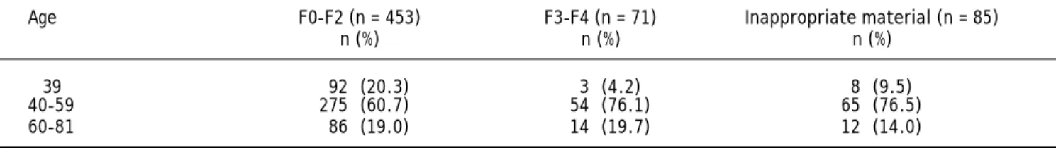
There was an association between age and fibro-sis in the HCV monoinfected group, observing that younger patients (≤ 39-years old) had milder degree of fibrosis than older than 40-years old (Table 2).
Liver steatosis was observed in 36.3% of HCV monoinfected patients and in 31.2% of HCV-HIV co-infected ones, without statistically significant diffe-rence. On the other hand, coinfected patients presented higher intra-hepatic iron rates than mo-noinfected ones (20.0 x 8.0%) (Table 3).
The material was considered inappropriate for analy-sis in 205 cases (10.48% of the sample). The average size of the assessed fragments was 10.51 ± 6.8 mm.
Major complications (other than pain) related to the procedure were observed in 07 (0.3%) cases, with no cases of death: 05 punctures where bile was aspi-red, 01 pneumothorax and 01 hemoperitoneum, which required surgery.
DISCUSSION
Despite the recent development of non-invasive methods to asses liver fibrosis, liver biopsy remains as an important tool to evaluate liver diseases, re-garding the diagnosis, staging, prognosis and mana-gement of patients with liver disorders.1,11,12
Considering the risk of adverse events and the de-velopment of non invasive methods for evaluation of liver fibrosis, the liver biopsy should only be indica-ted in cases in which the definition of the histopa-thology would modify the therapeutic/prognostic conduct.1,11
In the present study all the procedures were per-formed without ultrassonographic (US) guidance. Some authors suggest that procedures using US would be safer than blind PLB,13 but the complica-tion rate observed in the present casuistry was low even with blind PLB.
There was a predominance of men in the first as-sessed decade (63.3%), as it was also observed in other national and international studies;2,14 howe-ver, there was a reduction in the percentage of the second assessed decade (51.3%) (p < 0.001).
The mean age of the patients submitted to PLB was similar in both decades. Such findings are simi-lar to the results of other Brazilian studies.2,12
Chronic hepatitis C was the main indication for liver biopsy in the present study, representing 55% of the cases between 1993 and 2002, and 66% of the cases between 2003 and 2011, reflecting the national reality.2,12
The second most prevalent indication for PLB was HIV infection, including coinfected patients, with 18.3% between 1993 and 2002 and 18.8% between 2003 and 2011. HIV monoinfection was more frequent in the first decade (10.8% x 1.4%), while HCV-HIV coinfection was more prevalent in the second decade (7.5% x 17.4%), as presented in figure 1. The current expected tendency is that the number of coinfected patients will decrease in the PLB pool, once the procedure is no longer
Table 3. Assessment of liver steatosis and intra-hepatic iron in HCV ± HIV patients (2003-2011).
HCV (n = 609) HCV+HIV (n = 160) p
n (%) n (%)
Liver steatosis 221 (36.3) 50 (31.2) 0.235
Intra-hepatic iron 49 (8.0) 32 (20.0) < 0.001
Table 2. Age x fibrosis in patients with HCV (2003-2011).
Age F0-F2 (n = 453) F3-F4 (n = 71) Inappropriate material (n = 85)
n (%) n (%) n (%)
≤ 39 92 (20.3) 3 (4.2) 8 (9.5)
40-59 275 (60.7) 54 (76.1) 65 (76.5)
60-81 86 (19.0) 14 (19.7) 12 (14.0)
required for the treatment of this subpopulation.8 However, PLB still has an important role in the diagnosis of other as-sociated conditions, such as steatosis or toxicity by antiretroviral drugs.15,16
In a French study presented in the 2012 AASLD liver meeting,13 it was observed a marked reduction in the total number of liver biopsy in the last period studied, as well as for chronic viral hepatitis C.
When the fibrosis degree was assessed in such subgroup of patients, there was no difference when compared to monoinfected HCV (Figure 2). There were higher prevalence of patients with lower de-grees of fibrosis (F0-F2), when both HCV monoin-fected and HCV-HIV coinmonoin-fected patients were analyzed (Figure 2). Such findings can be explained by the fact that patients with clinical diagnosis of cirrhosis can be treated without the need for PLB, in accordance with the Brazilian Ministry of Health.8
The natural history of HCV chronic hepatitis and the liver fibrosis progression in patients carrying such disease has been studied, aiming at identifying factors related to the evolution to higher fibrosis de-grees (age over 40 years old in the infection, daily alcohol consumption and male gender).22 In the pre-sent study, the age group and degrees of liver fibro-sis were compared (Table 2), and a significant difference between patients over 40 years of age for higher fibrosis was observed, corroborating the lite-rature.17-19 It was not possible to assess whether such finding were due to the longer infection time or higher age at the time of the infection.
Liver steatosis is a histological finding commonly observed in patients with chronic hepatitis C infec-tion, with estimated prevalence of 30-70%,15,20,21 where some studies show genotype 3 as the most re-lated to non-alcoholic fatty liver disease (NAFLD). Such finding is frequently followed by insulin resis-tance, alterations in the glucose and lipids metabo-lism.15,20,22
The incidence of liver steatosis among HCV-HIV coinfected patients is estimated between 25-75%, and is usually attributed to the use of antiretroviral drugs.23 In the present study, the prevalence of liver steatosis corroborates the findings of previous stu-dies,15,20,21,23,24 both HCV monoinfected and HCV-HIV coinfected patients, without statistically significant difference between the groups.
Patients with chronic hepatitis C usually present greater amounts of iron storage in the liver paren-chyma, related to faster progression of liver fibrosis and development of hepatocellular carcinoma.25-27 This accumulation is attributed to several factors, such as the reabsorption of iron coming from
hepa-tocytes necrosis, hemochromatosis-related mutations, decreased expression of hepcidin or changes in the iron liver metabolism.28 In the present casuistry, iron accumulation was observed in 8% of the monoinfected patients, which is in accordance with the literature.2,3 Moreover it was observed that 20% of the HCV-HIV coinfected patients presented iron accumulation. These metabolic disturbances can also be related to insulin resistance, progression of HIV disease, and the type of antiretroviral therapy prescribed, increasing the risk of cardiovascular disease and mortality.12
The PLB is a safe procedure, specially in expe-rienced hands. Patients with malignant liver disease and/or advanced liver insufficiency present higher risk, and may have a mortality rate up to 19% in the first three months after biopsy.29 Overall, mortality directly related to the procedure is a rare event, va-rying from 0.01 and 0.1%. The main cause of death after liver biopsy is intraperitoneal hemorrhage, with 0.03 to 0.07% incidence.11,29 In the present stu-dy there were no deaths and the complications rate was low (0.3%).
In conclusion, percutaneous liver biopsy remains an important and widely used diagnostic tool in the survey of patients with liver disease, although the non-invasive methods establish a new paradigm in the diagnostic of liver diseases. The modification of the studied population over the last two decades reflects the real life, showing a greater access of the HCV and coinfected HCV/HIV patients to the public health system.
REFERENCES
1 . Rockey DC, Caldwell SH, Goodman ZD, Nelson RC, Smith AD. AASLD Position Paper. Liver biopsy. Hepatology
2009; 49: 1017-44.
2 . Lembrança L, Medina J, Portugal M, Gadelha R, de Freitas LA, Paraná R. Epidemiological assesment of li-ver disease in Northeastern Brazil by means of a standardized liver biopsy protocol. Ann Hepatol
2011; 10: 43-9.
3 . D’Souza RFC, Feakins R, Mears L, Sabin CA, Foster GR. Relationship between serum ferritin, hepatic iron staining, diabetes mellitus and fibrosis progression in patients with chronic hepatitis C. Aliment Pharmacol Ther 2005; 21: 519-24.
4 . Malik AH, Kumar KS, Malet PF, Jain R, Prasad P, Osta-powicz G.Correlation of percutaneous liver biopsy fragmentation and degree of liver fibrosis. Aliment Pharmacol Therap 2004; 19:545-9.
6 . Farrel RJ, Smiddy PF, Pilkington RM, Tobin AA, Mooney EE, Temperley IJ, McDonald GS, et al. Guided versus blind liver biopsy for chronic hepatitis C: clinical be-nefits and costs. J Hepatol 1999; 30: 580-7.
7 . Brasil. Ministério da Saúde. Secretaria de Vigilância em Saúde. Departamento de DST, Aids e Hepatites Vi-rais. Protocolo Clínico e Diretrizes Terapêuticas para Hepatite Viral B e Coinfecções. Brasília, Ministério da Saúde, 2010. 132 p. Série A. Normas e Manuais Técni-cos. In: http://portal.saude.gov.br/portal/arquivos/ pdf/prot_clinico_diretrizes_terapeuticas_hep_b.pdf 8 . Brasil. Ministério da Saúde. Secretaria de Vigilância
em Saúde. Departamento de DST, Aids e Hepatites Vi-rais. Protocolo Clínico e Diretrizes Terapêuticas para Hepatite Viral C e Coinfecções. Brasília, Ministério da Saúde, 2011. 144 p. Série A. Normas e Manuais Técni-cos. In: http://portal.saude.gov.br/portal/arquivos/ pdf/prot_clinico_diretrizes_tera_hepatite_c.pdf 9 . Sherlock S. Biopsy of the liver. In: Sherlock S, Dooley J
(eds.). Diseases of the liver and biliary system. 10th ed. 1998, p. 33-41.
10. Bedossa P, Poynard T. METAVIR Cooperative Study Group. An algorithm for the grading of activity in chronic hepatitis C. Hepatology 1996; 24: 289-93. 11. Grant A, Neuberger J. Guidelines on the use of liver
biopsy in the clinical practice. Gut 1999; 45 (Suppl. IV): IV1–IV11.
12. Almeida PRL, Tovo CV, Oliveira JF, Santos GC, Santos AP, Souza AR. Biópsia Hepática percutânea: estudo de 1037 casos. GED. Gastroenterologia e Endoscopia
2004; 23: 221-6.
13. Cadranel JFD, Nousbaum JB, Hanslik B. Major trends in liver biopsy in France: results of a nationwide 2009 survey. AASLD abstracts. Hepatology 2012; 1096A. 14. Focaccia R, Baraldo DC, Ferraz ML, Martinelli AL,
Carrilho FJ, Gonçales FL Jr, Pedroso ML, et al. Demo-graphic and anthropometrical analysis and genotype distribuition of chronic hepatitis C Patients Treated in public and private reference centers in Brazil. Braz J Infect Dis 2004; 8: 348-55.
15. Enjoji M, Kohjima M, Kotoh K, Nakamuta M. Metabolic Disorders and Steatosis in Patients with Chronic He-patitis C:Metabolic Strategies for Antiviral Treat-ments. Int J Hepatol 2012; 2012: 264017.
16. Tovo CV, Mattos AA, Souza AR, Ferrari de Oliveira Rigo J, Lerias de Almeida PR, Galperim B, Riegel Santos B. Impact of human immunodeficiency vírus infection in patients infected with the hepatitis C virus. Liver International 2007; 27: 40-6.
17. Poynard T, Bedossa P, Opolon P. Natural history of li-ver fibrosis progression in patients with chronic he-patitis C. The OBSVIRC, METAVIR, CLINIVIR, and DOSVIRC groups. Lancet 1997; 349: 825-32.
18. Costa LB, Ferraz ML, Perez RM, Ferreira AS, Matos CA, Lanzoni VP, Silva AE. Effect of host-related fac-tors on the intensity of liver fibrosis in patients with chronic hepatitis C virus infection. Braz J Infect Dis
2002; 6: 219-24.
19. Wright M, Goldin R, Fabre A, Lloyd J, Thomas H, Trepo C, Pradat P, et al. Measurement and determinants of the natural history of liver fibrosis in hepatitis C vi-rus infection: a cross sectional and longitudinal stu-dy. Gut 2003; 52: 574-9.
2 0 . Patel A, Harrison SA. Hepatitis C virus infection and nonalcoholic steatohepatitis. Gastroentero-logy & HepatoGastroentero-logy 2012; 8: 305-12.
2 1 . Asselah T, Rubbia-Brandt L, Marcellin P, Negro F. Steatosis in Chronic Hepatitis C: Why does it really matter? Gut 2006; 55: 123-30.
2 2 . Bugianesi E, Salamone F, Negro F. The interaction of metabolic factors with HCV infection: does it matter? J Hepatol 2012; 56(S1): S56-S65.
2 3 . Price JC, Thio CL. Liver disease in the HIV-infec-ted individual. Clin Gastroenterol Hepatol 2010; 8: 1002-12.
2 4 . Crum-Cianflone N, Dilay A, Collins G, Asher D, Campin R, Medina S, Goodman Z, et al. Nonalcoho-lic fatty liver disease among HIV-infected per-sons. J Acquir Immune Defic Syndr 2009; 50: 4 6 4 - 7 3 .
2 5 . Guyader D, Thirouard AS, Erdtmann L, Rakba N, Jacquelinet S, Danielou H, Perrin M, et al. Liver iron is a surrogate marker of severe fibrosis in chronic hepatitis C. J Hepatol 2007; 46: 587-95. 2 6 . Girelli D, Pasino M, Goodnough JB, Nemeth E,
Gui-do M, Castagna A, Busti F, et al. Reduced serum hepcidin levels in patients with chronic hepatitis C. J Hepatol 2009; 51: 845-52.
2 7 . Guyader D, Thirouard AS, Erdtmann L, Rakba N, Jacquelinet S, Danielou H, Perrin M, et al. Liver iron is a surrogate marker of severe fibrosis in chronic hepatitis C. J Hepatol 2007; 6: 587-95. 28. Bonkovsky HL, Banner BF, Rothman AL. Iron and chronic
viral hepatitis. Hepatology 1997; 25: 759-68.