1
Influence of the Substitution Pattern (ortho vs para) on the Structure and
Luminescence of Silver(I) Complexes Ligated by Diphenylphosphinobenzoic Acids
Ana B. Miguel–Coello and Manuel Bardají*
IU CINQUIMA/Química Inorgánica, Facultad de Ciencias, Universidad de Valladolid, E–47071
5Valladolid, Spain.
* Author to whom correspondence should be addressed: M. B. e–mail:
[email protected]
ABSTRACT
The reaction of AgCF3SO3with diphenylphosphinobenzoic acids
leads to a cyclic dinuclear or a polynuclear silver(I) compound, 10
depending on the relative position of phosphine versus carboxylic functional group. The dimers [Ag2(OSO2CF3)2(µ–O,P–
PPh2C6H4COOH–o)2] 1 or [{Ag(µ–O,P–PPh2C6H4COOH–
o)(OH2)2}2](CF3SO3)2.2CH2Cl2 1a are 12–membered
diargentacycles with a silver–silver distance of 3.982 or 3.754 Å, 15
while the polymer [Agn(OSO2CF3)n(µ–O,P–PPh2C6H4COOH–
p)n] 2 is a 1D type. The ligand is always acting as non–chelating
bridge, being the silver center coordinated to the phosphine phosphorous, and one oxygen (carbonyl) of the carboxylic acid of next fragment. One oxygen of the triflate anion or two water 20
molecules complete the coordination sphere. Besides, p– (diphenylphosphino)benzoic acid compound is emissive in solution and in the solid state at 298 and 77 K, while compound with the ortho ligand is not. At 298 K, the emissions are centered at 471 nm in the solid state and at 416 nm in solution.
25
Keywords: 2–(diphenyphosphino)benzoic acid, 4– (diphenyphosphino)benzoic acid, silver complexes, bridging ligand, luminescence, X–ray crystal structure.
1. Introduction
30
A key strategy in the design and synthesis of metal–organic frameworks (MOF’s) and other hybrid organic–inorganic molecular materials is the utilization of simple multifunctional building blocks that induce the formation of complex molecular structures by self–assembly [1]. Silver (I) centers 35
are particularly versatile as components of such building blocks because of their flexible coordination number and geometry [2], and the tendency to form Ag···Ag weak interactions [3]. Although there are many reports on other luminescent d10–metal complexes, emissive AgI complexes
40
have been less studied because of their potential photosensitivity and limited luminescence [4]. In this respect, the potentially polydentate o– and p– (diphenylphosphino)benzoic acids are interesting ligands combining soft (P) with hard (O) donor atoms. Moreover, the 45
carboxylic group is suitable to produce hydrogen bonds. In
fact 2–diphenylphosphinobenzoic acid is a dimer with the typical H–bonds of carboxylic acids (Scheme 1) [5].
50
Scheme 1. H–bonds between two carboxylic groups.
The ortho or para relative position of the phosphine versus the carboxylic acid can be used to engineer different three– dimensional geometries. For instance, the homoleptic gold 55
complex of 2–(diphenylphosphino)benzoate is a mixture of a dimer and a trimer (a trimer is isolated as crystal), while the corresponding 4–(diphenylphosphino)benzoate complex is a polymer; in both cases, the ligands are working as O, P– bridges [6]. Homoleptic silver complex of 2– 60
(diphenylphosphino)benzoate is a hexanuclear compound with the ligand acting as tri– and tetradentate bridge [7]. Depending on the metal center, 2– (diphenylphosphino)benzoate ligand leads to dimers via carboxylate or O, P–bridges, or to mononuclear compounds 65
acting as O, P–chelate (Scheme 2) [8]. 4– (diphenylphosphino)benzoate evolves again to a dimer with carboxylate bridges, which can be additionally bonded through the phosphorus atom; besides, polynuclear derivatives can be obtained by O, P–bridges (Scheme 2) [9].
70
PPh2
O
O M
M Ph2P
O O
M
PPh2
O
O M
M
M' Ph2P
O O
M
M a
c
b
d
Scheme 2. a) Chelate for 2–(diphenylphosphino)benzoate; b) carboxylate
bridges; c) O, P–bridges; d) more complex coordination mode (M’ can be the same metal fragment).
75
The related chloro gold(I) derivatives of 2– or 4– (diphenylphosphino)benzoic acid are dimers, by a
Ph2P
O O
H PPh2
symmetrical double H–bond between the two carboxylic groups [6, 10]. Similarly, Cu(I) complexes become dimers by a typical double carboxylic H–bond [11]. However, chloro palladium(II) compounds with 4–(diphenylphosphinobenzoic) acid are mononuclear with the carboxylic group making H– 5
bonds with solvent crystallization molecules [12].
Here we report the synthesis and structural characterization of silver(I) compounds with 2– and 4– (diphenylphosphino)benzoic acids, where the substitution 10
pattern on the ligand leads to different nuclearities and luminescence properties on the complexes.
2. Experimental
15
For general procedures see Supplementary data.
2.1. Synthesis of [Ag(OSO2CF3)(PPh2C6H4COOH)]; ortho (1), para (2).
20
To a diethyl ether solution (20 mL) of AgCF3SO3 (51 mg, 0.2
mmol) was added the corresponding phosphinobenzoic acid (61 mg, 0.2 mmol), and the reaction stirred for 2 h protected from the light. The insoluble compounds were filtered off, washed and dried. A second fraction was obtained by 25
evaporation ca. 2 ml and addition of hexane. Compounds 1–2
were obtained as whitesolids. Yield of 1: 78 mg, 70 %. Anal. Calc. for C40H30Ag2F6O10P2S2: C, 42.65; H, 2.68; N, 0.
Found: C, 42.37; H, 2.75; N, 0%. 1H NMR (d
6–acetone): δ
8.37 (d, JHH = 7.6 Hz, 1H, H6), 7.76 (td, JHH = 7.6 Hz, JHP =
30
1.4 Hz, 1H, H5), 7.70 (td, JHH = 7.6 Hz, JHP = 1.4 Hz, 1H, H4),
7.59–7.45 (m, 10H, Ph), 7.04 (t, JHP = JHH = 7.8 Hz, 1H, H3). 1H NMR (–50oC, d
6–acetone): δ12.49 (brs, 1H, COOH), 8.40
(ddd, JHH = 7.5 and 1.5 Hz, JHP = 4.4 Hz, 1H, H6), 7.82 (t, JHH
= 7.5 Hz, 1H, H5), 7.77 (t, JHH = 7.5 Hz, 1H, H4), 7.60 – 7.43
35
(m, 10H, Ph), 6.94 (ddd, JHP = 9.2 and 1.2 Hz, JHH = 7.5 Hz,
1H, H3).19F NMR (d
6–acetone): –77.58 (s). 31P NMR (d6–
acetone): 15.6 (brs). 31P NMR (–50oC, d
6–acetone): 15.5 (d, 1J
107Ag–31P = 736 and 1J109Ag–31P = 849 Hz). IR (KBr): 3060 ν(O–H), 1673 ν(C=O), 1257, 1223, 1209, 635 (CF3SO3) cm–1.
40
Yield of 2: 91 mg, 81 %. Anal. Calc. for C20H15AgF3O5PS: C,
42.65; H, 2.68; N, 0. Found: C, 42.60; H, 2.84; N, 0%. 1H
NMR (d6–acetone): δ 11.5 (brs, 1H, COOH), 8.13 (d, JHH =
8.0 Hz, 2H, H2), 7.68–7.55 (m, 12H, H3 + Ph). 1H NMR (–
50oC, d
6–acetone): δ 12.54 (brs, 1H, COOH), 8.15 (dd, JHP
45
=1.6 Hz, JHH = 8.4 Hz, 2H, H2), 7.68–7.53 (m, 12H, H3 + Ph). 19F NMR (d
6–acetone): –77.6 (s). 31P NMR (d6–acetone): 14.5
(d, 1J
Ag–P = 721 Hz). 31P NMR (–50oC, d6–acetone): 13.8 (d, 1J
107Ag–31P= 690 and 1J109Ag–31P = 795 Hz). IR (KBr): 3054 ν(O–H), 1687 ν(C=O), 1223, 635 (CF3SO3) cm–1.
50
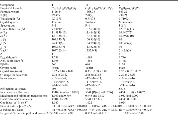
Table 1. Details of Crystal Data and Structure Refinement for Complexes 1, 1a and 2
Compound 1 1a 2
Empirical formula C40H30Ag2F6O10P2S2 C42H42Ag2Cl4F6O14P2S2 C20H15AgF3O5PS
Formula weigth 1126.44 1368.36 563.22
T (K) 298(2) 298(2) 298(2)
Wavelength (Å) 0.71073 0.71073 0.71073
Crystal system Triclinic Triclinic Monoclinic
Space group P–1 P–1 P 21/n
Unit cell dim.: a (Å) 7.8319(5) 10.5574(17) 12.8706(11)
b (Å) 11.8958(10) 11.4142(18) 10.4407(5)
c (Å) 12.1326(11) 13.1871(11) 16.3978(10)
α (º) 104.153(7) 100.856(10) 90
β (º) 94.333(6) 104.094(10) 103.466(7)
χ (º) 104.957(7) 113.623(16) 90
V (Å3) 1047.25(14) 1337.0(3) 2142.9(3)
Z 1 1 4
Dcalc (Mg/m3) 1.786 1.699 1.746
Abs. coeff. (mm–1) 1.195 1.151 1.168
F(000) 560 684 1120
Crystal habit Prism Tablet Plate
Crystal size (mm) 0.22 x 0.09 x 0.09 0.33 x 0.08 x 0.06 0.26 x 0.17 x 0.07 Θ range for data colln 2.72 to 28.62 2.06 to 27.83 2.29 to 28.76
Index ranges –10<=h<=6,
–15<=k<=15, –14<=l<=16
–12<=h<=13, –11<=k<=13, –17<=l<=15
–11<=h<=16, –12<=k<=13, –21<=l<=16
Reflections collected 7061 7344 8575
Independent reflections 4310 [R(int) = 0.0196] 5341 [R(int) = 0.0256] 4474 [R(int) = 0.0226] Maximum and minimum transmissions 1.000 and 0.707 0.943 and 0.803 0.933 and 0.797 Data/restraints/parameters 4310 / 0 / 290 5341 / 1 / 325 4474 / 0 / 280
Goodness–of–fit on F2 1.047 1.022 1.024
Final R indices [I > 2σ(I)] R1 = 0.0384, wR2 = 0.0704 R1 = 0.0669, wR2 = 0.1428 R1 = 0.0480, wR2 = 0.1083
R indices (all data) R1 = 0.0566, wR2 = 0.0786 R1 = 0.1243, wR2 = 0.1801 R1 = 0.0699, wR2 = 0.1204 Largest difference in peak and hole (e Å–3) 0.682 and –0.470 0.823 and –0.716 0.603 and –0.598
2.2. Crystal structure determination of compounds 1, 1a and
2. 55
The crystal was mounted on a glass fiber and transferred to
system software [13]. The structure was refined anisotropically on F2 [14]. All non–hydrogen atomic positions
were located in difference Fourier maps and refined anisotropically. The hydrogen atoms were placed in their geometrically generated positions. The presence of residual 5
electronic density in monocrystal 1 that cannot be taken into account by displacement parameters shows some delocalisation of the silver ions around the main site. To describe it, a second position (Ag1B) is introduced. The refinement of the occupancy of both sites leads to an overall 10
composition of 0.935 mainly located on the first one. H of water in 1a were localized in Fourier maps, then the water molecules were refined as rigid units (1a is: [{Ag(µ–O,P– PPh2C6H4COOH–o)(OH2)2}2](CF3SO3)2.2CH2Cl2). The
triflate anion of monocrystal 2 is ‘incipiently’ disordered. 15
20
25
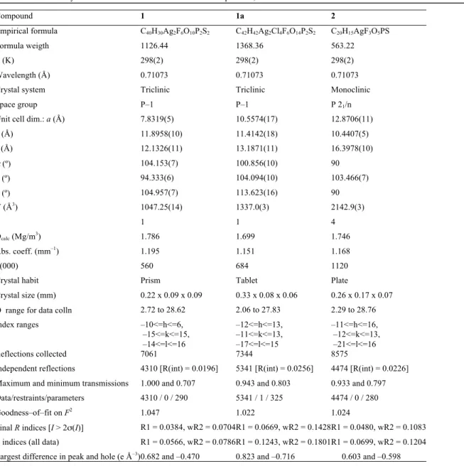
Table 1. Details of Crystal Data and Structure Refinement for Complexes 1, 1a and 2.
Compound 1 1a 2
Empirical formula C40H30Ag2F6O10P2S2 C42H42Ag2Cl4F6O14P2S2 C20H15AgF3O5PS
Formula weigth 1126.44 1368.36 563.22
T (K) 298(2) 298(2) 298(2)
Wavelength (Å) 0.71073 0.71073 0.71073
Crystal system Triclinic Triclinic Monoclinic
Space group P–1 P–1 P 21/n
Unit cell dim.: a (Å) 7.8319(5) 10.5574(17) 12.8706(11)
b (Å) 11.8958(10) 11.4142(18) 10.4407(5)
c (Å) 12.1326(11) 13.1871(11) 16.3978(10)
α (º) 104.153(7) 100.856(10) 90
β (º) 94.333(6) 104.094(10) 103.466(7)
χ (º) 104.957(7) 113.623(16) 90
V (Å3) 1047.25(14) 1337.0(3) 2142.9(3)
Z 1 1 4
Dcalc (Mg/m3) 1.786 1.699 1.746
Abs. coeff. (mm–1) 1.195 1.151 1.168
F(000) 560 684 1120
Crystal habit Prism Tablet Plate
Crystal size (mm) 0.22 x 0.09 x 0.09 0.33 x 0.08 x 0.06 0.26 x 0.17 x 0.07
Θ range for data colln 2.72 to 28.62 2.06 to 27.83 2.29 to 28.76
Index ranges –10<=h<=6,
–15<=k<=15, –14<=l<=16
–12<=h<=13, –11<=k<=13, –17<=l<=15
–11<=h<=16, –12<=k<=13, –21<=l<=16
Reflections collected 7061 7344 8575
Independent reflections 4310 [R(int) = 0.0196] 5341 [R(int) = 0.0256] 4474 [R(int) = 0.0226] Maximum and minimum transmissions 1.000 and 0.707 0.943 and 0.803 0.933 and 0.797 Data/restraints/parameters 4310 / 0 / 290 5341 / 1 / 325 4474 / 0 / 280
Goodness–of–fit on F2 1.047 1.022 1.024
Final R indices [I > 2σ(I)] R1 = 0.0384, wR2 = 0.0704 R1 = 0.0669, wR2 = 0.1428 R1 = 0.0480, wR2 = 0.1083
R indices (all data) R1 = 0.0566, wR2 = 0.0786 R1 = 0.1243, wR2 = 0.1801 R1 = 0.0699, wR2 = 0.1204 Largest difference in peak and hole (e Å–3) 0.682 and –0.470 0.823 and –0.716 0.603 and –0.598
30
text.
The reaction of silver(I) triflate with 2– and 4– (diphenylphosphino)benzoic acid in a 1:1 molar ratio (Scheme 3) yields [Ag(OSO2CF3)(PPh2C6H4COOH)].
5
10
15
Scheme 3. Preparation of compounds.
Compounds 1 – 2 are air–stable white solids at room temperature, and were characterized by elemental analysis, IR 20
and NMR spectroscopy. The complexes display one ν(C=O) band from the carboxylic group at about 1680 cm–1, and a
broad ν(O–H) band at about 3060 cm–1. In their 1H NMR
spectra, the aromatic benzoic protons are slightly shifted compared to the free phosphine ligand. The largest shifts are 25
about +0.27 ppm for H5 in the ortho ligand, and +0.28 ppm
for H3 in the para ligand. The assignment was confirmed by
COSY and 1H ⎨31P⎬ NMR spectra. The 19F NMR spectra show
a singlet at about –78 ppm because of the triflate anion. A broad resonance for the coordinated phosphine, because of 30
unresolved coupling to the two silver isotopes, was observed in the 31P{1H} NMR spectra of compound 1, while for compound 2 a doublet is seen. The resonance is low–field shifted about 19 ppm for compounds compared to the free phosphine ligand. At –50 ºC, the two Ag–P couplings are 35
resolved and two doublets are observed centered at 15.5 ppm (1J
107Ag–31P = 736 Hz, 1J109Ag–31P = 849 Hz) for complex 1, and
13.8 (1J
107Ag–31P= 690 Hz, 1J109Ag–31P = 795 Hz) for 2.
3.2. Solid–state structures 40
The solid–state structures of compounds 1 and 2 were solved by single–crystal X–ray diffraction studies and confirmed the bridging coordination of diphenylphosphinobenzoic acid. The crystals studied were obtained by slow diffusion of petroleum 45
ether (60–95o) into a solution of 1 in acetone/toluene or 2 in
acetone at –18oC. By slow evaporation of a diethyl ether
solution of 1, the same single crystals were obtained, which was verified by determining the unit cell. Slow diffusion of hexane into a solution of 1 in dichloromethane at –18oC led to
50
a different complex, denoted as 1a and being its formula: [{Ag(2–PPh2C6H4COOH)(OH2)2}2](CF3SO3)2.2CH2Cl2.
Compounds 1 and 1a crystallized as a P–1 triclinic single crystal, while compound 2 crystallized as a P21/n monoclinic
single crystal. The structures are shown in Figures 1–3, with 55
selected bond lengths and angles in Table 2. The asymmetric unit of 1 and 2 contained the fragment Ag(OSO2CF3)(PPh2C6H4COOH), with the silver center
coordinated to the phosphine through the P atom and to the
triflate anion via an oxygen. On the other hand, the 60
asymmetric unit of 1a displayed the fragment [Ag(OH2)2(PPh2C6H4COOH)](CF3SO3), again with the silver
center coordinated to the phosphine through the P atom, but now is coordinated to two water molecules (loosely to one) via their oxygen atom and with free triflate anion. The silver 65
center is coordinated to another oxygen atom in the three structures: the carbonyl of the carboxylic acid of another equal fragment. Therefore, the ligand is always acting as a bidentate non–chelating bridge. In that way the ortho benzoic acid leads to a cyclic dimer, which consists of a 12–membered 70
dimetallacycle with tri– or tetracoordinated silver centers. On the other hand, the para benzoic acid leads to a 1D–polymer because of the relative position of the carboxylic and the phosphine functional groups. The close compound [Ag6(o–
PPh2C6H4COO)6] shows two silver triangles (Ag–Ag
75
distances of 3.244, 3.756 and 3.805 Å) related by a symmetry center, with the anionic ligand acting as tri– or tetradentate non–chelating bridge [7].
Table 2. Selected Bond Lengths [Å] and Angles [deg] for Complexes 1,
80 1a and 2.
1 1a 2
Ag–P 2.4127(9) 2.3623(16) 2.3617(11)
Ag–O
(carbox.) 2.482(2) 2.362(5) 2.272(3) Ag–O
(triflate) 2.318(3) – 2.398(5) Ag–O
(water) – 2.2474(18) 2.7187(19) –
O=C 1.211(4) 1.207(8) 1.200(5)
O–C 1.308(4) 1.315(8) 1.308(5)
P–Ag–O
(triflate) 129.09(8) – 129.77(12) P–Ag–O
(water) – 136.18(8) 100.46(6) – P–Ag–O
(carbox.) 112.49(6) 138.09(13) 146.08(11) O–Ag–O 97.07(9) 80.02(13)
84.46(6) 85.74(13)
78.34(17)
C–O–C 123.6(3) 122.8(6) 123.1(4)
C–P–Ag 108.65(10) 114.49(10) 118.66(10) 107.3(2) 116.7(2) 119.92(19) 112.05(12) 114.52(14) 116.51(14)
The silver atom of compound 1 displays a distorted trigonal geometry, with a large P–Ag–O (triflate) angle of 129.09(8)o,
and smaller P–Ag–O (carboxylic) and O–Ag–O angles of 85
112.49(6) and 97.07(9)o, respectively. However, the silver
atom of compound 1a displays a highly distorted tetrahedral geometry, with a large P–Ag–O (carboxylic) angle of 138.09(13)o, smaller P–Ag–O (water) angles of 136.18 (8) and
100.46 (6)o, and the smallest angles are O–Ag–O: 80.02(13),
90
84.46(6) and 85.74(13)o. On the other hand, the silver atom of
compound 2 shows a highly distorted trigonal geometry, being the largest angle of 146.08(11)o corresponding to P–Ag–O (carboxylic); there is an intermediate angle P–Ag–O (triflate) of 129.77(12)o, and a small angle of 78.34(17)o for O–Ag–O.
95
The Ag–O (triflate) distance found for 1 (2.318(3) Å) is slightly shorter than found for 2 (2.398(5) Å). However, the Ag–O (carboxylic) distance of 1 (2.482(2) Å) is slightly longer than found for 1a (2.362(5) Å), and both are longer than found for 2 (2.272(3) Å). For 1a there is a short and a 100
Ag(O3SCF3)
PPh2C6H4COOH-o PPh2C6H4COOH-p
P Ph2
O O
Ag
F3CO2SO
H Ph2 P O O Ag
OSO2CF3
H
Ph2P
O O
H Ag
OSO2CF3
long Ag–O (water) distances of 2.2474(18) and 2.7187(19) Å, respectively. Finally, the Ag–P distance observed for 1 is slightly longer than observed for 1a or 2: 2.4127(9) versus 2.3623(16) or 2.3617(11) Å. The related derivative [Ag6(o–
PPh2C6H4COO)6] displays Ag–P intermediate distances in the
5
range 2.376(4)–2.381(4) Å, while displays a short Ag–O (carboxylate) distance (2.176(9) – 2.261(9) Å) and one or two longer Ag–O (carboxylate) distances from 2.394(9) to 2.619(9) Å per silver(I) center (in the asymmetric unit there is one tetracoordinate and two tricoordinate silver atoms) [7]. 10
The intramolecular silver–silver distances are 3.982 (compound 1) and 3.754 (compound 1a) Å, long to be considered as an Ag···Ag interaction (typically in the range 2.88–3.44 Å: from metallic distance to sum of van der Waals radii [3]. The shortest nonbonding intermolecular Ag–Ag 15
distance is 7.832 Å for dimer 1, 8.213 Å for dimer 1a, while the shortest Ag–Ag distance for polymer 2 is 4.467 Å. In addition, the O–H of the carboxylic functional group forms an intramolecular H–bond with an oxygen of the coordinated triflate anion for 1 or with the loosely coordinated water for 20
1a (Table 3). Therefore, there are two equal H–bonds per dimer. Similarly, compound 2 forms an additional donor H– bond between the O–H and an oxygen of another triflate (of a second monomer), and also forms the symmetrical acceptor H–bond between an oxygen of its triflate and the O–H of the 25
same second monomer. In this way, each monomer of the 1D polymer associates doubly to another monomer of next polymer chain, leading to a double chain polymer (Supplementary Information). This structure demonstrates the remarkable ability of silver(I) to form double polymeric 30
structures through H–bonding. Besides, longer H–bonds can be proposed for structures 1, 1a and 2 (see Tables in Supplementary Information).
35
Figure 1. Structure of the dimeric molecule of compound 1. Ellipsoids are at 25% probability level (H atoms omitted except carboxylic groups).
As stated in the introduction, the related gold(I) derivatives 40
[AuCl(PPh2C6H4COOH)] are monomers, which display a
typical linear coordination for gold, being the free carboxylic acid groups associated by a double symmetric H–bond to give dimers, the same for the ortho and the para ligands [6, 10]. The explanation must be related to the fact that the hard donor 45
oxygen atom of the carbonyl group bonds stronger for silver than for gold, and also to the tendency of silver(I) to higher coordination numbers than gold(I).
Figure 2. Structure of the dimeric cation of compound 1a. Ellipsoids are
50
at 30% probability level (H atoms omitted except carboxylic groups).
Figure 3. Polymeric structure of compound 2. Additional black lines to show the polymer growth. Ellipsoids are at 25% probability (most H atoms omitted for clarity): above, two consecutive units of the 1D chain (Ag–O bonds); below, fragment showing inter–chains H–bonds, giving rise to double chains.
5
Table 3. Hydrogen Bonds for Dimers 1 and 1a and polymer 2 [Å and deg] involving the carboxylic group.
D–H...A d(D–H) d(H...A) d(D...A) <(DHA) Compound 1
O(2)–H(2)...O(4A) 0.820 1.839 2.646 168.39 O(2A)–H(2A)...O(4) 0.820 1.839 2.646 168.39 Compound 1a
O(2)–H(2)...O(7A) 0.820 1.819 2.610 161.69 O(2A)–H(2A)...O(7) 0.820 1.819 2.610 161.69 Compound 2
O(1)–H(1)...O(4A) 0.820 1.828 2.626 164.09 O(1A)–H(1A)...O(4) 0.820 1.828 2.626 164.09 Symmetry transformations used to generate equivalent atoms: for compound 1 are 1–x, 2–y, 1–z; for compound 1a are –x, –y, 1–z; for 10
compound 2 are 1–x, 2–y, 1–z.
The powder X–ray diffraction (XRD) patterns of compounds 1
and 2 are in good agreement with that simulated from the corresponding single–crystal diffraction data (see 15
Supplementary Information) confirming that the latter accurately represents the structure of the bulk solid (and not
1a).
3.3. Luminescence studies 20
The emission and excitation spectra of the free ligands and the silver complexes were recorded in the solid–state and in CH2Cl2 solution, at 298 K and 77 K. The spectra of compound 2 at 298 K are shown in Figure 4. The 4– 25
(diphenylphosphino)benzoic compound 2 and the corresponding free ligand emit, while the 2– (diphenylphosphino)benzoic acid and its derivative 1 are non– emissive. The emission maximum in the solid state is at 471 nm at 298 K, similar to the free ligand (474 nm), but at 457 30
nm at 77 K, blue–shifted compared to the free ligand (479 nm) and to 298 K emission. This emission could be assigned as ligand centered, although slightly modified by the metal fragment. Compound 2 shows an emission in CH2Cl2 solution
at 298 K centered at 416 nm, while compound 1 and the two 35
free ligands are non–emissive. At 77 K the emission maximum show little change, and is observed at 420 nm, blue–shifted compared to the corresponding free ligand emission (486 nm). Emission peaks of compound 2 are strongly blue–shifted compared to these observed in the solid 40
state. Again, the emission could be assigned as ligand centered modified by the silver fragment.
45
50
55
Figure 4. Solid state (regular line) and dichloromethane solution (bold line) excitation and emission spectra of compound 2 at 298K.
60
4. Conclusions
2– or 4–(diphenylphosphino)benzoic acids act as non– chelating bridge to give di– or polymeric silver(I) compounds, respectively, demonstrating that different geometrical 65
orientation of the substituent induces entirely different connectivity patterns. The triflate anion or water molecules complete the coordination sphere. On the one hand, the carboxylic functional group is bridging monomers, on the other hand is making H–bonds: intramolecularly for the 70
dimer, and associating 1D chains in pairs for the polymer. Besides, the p–(diphenylphosphino) benzoic ligand and its corresponding silver complex are emissive, while the ortho ligand and complex are non–emissive.
75
Acknowledgements
We thank the Spanish Comisión Interministerial de Ciencia y Tecnología (Project CTQ2011–25137) and the Junta de Castilla y León (Project VA302U13) for financial support. 80
Appendix A. Supplementary material
CCDC 1004945–1004947 contains the supplementary crystallographic data for this paper. These data can be obtained free of charge from The Cambridge Crystallographic Data Centre via www.ccdc.cam.ac.uk/data_request/cif. 85
Supplementary data associated with this article can be found, in the online version, at doi: xxx.
References
[1] (a) S. Leininger, B. Olenyuk, P. J. Stang, Chem. Rev. 100 (2000) 853; (b) G. Ferey, Chem. Soc. Rev. 37 (2008) 191. (c) G. A. Santillan, C. J. Carrano, Cryst. Growth Des. 9 (2009) 1590; (d) J. Ni, K.–J. Wei, Y. Liu, X.–C. Huang, D. Li, Cryst. Growth Des. 10 (2010) 3964; (e) A. M. Kirillov, S. W. Wieczorek, M. F. C. Guedes da Silva, J. Krol, Z. Staroniewicz, P. Smolenski, A. J. L. Pombeiro, Cryst. Growth Des. 11 (2011) 2711; (f) X.–L. Wang, Q. Gao, A.–X. Tian, G.–C. Liu, Cryst. Growth Des. 12 (2012) 2346; (g) L. Cunha–Silva, M. J. Carr, J. D. Kennedy, M. J. Hardie, Cryst. Growth Des. 13 (2013) 3162.
[2] (a) A. Laguna, M. C. Gimeno, Silver and Gold In Comprehensive Coordination Chemistry II: From Biology to Nanotechnology McCleverty, J. A., Meyer, T. J., Eds.; Elsevier, Oxford, UK, 2003; Vol. 6, Transition Metal Groups 9–12, Fenton, D. E., ed. Chapter 6.7, p. 911; (b) J.-R. Li, R.-H. Zhang, X.-H. Bu, Cryst. Growth Des. 3 (2003) 829; (c) C. S. Liu, P. Q. Chen, E. C. Yang, J. L. Tian, X. H. Bu, Z. M. Li, H. W. Sun, Z. Lin, Inorg. Chem. 45 (2006) 5812; (d) P. Yang, F. Cui, X.–J. Yang, B. Wu, Cryst. Growth Des. 13 (2013) 186; (e) X. Ma, L.-H. Huo, Z.-P. Deng, T.-P. Liu, H. Zhao, S. Gao, Inorg. Chem. Commun. 43 (2014) 94.
[3] (a) ) K. Singh, J.R. Long, P. Stavropoulos, J. Am. Chem. Soc. 119 (1997) 2942; (b) Q. M. Wang, T. C. W. Mak, J. Am. Chem. Soc. 123 (2001) 7594; (c) B. Liu, W. Chen, S. Jin, Organometallics 26 (2007)
3660; (d) L. Ray, M. M. Shaikh, P. Ghosh, Inorg. Chem. 47 (2008)
230;(e) X. D. Zheng, L. Jiang, X. L. Feng, T. B. Lu, Inorg. Chem. 47 (2008) 10858; (f) R. Santra, K. Biradha, Cryst. Growth Des. 10 (2010) 3315; (g) B. Li, S. Q. Zang, R. Liang, Y. J. Wu, T. C. W. Mak, Organometallics 30 (2011) 1710; (h) A. Serpe, F. Artizzu, L. Marchio, M. L. Mercuri, L. Pilia, P. Deplano, Cryst. Growth Des. 11
0,0 0,2 0,4 0,6 0,8 1,0
N
orma
lize
d
In
te
nsi
ty/
a.
(2011) 1278; (i) P.–S. Cheng, S. Marivel, S.–Q. Zang, G.–G. Gao, T. C. W. Mak, Cryst. Growth Des. 12 (2012) 4519.
[4] (a) M. A. Omary, H. H. Patterson, Inorg. Chem. 37 (1998) 1060; (b) M. L. Tong, B. H. Ye, X. M. Chen, L. N. Ji, Angew. Chem. Int. Ed. 38 (1999) 2237; (c) X.H. Bu, H. Liu, M: Du, K. M. Wong, V. W. Yam, M. Shionoya, Inorg. Chem. 40 (2001) 4143; (d) T.-L. Hu, J.-R. Li, Y.-B. Xie, X.-H. Bu, Cryst. Growth Des. 6 (2006) 648; (e) M.–S. Wang, S.–P. Guo, Y. Li, L.–Z. Cai, J.–P. Zou, G. Xu, W.–W. Zhou, F.–K. Zheng, G.–C. Guo, J. Am. Chem. Soc. 131 (2009) 13572; (f)
K. Matsumoto, T. Shindo, N. Mukasa, T. Tsukuda, T. Tsubomura,
Inorg. Chem. 49 (2010) 805; (g) M. Bardají, M. Barrio, P. Espinet,
Dalton Trans. 40 (2011) 2570;(h) D. Pucci, A. Crispini, M. Ghedini, E.I. Szerb, M. La Deda, Dalton Trans. 40 (2011) 4614; (i) M. G. Babashkina, D. A. Safin, M. Bolte, Y. Garcia, Dalton Trans. 40 (2011) 8523; (j) A. Hameau, F. Guyon, A. Khatyr, M. Knorr, C. Strohmann, Inorg. Chim. Acta 388 (2012) 60; (k) M. Bardají, A. B. Miguel–Coello, P. Espinet, Inorg. Chim. Acta 386 (2012) 93; (l) C.-F. Yan, Y.-X. Lin, C.-F.-L. Jiang, M.-C. Hong, Inorg. Chem. Commun.
43 (2014) 19.
[5] A. Chandrasekaran, R. O. Day, R. R. Holmes, Inorg. Chem. 40 (2001) 6229.
[6] F. Mohr, M. C. Jennings, R. J. Puddephatt, Angew. Chem. Int. Ed. 43 (2004) 969.
[7] W.-K. Wong, L. Zhang, W.–T. Wong, Chem. Commun. (1998) 673. [8] (a) Z. J. A. Komon, X. Bu, G. C. Bazan, J. Am. Chem. Soc. 122
(2000) 12379; (b) P. Barbaro, C. A. Ghilardi, S. Midollini, A. Orlandini, J. A. Ramirez, G. Scapacci, J. Organomet. Chem. 555 (1998) 255; (c) S.-M. Kuang, P. E. Fanwick, R. A. Walton, Inorg. Chem. (41) 2002 405; (d) S.-M. Kuang, P. E. Fanwick, R. A. Walton, Inorg. Chem. Commun. 5 (2002) 134; (e) W. Uhl, H. R. Bock, J. Kosters, M. Voss, Z. Anorg. Allg. Chem. 636 (2010) 1851; (f) G. Sanchez, J. Garcia, D. Meseguer, J. L. Serrano, L. Garcia, J. Perez, G. Lopez, Dalton Trans. (2003) 4709; (g) T. Schultz, A. Pfaltz, Synthesis (2005) 1005; (h) P. P. Phadnis, S. Dey, V. K. Jain, M. Nethaji, R. J. Butcher, Polyhedron 25 (2006) 87; (i) H. Schumann, H. Hemling, V. Ravindar, Y. Badrieh, J. Blum, J. Organomet. Chem. 469 (1994) 213. (j) J. S. M. Samec, R. H. Grubbs, Chem. Eur. J. 14 (2008) 2686; (k) M. Kawatsura, F. Ata, S. Wada, S. Hayase, H. Uno, T. Itoh, Chem. Commun. (2007) 298; (l) J. D. G. Correia, A. Domingos, I. Santos, C. Bolzati, F. Refosco, F. Tisato, Inorg. Chim. Acta 315 (2001) 213.
[9] (a) S.-M. Kuang, P. E. Fanwick, R. A. Walton, Inorg. Chem. 41 (2002) 1036; (b) S.-M. Kuang, P. E. Fanwick, R. A. Walton, Inorg. Chim. Acta 338 (2002) 219; (c) W. Uhl, H. R. Bock, A. Hepp, F. Rogel, M. Voss, Z. Anorg. Allg. Chem. 636 (2010) 1255; (d) J.-M. Rueff, O. Perez, A. Leclaire, H. Couthon-Gourves, P.-A. Jaffres, Eur. J. Inorg. Chem. (2009) 4870.
[10] B. P. Howe, R. V. Parish, R. G. Pritchard, Quim. Nova 21 (1998) 564.
[11] M. Pellei, G. G. Lobbia, C. Santini, R. Spagna, M. Camalli, D. Fedeli, G. Falcioni, Dalton Trans. (2004) 2822.
[12] S. J. Carlson, T. Lu, R. L. Luck, Inorg. Chem. Commun. 6 (2003) 455.
[13] CrysAlisPro Software system, Version 1.171.32; Oxford Diffraction; Oxford Ltd., Xcalibur CCD system, 2007.
![Table 2. Selected Bond Lengths [Å] and Angles [deg] for Complexes 1, 80 1a and 2. 1 1a 2 Ag–P 2.4127(9) 2.3623(16) 2.3617(11) Ag–O (carbox.) 2.482(2) 2.362(5) 2.272(3) Ag–O (triflate) 2.318(3) – 2.398(5) Ag–O (water) – 2.2474(18) 2.71](https://thumb-us.123doks.com/thumbv2/123dok_es/6540632.228562/4.892.57.420.177.348/table-selected-bond-lengths-angles-complexes-carbox-triflate.webp)

![Table 3. Hydrogen Bonds for Dimers 1 and 1a and polymer 2 [Å and deg] involving the carboxylic group](https://thumb-us.123doks.com/thumbv2/123dok_es/6540632.228562/6.892.59.404.905.1137/table-hydrogen-bonds-dimers-polymer-involving-carboxylic-group.webp)