Tumor Growth Inhibition by Local Activation of Cellular Sensitivity to Drugs by
Focused Acoustic Shock Waves
PACS: 43.25.Yw
V. S. Teslenko, Larentiev Institute of Hydrodynamics SD RAS, V. P. Nikolin, Institute of Cytology and Genetics SD RAS, I. V. Mastikhin, International Tomographic Center SD RAS,
N. G. Kolosova, Institute of Physiology SD RAMS, V. N. Gorchakov, Institute of Limfology SD RAMS
Lavrentyev Institute of Hydrodynamics SD RAS 15, Lavrentiev str.,
Novosibirsk 630090 Russia
Fax: (3832)331612 Phone: (3832)333047
vteslenko@mail.ru
ABSTRACT
Development of methods for efficient delivery of drugs to the cells of certain tissues or organs is of high priority in up-to-date medicine and biology. These methods are of essential importance for chemotherapy and gene therapy of malignancy because they employ organ-targeted delivery of drugs to certain cells. A possible approach to the problem of targeted delivery is a local activation of cell membrane permeability under physical actions. To implement this approach, it is possible to regulate the cell membrane permeability by focused acoustic shock waves (ASW). At present, the methods for focusing of ASW that allow localize the impact within a focal region in a body are developed in much details. It is shown experimentally that the use of ASW increases considerably in vivo and in vitro cell membrane permeability and can be used to promote targeted delivery of drugs, including fragments of nucleic acids, to cells.
INTRODUCTION
Development of installation devices for generating and focusing acoustic shock waves (ASW) offered novel perspectives for using of these waves in biology and medicine. At first, ASW were successfully implemented to disintegrate hepatic and renal calculi. After that, the first experimental results on the influence of focused of ASW on malignant tumors were reported. However, the use of lithotriptors to influence locally a tumor caused tumor cell necrosis [1]. It was supposed that a destructive effect of ASW on tumors was due to mechanical failure of cells caused by induction of ASW cavitation process, i.e., formation and pulsation of gas bubbles dissolved in a liquid [2–5].
To verify this hypothesis, we studied changes in tumor tissue after the ASW action using NMR-tomography followed by histological analysis.
EXPERIMENTAL RESULTS
placed in the receiver of microtomographic indicator of NMR-spectrometer. In NMR-tomography, we used spin- and gradient-echo methods with the following parameters: the field of vision was 30x30 mm, section thickness was 1 mm, and plane resolution was 0.1x0.1 mm. In the region that geometrically coincided with the focal zone of a radiator, we observed a zone of signal reduction (on the tomogram this zone is shown with a circle, Fig. 1), which was due to both decrease in the proton density and tissue loosening after ASW action. Failure of the capillary structure caused microhemorrhage recorded on the tomogram.
Fig. 1. NMR-tomogram of a tumor tissue recorded after 10 ASW pulses, the focal zone is shown with a circle (to the left); photograph of a histologic section of the zone corresponding to that shown with
a circle on a tomogram: white beads show cavitation processes in the tissue (to the right).
A histologic section of the focal ASW zone shows the presence of extensive necrosis regions with hemorrhage zones. On the boundary between the preserved tumor tissue and the necrosis zone, one can see cavities with diameter of several tenths of microns which are due to formation of cavitation bubbles. Obviously, cavitation processes occurred in the focal ASW zone cause formation and extension of cavitation bubbles. However, because of high medium density and considerable energy losses, the bubbles strengthen and are filled with an intertissue liquid. Such formations, known as “false bubbles”, are reported in literature as resulting from cavitation in biological tissues after strong ultrasonic action.
Thus, histologic data on the tumor tissue from the focal ASW region show that in biological tissues as well as in solutions under weak ASW action cavitation occurs and is accompanied by tissue failure.
Studies of hydrodynamic and chemical processes caused by ASW in liquid media revealed that cavitation under weak ASW action leads to water hydrolysis and, consequently, to the induction of various radical reactions and decomposition of unstable compounds. Cavitation processes in tumor tissues under the ASW action suggest that radical generation and induction are possible to occur in tumor tissues. As the formation of free radicals occurs via the cavitation mechanism, these radicals can appear primarily on the boundary between the media: a denser medium (cells) and a looser medium (extracellular). Under these conditions biological membranes can be the main targets. It is known that a universal response of oxidative modification of proteins and celle membrane lipids. The energy cellular systems are particularly sensitive to the damaging effect of lipid peroxidation (LPO).
We studied the structural-functional characteristics of cellular membranes, such as the membrane potential (∆ψ), lipid-protein interactions, lipid viscosity, and LPO products (conjugated diene and malonaldehyde) for two types of cells: Krebs-2 tumor cells and Wistar rat thymocytes. The cells were loaded with 5–70 ASW pulses of fixed amplitude (Ð=25-45 MPa) and time (t = 0.5ìs); the interval between the pulses was 5 sec. Membrane lipid viscosity was determined by a pair of pyrene, and DSP-12 fluorescent probes (DSP-DSP-12 is 4-(p-dimethylaminostyrene)-1-dodecyl pyridine). Lipid-protein interactions were indicated by recording the energy transfer from the tryptophane residues of membrane proteins to pyrene localized in lipids. The measurements of the membrane potential ∆Ψ were performed with 4-n-(dimethylaminostiryl)-1-methylpyridinium as a fluorescent probe in a Hitachi MPF-4 spectrofluorimeter (Japan), and spectrophotometric measurements were conducted using a Hitachi-556 spectrophotometer.
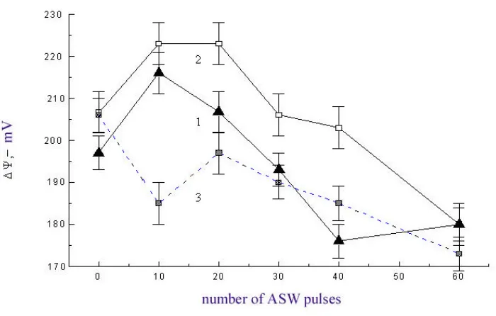
products and lipid viscosity are observed; the proteins aggregate and the membrane potential ∆ψ increases. As shown in Fig. 2, the changes noted depend nonlinearly on the number of ASW pulses. Maximal changes are observed for 10–20 ASW pulses. Further increase in the number of pulses yields poorer results.
Fig. 2. Membrane electric potential ∆ψ of Krebs-2 cells (1) and thymocytes (2) 30 minutes, and that of thymocytes 60 minutes (3) after the impact of different number of ASW pulses.
The appearance of free radicals induced by ASW can cause the changes described above. However, we suppose that the nonlinear nature of these effects can be due not only to the free radicals generated by ASW, but also the free radicals generated by mitochondria of the cells. At 10–20 ASW pulses an increase in the membrane potential suggests the activation of oxidative phosphorylation, and an increase in the LPO product content results from the mechanical failures that hamper normal mitochondrial performance. We may suppose that for a large number of ASW pulses, a complete inhibition of phosphorylation mitochondrial activity occurs, which is confirmed by low values of the membrane potential.
Enhancement of LPO and accumulation of lipid peroxides lead to inactivation of the membrane– bound enzymes to different extents and to the changes in cell membrane permeability for various ions, non-electrolytes, and macromolecules [7].
Obviously, enhancement of LPO after the effect of weak ASW on cells should have resulted in analogous variation in the cell membranes and an increase in their permeability for different substances, probably including anti-tumor drugs. This suggested further studies of the effect of weak ASW on the sensitivity of tumor cells to chemical drugs.
We studied the sensitivity of mice Krebs-2 tumor cells to cyclophosphane (CF), an anti-tumor drug widely used in clinical practice. A tumor cell suspension was subjected to 5–70 ASW pulses of a constant amplitude (Ð=25-45 MPa) and time (t = 0.5 ìs.) with an interval between the pulses of 5 sec. Then, the cells were inoculated in mice leg muscles. The mice received cyclophosphane at 100 mg/kg body weight intraperitoneal injection 30 minutes after inoculation. The ASW action resulted in an increase in the number of nonviable cells proportional to the number of pulses; however, even after 70 pulses, the number of killed cells did not exceed 30%. After 12 days, the mice were killed to determine the mass of grown tumors.
to quantitative parameters of the structural–functional changes in the tumor cell membranes after treatment with ASW, and obviously connected with an enhancement of tumor cell permeability for CF and an increase in its intracellular concentration.
Fig. 3. Inhibition of inoculated tumor growth versus the number of ASW pulses. The upper curve corresponds to the combined use of ASW with cyclophosphane (CF), the middle curve, to the use of CF
only, and the lower curve refers to the use of ASW exclusively.
Figure 4 shows results of in vivo experiments in 2-month-old female mice (SVA line) inoculated with Krebs-2 tumor cells (2 million cells) in their feet pads. After the tumor reached 0.4–0.5 cm in diameter, the mice were divided into six groups:
I. Control group (16 mice).
II. 10 mice with tumors treated locally with ASW;
III. 18 mice received cyclophosphane at 100 mg/kg body weight intraperitoneal injection simultaneously with Groups 4, 5, and 6;
IV. 9 mice were treated with 10 ASW pulses; V. 9 mice were treated with 20 ASW pulses; VI. 9 mice were treated with 40 ASW pulses.
The mice of Group 4, 5, and 6 received cyclophosphane at 100 mg/kg body weight intraperitoneal injection 30 min after the local treatment of tumor with ASW.
The tumor weight was determined 7 days later.
Figure 4 shows that the combined use of ASW and CF inhibits more effectively tumor growth than CF applied exclusively. This suggests that the sensitivity of tumor cells to cytostatic increases not only upon the in vitro treatment with ASW, but also upon the in vivo treatment.
RESUME
Until present, the efficacy of the combined treatment of tumors with ASW and chemical drugs is reported by several foreign authors. They note that the use of ASW may promote overcoming the resistance of some tumors to a chemical drug. The sphere of ASW application is constantly extending. In particular, by increasing cellular permeability with ASW, it is possible to deliver in vivo and in vitro
some large molecules, for example DNA, which is able to express in transfected cells (transfection by sonophoresis). This suggests the potential therapeutic use of ASW in gene therapy.
Further research will require determination of affecting and non-affecting parameters of ASW for various cells, investigation of the mechanism of the ASW effect on cell membrane permeability, and determination of optimal ASW parameters providing for the maximum concentration level of cytostatic (and other drugs) in cells. These questions are closely connected with the quantitative parameters of ASW, such as amplitude, pulse length, relative pulse duration, and the number of pulses (“dose”).
EXPERIMENTAL RESULTS
This work is supported by RFBR, projects ¹ 96-02-19329, ¹ 00-02-17992.
BIBLIOGRAPHICAL REFERENCES
1. Gamarra, F.,Spelsberg, F., Dellian, M., and Goetz, A. E. Complete local tumor remission after therapy with extra-corporeally applied high-energy shock waves (HESW). Int. J. Cancer, 1993, 55, pp. 153–156.
2. Mastikhin I.V., Nikolin V.P., Teslenko V.S., Zelentsov E.L., Mayer V.A., Salganik R.I., and Dikalov S.I. Increase in sensitivity of tumor cells to cyclophosphane after therapy with shock-waves. Doklady Akad. Nauk, 1995, vol. 342, No. 2, pp. 262-264.
3. Mastikhin I.V., Gorchakov V.N., Teslenko V.S., Nikolin V.P., and Kolosova N.G. Recoring of cavitation processes in biological bodies upon shock-wave action. In Dynamics of Continuous Media, Novosibirsk, 1999, No. 115, pp. 151–154.
4. Teslenko V.S., Mastikhin I.V., Nikolin V.P., and Kolosova N.G. Variations in structural–functional characteristics of cell membranes upon pulse acoustic action. Doklady Akad. Nauk, 1999, vol. 369, No. 5, pp. 698–700. 5. Shipping Bao, Brain D. Thrall, Richard A. Gies, and Douglas L. Miller. In vivo transfection of melanoma cells by
lithotripter shock waves. Cancer research,1998, vol. 58, No. 2, pp. 219–221.
6. Timashev S.F. Physical Chemistry of Membrane Processes. Moscow, Khimiya (1988).