Altered intestinal transport of amino acids in cirrhotic rats : the effect of insulin like growth factor I
6
0
0
Texto completo
(2) G320. IGF-I AND INTESTINAL AMINO ACID TRANSPORT IN CIRRHOSIS. bo,⫹ system for neutral amino acids and the y⫹ system for basic amino acids (9, 10, 15, 16, 31, 40). Here we have investigated the intestinal transport of amino acids in cirrhotic rats and the effect of IGF-I treatment on this function. For this purpose, the four major types of amino acid transport system were studied by testing the uptake of representative amino acids L-leucine (neutral amino acid), L-proline (IMINO acid), L-glutamic acid (acidic amino acid), and L-cysteine (sulfurate amino acid) in brush-border membrane vesicles (BBMV) from control rats, cirrhotic rats, and cirrhotic rats treated with IGF-I.. Induction of cirrhosis and experimental design. Cirrhosis was induced in male Wistar rats, as previously described (4, 5, 6, 34), by inhalation of CCl4 and administration of phenobarbital (Luminal; Bayer, Leverkusen, Germany) in drinking water (400 mg/l), for 11 wk. Age-matched healthy control rats (group CO, n ⫽ 12), which did not receive any treatment, were studied in parallel. One week after stopping CCl4 administration, cirrhotic animals subcutaneously received either saline (group CI, n ⫽ 12) or IGF-I (2 g 䡠 100 g body wt⫺1 䡠 day⫺1, group CI⫹IGF, n ⫽ 12) for 2 wk. The CO group received saline during these last 2 wk of the study. The day before starting treatment (day 0), blood (⬃4 ml) was collected using capillary tubes (70 mm, Marienfeld, Mergenheim, Germany) from the retroocular plexus of all rats. Serum samples were divided into aliquots and stored at ⫺20°C until used. At the end of the experimental period (day 15 after initiation of the treatment), blood was collected again and animals were killed by decapitation. None of the animals presented ascites on examination at necropsy at the end of the study. Jejunal specimens (⬃20 cm) were immediately frozen by immersion in liquid N2 and stored at ⫺80°C. Liver and spleen were dissected out and weighed. Liver samples were processed for histological examination. All procedures were performed in conformity with “The Guiding Principles for Research Involving Animals and Human Beings”. Biochemical determinations. Serum levels of albumin, total proteins, and glucose were determined by laboratory methods using a Cobas-Mira autoanalyzer (⌬Bx Systems, Madrid, Spain). Preparation of BBMVs. BBMVs were isolated using the magnesium precipitation method (19) as previously described (6). The procedure was carried out at 0–4°C. The everted jejunum was placed in 2 mmol/l Tris 䡠 HCl (pH 7.4) containing 100 mmol/l mannitol, stirred for 3 min (Vibromixer, E-1, Sorvall), and the scraped jejunal mucosa was removed. The mucosal suspension was mixed with 10 mmol/l MgCl2 (final concentration) and centrifuged at 10,000 g (15 min). The supernatants were centrifuged again at 26,000 g (30 min). Pellets were resuspended in 10 mmol/l MgCl2 and centrifuged at 26,000 g (30 min). The final pellets were resuspended in the desired volume of 300 mmol/l mannitol, 0.1 mmol/l MgSO4, and 10 mmol/l Tris-HEPES buffer (pH 7.4) (load solution), so that a final protein concentration of 8–10 mg/ml was obtained. Isolated membranes were circled using an N 27-gauge needle. Sucrase (EC 3.2.1.48, enterocyte apical membrane marker) and Na⫹-K⫹-ATPase (EC 3.6.1.3, enterocyte basolateral membrane marker) activities were determined in BBMV suspension (6). The BBMV activities of sucrase and Na⫹-K⫹-ATPase were 11-fold higher and 20-fold. RESULTS. The presence of cirrhosis in all animals that received CCl4 was confirmed by histological examination of the liver samples obtained at the end of the study. At day 0 (the day before initiation of treatment with IGF-I or saline) all cirrhotic rats showed significant hypoalbuminemia, hypoproteinemia, and hypoglycemia compared with controls (Table 1). At the end of the treatment, body weight of untreated cirrhotic rats (459 ⫾ 10 g) was similar to that of IGF-I-treated cirrhotic animals (459 ⫾ 9 g), and in these two groups body weight was significantly lower than that of normal controls (555 ⫾ 16; P ⬍ 0.001). However, at the end of the study, total serum proteins, serum albumin, and glycemia were similar in healthy controls and in cir-. Downloaded from http://ajpgi.physiology.org/ at Mt Sinai School of Medicine on February 25, 2013. MATERIALS AND METHODS. lower, respectively, than those found in the initial mucosal homogenate. The total protein content of BBMVs was determined using the Bradford method (3). Vesicles were stored in liquid N2. Assessment of amino acid uptake by BBMVs. Determination of amino acid uptake by BBMVs was performed at 25°C using the rapid filtration technique described by Hopfer et al. (19) with slight modifications. BBMV suspensions (5 l) were added to the incubation medium (45 l) containing 1 mmol/l of unlabeled amino acid, 25 Ci/ml of radiolabeled substrate 14 14 14 L-[U- C]leucine, L-[U- C]proline, L-[U- C]glutamic acid, or 35 L-[ S]cysteine (Amersham, Little Chalfont, UK), 100 mmol/l NaSCN or KSCN, 100 mmol/l mannitol, 0.1 mmol/l MgSO4 and 10 mmol/l HEPES (pH 7.4). The time courses of the uptake of amino acids were measured in the presence of Na⫹ gradient (using medium containing NaSCN) and in the absence of Na⫹ gradient (medium containing KSCN). At specific time intervals, the uptake process was ended by adding 5 ml of ice-cold stop solution containing 150 mmol/l KSCN and 10 mmol/l Tris-HEPES (pH 7.4). The suspension was immediately poured onto a prewetted Millipore filter that was washed three times with 3 ml of ice-cold stop solution and immersed in 5 ml of scintillator Hisafe 3 fluid (LKB Products, Bromma, Sweden). The filter was then counted in a Counter Wallac 1409 (Pharmacia, Turku, Finland). Nonspecific binding to the filter was previously measured and subtracted from the total uptake. Results were expressed as picomoles of amino acid uptake per milligram of protein. The kinetic constants of the uptake of amino acids by BBMV were evaluated as previously reported (6, 14). For this purpose, the assays of amino acid uptake by BBMV were performed in the presence of several concentrations of substrate, from 0.025 to 7 mmol/l, at a fixed transport time of 3 s (19). Each assay was performed in triplicate using the pool of BBMV (n ⫽ 12) from each experimental group. Maximal velocity (Vmax) was expressed as picomoles of substrate per milligram of protein in 3 s, and the transporter affinity constant (Kt) was expressed as millimoles per liter. Statistical analysis. Data are given as means ⫾ SE. To analyze the homogeneity among groups, a Kruskall-Wallis test was used, followed by multiple post hoc comparisons using Mann-Whitney U-tests with Bonferroni adjustment. A Wilcoxon signed-rank test was used to compare data before and after IGF-I treatment (days 0 and 15) in the same group. Any P value ⬍ 0.05 was considered statistically significant. Calculations were performed with SPSS version 6.0 (SPSS, Chicago, IL). The SigmaPlot Program (version 3.02 for PC) was used to process data of the kinetic study of amino acid uptake by BBMV..
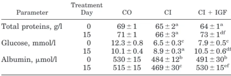
(3) IGF-I AND INTESTINAL AMINO ACID TRANSPORT IN CIRRHOSIS. Table 1. Serum levels of total proteins, albumin, and glucose in the three experimental groups at baseline (day 0) and at the end of the treatment (day 15) Parameter. Total proteins, g/l Glucose, mmol/l Albumin, mol/l. Treatment Day. CO. CI. 0 15 0 15 0 15. 69 ⫾ 1 71 ⫾ 1 12.3 ⫾ 0.8 10.1 ⫾ 0.4 530 ⫾ 15 515 ⫾ 15. 65 ⫾ 2 66 ⫾ 3a 6.5 ⫾ 0.3c 8.9 ⫾ 0.3a 484 ⫾ 12b 469 ⫾ 30c. CI ⫹ IGF a. 64 ⫾ 1a 73 ⫾ 1df 7.9 ⫾ 0.5c 10.5 ⫾ 0.6df 491 ⫾ 30b 530 ⫾ 15ef. rhotic rats that received IGF-I, but these parameters were significantly reduced in untreated cirrhotic rats compared with the two other groups (Table 1). The improvement of liver function in cirrhotic rats treated with IGF-I confirms previous published data (4) dem-. onstrating a hepatoprotective effect of IGF-I in experimental cirrhosis. Uptake of amino acids by jejunal BBMVs. No differences between groups were found in enzyme membrane marker activities in BBMV (sucrase: CO ⫽ 456 ⫾ 5; CI ⫽ 496 ⫾ 4; and CI ⫹ IGF ⫽ 427 ⫾ 2 mol hydrolyzed sucrase 䡠 min⫺1 䡠 mg protein⫺1; Na⫹-K⫹ATPase: CO ⫽ 9.81 ⫾ 0.2; CI ⫽ 10.2 ⫾ 0.3; CI ⫹ IGF ⫽ 9.1 ⫾ 0.3 mol Pi 䡠 mg protein⫺1 䡠 min⫺1). Results of L-leucine, L-proline, L-glutamic acid, and L-cysteine uptake by BBMVs are shown in Fig. 1. The uptake of amino acids was assessed in the presence and in the absence of Na⫹ gradient. Na⫹-independent uptake of all amino acids was similar in all groups of animals. In the presence of Na⫹ gradient, however, the uptake of amino acids by BBMV from untreated cirrhotic rats was significantly lower than that obtained with BBMV from healthy controls, whereas BBMV from IGF-Itreated cirrhotic animals exhibited uptake values similar to those of BBMV from normal controls and significantly higher than those of BBMV from untreated cirrhotic rats. These differences in the uptake of amino acids were mainly noticeable for L-leucine, L-proline,. Fig. 1. Time course of amino acid (1.0 mM) uptake into brush-border membrane vesicles (BBMVs) at 25°C, with (in the presence of NaSCN) or without (in the presence of KSCN) Na⫹ gradient. Each point represents the mean ⫾ SE of 3 different experiments, using a pool of BBMVs from 12 animals from each experimental group: controls (CO), untreated cirrhotic rats (CI), and cirrhotic rats treated with IGF-I for 2 wk (CI ⫹ IGF). *** P ⬍ 0.001 for CI vs. other groups in the presence of Na⫹ gradient. No differences were found between CO and CI⫹IGF groups in Na⫹-dependent transport. In the absence of Na⫹ gradient (with K⫹ gradient), values from the 3 experimental groups are superimposed. A: L-leucine. B: L-proline. C: L-glutamic acid. D: L-cysteine.. Downloaded from http://ajpgi.physiology.org/ at Mt Sinai School of Medicine on February 25, 2013. Values are means ⫾ SE; n ⫽ 12. No differences were found between untreated cirrhotic rats (CI) and cirrhotic rats treated with insulin-like growth factor-I (IGF-I) (CI ⫹ IGF) before the treatment (day 0). a P ⬍ 0.05, b P ⬍ 0.01, and c P ⬍ 0.001 for control (CO) vs. cirrhotic groups. d P ⬍ 0.05 and e P ⬍ 0.001 for CI vs. CI ⫹ IGF groups at the end of the treatment. f P ⬍ 0.05 between values before vs. after treatment (day 0 vs. day 15) in the same group.. G321.
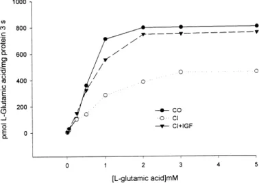
(4) G322. IGF-I AND INTESTINAL AMINO ACID TRANSPORT IN CIRRHOSIS. and L-glutamic acid and less for L-cysteine, in which significant differences were observed only at 60 s but not at other time points (Fig. 1). Thus the initial rate of uptake at 5 s (expressed as pmol/mg protein) for Lleucine was lower in untreated cirrhotic rats than in control rats and cirrhotic animals treated with IGF-I (Fig. 1A; CO ⫽ 332 ⫾ 12, CI ⫽ 98 ⫾ 10, and CI ⫹ IGF ⫽ 332 ⫾ 8; P ⬍ 0.001 for CI vs. other groups). A similar profile was found for L-proline (Fig. 1B; CO ⫽ 171 ⫾ 10, CI ⫽ 121 ⫾ 2, and CI ⫹ IGF ⫽ 163 ⫾ 3; P ⬍ 0.001 for CI vs. other groups) and L-glutamic acid (Fig. 1C; CO ⫽ 413 ⫾ 12, CI ⫽ 320 ⫾ 10, and CI ⫹ IGF ⫽ 387 ⫾ 8; P ⬍ 0.001 for CI vs. other groups). For L-cysteine (Fig. 1D), the initial rate of uptake did not show significant differences in the three experimental groups (CO ⫽ 25 ⫾ 2, CI ⫽ 18 ⫾ 4, and CI ⫹ IGF ⫽ 23 ⫾ 2). The peak uptake values (expressed as pmol/mg protein) showed significant differences between CI and the other groups for L-leucine (CO ⫽ 482 ⫾ 11, CI ⫽ 264 ⫾ 12, and CI ⫹ IGF ⫽ 471 ⫾ 6; P ⬍ 0.001 for CI vs. other groups), L-proline (CO ⫽ 302 ⫾ 12, CI ⫽ 151 ⫾ 5, and CI ⫹ IGF ⫽ 286 ⫾ 10; P ⬍ 0.001 for CI vs. other groups), and L-glutamic acid (CO ⫽ 676 ⫾ 11, CI ⫽ 508 ⫾ 12, and CI ⫹ IGF ⫽ 650 ⫾ 6; P ⬍ 0.001 for CI vs. other groups) at 30 s. However, differences between groups for uptake of L-cysteine were only significant at 60 s (CO ⫽ 108 ⫾ 4, CI ⫽ 92 ⫾ 8, and CI ⫹ IGF ⫽ 111 ⫾ 9; P ⬍ 0.05 for CI vs. other groups). The time course from the peak to the equilibrium uptake (as the ratio between these two values) was similar in control animals and in CI ⫹ IGF rats for the four amino acids tested (L-leucine ⫽ 5.02 ⫾ 0.11, L-proline ⫽ 3.08 ⫾ 0.12, L-glutamic acid ⫽ 6.09 ⫾ 0.09, and L-cysteine ⫽ 3.27 ⫾ 0.05 in the CO group and L-leucine ⫽ 5.01 ⫾ 0.06, L-proline ⫽ 3.07 ⫾ 0.10, L-glutamic acid ⫽ 5.65 ⫾ 0.05, and L-cysteine ⫽ 3.29 ⫾ 0.20 in the CI⫹IGF group). However, in untreated cirrhotic rats this ratio was significantly lower than in. DISCUSSION. The intestinal transport of amino acids involves different carrier proteins. In this study, we investigated a representative substrate for each of the four well-characterized Na⫹-dependent amino acid transport systems (8, 14): L-leucine as substrate for the B system, L-proline for the IMINO system, L-glutamic acid for the ⫺ Xag system, and L-cysteine for the Bo,⫹ system. Data from this study demonstrating altered transport of the four amino acids and our previous observations showing diminished intestinal D-galactose absorption in cirrhotic rats (5, 6) indicate the presence of a generalized defect in the intestinal transport of nutrients in cirrhotic animals.. Table 2. Kinetic constants of amino acid uptake into brush-border membrane vesicles in the three experimental groups Amino Acid L-Leucine L-Proline L-Glutamic L-Cysteine. acid. Parameters. CO. CI. CI ⫹ IGF. Vmax Kt Vmax Kt Vmax Kt Vmax Kt. 512 ⫾ 8 3.0 ⫾ 0.1 325 ⫾ 29 5.4 ⫾ 0.4 1,024 ⫾ 46 0.8 ⫾ 0.0 513 ⫾ 27 7.5 ⫾ 0.1. 326 ⫾ 19† 3.5 ⫾ 0.2† 160 ⫾ 13† 5.8 ⫾ 0.4* 615 ⫾ 31† 1.3 ⫾ 0.0† 285 ⫾ 14† 8.2 ⫾ 0.2*. 487 ⫾ 16 3.2 ⫾ 0.1 300 ⫾ 26 5.6 ⫾ 0.2 966 ⫾ 38 0.9 ⫾ 0.0 501 ⫾ 22 7.6 ⫾ 0.1. Values are means ⫾ SE of 3 independent experiments. Study was performed in pooled brush-border membrane vesicles from 12 animals from each group. Maximal velocity (Vmax) is expressed as pmol substrate/mg protein 䡠 3 s, and the transporter affinity constant (Kt) is expressed as mmol/l. * P ⬍ 0.01 and † P ⬍ 0.001 for CO vs. other groups.. Downloaded from http://ajpgi.physiology.org/ at Mt Sinai School of Medicine on February 25, 2013. Fig. 2. L-Glutamic acid uptake into BBMVs from CO (closed circles), CI (open circles), and CI ⫹ IGF (closed triangles) rats. Each point represents the initial rate of L-glutamic acid uptake obtained from 3 different experiments for a given substrate concentration using a pool of BBMVs from 12 animals from each group (results expressed as means; SE was ⬍10%).. the other two groups (L-leucine ⫽ 3.02 ⫾ 0.13, P ⬍ 0.001; L-proline ⫽ 1.93 ⫾ 0.06, P ⬍ 0.001; L-glutamic acid ⫽ 4.66 ⫾ 0.11, P ⬍ 0.001; and L-cysteine ⫽ 2.87 ⫾ 0.13, P ⬍ 0.05). Kinetic analysis of the uptake of amino acids by jejunal BBMV. For kinetic analysis, the uptake of amino acids by a pool (n ⫽ 12 animals) of BBMVs from each experimental group was determined at 3 s in the presence of different concentrations of substrate (from 0.025 to 7 mmol/l). Figure 2 represents, as an example, the kinetic study of the initial rate of uptake of Lglutamic acid. Each point represents the mean (SE was ⬍10%) of three experiments using a pool of BBMV from each experimental group of rats. Table 2 shows Vmax and Kt for L-leucine, L-proline, L-glutamic acid, and L-cysteine. Clearly, Vmax for the four amino acids tested was significantly diminished in untreated cirrhotic animals compared with healthy controls. Also, Kt for the four amino acids was increased in untreated cirrhotic rats compared with normal animals. However, both Vmax and Kt for all amino acids showed values similar to normal controls in cirrhotic rats that received IGF-I treatment (Table 2). These data indicate the existence of a disturbance in amino acid transport by the jejunum of cirrhotic rats, which can be corrected by low doses of IGF-I..
(5) IGF-I AND INTESTINAL AMINO ACID TRANSPORT IN CIRRHOSIS. transport of galactose found in previously published work (5, 6). Our previous observations have shown that the absorption of nitrogen from food was roughly preserved in rats with early compensated cirrhosis (34). Although there is an apparent contradiction between these findings and those reported here, it must be considered that in the present study the animals showed a more advanced cirrhosis than those analyzed in our first report (although the rats were the same age, rats in Ref. 34 weighed 563 ⫾ 7 g , whereas in the present study body weight was 459 ⫾ 10 g), and it seems possible that amino acid absorption may not be impaired until more advanced stages of the disease. On the other hand, nitrogen is absorbed as amino acids as well as di- and tripeptides. In fact, patients with genetically determined amino acid malabsorption such as Hartnup’s disease (malabsorption of neutral amino acids) (32) or cystinuria (malabsorption of cystine and cationic amino acids) (18) usually do not show obvious symptoms of protein malnutrition. This has been attributed to the preservation of dipeptide absorption in these patients that compensates for the malabsorption of a specific kind of amino acid (16). The absorption of di- and tripeptides has never been evaluated in cirrhosis, and therefore the influence of the intestinal transport of di- and tripeptides on nitrogen balance in cirrhotic patients has yet to be defined. Also, further studies should be done to determine the implication of disturbed intestinal transport of amino acids on the nutritional status of patients with cirrhosis. In summary, in cirrhotic rats there is a defect in the transport of amino acids by jejunal brush-border membrane. This defect is corrected by treatment with low doses of IGF-I. Our results offer grounds for consideration of IGF-I as a possible treatment to improve intestinal function and nutritional status in cirrhosis. We wish to express our gratitude to Dr. Bruce Scharschmidt, Chiron (USA), for generously granting the IGF-I used in this study. We are also deeply indebted to Cristina Chocarro for her expert technical assistance and the grants from J. Celaya, C. Alonso-Borrás, I. Sanz, and Fundación Echébano. This work was supported by the Program I⫹D, Comisión Interministerial de Ciencia y Tecnologı́a (CICYT), Gobierno de España, SAF 99/0072. REFERENCES 1. Abad A, Cabré E, González-Huix F, Dolz C, Xiol X, and Gassull MA. Influence of the nutritional status in the prognosis and clinical outcome of hospitalized patients with liver cirrhosis, preliminary report. J Clin Nutr Gastroenterol 2: 63–68, 1987. 2. Adibi SA. Intestinal oligopeptide transporter: from hypothesis to cloning. News Physiol Sci 11: 133–137, 1996. 3. Bradford MM. A rapid and sensitive method for the quantitation of microgram quantities of protein utilizing the principle of protein-dye binding. Anal Biochem 72: 248–254, 1976. 4. Castilla-Cortázar I, Garcı́a M, Muguerza B, Quiroga J, Pérez R, Santidrián S, and Prieto J. Hepatoprotective effects of insulin-like growth factor I in rats with carbon tetrachlorideinduced cirrhosis. Gastroenterology 113: 1682–1691, 1997. 5. Castilla-Cortázar I, Picardi A, Ainzua J, Tosar A, Urdaneta E, Pascual M, Garcı́a M, Quiroga J, and Prieto J. Effect of insulin-like growth factor I on in vivo intestinal absorption of D-galactose in cirrhotic rats. Am J Physiol Gastrointest Liver Physiol 276: G37–G42, 1999.. Downloaded from http://ajpgi.physiology.org/ at Mt Sinai School of Medicine on February 25, 2013. The decrease in Vmax was similar for all four amino acids tested, suggesting that a nonspecific mechanism is responsible for amino acid malabsorption. The previous report by our group of a quantitatively similar reduction of D-galactose uptake (35%) in BBMV from cirrhotic rats is in agreement with these data (6). This defect might be implicated in the altered nutritional status commonly found in patients with cirrhosis. Enhancement of intestinal nutrient transport by IGF-I may be involved in correcting nutrition in cirrhotic rats, although other effects of IGF-I, such as improvement of liver biosynthetic ability (4), may contribute to the correction of malnutrition in cirrhosis. The mechanisms responsible for altered transport of amino acids by jejunal brush-border membranes in cirrhotic rats remain to be defined. In these animals, intestinal microvilli exhibit morphological changes that appear to be related to altered cytoskeletal organization in enterocytes (5, 6). In untreated cirrhotic rats, in addition to diminished Vmax, we found increased Kt for the four amino acids tested. The alteration of Kt indicates the existence of reduced affinity of the transporters for their substrates and suggests an abnormal interaction of the carrier proteins with the corresponding amino acids. Altered carrier-substrate interaction might be due to abnormalities in the plasma membrane of enterocytes, to changes in the position of the carriers in the intestinal brush border, or to altered anchorage of the carriers on the underlying cytoskeleton (5, 6). In any case, the responsible mechanism should explain the generalized defect in the transport of nutrients found in cirrhotic rats. In cirrhotic patients, clinically apparent intestinal malabsorption is not frequent, but the existence of low-grade malabsorption is thought to adversely affect nutrition (11, 30, 36). As mentioned, in cirrhosis there is a progressive reduction in the bioavailability of IGF-I (7, 12, 17, 37), a hormone that exerts important trophic activities on the intestine (25, 39, 42). IGF-I receptors are densely expressed in the intestinal tract (24, 27, 33, 35, 41), and it has been reported that IGF-I stimulates intestinal function and increases nutrient absorption in different experimental settings (8, 35, 38, 39). Treatment of cirrhotic rats with IGF-I has been shown to correct the structural changes of microvilli by influencing the organization of the cytoskeleton at the brush border and to improve the intestinal absorption of monosaccharides (5, 6). In this study, we show that IGF-I given daily at low doses to rats with cirrhosis also improves the intestinal transport of amino acids and reverts to normal both Kt and Vmax for all amino acids tested. In fact, IGF-I has been shown to have a similar stimulatory activity on amino acid transport by cultured human trophoblasts by an as yet undefined mechanism (21). It seems possible that the action of IGF-I on the organization of brush border cytoskeleton (5, 6) and/or on protein synthesis in enterocytes (25, 26) might explain the positive effect of this hormone on intestinal transport of amino acids observed in cirrhotic rats in the present study and on the intestinal. G323.
(6) G324. IGF-I AND INTESTINAL AMINO ACID TRANSPORT IN CIRRHOSIS. 25.. 26.. 27.. 28.. 29. 30.. 31.. 32.. 33.. 34.. 35.. 36.. 37.. 38.. 39.. 40.. 41.. 42.. epithelium. Am J Physiol Gastrointest Liver Physiol 254: G457– G462, 1988. Lemmey AB, Ballard FJ, Martı́n AA, Tomas FM, Howarth GS, and Read LC. Treatment with IGF-I peptides improves function of the remnant gut following small bowel resection in rats. Growth Factors 10: 243–252, 1994. Lo CH and Ney DM. GH and IGF-I differentially increase protein synthesis in skeletal muscle and jejunum of parenterally fed rats. Am J Physiol Endocrinol Metab 271: E872–E878, 1996. MacDonald RS, Park JHY, and Thornton WH. Insulin, IGF-1, and IGF-2 receptors in rat small intestine following massive small bowel resection. Dig Dis Sci 38: 1658–1669, 1993. McCullough AJ. Disorders of nutrition and intermediary metabolism in cirrhosis. In: Complications of Chronic Liver Disease, edited by Rector WG. St. Louis, MO: Mosby Year Book, 1992, p. 182–211. McCullough AJ and Tavill AS. Disordered energy and protein metabolism in liver disease. Semin Liver Dis 11: 265–277, 1991. Morgan MY. Nutritional aspects of liver and biliary disease. In: Oxford Textbook of Clinical Hepatology (2nd ed.), edited by Bircher J, Benhamou J-P, McIntyre N, Rizzetto M, and Rodés J. New York: Oxford Medical, 1999, p. 1923–1981. Munck LK and Munck BG. The rabbit jejunal “imino carrier” and the ileal “imino carrier” describe the same epithelial function. Biochim Biophys Acta 1116: 91–96, 1992. Navab F and Asatoor AM. Studies on intestinal absorption of amino acids and a dipeptide in Hartnup disease. Gut 11: 373– 376, 1970. Park JHY, Vanderhoof JA, Blackwood A, and MacDonald RS. Characterization of type I and type II insulin-like growth factor receptors in an intestinal epithelial cell line. Endocrinology 126: 2998–3005, 1990. Picardi A, Costa de Oliveira A, Muguerza B, Tosar A, Quiroga J, Castilla-Cortázar I, Santidrián S, and Prieto J. Low doses of insulin-like growth factor-I improve nitrogen retention and food efficiency in rats with early cirrhosis. J Hepatol 24: 267–279, 1996. Remade-Bonnet MF, Garrouste F, Atiq FE, Marvaldi G, and Pommier G. Cell polarity of the insulin-like growth factor system in human intestinal epithelial cells. J Clin Invest 96: 192–200, 1995. Romijn JA, Endert E, and Sauerwein HP. Glucose and fat metabolism during short term starvation in cirrhosis. Gastroenterology 100: 731–737, 1991. Schimpf RM, Lebrec D, and Donadieu M. Somatomedin production in normal adults and cirrhotic patients. Acta Endocrinol 86: 355–362, 1977. Steeb CB, Trahair JF, and Read LC. Administration of insulin-like growth factor-I (IGF-I) peptides for three days stimulates proliferation of the small intestinal epithelium in rats. Gut 37: 630–638, 1995. Steeb CB, Trahair JF, Tomas FM, and Read LC. Prolonged administration of IGF peptides enhances growth of gastrointestinal tissues in rats. Am J Physiol Gastrointest Liver Physiol 266: G1090–G1098, 1994. Stevens BR. Amino acid transport in intestine. In: Mammalian Amino Acid Transport: Mechanisms and Control, edited by Kilberg M and Haussinger D. New York: Plenum, 1992, p. 149–163. Termanini B, Nardi RV, Finan TM, Parikh I, and Korman LY. Insulin like growth factor I receptors in rabbit gastrointestinal tract. Gastroenterology 99: 51–66, 1990. Vanderhoof JA, McCusker RH, Clark R, Mohammadpour H, Blackwood DJ, and Harty RF. Truncated and native insulin-like growth factor I enhance mucosal adaptation after jejunoileal resection. Gastroenterology 102: 1949–1956, 1992.. Downloaded from http://ajpgi.physiology.org/ at Mt Sinai School of Medicine on February 25, 2013. 6. Castilla-Cortázar I, Prieto J, Urdaneta E, Pascual M, Núñez M, Zudaire E, Garcı́a M, Quiroga J, and Santidrián S. Impaired intestinal sugar transport in cirrhotic rats: correction by low doses of insulin-like growth factor I. Gastroenterology 113: 1180–1187, 1997. 7. Caufried A, Reding P, Urbain D, Goldstein J, and Copinschi G. Insulin-like growth factor-I: a good indicator of functional hepatocellular capacity in alcoholic liver cirrhosis. J Endocrinol Invest 14: 317–321, 1991. 8. Chaurasia OP, Marcuard SP, and Seidel ER. Insulin-like growth factor I in human gastrointestinal exocrine secretions. Regul Pept 50: 113–119, 1994. 9. Christensen HN. On the strategy of kinetic discrimination of amino acid transport systems. J Membr Biol 84: 97–103, 1985. 10. Christensen HN. Role of amino acid transport and counter transport in nutrition and metabolism. Physiol Rev 70: 43–77, 1990. 11. Crawford DHG, Shepherd RW, Halliday JW, Cookley GWGE, Golding SD, Cheng WSC, and Powell LW. Body composition in nonalcoholic cirrhosis: the effect of disease etiology and severity on nutritional compartments. Gastroenterology 106: 1611–1617, 1994. 12. Donaghy A, Ross R, Gimson A, Hughes SC, Holly J, and Williams R. Growth hormone, insulin-like growth factor-I, and insulin-like growth factor binding proteins 1 and 3 in chronic liver disease. Hepatology 21: 680–688, 1995. 13. Fei YJ, Kanal Y, Nussberger S, Ganapathy V, Leibach FH, Romero MF, Singh SK, Boron WF, and Hediger AA. Expression cloning of a mammalian proton-coupled oligopeptide transporter. Nature 368: 563–566, 1994. 14. Fersht A. The basic equation of enzyme kinetics. In: Enzyme Structure and Mechanism. New York: Freeman. 1985, p. 98– 121. 15. Ganapathy V, Brysch M, and Leibach FH. Intestinal transport of amino acids and dipeptides. In: Physiology of the Gastrointestinal Tract, edited by Johnson LR. New York: Raven, 1994, p. 1773–1794. 16. Ganapathy V and Leibach FH. Protein digestion and Assimilation. In: Textbook of Gastroenterology (3rd ed.), edited by Yamada T, Alpers DH, Laine L, Owyang C, and Powell DW. Philadelphia, PA: Lippincott Williams & Wilkins, 1999, p. 456– 467. 17. Hattori N, Kurahachi H, Ikekubo K, Ishihara T, Moridera K, Hino M, Saiki Y, and Imura H. Serum growth hormonebinding protein, insulin-like growth factor-I, and growth hormone in patients with liver cirrhosis. Metabolism 41: 377–381, 1992. 18. Hellier MD, Holdsworth CD, Perrett D, and Thirumalai C. Intestinal dipeptide transport in normal and cystinuric subjects. Clin Sci 43: 659–661, 1972. 19. Hopfer U, Sigrist-Nelson K, Perrotto J, and Isselbacher KL. Glucose transport in isolated brush border membranes from rat small intestine. J Biol Chem 248: 25–32, 1973. 20. Jones JI and Clemmons DR. Insulin-like growth factors and their binding proteins: biological actions. Endocr Rev 16: 3–34, 1995. 21. Karl PI. Insulin-like growth factor I stimulates amino acid uptake by the cultured human placental trophoblast. J Cell Physiol 165: 83–88, 1995. 22. Kondrup J, Nielsen K, and Hamberg O. Nutritional therapy in patients with liver cirrhosis. Eur J Clin Nutr 46: 239–246, 1992. 23. Kondrup J, Nielsen K, and Juul A. Effect of long-term refeeding on protein metabolism in patients with cirrhosis of the liver. Br J Nutrition 77: 197–212, 1997. 24. Laburthe M, Rouyer-Fessard C, and Gammeltoft S. Receptors for insulin-like growth factors I and II in rat gastrointestinal.
(7)
Figure
Documento similar
Tumor cells (and other cells in the tumor) deplete nutrient levels (glucose, glutamine, amino acids, O 2 , etc.) in the TME, increase the levels of some metabolites, such as lactic
Mapping of the broad-spectrum-linked amino-acid loci to the BsrV structure revealed that these amino acids affect or reside within features that distinguish BsrV from AlrV,
The stability and fragmentation dynamics of several positively charged molecules in the gas phase have been studied: thymidine, glycine, β-alanine, γ -aminobutyric acid,
The characterization in amino acids, organic acids, sugars, trigonelline, volatiles compounds, fatty acids, total phenolic, carotenoids, vitamin C content, and antioxidant capacity
(A) Levels of GLAST in primary hypothalamic astrocyte cultures treated with saline (control) or leptin (Lep, 100 ng/ml) for 1 or 24 hours.. (B) Glutamate uptake in
The origin and amino acid composition of the proteins would influence the digestibility, absorption and the utilization of the amino acids in the host, and therefore
High-performance liquid chromatography- ultraviolet detection method for the simultaneous determination of typical biogenic amines and precursor amino acids. applications in
As previously shown for nevirapine [17], amino acid substitutions N348I/T369I reduced the efficiency of the WT enzyme in PPT cleavage assays carried out in the presence of
