Coagulation factor Xa promotes metastasis through endothelial cell activation
78
0
0
Texto completo
(2) i. DEDICATORY. To my family, friends and colleagues for their constant support and love..
(3) ii. ACKNOWLEDGMENTS. This study was funded by the grants issued by CONICYT, Government of Chile: Beca doctorado nacional n° 21160705, CONICYT FONDAP-15130011, Millennium Institute on Immunology & Immunotherapy IMII P09/016-F, Basal AFB 170004, FONDECYT 1140970, 1170925, 1171703, 1150744, 1161115, 15130011 and 1180241 and the Swedish Cancer Society (CAN 2017/502). We especially thanks to Dr Alejandro Godoy and Patricia Fuenzalida (PUC) for their kindly support in HUVEC obtention and cultivation. We also want to thank to Rocío Artigas, coordinator of the Biodata Core facility (ACCDiS) for her scientific support in the microarray analysis and to Tania Koning, Pamela Orellana, Lorena Lobos and Veronica Silva for their excellent support in the development of in vivo models in this study..
(4) iii. MAIN INDEX DEDICATORY…………………………………………………………………………………i ACKNOWLEDGMENTS……………………………………………………………………...ii MAIN INDEX…………………………………………………………………………………iii ABBREVIATION LIST………………………………………………………………………..v FIGURES INDEX………………………………………………………...……………...…...vii RESUMEN………………………………………………………………………...…………..ix ABSTRACT……………………………………………………………………………...…….x 1-. INTRODUCTION…………………………………………………………………..…1 1.1. Coagulation and cancer…………………………………………………….…1. 1.2. Coagulation cascade proteins in tumor cells and in their. microenvironment……………………………………………………………………..2 1.3. Non-hematological functions of coagulation factors………………………...2. 1.4. Hematogenous metastasis and endothelial activation………………………3. 1.5. Anticoagulants regime in cancer patients……………………………………5. 2-. HYPOTHESIS……………………………………………………………...………….7. 3-. OBJECTIVES…………………………………………………………………………7. 4-. 3.1. GENERAL OBJECTIVE………………………………………………….….7. 3.2. SPECIFIC OBJECTIVES………….……………………………………........7. MATERIALS AND METHODS………………………………….………………….9 4.1. Reagents and cell culture……………………………………………………...9. 4.2. Flow cytometry……………………………………………………………….10. 4.3. Regression assay……………………………………………………………...10. 4.4. In vitro/ in vivo permeability assay…………………………………………11. 4.5. Immunofluorescence…………………………………………………………12. 4.6. Underflow and static adhesion assay……………………………………….12.
(5) iv. 5-. 4.7. Cell migration and invasion…………………………………………………13. 4.8. WST-1 proliferation assay…………………………………………………..14. 4.9. Western Blot………………………………………………………………….14. 4.10. Metastasis and tumor growth in vivo experiments………………………...15. 4.11. Microarray analysis………………………………………………………….16. 4.12. Statistical analysis……………………………………………………………16. RESULTS……………………………………………………...……………………..17 5.1. FXa increased tumor growth and metastasis in vivo………………………17. 5.2. The anticoagulant Dalteparin inhibits the pro-metastatic effect of FXa…20. 5.3. FXa did not modify cancer cell proliferation, migration, invasion nor homing-related proteins in vitro…………………………………………….22. 5.4. FXa disrupts tubular like structure formation on Matrigel………………26. 5.5. FXa increases stress fiber formation and permeability-related protein response over endothelial cells……………………………………….………30. 5.6. FXa induces endothelial hyperpermeability………………………………..34. 5.7. An acute regime of FXa is enough to increases metastasis of B16F10……35. 5.8. FXa changes mRNA expression within endothelial cells…………………..39. 5.9. FXa increases immune infiltration, a process reverted by dalteparin…...43. 5.10. FXa increases cancer cell adhesion to the endothelium…………………...45. 6-. DISCUSSION………………………………………………………………………...52. 7-. CONCLUSIONS AND PROJECTIONS…………………………………………...58. 8-. REFERENCES………………………………………………………...……………..59. 9-. APPENDIX………………………………………………………………...…………64.
(6) v. ABBREVIATION LIST. BSA. : Bovine Serum Albumin. DMEM. : Dulbecco's Modified Eagle Medium. FAK. : Focal Adhesion Kinase. FBS. : Fetal Bovine Serum. FGF-2. : Fibroblast Growth Factor 2. FVII. : Coagulation Factor VII. FVIIa. : Activated Coagulation Factor VII. FX. : Coagulation Factor X. FXa. : Activated Coagulation Factor X. GFP. : Green fluorescent Protein. HUVEC. : Human Umbilical Vein Endothelial Cell. HPMEC. : Human Pulmonary Microvascular Endothelial Cell. IL-1β. : Interleukin 1 Beta. IMDM. : Iscove's Modified Dulbecco's Medium. IOI. : Integrated Optical Intensity. L. : Liter. LPS. : Lipopolysaccharide.
(7) vi. M. : Molar. m. : meter. mRNA. : Messenger RNA. min. : minute. PAR. : Protease Activated Receptor. PBS. : Phosphate Buffered Saline. Rmp. : Revolutions per minute. RNA. : Ribonucleic Acid. RPMI. : Roswell Park Memorial Institute medium. RT-PCR. : Reverse Transcription Polymerase Chain. s. : seconds. SDS. : Sodium Dodecyl Sulfate. T. : Thrombin. TF. : Tissue Factor. TNF-α. : Tumor Necrosis Factor Alpha. U. : Unit. UI. : International Units. VE-cad. : Vascular-Endothelial Cadherin. VEGF. : Vascular Endothelial Growth Factor.
(8) vii. FIGURE INDEX. Figure 1. Endothelial ICAM-1 / VCAM-1 and their ligands: LFA-1, Mac-1 and VLA-4.………5 Figure 2. FXa injection increases tumor size in human endometrial cancer and melanoma models and promotes metastasis of B16F10 melanoma cells …………………….…19 Figure 3. FXa promotes melanoma metastasis, an effect reduced by Dalteparin………………21 Figure 4. FXa did not change cell cycle, migration or Epithelial Mesenchymal Transition (EMT) markers in Ishikawa and Mel-1 cells ………………………………………………...23 Figure 5. FXa does not alter cancer cell biology in vitro ………………………………………24 Figure 6. Endothelial activation: A link between coagulation and the inflammatory pathway………………………………………………………………………….….25 Figure 7. FXa disrupts tubular structures in Matrigel and promotes endothelial cell contraction…………………………………………………………………………..28 Figure 8. FXa, but not the zymogen, disrupts tubular structures formed by endothelial cells in Matrigel. ……………………………………………………………………………29 Figure 9. FXa promotes stress fiber formation and endothelial cell contraction …………….…31 Figure 10. FXa-treatment promotes active RhoA localization at the edge of cells ………….…32 Figure 11. FXa promotes pFAK distribution to the cellular border ……………………………33 Figure 12. FXa promote VE-Cadherin disruption in HUVEC and in the Human Pulmonary Microvasculature Endothelial Cells, HPMEC-1……………………………………36.
(9) viii. Figure 13. FXa induces endothelial hyperpermeability in vitro……………………………..…37 Figure 14. FXa induces endothelial hyperpermeability in vivo. …………………………….…38 Figure 15. FXa increases metastasis in a short-term treatment…………………………………………………...…………………...…40 Figure 16. FXa induces changes in endothelial cell mRNA expression …………………….…41 Figure 17. FXa treatment promotes a pro-inflammatory response in mice, an effect reduced by Dalteparin treatment…………………………………………………………….…44 Figure 18. FXa promotes adhesion molecule expression in HUVEC …………………….……46 Figure 19. Thrombin, but not FXa, increases ICAM-1 protein levels in HPMEC-1………...…47 Figure 20. FXa and Thrombin increases the adhesion of B16F10 under static conditions. ……48 Figure 21. FXa increases the adhesion of B16F10 under flow conditions………………...……49 Figure 22. RNA levels of adhesion molecule ligands are increased in metastasis samples compared to primary tumors ………………………………………………………51 Figure 23. Proposed model to FXa action upon endothelial cell and consequently increases circulating tumor cell metastasis……………………………………………….…57.
(10) ix. RESUMEN. Diversos estudios han demostrado una alta asociación entre hipercoagulabilidad sanguínea y la progresión del cáncer. La principal consecuencia de esta asociación es el desarrollo de cuadros de trombosis venosa, la cual es la segunda causa de muerte en pacientes oncológicos. Sin embargo, se desconocen ciertos mecanismos asociados al efecto de la vía de coagulación en diversos aspectos relacionados a la progresión del cáncer. Por lo tanto, analizamos el efecto del factor de coagulación Xa (FXa), una proteína central de la vía de coagulación, sobre el crecimiento tumoral y en la metástasis experimental de células de cáncer. El tratamiento intravenoso con FXa aumentó el crecimiento tumoral y la metástasis de células de melanoma murino (B16F10) al hígado, riñón y ganglios linfáticos. Tras la administración concomitante del anticoagulante anti-FXa Dalteparina, la metástasis pulmonar se redujo significativamente y no se observó metástasis en otros órganos. FXa no alteró directamente la proliferación, migración o invasión de células cancerosas in vitro. Por otro lado, FXa aumentó la formación de fibras de estrés, promoviendo así la disrupción de la VEcadherina en la membrana y en consecuencia un aumento en la permeabilidad endotelial. Adicionalmente, células endoteliales tratadas con FXa mostraron un aumento en las moléculas de adhesión ICAM-1 y VCAM-1, lo cual promovió la adhesión de células B16F10. Un análisis realizado mediante un Microarray demostró que células endoteliales tratadas con FXa expresaron mayores niveles de transcritos inflamatorios en comparación al control, lo cual se relacionó con un incremento en la infiltración de células inmunes en los pulmones de ratones tratados con FXa. En conjunto, nuestros resultados sugieren que FXa incrementa la metástasis de células B16F10 a través de la activación de células endoteliales, lo cual apoya el concepto de que el sistema de coagulación no es simplemente un espectador en el proceso de metástasis del cáncer..
(11) x. ABSTRACT. Hypercoagulable state has been intimately linked to cancer progression, however, the precise role of the coagulation cascade is poorly described. Herein, we examined the contribution of a hypercoagulative state through the administration of intravenous Coagulation Factor Xa (FXa), on the growth of solid tumors in mouse models of two human cancers and the experimental metastasis of the B16F10 mouse melanoma in a syngeneic model. The activated coagulation factor increased solid tumor volume and increased lung, liver, kidney and lymph node metastasis of tail-vein injected B16F10 cells. Concentrating on the metastasis model, upon coadministration of the anticoagulant Dalteparin, lung metastasis was significantly reduced, and no metastasis was observed in other organs. FXa did not directly alter proliferation, migration or invasion of cancer cells in vitro. Alternatively, FXa acted upon endothelial cells to promote cytoskeleton contraction, disrupt membrane VE-Cadherin pattern, to heighten endothelialhyperpermeability, to increase the inflammatory adhesion molecules ICAM-1 and VCAM-1 and enhance B16F10 adhesion under flow conditions. Microarray analysis of endothelial cells treated with FXa demonstrated elevated expression of inflammatory transcripts. Accordingly, FXa treatment increased immune cell infiltration in mouse lungs, an effect reduced by Dalteparin. Taken together, our results suggest that FXa-increases B16F10 metastasis via endothelial cell activation and enhanced cancer cell-endothelium adhesion, which support the concept that the coagulation system is not merely a bystander in the process of cancer metastasis..
(12) 1. 1. INTRODUCTION. 1.1 Coagulation and cancer The first observation linking exacerbated thrombus generation in cancer patients came from Dr. Armand Trousseau in 1865. Cancer presence has been related to coagulation cascade promotion and platelet activation, which can impinge tumor biology and generates a chronic hypercoagulable state in patients (Noble et al., 2010). As a consequence, venous thrombosis (VT) is the second leading cause of death among cancer patients worldwide (Elyamany et al., 2014; Falanga et al.,2013). The probability to suffer a thrombotic event is associated with the cancer type (e.g: brain, gastric and pancreatic cancer patients are more likely to develop VT) and the presence of risk factors, including chemotherapy regimens, high platelet count, and obesity favor such events (Muñoz Martín et al., 2018). Altogether, environmental factors and co-morbidities play a role in the development of a thrombotic event in cancer patients. Several studies in the field identified a relationship between the cancer stage and the development of VT, in addition to the tumor origin. Interestingly, a high occurrence of venous thromboembolism in stage IV melanoma patients was found in a retrospective study (Sparsa et al., 2011), showing similar percentages of affected patients with lung and gastrointestinal cancers (both with high prevalence of VT). Moreover, in melanoma patients there is an association of high D-dimer levels (final product of the coagulation cascade activation) with an advance cancer stage and a positive correlation with poor prognosis and survival (Desch et al., 2016). Altogether these findings suggest a strong contribution of coagulation cascade activation and thrombosis to cancer progression..
(13) 2. 1.2 Coagulation cascade proteins in tumor cells and in their microenvironment Tissue factor (TF) is a membrane protein, which, when in contact with blood, initiates the extrinsic pathway of the coagulation cascade by binding to Coagulation Factor VIIa (FVIIa) (Kasthuri et al., 2013; Henriquez et al.,2011). The complex TF-FVIIa promotes the formation of Coagulation Factor Xa (FXa) via proteolytic cleavage and the subsequent initiation of the common pathway of the coagulation cascade (Krupiczoic et al., 2008). TF is expressed in response to pro-inflammatory and pro-apoptotic factor exposure in endothelial cells and in the cell-microenvironment, like macrophages, neutrophils and fibroblasts. Additionally, Tissue Factor has been reported to be overexpressed in different tumor types (and in non-adherent cancer cells), suggesting a contribution to coagulation cascade activation in tumor biology. Moreover, chemotherapeutic drugs can stimulate the activation of the coagulation cascade by increasing TF levels or by decreasing Tissue Factor Protein Inhibitor-1 (TFPI-1), a protein that specifically binds TF acting as an inhibitor of the proteolytic activity of the TF-FVIIa complex (Haddad et al., 2006) Additionally, other mechanisms have been proposed associated to chemotherapy-procoagulant effect, including cellular stress and apoptosis of endothelial cells (Kirwan et al., 2015). Isolation of platelet and endothelial-derived microvesicles from melanoma patients showed a procoagulant potential compared to healthy subjects, suggesting an association of melanoma presence with thrombogenesis (Laresche et al., 2014).. 1.3 Non-hematological functions of coagulation factors Coagulation factor X (FX) plays a central role in the coagulation cascade. Synthesized in the liver, FX and can be proteolytically activated to form FXa. Activation may occur via the extrinsic or the intrinsic coagulation pathways. Once activated FXa unleashes a.
(14) 3. proteolytic cascade that converts prothrombin into Thrombin, leading to the formation of fibrin networks, platelet activation eventually generating a blood clot. Besides their role in the coagulation cascade, both FXa and Thrombin can also act as activators of Protease Activated Receptors (PARs) triggering different responses in several cell types such as: fibroblasts, platelets, endothelial and cancer cells (Zigler et al.,2011; Han et al, 2011). Accordingly, FXa via PAR-1 activation can inhibit breast, colon and lung cancer cell line migration (Borensztain et al., 2011). In endothelial cells, FXa and Thrombin increase the expression of proinflammatory molecules, such as interleukin 6 and 8 (IL-6 and IL-8) (Zigler et al., 2011; Senden et al., 1998). Furthermore, the anti-inflammatory effect of human-FVIIa has been demonstrated through intravenous administration in mice (Kondreddy et al., 2018), suggesting an independent role of coagulation factors over inflammatory responses. The relation between the coagulation pathway and inflammation has been addressed extensively, in which Thrombin (independent of FXa) appears to be the key molecule in this association (Ebrahimi et al., 2017).. 1.4 Hematogenous metastasis and endothelial activation Hematogenous metastasis is a highly ordered, stepwise process that starts when cancer cells acquire an invasive phenotype, detach from the primary tumor and enter the bloodstream, in a process termed hematogenous intravasation (Nguyen et al., 2009). The cancer cells subsequently can migrate to distant organs and establish metastatic foci after extravasation from the bloodstream. Extravasation first requires the attachment and firm adhesion of the cancer cell to the endothelium. This process is mediated principally by integrins and selectins expressed both in the cancer and on the endothelial cell (Läubli et al., 2010; Bendas et al., 2012; Ferjancic et al., 2013). Endothelial selectins, such as: P-selectin, E-selectin, and the intercellular adhesion.
(15) 4. molecules ICAM-1, VCAM-1 and their ligands, have been related to immune cell and cancer cell adhesion to the vasculature (Kolaczkowska et al., 2013). All these adhesion molecules are expressed in response to a pro-inflammatory stimulus (such as TNF-α, LPS or IL-1β) from a damaged tissue, triggering the extravasation and infiltration of monocytes and neutrophils into the target tissue. The ligands of these adhesion molecules are expressed mainly in immune system cells; however, their expression has also been observed in cancer cells. Schlesinger et al (2014) demonstrated the expression of VLA-4 in B16F10 murine melanoma cells, also elucidating their contribution to the metastasis. After firm adhesion, cancer cells must transverse the highly selective endothelial barrier. The permeability of the endothelial barrier is principally maintained by adherence junctions and occludines, of which the most studied protein is the Vascular Endothelial cadherin (VE-cadherin) (Vestweber et al., 2008). Under inflammatory conditions, the VE-cadherin complex disassembles (Lampugnani and Dejana, 2007), promoting vascular hyperpermeability and allowing the extravasation of immune cells into the target tissue. Cancer cells can exploit this physiological process to facilitate metastasis (Reymond et al., 2013). Interestingly, in vitro observations showed that thrombin promotes endothelial hyperpermeability through the activation of the small GTPase RhoA (van Nieuw et al., 2000)..
(16) 5. Figure 1. Endothelial ICAM-1 / VCAM-1 and their ligands: LFA-1, Mac-1 and VLA-4. Adhesion molecules are expressed on endothelial cells in response to pro-inflammatory stimuli (eg. TNF-α, IL-1 or LPS) or upon activation of the coagulation cascade (Thrombin). The ligands of these molecules are principally expressed in monocytes or neutrophils and mainly serve as an anchor of adhesion to the endothelium and thus facilitate its extravasation. A cumulative number of studies suggest that the VCAM-1/ICAM-1 ligands could be expressed in cancer cells and participate in the metastasis process.. 1.5 Anticoagulants regime in cancer patients Previous reports suggest that Direct Oral Anticoagulants (DOACs, specific protease inhibitors of the coagulation cascade) increase survival rates among cancer patients (Gerotziafas et al., 2008). In contrast, the FRAGMATIC trial found that the adjuvant Dalteparin (a low molecular weight heparin anticoagulant) had no effect upon survival rates in advanced lung cancer patients (Griffiths et al., 2009). Despite the negative results with Dalteparin, a similar trial found that its.
(17) 6. use in combination with chemotherapy increased survival rates among lung small-cell carcinoma patients (compared to patients that only received chemotherapy) (Altinbas et al, 2004). Significantly, a twelve-month treatment with Dalteparin in a multiple-cancer clinical trial showed an increase in the overall survival in a subgroup of patients with better prognosis, suggesting a potential role of this drug over metastasis and tumor biology (Kakkar et al., 2004). The use of anticoagulants in cancer patients is still controversial and their use as an adjuvant treatment is under debate, in part due to the potential side effects, such as thrombocytopenia and spontaneous hemorrhage. However, their safeness has been demonstrated over numerous clinical trials on cancer patients, therefore the possibility to use anticoagulants as a prophylactic treatment is still under evaluation. The current clinical and in vivo/viro studies suggests that the coagulation system may not be merely a bystander in the process of cancer metastasis, indeed is an escential physiological axis. In that line, we propose that the hypercoagulable state observed in many cancer patients, not only increases the risk of thrombosis, but may alter endothelial barrier properties and thus promoting cancer cell metastasis.. Taken together, clinical and in vivo/vitro studies suggest a role of the coagulation system in inflammation and metastasis; however, the mechanisms are still elusive. Thus, we studied the contribution of the central protein of the coagulation cascade, FXa, to metastasis and the role of endothelial cells in this process..
(18) 7. 2. HYPOTHESIS The Coagulation Factor Xa (FXa) promotes melanoma metastasis by increasing endothelial permeability and enhancing tumor-endothelial cell adhesion.. 3. OBJECTIVES. 3.1 General Objective Analyze the contribution of FXa to the metastasis of B16F10 cells and its effect on the activation of endothelial cells.. 3.2 Specific Objectives. 1- To determine the contribution of FXa to the metastasis of B16F10 cells in C57BL/6 mice.. 1.1. Analyze lung and other organ metastasis of B16F10 cells injected into C57BL/6 mice. intravenously treated with FXa using short-term and chronic regimens.. 1.2. Analyze lung and other organ metastasis of B16F10 cells injected into C57BL/6 mice intravenously treated with FXa in combination with the anticoagulant Dalteparin.. 1.3 Evaluate the immune infiltration (over lungs) in response to FXa and Dalteparin treated mice.. 2- Determine the effect of FXa on B16F10 in vitro.. 2.1. Evaluate the effect of FXa on cell viability and proliferation, using WST-1 and flow cytometry.. 2.2. Determine changes in the migration and invasion of B16F10 cells treated with FXa, using wound closure assays and Transwell systems, respectively..
(19) 8. 2.3. Analyze changes in the expression of homing-related molecules in B16F10 with FXa by flow cytometry.. 3. Analyze the endothelial activation in response to FXa exposure.. 3.1. Determine the effect of FXa treatment over already formed tubular like structures formed by endothelial cells on Matrigel.. 3.2. Evaluate the formation of stress fibers and determine the cell area in endothelial cells treated with FXa by immunofluorescence.. 3.3. Evaluate changes in the VE-cadherin, RhoA and pFAK pattern in endothelial cells treated with FXa.. 3.4. Determine the effect of FXa treatment over different genes related to endothelial cell biology using microarrays.. 3.5 Analyze the endothelial barrier permeability to macromolecules in response to FXa in vitro and in vivo.. 4. Determine the effect of FXa on the adhesion of B16F10 and endothelial cells co.-cultures.. 4.1. Determine the changes in VE-cadherin, ICAM-1 and VCAM-1 total protein levels in endothelial cells treated with FXa.. 4.2. Evaluate changes in the adhesion of B16F10 to endothelial cells pre-treated with FXa under static/flow conditions.. 4.3. Evaluate the mRNA levels of ICAM-1/VCAM-1 ligands on primary tumor and metastasis samples of melanoma patients using the TCGA-TIMER data base..
(20) 9. 4.. MATERIALS AND METHODS. 4.1. Reagents and cell culture FX(a) were purchased from Haematologic Technologies (CXA-0060). Antibodies were purchased from the following companies: VCAM-1 and ICAM-1 (Santa Cruz), pFAK (Abcam), Vimentin (Abcam), Actin (Sigma), VE-, E- and N-cadherin (Cell Signaling). Matrigel Growth Factor Reduced (Corning 354230) and Phalloidin-Rhodamine (ThermoScientific) were purchased from local distributors. Dalteparin 2500UI (Pfizer) was purchased directly from the pharmacy and diluted in sterile saline solution. The endothelial cell line EAhy926 (CRL-2922) and the melanoma cell line B16F10 (CRL-6475) cells were purchased from the ATCC. B16F10-eGFP cells were purchased from Imanis Life Science (CL053, Rochester, USA). The human melanoma cell line Mel-1 was kindly donated by Dr Lopez and Dr. Salazar (University of Chile). Ishikawa cells were donated by Dr. John While (Hammersmith Hospital, London UK) and HPMEC-1 were kindly provided by Dr. Claudia Sáez (Catholic University of Chile). Primary cultures of Human Umbilical Vein Endothelial Cells (HUVECs) were isolated from fresh umbilical cords and obtained as previously described (Torres-Estay et al., 2016). All procedures were approved by the Ethics committee of Pontifical Catholic University. All cells were cultured at 37°C, 5% CO 2 in DMEM-F12 medium (Dulbecco's Modified Eagle Medium), IMDM (Iscove's modified Dulbecco's medium), Growth Medium MV supplemented with endothelial growth factors or SFM (Serum free media supplemented with Endothelial cell growth factors) and 10% FBS (S1810, Biowest, France)..
(21) 10. 4.2. Flow cytometry Cell cycle and chemokine receptor analysis was done as previous described (Oyarce et al., 2017). Briefly, B16F10 and Mel-1 cells were treated with FXa or vehicle (water) for 24h, then fixed with ice-cold 70% ethanol and incubated with propidium iodide (0.5 mg/ml). Stained nuclei cells were analyzed by flow cytometry (FACSsan) and the fraction (sub-G0/G1, G0/G1, G2/M and S phase) determined as a percentage of the total cell population using the Cell-Quest program (Beckton-Dickinson). For the analysis of chemokine receptor expression in B16F10, cells were treated with FXa for 24 h and harvested after 3 min incubation with 0.25% trypsin-EDTA solution. Then cells were washed once with PBS/FBS 2%. Samples were stained with antibodies for 30 min (αCD62LPE, αCXCR3-Percp, αCCR8-APC, αCCR7-Pacific Blue and Zombie-aqua for cell viability). Cells were washed two times with PBS / FBS 2% and then analyzed by flow cytometry.. 4.3. Regression assay Eahy926 and HUVECs (30.000 cells) were seeded onto 48 well pre-coated with non-diluted Matrigel. To promote tubular like structure formation, VEGF 10ng/mL (in serum free media) was added. After 8 h, the maximum formation of capillary like structures was observed and FX(a) was added. Several images from same fields were taken at 0, 2 and 4 h after the addition of FXa and the disassembly of the already-formed tubular like structures was determine using the Angiogenesis Analyzer plugin from ImageJ. Master segments were quantified and expressed as the percentage of linked networks, assuming 0h as a 100%..
(22) 11. 4.4. In vitro/ in vivo permeability assay EA.hy926 cells were grown to a 100% confluent monolayer in 3 µm pore Transwell inserts (PITP01250, Merck-Millipore) pre-coated with 1% gelatin. Then, permeability was analyzed using Evans Blue as described by Patterson et al (1992) with minor modifications. Briefly, the upper compartment was filled with 400µL of serum-free media in the presence of FXa. After 30 min of incubation, upper media was replaced with Evans Blue (0.67mg/mL) and 4% BSA dissolved in serum free culture media and 1.5mL of fresh culture media was added into the lower chamber. 100µL aliquots were collected from the lower chamber every 15 min and optical density was analyzed at 650nm in a 1:3 dilution. Animal protocols were approved by the Institutional Bioethics and Biosecurity Committee at the Universidad Austral de Chile and conducted according to NIH Guidelines. Microvascular permeability to macromolecules was measured by intravital microscopy in the mouse cremaster muscle according to published methods (Zamorano et al., 2012; Durán et al., 2000). Briefly, C57BL/6 male mice (30-50g) were anesthetized with Ketamine 80 mg/kg Xilazine, 8 mg /kg I.P; and the left cremaster muscle was prepared for intravital microscopy, by exposing the tissue and placing it in an ad hoc observation chamber, under constant perfusion with bicarbonate buffer as described. FITC-Dextran 70 kDa (100 mg/kg i.v.) was injected as a fluorescent macromolecular tracer. Plasma to tissue transport was evaluated in situ by integrated optical intensity (IOI). FXa was added topically in the cremaster for 15 min. At the end of the experiment, the animal was sacrificed by an anesthetic overdose (~300 mg/Kg iv), followed by euthanasia..
(23) 12. 4.5. Immunofluorescence HUVECs cells were grown to high/low confluence state on glass coverslips. Then treated with FXa (20 and 130nM) in serum free media and fixed with 4% PFA at room temperature. Cells were then washed with 1X PBS and incubated with PBS-Triton 0.1% for 5 min and blocked with 2% BSA-PBS for 30 min at room temperature. Primary antibody was incubated overnight in a humid chamber at 4 °C and the secondary antibodies (Anti-rabbit Alexa Fluor 488 and 555) and Phalloidin-Rhodamine were incubated for 1h at room temperature. Cell nuclei were stained by DAPI and coverslips were mounted using Fluoromount (1798425, Fisher Scientific, USA). Images were obtained from independent fields for each coverslip in a Nikon C2s1 microscope and processed/analyzed in ImageJ (NIH). To evaluate the localization and recruitment of the active form of RhoA in response to FXa, HUVECs were treated in serum free media for 5 min, fixed with Trevors fixation buffer and maintained in Universal buffer. After that, fixed cells were subjected to Far-IFI to analyze the active form of RhoA (Bustos et al., 2012). Cells were permeabilized with Triton 0.1% and blocked using BSA 2% for 30 min. Cells were incubated with anti RhoA-GST cassette (dilution 1:25) for 2h at room temperature in humid chamber. Anti-GST (1:100), anti-mouse Alexa Fluor 488 (1:300) and Phalloidin-Rhodamine (1:500) were incubated for 1h at room temperature in a humid chamber. Cells were mounted with Mowiol embedding medium and visualized using epifluorescence microscopy (Nikon C2s1).. 4.6. Underflow and static adhesion assay HUVECs were plated and grown to 100% confluence on a 48-well plate. Cells were treated with FXa for 4h. 10,000 B16F10-eGFP cells were added on top of the endothelial cell monolayer and incubated for 30 min. The well was washed with warm 1X PBS to remove unattached B16F10-.
(24) 13. eGFP cells, before fixing for 20 min with PFA 4%. Visualization was by epifluorescence microscopy Nikon C2s1 and quantification was with the ImageJ software. HUVEC were seeded in a gelatin-coated microchannel slide (ibiTreat µSlide Luer I0.4; Ibidi; Ref.80176) and cultured in Endothelial Cell Basal Medium with full supplements (PromoCell, EBM-MV2) until reaching confluency at 37°C and 5% CO2/95% air in a humidified chamber. B16F10 cells (1.7x10⁶ cells/mL) were pumped into the microchannel slide under 50ul/min flow speed using a syringe pump (NE-1200X; New Era Pump System) enabling B16F10 cells to interact with the monolayer of control or FXa pre-treated HUVEC. Adhesion of B16F10 cells to the endothelial monolayer was monitored using a Leica SP8 microscope equipped with a motorized xyz stage and with a temperature-controlled CO2-incubation system for live cell timelapse imaging. Differential interference contrast (DIC) images of 3 distinct areas along the microchannel were taken using 10x objective every 10s during a total of 25 min. The number of B16F10 cells adhering to the HUVEC monolayer was quantified by ImageJ software.. 4.7. Cell migration and invasion For the Scratch assay (wound healing assay), 6-well plates with 100% confluent B16F10 cells were used. A scratch was made with a yellow pipette tip. The plate was treated with FXa or its vehicle in serum free medium. The plate was photographed at different time points until total wound closure was observed. The area of wound closure was quantified with ImageJ. To analyze cell invasion, Transwell inserts containing membranes with 8 μm pore size (Millicel) were coated with growth factor reduced Matrigel as described (Erices et al., 2017). 50.000 B16F10 cells were seeded over the matrigel (1:5 dilution in serum free media) and 800 μL of.
(25) 14. 10% of serum medium was added to the lower chamber as a chemoattractant. FXa was added to the upper chamber together with cells, at a final concentration of 130nM and cells were allowed to invade for 8h. To assess the number of invaded cells, transwells were fixed with warm 4% PFA for 20 min at room temperature and stained with crystal violet for 10 min. The membranes were removed and mounted on cover slides with warm 10% gelatin (Merck). At least 10 images at 10X magnification were taken per treatment and the number of cells on the underside of the filter were counted.. 4.8. WST-1 proliferation assay B16F10 were seeded into 96-well dishes and treated with FXa for 24h. After that, cells where incubated with the colorimetric reagent WST-1 for 15 min approximately. The absorbance (at 490nm vs 620nm) was analyzed and graphed as the percentage of cell viability.. 4.9. Western Blot HUVECs were incubated with FXa in serum free media before protein extraction using lysis buffer supplemented with protease inhibitors. Proteins were quantified by the Bradford method and separated by SDS-PAGE (in 12% gels) for 1.5h at 100 volts. Samples were transferred to nitrocellulose membranes for 2h at 300mA (Wet/Tank Blotting Systems, Bio-Rad). The membranes were blocked with TBS-Tween 5% Milk and the primary antibodies (1:500 dilution) were incubated overnight at 4 °C. The secondary antibody (anti rabbit/mouse-HRP 1:3000 dilution) was incubated for 1h at room temperature and labeled proteins were identified using the myECL system (Pierce Biotechnology)..
(26) 15. 4.10. Metastasis and tumor growth in vivo experiments All protocols and animal management were performed in accordance with guidelines established by the Bioethics Committee of the respective institutions. Animal studies using Ishikawa endometrial cells in NIH nu/nu mice (purchased from the Universidad Nacional de La Plata, Argentina) were performed at the animal facility of the Pontificia Universidad Católica de Chile. Mel-1 melanoma studies were performed in NOD/SCID mice purchased from the animal facility at the University of Chile. B16F10 melanoma cells were injected into C56BL/6 mice at the animal facility of Andes Biotechnologies within the Science and Life Foundation, Santiago, Chile. Briefly, to analyze metastasis, 7-9 week old male and female C56BL/6 mice were injected via the tail vein with 200,000 B16F10 (in 400μL of sterile saline solution). Analysis of a hypercoagulative state through the administration of intravenous coagulation factors has been reported previously (28). As addition was via intravenous injection, FXa was added at a circulatory concentration of 130nM into the tail vein twice a week for up to 21 days. Dalteparin (final concentration 200UI/kg, diluted in 150 µL of saline solution) was injected subcutaneously 30 min before FXa administration. For the analysis of solid tumors of human cancer cell lines, Ishikawa cells (5x10⁶ cells) or Mel-1 cells (2x10⁶ cells) were injected subcutaneously into the flank of the immunocompromised female NIH nu/nu mice and NOD/SCID mice respectively. FXa, FX or vehicle (PBS) was added intravenously every 2 days for 21 days. Animals were sacrificed and autopsied before they reached the maximum tumor size established by the bioethics protocol..
(27) 16. 4.11. Microarray analysis Endothelial cells (EA.hy926) were treated with FXa 130nM for 4h in serum free media. The RNA extraction was performed using an RNA isolation kit. A total of 6 samples were assayed on Affymetrix whole transcriptome expression (Affymetrix HuGene-2_0-st-v1) in which 53.617 transcripts were analyzed. The data was processed at the Microarray Facility at The Centre for Applied Genomic, Sick Kids hospital, Toronto. All steps were performed with the statistical analysis program R version 3.2.0 (https://cran.rproject.org/), using the Bioconductor package oligo version 1.32.0. Once the samples were filtered, the Differential Expression Analysis was carried out, contrasting the two study groups, using Linear Models for Microarray Analysis, Limma. A Fold Change, FC = 1 and a p value <0.05 were established, thus obtaining 127 overexpressed genes and 37 genes were down-regulated in the cells. The algorithm of Benjamini and Hochberg was used to adjust the p-value and eliminate the false positives from the analysis. The whole final data analysis was performed in the CORE biodata facility of Advance Center for Chronic Diseases (ACCDiS) center.. 4.12. Statistical analysis For the comparison of the data obtained between treatments, an ANOVA or t-test statistical analysis was performed followed by a Bonferroni post-test, considering a value of P <0.05 as statistically significant. For the in vivo experiments a minimum of 3 animals were used per group..
(28) 17. 5.. RESULTS. 5.1 FXa increased tumor growth and metastasis in vivo To determine the effect of FXa on tumor growth, we examined the treatment of this activated coagulation factor on the growth of solid tumors in mice. To this end, we subcutaneously introduced the Ishikawa human endometrial cancer cells into the flank of immunosuppressed NIH nu/nu mice and the human melanoma cell line Mel-1 into the flank of NOD/SCID mice. As shown in figure 1, intravenous FXa increased the solid tumor mass of both the endometrial cancer model and the human melanoma cell tumor (Fig 2 A and B). No metastases were observed upon autopsy in either model. Recombinant zymogen FX did not influence tumor growth (Fig 2A). To assess the contribution of the FXa in the metastasis, B16F10 cells were injected into the tail vein of C57BL/6 and mice were treated with FXa or vehicle intravenously three times a week until completing the 21 days of experimentation. Representative images of the lung metastasis (Fig 2C), reveal an increased tumor burden in the FXa group. Significantly, FXa increases the metastasis to other organs (spleen and lymph node), which is uncommon in this melanoma metastasis model. Mice that were treated with FXa showed a similar behavior and phenotype as the control mice and only in the last day of experimentation showed features related to advanced cancer. Our next aim was to elucidate a possible mechanism of action by which FXa was increasing metastasis and tumor burden. Therefore, our first approximation was evaluated the response of cancer cells to FXa exposure in vitro..
(29) 18. C.
(30) 19. Figure 2. FXa injection increases tumor size in human endometrial cancer and melanoma models and promotes metastasis of B16F10 melanoma cells. (A) Human endometrial cancer (Ishikawa-ZsGreen) and (B) human melanoma cells (Mel-1-Luciferase) were injected subcutaneously in the flank of NIHnu/nu and NOD/SCID immunocompromised mice, respectively. FXa (at 130nM final concentration) was administrated intravenously every two days throughout the experiment. Tumor volume was determined using calipers in the endometrial cancer model and by IVIS chemiluminescence and calipers in the melanoma model. The equation TV (mm3) = (long x width2)/2 was used and results obtained in both models are shown. n=4 p<0.05 ANOVA. (C) B16F10 cells injected into the tail vein of C57BL6 mice. FXa or vehicle was administered intravenously every 48 h for 3 weeks n=4..
(31) 20. 5.2 The anticoagulant Dalteparin inhibits the pro-metastatic effect of FXa in mice. B16F10 cells were injected into the tail vein of C57BL/6. Mice were treated with FXa or vehicle intravenously three times a week until completing 21 days of experimentation (chronic regime). Representative images of the lung metastasis(Fig 3A), revealed an increased tumor burden in the FXa group. Additionally, other organs were affected only in the FXa group, suggesting that FXa is increasing the metastasis and changing the homing of B16F10 cells. An anticoagulant treatment (with the low molecular weight heparin Dalteparin, 200UI/kg) was administered sub-cutaneously to mice alone and in combination with FXa intravenous injections. Dalteparin treatment inhibited the pro-metastatic effect of FXa in this mouse model (Fig 3A & B). The effect of the zymogen FX injection showed no statistical differences compared to the control group (data not shown). Notably, liver, kidney, spleen and lymph nodes metastasis were observed only in mice treated with FXa (Fig 3C) and Dalteparin treatment prevented the appearance of metastasis outside the lung. The anticoagulation regime was well tolerated during the experiment..
(32) 21. Figure 3. FXa promotes melanoma metastasis, an effect reduced by Dalteparin. (A) 200.000 B16F10 cells were injected into the tail vein of C57BL/6 mice and FXa (or vehicle, control) was administrated intravenously twice a week until euthanasia at 21 days. The anticoagulant Dalteparin was injected sub-cutaneously 30 min before FXa administration. Representative images from metastasis-affected lungs are shown. (B) Quantification of lung tumor burden was assessed through the weighing of total lung mass and the dissected tumor mass. p<0.05 ANOVA, post-test Bonferroni. (C) Representative images of organ metastasis found exclusively in mice from the FXa group.
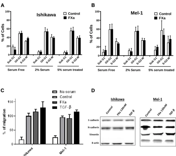
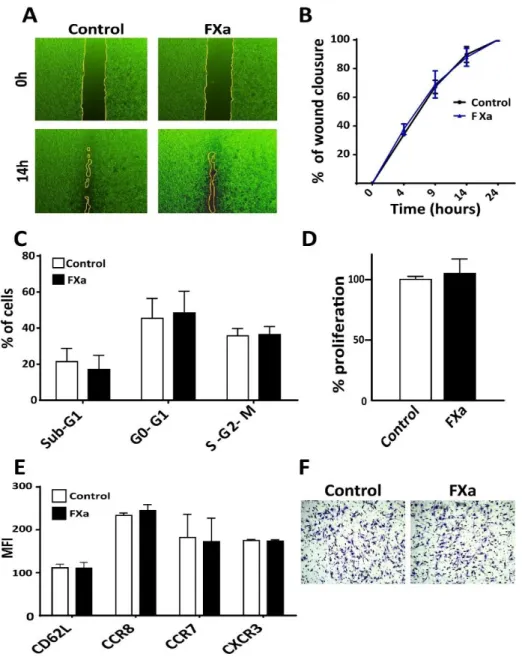
(33) 22. 5.3. FXa did not modify cancer cell proliferation, migration, invasion nor homing-. related proteins in vitro. Next, we sought to determine the mechanism by which FXa increases tumor growth and metastasis. First, we hypothesized that FXa has an effect upon cancer cells. As a first approximation, Ishikawa and Mel-1 were treated with FXa (130 nM) for 24h to analyze cell cycle using the cellular DNA content as a parameter (Fig 4A & B). There were not statistical differences between sub G1, G0-G1, S-G2-M phases. This result suggests that cancer cells are not proliferating or entering into apoptosis/necrosis (peak in the sub G0/G1 phase) in response to FXa. Tumor cells that were subjected to FXa treatment did not exhibit changes in the Endothelial to Mesenchymal Transition (EMT) markers E-cadherin, N-cadherin and Vimentin (Figure 4D). Consequently, cell migration was not affected in cancer cells exposed to FXa (Fig 4C). The migration of the cancer cells was evaluated using the Transwell system, in which 10% FBS was seeded in the lower chamber as a chemoattractant and FXa was added to cells in the upper compartment as a treatment. Next, the effect of FXa-treatment was evaluated in the murine melanoma cell line B16F10 in vitro. FXa addition (130nM) did not alter B16F10 cell migration (Fig 5A & B). Moreover, FXa treatment did not alter cell death (appearance of a sub G0/G1 peak by flow cytometry) or bring about significant changes in the cell cycle (Fig 5C). The lack of changes in proliferation was further confirmed using the colorimetric reagent WST-1, in which an increase in the absorbance is associated to a higher cell number (Fig 5D). Flow cytometry analysis confirmed that FXa did not change the levels of the cytokine receptors: CCR7, CCR8, CXCR3 and CD62L (Fig 5E). These receptors have been related to differential homing of cancer cells to distant organs, similarly to the figure 1C. Furthermore, invasion capabilities (evaluated using Transwells.
(34) 23. system with Matrigel coating) were not affected in response to FXa (Fig 5F). Additionally, we tested whether FXa could stimulate pro-angiogenic factors when in contact with cancer cells; however the conditioned medium from cancer cells treated with FXa did not alter capillary-like formation by the endothelial cells.. B. A 100. 60 40 20. Control FXa. 60 40 20. 0. b Su. Mel-1. 80. % of Cells. 80. % of Cells. 100. Control FXa. Ishikawa. G. 1. 1 -M G 0- -G2 G S. Serum Free. b Su. G. 1. 1 -M G 0- -G2 G S. 2% Serum. b Su. G. 1. 0. 1 -M G 0- -G2 G S. b Su. 5% serum treated. C. G. 1. 1 M G 20G S-G. Serum Free. b Su. G. 1. 1 M G 20G S-G. 2% Serum. b Su. G. 1. 1 M G 20G S-G. 5% serum treated. D β. Figure 4. FXa did not change cell cycle, migration or Epithelial Mesenchymal Transition (EMT) markers in Ishikawa and Mel-1 cells. Ishikawa (A) and Mel-1 (B) were treated with FXa for 24h and the subjected to cell cycle analysis using flow cytometry. (C) The migration of both cell lines was analyzed using transwell systems, promoting migration with 10% FBS in the lower chamber. Cells were co-treated with FXa or TGF-B and after 8h were fixed and stained with Violet Crystal. (D) Ishikawa and Mel-1 were treated with FXa or TGF- B (inductor of EMT) for 24h. EMT markers were visualized using Western Blotting in three different experiments. Representative images are shown. P>0.05, n=3..
(35) 24. Figure 5. FXa does not alter cancer cell biology in vitro. (A) A scratch was generated upon a confluent B16F10 cell monolayer. After that, FXa or vehicle were added and (B) wound closure was evaluated through the time, n=3. (C) Cells were treated with FXa, fixed and stained with propidium iodide. Cell cycle was analyzed with flow cytometry. (D) Semi-confluent B16F10 were treated with FXa for 24h and proliferation was evaluated using WST-1, n=3. (E) The adhesion molecule CD62L and the chemokines receptors CCR8, CCR7 and CXCR3 levels in cancer cells were analyzed by flow cytometry in response to FXa treatment, n=3. (F) B16F10 were treated for 24h with FXa and placed to invade for 8h. Cells were fixed and stained with violet crystal. P>0.05.
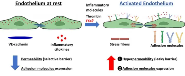
(36) 25. Figure 6. Endothelial activation: A link between coagulation and the inflammatory pathway. Pro-inflammatory molecules or Thrombin are released from an injury tissue or from an acute infection and trigger endothelial cell activation. Distinct chemokines (such as: IL-8, IL-6 and MCP-1) are released, and adhesion molecules (such as: VCAM-, ICAM-1 and Eselectin) are exposed to the bloodstream. Additionally, endothelial-hyperpermability occurs through stress fiber formation and the uncoupling of tight juctions (such as VE-cadherin). Under physiological conditions, this process promotes monocyte/neutrophil attachment, firm adhesion and extravasation to the inflammated tissue. The importance of FXa in endothelial activation and its relevance to cancer cell metastasis are poorly understood..
(37) 26. 5.4. FXa disrupts tubular like structure formation in Matrigel. An increasing number of publications are suggesting a contribution of tumor microenvironment to cancer progression and metastasis. The hemostatic role of coagulant proteins and their upregulation in cancer has been proposed as a main cause of Thrombosis in patients; however, the non-hemostatic effect on the pathogenesis and evolution of the disease is still unknown. The non-hemostatic function of coagulation proteins has been principally related to endothelial cells. Therefore, we proposed that this component of the tumor microenvironment represents a main actor in the pro-metastatic effect of FXa (Fig 6). Of note, Thrombin was employed in our experiments as a positive control for endothelial activation. As shown in figure 6, there is a well-described link between the coagulation system and the inflammatory pathway, in which Thrombin represents the most studied protein. Here, we postulate that FXa independently of Thrombin is promoting endothelial cell activation and thus increasing the metastasis of tumor cells. The main aim of the following experiments was to determine the effect on the metastasis of the B16F10 melanoma cells, principally because metastasis is the main cause of death in cancer patients. Therefore , it was interesting to us to study the importance of the coagulation cascade pathway in this pathological process. As a first approximation, changes in endothelial cell phenotype were analyzed in response to FXa. First, we used an angiogenesis assay culturing HUVECs in 3D matrigel wells. Under these conditions, endothelial cells form tubular-like structures, in which cells connect to each other and form polygonal elongations (Fig. 7). Our data showed a significant reduction in the percentage of tubular linked network after a 2h incubation with FXa (please refer to a time-lapse video. of. FXa-treated. endothelial. cells,. http://www.labowen.cl/cgi-. bin/Website/Pages/Internal.html.pl?PagId=51&). While cells were still attached to the Matrigel.
(38) 27. and alive, neither elongation or migration was observed. Furthermore, this result was maintained 24h post-treatment (not shown). The aforementioned results strongly suggest that FXa is increasing endothelial cell contraction. This effect is specific for the protease activity of FXa since the treatment with the zymogen (FX) did not lead to differences compared to the control (Fig 8). Given these findings, we next assessed the response of endothelial cells at cellular level..
(39) 28. Figure 7. FXa disrupts tubular structures in Matrigel and promotes endothelial cell contraction. (A) HUVECs were seeded in Matrigel in the presence of VEGF (10ng/mL). 15 h later, FXa, in two different concentrations, was added to the tubular structures already formed. Images were taken and representative pictures in 10X are shown. (B) The contraction of this already formed tubular network was quantified 2 h (white bars) and 4 h (black bars) after FXa addition, n=4 p<0.05..
(40) 29. Figure 8. FXa, but not the zymogen, disrupts tubular structures formed by endothelial cells in Matrigel. EA.hy cells were seeded in Matrigel in the presence of VEGF (10ng/mL). 15 h later, FX (zymogen) or FXa were added to the tubular structures already formed. The disassembly of this established tubular network was evaluated after 4 h, using the master segments index from ImageJ software quantification tool, n=3 p<0.05..
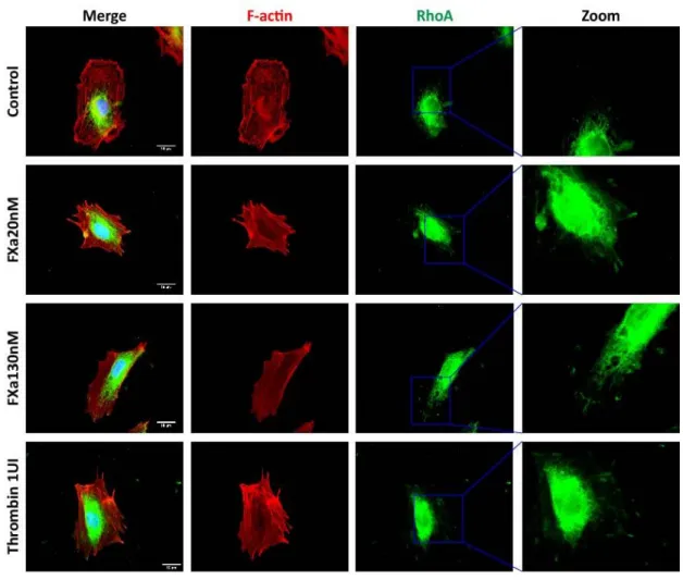
(41) 30. 5.5 FXa increases stress fiber formation and permeability-related protein responses in endothelial cells Based on the finding suggesting that FXa is promoting endothelial cell contraction, we analyzed the F-actin stress fiber formation (indicator of cell contraction) in HUVEC seeded at high/low confluency. In a high-confluency state, F-actin-rich rings were present in control conditions; however, treatment with FXa promoted the loss of F-actin-rich rings and an increase in stress fiber formation throughout the monolayer (Fig 9A). Moreover, in a low-confluency state, there was a loss of lamellipodia at the leading edge and an increment in F-actin enriched stress fiber formation in response to FXa. Also, a decrease in the cell area was observed, suggesting that cell contraction was occurring (Fig 9B). Additionally, the active form of RhoA (RhoA-GTP), a small GTPase identified as a regulator of the actin cytoskeleton and implicated in endothelial hyperpermeability, was observed in highly F-actin-enriched zones surrounding the edge of cell membrane in response to FXa treatment (Figure 10). The activation of this protein is related to cell contraction and cytoskeleton remodeling, suggesting that FXa is promoting a fast remodeling in endothelial cells. Furthermore, phosphorylation of Focal Adhesion Kinase (pFAK, Y397), a protein involved in the assembly/disassembly of focal adhesions, was increased in response to FXa after 5 min of treatment (Fig 11). The Y397 phosphorylation site has been described as an essential autophosphorylation residue, which is one of the first targets of FAK activation, and thus promoting the down-stream phosphorylation of the other residues involved in the cellular response and cytoskeleton remodeling..
(42) 31. Figure 9. FXa promotes stress fiber formation and endothelial cell contraction (A) High/low confluency HUVECs were treated with FXa and thrombin, fixed and stained with Phalloidin-rhodamine to observe endothelial phenotype and stress fiber content. (B) Cell area was quantified using ImageJ software, p<0.05. Magnification bar = 10 µm.
(43) 32. Figure 10. FXa-treatment promotes active RhoA localization at the edge of cells. HUVECs were treated for 5 min with FXa, fixed and prepared for the analysis of RhoA-GTP (active form) using the Far-IFI technique as described in methods. Stress fiber content was analyzed using Phalloidin-Rhodamine. Representative images at 100X are shown. Magnification bars in panels to the left = 10 µm..
(44) 33. Figure 11. FXa promotes pFAK distribution to the cellular border. Endothelial cells were treated for 5 min with FXa, fixed and prepared for the analysis of the phosphorylated Focal Adhesion Kinase (pFAK, green stain) by Immunofluorescence. Stress fiber content was analyze using Phalloidin-Rhodamine. Representative images at 100X are shown. Magnification bar = 10 µm..
(45) 34. 5.6 FXa induces endothelial hyperpermeability Based on the previous results, in which FXa was triggering changes in the endothelial cytoskeleton and in cell-contraction related proteins, we assessed the effect of FXa on the permeability of an endothelial monolayer. First, FXa at both concentrations induced the disruption of an important member of adherent junctions family that controls endothelial permeability, VE-cadherin. The pattern distribution of VE-cadherin within a confluent HUVEC monolayer (Fig 12 A) and in the Human Pulmonary Microvasculature Endothelial cell line, HPMEC-1 (Fig 12 B) was disrupted in response to 30 min of FXa exposure, suggesting that FXa can change endothelial permeability. It is important to note that all these effects were due to FXa, as prothrombin or Thrombin were not present in this in vitro system. The effect of FXa treatment on the permeability of an endothelial monolayer was evaluated using the Transwell system (Fig 13A). Briefly, endothelial cells were treated for 30 min with FXa, FX or Thrombin; and the permeability to the macromolecule BSA coupled to Evans Blue as a dye was evaluated. The pre-treatment with FXa, but not the zymogen FX, increased the permeability to Evans Blue-BSA (Figure 13B), as did also Thrombin (positive control). This increase in endothelial permeability was maintained for at least 30 min. After showing that FXa promotes endothelial hyperpermeability, we assessed the effect of FXa on vascular permeability to macromolecules in vivo using the cremaster model in C57BL/6 mice. The mice were anesthetized and the cremasteric muscle was exposed, prepared and treated topically with FXa (130nM). The mice were perfused with the macromolecule Dextran coupled with the green fluorescent molecule FITC to identify blood vessels and to analyze the extravasation of this dye. After 6 min of topical treatment, FXa significantly increased the FITC-.
(46) 35. dextran extravasation to interstitial tissue (Fig 14), visible as an increase background fluorescence near of blood vessels.. 5.7 An acute regime of FXa is enough to increase metastasis of B16F10 The aforementioned results suggest that FXa is increasing endothelial cell permeability, thus allowing circulating cancer cells to extravasate. Therefore, we hypothesized that FXa when applied only during the first 16h after cancer cell injection, when the cancer cells are attaching and crossing the endothelium into the pulmonary tissue, would result in increased metastasis 21 days later. Repeating the experiment detailed in figure 1, but now applying FXa only after injection of the cancer cells and /or 16h later (see schematic in Fig 15), we observed a clear increase in cancer metastasis at 21 days. In this case, we did not recreate the previous findings, in which FXa treatment increased the metastasis to different organs. Our results suggest that endothelial permeability is a potential mechanism by which FXa increases metastasis in this model..
(47) 36. A. B. Figure 12. FXa promote VE-Cadherin disruption in HUVEC and in the Human Pulmonary Microvasculature Endothelial Cells, HPMEC-1. Distribution of VE-cadherin pattern was analyzed after 30 min of treatment with FXa and Thrombin as a positive control. Representative images at 100X amplification are shown. Magnification bars = 10 µm..
(48) 37. Figure 13. FXa induces endothelial hyperpermeability in vitro. (A) Endothelial cells were seeded onto 1% gelatin pre-coated Transwells (3µm) and allowed to proliferate until generating a 100% confluent monolayer. Cells were then treated with FXa or FX (zymogen) and thrombin for 30 min in serum free media (A). Evans Blue-BSA was added onto the upper chamber, and 100µL aliquots were taken from the lower chamber. (B) Evans Blue-BSA was measured as an indicator of hyperpermeability (Absorbance at 620nm) in time, n=5 p<0.05..
(49) 38. Figure 14. FXa induces endothelial hyperpermeability in vivo. Cremaster muscle from mice was exposed, prepared and treated topically with FXa. Vascular hyperpermeability to FITCDextran was evaluated using intravital microscopy and quantifying the integrated optical intensity (IOI) in the tissue for 10 min, n=3 p<0.05..
(50) 39. 5.8. FXa changes mRNA expression within endothelial cells. Given the observed changes in the endothelial phenotype promoted by FXa, we assessed whether this was reflected in alterations at the transcriptional level using a GeneChip Human Gene 2.0 ST Array. After 4 h of treatment (EA.hy926 endothelial cell line), 127 genes were significantly up-regulated and 36 were down-regulated in response to FXa 130nM. These differences between treatments are represented in a heatmap (Fig 16A). A protein interaction network was generated based on the data from up-regulated genes and using the Gene Ontology Consortium database, showing a main cluster of proteins associated with the Interferon (IFN) response pathway. Transcripts of proteins associated with angiogenesis (FGF-2), inflammation (IL-6, ICAM-1), extracellular matrix degradation (PLAUR) and immune chemokines (CCL5) were also up-regulated in response to FXa (Fig 16B). In summary, the response triggered by FXa in endothelial cells matched to an inflammatory signature, validating the literature observations suggesting a role of the coagulation system in the inflammatory pathway. The entire list of up/down regulated genes is shown in Table 2 and 3 (Appendix)..
(51) 40. Figure 15. FXa increases metastasis in a short-term treatment. Schematic representation of the regimens of FXa administration in mice. FXa A & B groups were treated with FXa immediately after cancer cell injection but only FXa B group received a reinforcement 16h later. Representative images of lungs 21 days post-injection are shown and tumor mass referred to total lung mass was assessed..
(52) 41. Figure 16. FXa induces changes in endothelial cell mRNA expression. (A) Gene expression signature of endothelial cells treated with FXa was evaluated with the Affymetrix Human Genome 2.0 ST Array. Heatmap shows the 6 analyzed samples (EA.hy control vs EA.hy treated with FXa130nM for 4h) clustered by condition at the left side of the image. Red and green colors represent up and downregulated genes respectively. Gene clusters are at the top of the heatmap. (B) Protein Interaction Network based on the top 10 Upregulated genes and some of our genes of interest. Further information on the main pathways involved in FXa treatment is shown in Table 1..
(53) 42. Table 1. Top 5 up-regulated and top 5 “interesting” genes in response to FXa treatment in endothelial cells..
(54) 43. 5.9 FXa increases lung immune infiltration, a process reverted by Dalteparin To understand the contribution of FXa to inflammation in vivo, we analyzed the immune cell infiltration in lungs from mice treated with FXa alone or in combination with Dalteparin. In concordance with the microarray result, analysis of the non-cancer containing sections of the mice lungs showed that FXa treatment evoked an inflammatory response near blood vessels (Fig 17). As detected by the H&E stain, the immune infiltration was observed principally close to large blood vessels (based on the architecture, principally corresponding to veins and venules). This process was reversed or prevented by the presence of the anticoagulant Dalteparin, suggesting that anticoagulant treatment can have a potent anti-inflammatory effect..
(55) 44. Figure 17. FXa treatment promotes a pro-inflammatory response in mice, an effect reduced by Dalteparin treatment. Lungs from mice treated with Vehicle, FXa and FXa+Dalteparin were stained with H&E to analyze immune cell infiltration. Immune infiltration was quantified and expressed as the percentage of total area per lung, p<0.05..
(56) 45. 5.10. FXa increases cancer cell adhesion to the endothelium. Prior to cancer cell extravasation during metastasis, cancer cells must be attached firmly to the endothelium which allows them to withstand the pressure delivered by blood flow. As adhesion molecules allow cancer cell capture at the endothelial wall prior to extravasation, we assessed whether HUVEC increased adhesion molecules when treated with FXa. Figure 18 shows that FXa increased the protein levels of ICAM-1 and VCAM-1 after 4 and 8h of treatment. NO detectable levels were found at 2h post-treatment. Changes in VE-Cadherin total protein levels were not detected. Additionally, we also tested the response of the Human Pulmonary microvasculature Endothelial Cells (HPMEC-1) treated with FXa and Thrombin (as a positive control). We also used the monocyte cell line THP-1 treated with PMA as a control of ICAM-1 expression (Fig 19). We did not find statistical differences between cells treated with FXa, nevertheless we found statistical differences in the VCAM-1 levels of cells treated with Thrombin (Figure 19). A tendency to increased VCAM-1 total protein levels was observed in FXa-treated HPMEC-1, suggesting that Thrombin, instead of FXa, has a role in inflammation in this cell line. As a proof of concept that FXa con enhance the adhesion of cancer cells to an endothelium, we pre-treated a confluent monolayer of HUVEC with FXa for 4h (Fig 20). After that, tumor cells (B16F10-eGFP) were added and co-incubated for 30 min, washed with warm PBS and fixed for visualization. There was a significant increase in the number of attached B16F10-eGFP cancer cells to endothelial cells treated with FXa and Thrombin under static conditions (Fig 21). Interestingly, to assess the increase in adhesion under “physiological” conditions, we used a microfluidic microchannel, where B16F10 cells were pumped over a FXa pre-treated HUVEC.
(57) 46. monolayer. In line with our previous experiment, we observed that under flow conditions FXa increased adhesion of B16F10 to endothelial cells (Fig. 20).. Figure 18. FXa promotes adhesion molecule expression in HUVEC. 100% confluent HUVECs were treated with thrombin, FXa or vehicle in no serum media and total protein levels of the adhesion molecules ICAM-1 and VCAM-1 were analyzed. Representative images of 4 independent HUVEC primary cultures are shown..
(58) 47. VCAM-1 Normalized fold change. * 25 20. ns. 15 4 2. 1U I n bi. Th ro m. a1 30 nM FX. FX a2 0n M. C. on tr ol. 0. Figure 19. Thrombin, but not FXa, increases ICAM-1 protein levels in HPMEC-1. 100% confluent HPMEC-1 cells were treated with thrombin, FXa or vehicle in no serum media and total protein levels of the adhesion molecules ICAM-1 and VCAM-1 were analyzed. Representative images of 4 independent HUVEC primary cultures are shown. Quantification of normalized relative levels is shown..
(59) 48. Figure 20. FXa and Thrombin increases the adhesion of B16F10 under static conditions. (A) B16F10-eGFP cells were seeded for 30 min over a confluent monolayer of HUVEC cells pretreated with either vehicle or FXa for 4h. Subsequently, cells were washed with warm PBS, fixed and mounted for visualization. The number of B16F10-eGFP cells that remained attached were determined by fluorescence microscopy and using the particle counter of ImageJ software. n=4 p<0.05..
(60) 49. C o n tro l. A n g io g e n ic S c o r e. 6. FXa 4. 2. 0. C. o. n. tr. o. l F. X. a. Figure 21. FXa increases the adhesion of B16F10 under flow conditions. A microfluidic adhesion assay was developed, where HUVEC cells were treated for 6h with either control or FXa (130nM) prior to underflow conditions (flow 50µL/min, 37°C and 5% CO2), delivering 175.000 B16F10 cells per min. Resulting adherent cells were evaluated with live imaging for 20 min and then quantified using ImageJ software n=3 p<0.05..
(61) 50. Taking in consideration that FXa elicited an inflammatory response over endothelial cells, and thus promoting cancer cell adhesion, our next objective was to relate these findings to clinical observations. To assess that, we performed an analysis of the ICAM-1 and VCAM-1 ligands transcripts levels in melanoma patients using the TCGA-TIMER database. Consistent with an important role of the cell adhesion molecules ICAM-1 and VCAM-1 in melanoma metastasis, we observed that there is a significant increase of the adhesion molecules ligands: ITGAL, ITGAM, ITGB1, ITGB2 and ITGA4 mRNA levels in metastasis samples in comparison to primary tumor samples (Fig 22). This result suggests that melanoma cells that express these adhesion molecule ligands could have an advantage to metastasize, thus promoting their clonal expansion and maintaining the expression of molecules that facilitate the metastatic process..
(62) 51. Figure 22. RNA levels of adhesion molecule ligands are increased in metastasis samples compared to primary tumors. ICAM-1 ligands (LFA-1 and MAC-1) and VCAM-1 (VLA-4) were analyzed using TCGA-TIMER database to evaluate the relative RNA expression of endothelial adhesion molecules ligands (red boxplot represents primary tumors from melanoma patients, mine while purple boxplots represent metastasis samples). Data was extracted from 333 primaries and 331 metastatic melanomas patients (https://cistrome.shinyapps.io/timer/)..
Figure


+7


Documento similar