TítuloExploring genetic outcomes as frailty biomakers
16
0
0
Texto completo
(2) The concept of “frailty” has emerged in the last years as a more accurate and reliable measure of the aging process than the traditional chronological age. Frailty is a multidimensional geriatric syndrome of decreased reserves and resistance to stressors, resulting from cumulative declines across multiple physiological systems and causing vulnerability to adverse health outcomes, including dependency and death (1). Prevalence of frailty increases with age (2), and it has been reported to vary between 4.0% and 59.1% across different studies, due to factors such as criteria employed for frailty identification, gender, race, socioeconomic conditions, or education (1,3). Despite being phenotypically well characterized, the biological basis of frailty still remains fairly unknown. This is due to the fact that this syndrome is not characterized by impairment of a single system, but by several events and anomalies in multiple physiological systems in an intricate process that leads to frailty (4). To date, identification of frail individuals is mainly performed in health care settings by using clinical features, although other approaches using standard laboratory tests have been reported (5,6). The most commonly employed diagnostic criteria, due to their simplicity of implementation, are those proposed by Fried and colleagues in 2001 (1), based on five phenotypic characteristics (muscle weakness, low gait speed, unintentional weight loss, exhaustion, and low physical activity). Another frequent approach is to calculate a frailty index, based on the accumulation of health deficits (7). Detection of frailty as soon as possible is crucial, because it is potentially preventable and can be even reverted in its early stages (8). Development of biomarkers for this syndrome would allow an earlier and more objective identification of frail individuals, but a deeper knowledge on the biological basis of frailty is required for this aim. In this regard, frailty is commonly accepted to have a strong biological component resulting from cumulative cellular damage over the life course (9), although other aspects of aging involving intercellular communication, breakdown of homeostasis at the organism level, and so on probably participate in frailty development. Increased levels of damage can lead to different cellular alterations, including genomic instability, mutations, altered gene expression, loss of cell division potential, cell death, or impaired intercellular communication, among others (10). These alterations at the cellular and molecular levels could be a good basis to establish frailty biomarkers. T-cell receptor (TCR) is a complex of integral membrane proteins that participate in the activation of T cells in response to an antigen. Induced or spontaneous mutations in TCR genes could result in the phenotypic expression of TCR-defective T cells and thus contribute to impairment of T-cell response. It has been suggested that TCR variant frequency might be a particularly relevant end point in population monitoring for genetic damage (11). Accordingly, this end point has been previously employed as a mutagenicity biomarker in biomonitoring studies of occupationally or medically exposed individuals (12,13), as well as a predictor of cancer risk (14). The alkaline single-cell gel electrophoresis (comet) assay is a simple and sensitive method to measure primary DNA damage in individual cells. Strand breaks cause relaxation of the supercoiling in the DNA, and free DNA loops are pulled toward the anode during electrophoresis, giving the appearance of a comet tail (15). The phosphorylation of the C-terminal of the variant core histone protein H2AX (γH2AX) at the highly conserved amino acid Ser139 is a quickly occurring event in the early DNA damage response to double-strand breaks (16). The half-life of γH2AX after DNA damage induction has been estimated to be 2–7 hours; after this time, H2AX is again dephosphorylated (17). Still, it has been reported that γH2AX persistent in time represent DNA lesions with unrepairable double-strand breaks (18). DNA repair is one of the most important mechanisms to maintain genome integrity. Consequently, deficiencies in this process are often considered one of the key processes in the development of diseases such as cancer and other age-related pathologies (19). Indeed, it was previously suggested that one of the possible causes or events involved in frailty syndrome is the alteration of the cellular repair mechanisms that would result in the accumulation of genetic damage (9,10)..
(3) Hence, to improve the understanding of the biological features associated with frailty status, and consequently set the basis to identify potential biomarkers of frailty, in the present study, several genetic parameters were analyzed in a population of older adults (aged 65 and older) classified into frail, prefrail, and nonfrail according to Fried’s criteria (1). Genetic outcomes analyzed included mutation rate (by means of the TCR mutation assay), different types of genetic damage (by employing the comet assay and the γH2AX assay), and cellular repair capacity (by the DNA repair competence assay). Besides, the influence of clinical parameters, namely nutritional status and cognitive status, was also evaluated.. Method Participant Recruitment After obtaining approval from the University of A Coruña Ethics Committee (CE 18/2014), and following the principles embodied in the Declaration of Helsinki, peripheral blood samples were obtained by venipuncture from 250 volunteer donors, aged at least 65 years old, recruited from associations of elder people and nursing homes located in Galicia (NW of Spain). An informed consent form was signed by all participants in the study or by their relatives in case of inability. A questionnaire with information about demographics, lifestyle, and medical information was completed for each donor. The clinical evaluation of the participants was performed by qualified staff with extensive experience in the gerontology field, equally trained prior to the start of the study to unify criteria. Exclusion criteria included taking medications known to affect the immune system and having had cancer or any chronic infection (eg, human immunodeficiency virus, hepatitis B virus, hepatitis C virus). Characteristics of the study population are presented in Table 1..
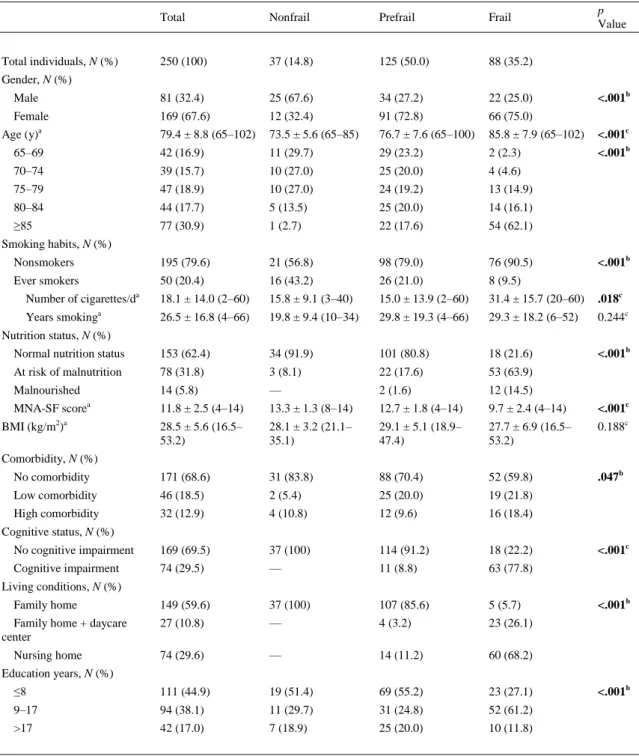
(4) Table 1. Description of the Study Population p Value. Total. Nonfrail. Prefrail. Frail. 250 (100). 37 (14.8). 125 (50.0). 88 (35.2). Male. 81 (32.4). 25 (67.6). 34 (27.2). 22 (25.0). Female. 169 (67.6). 12 (32.4). 91 (72.8). 66 (75.0). 79.4 ± 8.8 (65–102). 73.5 ± 5.6 (65–85). 76.7 ± 7.6 (65–100). 85.8 ± 7.9 (65–102). <.001c. 65–69. 42 (16.9). 11 (29.7). 29 (23.2). 2 (2.3). <.001b. 70–74. 39 (15.7). 10 (27.0). 25 (20.0). 4 (4.6). 75–79. 47 (18.9). 10 (27.0). 24 (19.2). 13 (14.9). 80–84. 44 (17.7). 5 (13.5). 25 (20.0). 14 (16.1). ≥85. 77 (30.9). 1 (2.7). 22 (17.6). 54 (62.1). Nonsmokers. 195 (79.6). 21 (56.8). 98 (79.0). 76 (90.5). Ever smokers. 50 (20.4). 16 (43.2). 26 (21.0). 8 (9.5). 18.1 ± 14.0 (2–60). 15.8 ± 9.1 (3–40). 15.0 ± 13.9 (2–60). 31.4 ± 15.7 (20–60). .018c. 26.5 ± 16.8 (4–66). 19.8 ± 9.4 (10–34). 29.8 ± 19.3 (4–66). 29.3 ± 18.2 (6–52). 0.244c. Normal nutrition status. 153 (62.4). 34 (91.9). 101 (80.8). 18 (21.6). <.001b. At risk of malnutrition. 78 (31.8). 3 (8.1). 22 (17.6). 53 (63.9). Malnourished. 14 (5.8). —. 2 (1.6). 12 (14.5). MNA-SF scorea. 11.8 ± 2.5 (4–14). 13.3 ± 1.3 (8–14). 12.7 ± 1.8 (4–14). 9.7 ± 2.4 (4–14). <.001c. 28.5 ± 5.6 (16.5– 53.2). 28.1 ± 3.2 (21.1– 35.1). 29.1 ± 5.1 (18.9– 47.4). 27.7 ± 6.9 (16.5– 53.2). 0.188c. No comorbidity. 171 (68.6). 31 (83.8). 88 (70.4). 52 (59.8). .047b. Low comorbidity. 46 (18.5). 2 (5.4). 25 (20.0). 19 (21.8). High comorbidity. 32 (12.9). 4 (10.8). 12 (9.6). 16 (18.4). No cognitive impairment. 169 (69.5). 37 (100). 114 (91.2). 18 (22.2). Cognitive impairment. 74 (29.5). —. 11 (8.8). 63 (77.8). 149 (59.6). 37 (100). 107 (85.6). 5 (5.7). 27 (10.8). —. 4 (3.2). 23 (26.1). 74 (29.6). —. 14 (11.2). 60 (68.2). ≤8. 111 (44.9). 19 (51.4). 69 (55.2). 23 (27.1). 9–17. 94 (38.1). 11 (29.7). 31 (24.8). 52 (61.2). >17. 42 (17.0). 7 (18.9). 25 (20.0). 10 (11.8). Total individuals, N (%) Gender, N (%). Age (y)a. <.001b. Smoking habits, N (%). Number of cigarettes/da a. Years smoking. <.001b. Nutrition status, N (%). BMI (kg/m2)a Comorbidity, N (%). Cognitive status, N (%) <.001c. Living conditions, N (%) Family home Family home + daycare center Nursing home. <.001b. Education years, N (%). Note: BMI = body mass index; MNA-SF = Mini-Nutritional Assessment–Short Form. Bold values mean statistically significant, i.e., P < .05. a Mean ± SD (range). bChi-square test (bilateral). cAnalysis of variance test (bilateral).. <.001b.
(5) Frailty Criteria Frailty status of the study participants was determined according to the five Fried’s phenotypic criteria (1): (a) shrinking or unintentional weight loss; (b) muscular weakness (grip strength); (c) self-reported exhaustion; (d) slow walk; and (e) low physical activity level. Individuals were classified as nonfrail, prefrail, or frail depending on the number of items they presented (none, 1–2, or ≥3, respectively). Clinical Evaluation The Spanish version (20) of the Mini-Nutritional Assessment–Short Form (21) was employed to assess the nutritional status of the participants. The score obtained in the questionnaire was used to classify participants with normal nutritional status (12–14 points), at risk of malnutrition (8–11 points), and malnourished (0–7 points). Cognitive status was evaluated using the Mini-Mental State Examination scale in its Spanish version (22). Scores of this questionnaire, ranging from 0 to 30, were adjusted for age and education level. Participants who scored less than or equal to 24 were considered as cognitively impaired (22). Charlson’s comorbidity index (23) was employed to determine general comorbidity and number of comorbid diseases. For each patient, an age-adjusted score was computed, coding comorbid diseases as 0 = absent and 1–6 = present. This index was also used to examine whether the 10-year mortality rates from comorbid disease were significantly different among frailty groups. A composite comorbidity-age score was computed for each participant, and the 10-year mortality was evaluated using a theoretical low-risk population, whose 10-year survival was 98.3% (24). Blood Sample Collection and Lymphocyte Isolation Whole blood was collected into BD Vacutainer CPT Cell Preparation Tubes with sodium heparin (Becton Dickinson), for the isolation of peripheral blood mononuclear leukocytes (lymphocytes + monocytes) following manufacturer’s instructions. Fresh leukocytes were employed in the TCR mutation assay. For the comet, γH2AX, and DNA repair competence assays, isolated leukocytes were frozen at −80°C in a solution composed of 50% fetal calf serum, 40% RPMI 1640, and 10% dimethyl sulfoxide, at 107 cells/mL, and stored until analysis. All samples were coded at the moment of collection, to ensure a blind study. TCR Mutation Assay TCR mutation assay was conducted in duplicate following the protocol proposed by Akiyama and colleagues (25). In brief, leukocytes were stained with 7-amino-actinomycin D as a viability marker and with fluorescein isothiocyanate-labeled antiCD3 and phycoerythrin-labeled antiCD4 antibodies (Becton Dickinson). Cell suspensions were analyzed using a FACSCalibur flow cytometer (Becton Dickinson) with Cell Quest Pro software (Becton Dickinson). The lymphocyte population was gated according to size and complexity. A minimum of 2.5 × 10⁵ lymphocyte-gated events were acquired, and TCR mutation frequencies (TCR-Mf) were calculated as the number of events in the mutant cell window (CD3 −CD4+ cells) divided by the total number of events corresponding to CD4+ cells..
(6) Alkaline Comet Assay To conduct the alkaline comet assay, following the protocol previously described in Laffon and colleagues (26), leukocytes were rapidly thawed at 37°C and subsequently centrifuged. The remaining pellet was suspended in phosphate-buffered saline solution. Cell viability was assessed by trypan blue exclusion technique being, in all cases, higher than 85%. An internal standard (leukocytes isolated from whole blood extracted once from a single donor and stored aliquoted at −80°C) was introduced in every electrophoresis run as described by Cebuslka-Wasilewska (27). Comet IV software (Perceptive Instruments) was used for image capture and analysis. For all donors and standards, 50 cells were scored from each replicate slide (ie, 100 cells in total) by a single scorer. The percentage of DNA in the comet tail (%TDNA) was evaluated as DNA damage parameter. γH2AX Assay γH2AX analysis was performed in duplicate following the protocol previously described by SánchezFlores and colleagues (28). Briefly, after being thawed, cell suspensions were centrifuged, and supernatants were removed. Remaining pellets were suspended in culture medium containing 1% phytohemagglutinin and incubated for 24 hours at 37°C. After fixation, cell suspensions were incubated with anti-human γH2AX-Alexa Fluor 488-conjugated antibody (Becton Dickinson) and stained with propidium iodide. Flow cytometry analysis was performed in a FACSCalibur flow cytometer (Becton Dickinson) with Cell Quest Pro software (Becton Dickinson). A minimum of 10,000 events in the lymphocyte region (gated according to size and complexity) were acquired, obtaining data from FL1 (γH2AX-Alexa Fluor 488) and FL2 (propidium iodide) detectors. The percentage of gated events positive for both γH2AX and PI was calculated with respect to the total lymphocytes gated and indicated as %γH2AX. DNA Repair Competence Assay DNA repair competence assay was performed as previously described by Laffon and colleagues (29). In brief, after being rapidly thawed, leukocytes were centrifuged, and the remaining pellet was incubated for 24 hours at 37°C in culture medium containing 1% phytohemagglutinin. Cells were then treated with the challenging agent bleomycin for 30 minutes at 37°C to induce DNA damage. Two duplicate slides were prepared for each donor: The first ones from cells that continued the comet assay protocol as previously described directly after bleomycin treatment (labeled as before repair) and the second ones from cells that, after bleomycin treatment, were incubated in fresh culture medium for 15 minutes at 37°C to allow DNA repair (labeled as after repair), before being processed following the comet assay protocol. An internal standard was introduced as well in each experiment as described by Cebulska-Wasilewska (27). Final data are shown as percentage of repair capacity (%RC), calculated as follows: (%TDNA BR − %TDNAAR) × 100/%TDNABR, where “BR” is before repair and “AR” is after repair. Leukocyte samples from several individuals (mostly frail and a few prefrail) were lost due to unexpected problems in storage. Hence, the number of data available in comet assay and DNA repair capacity evaluation is lower than in the rest of assays. Statistical Analysis Characteristics of the study population, namely sociodemographic factors (gender, age, living conditions, and years of education), lifestyle factors (smoking habit), and clinical characteristics (body mass index [BMI], nutrition status, comorbidity, and cognitive status), were compared in the three groups of older adults according to their frailty status. Analysis of variance and chi-square test were applied for continuous variables and categorical variables, respectively..
(7) The effect of frailty status on the genetic parameters was preliminarily tested through univariate analyses. Analysis of variance and Tukey’s post hoc test were applied to normally distributed parameters, according to Kolmogorov–Smirnov goodness-of-fit test (%γH2AX and log-transformed TCR-Mf). Kruskal–Wallis test and Bonferroni’s correction were applied to %TDNA and %RC because no better approximation to the normal distribution was achieved with transformation. Negative binomial regression models were used to estimate the effect of frailty status and other clinical parameters on TCR-Mf because this parameter is a count. The same effects on %γH2AX, %TDNA, and %RC were assessed by applying linear regression models on the log-transformed data. All models included gender, age, and smoking habit (never/ever smokers). Similar models were run adjusting also by BMI, but results obtained were very similar in all cases. The results are presented as mean ratios and 95% confidence intervals. Associations between genotoxicity parameters were estimated by Spearman’s rank correlation. Receiver-operating characteristic curves were computed to assess the biomarkers discriminating ability. The critical limit for significance was set at p less than .05. The statistical software used for the analyses were the IBM SPSS software package V. 20 (SPSS, Inc.) and the STATA/SE software package V. 12.0 (StataCorp LP).. Results The total population analyzed consisted of 250 individuals aged 65 and older (range 65–102; Table 1). Prefrail individuals were the most prevalent (half of the population), followed by frail participants (35.2%). Female gender was more frequent in the prefrail and frail groups, but the opposite occurred in the nonfrail group. As expected, mean age increased with frailty severity, and so did the prevalence of nonsmokers and individuals at risk of malnutrition and malnourished. Both the number of years smoking and BMI were homogeneously distributed among groups. Cognitive impairment was very frequent in frail individuals (77.8%), but it was absent in nonfrail participants. Most frail individuals lived in nursing homes, whereas all nonfrail individuals lived at family homes. Regarding years of education, distribution in the three education categories established was very similar for nonfrail and prefrail participants, with a higher prevalence of lower education level (51.4% and 55.2%, respectively), but middle education level was more prevalent among frail individuals (61.2%). Figure 1 shows the results of the different genetic parameters tested in the study population. According to the univariate analyses, no significant influence of frailty status on TCR-Mf, %TDNA, or %RC was obtained, although a clear tendency to decline in repair capacity with increasing frailty status was observed. Moreover, a significant (p < .01) and progressive increase of %γH2AX with frailty severity was also detected..
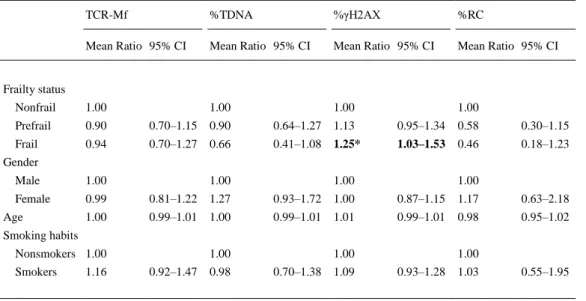
(8) Figure 1. Results of biomarkers analyzed in the study group, classified according to frailty status. Bars represent SEM. Different letters indicate statistically different groups (ANOVA and Bonferroni’s correction). ANOVA = analysis of variance; RC = repair capacity; TCR-Mf = T-cell receptor mutation frequency; TDNA = DNA in the comet tail; γH2AX = phosphorylated H2AX histone.. Associations between genetic parameters tested were not obtained according to Spearman’s correlation. Nevertheless, when associations with micronucleus rate in peripheral lymphocytes previously determined in the current population (30) were tested, a significant correlation was obtained for the association with %γH2AX (r = .252, p < .001). Besides, %γH2AX and the number of positive frailty criteria also showed to be significantly correlated (r = .201, p < .01). Results obtained in the multivariate statistical analyses, adjusting by gender, age, and tobacco consumption (and alternatively adjusting by BMI), confirmed previous results from univariate analyses on the influence of frailty (Table 2), that is, increasing frailty severity was accompanied by a progressive decrease in repair capacity and increase in H2AX phosphorylation; significance was observed for this last parameter in frail individuals with regard to nonfrail (p < .05). TCR-Mf and comet assay results did not show any significant effect. No significant influences were obtained for gender, age, or smoking on any parameter tested, and including BMI in the models scarcely changed the results..
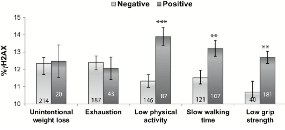
(9) Table 2. Effect of Frailty Status on the Biomarkers Analyzed TCR-Mf. %TDNA. %γH2AX. %RC. Mean Ratio 95% CI. Mean Ratio 95% CI. Mean Ratio 95% CI. Mean Ratio 95% CI. Nonfrail. 1.00. 1.00. 1.00. 1.00. Prefrail. 0.90. 0.70–1.15. 0.90. 0.64–1.27. 1.13. 0.95–1.34. 0.58. 0.30–1.15. Frail. 0.94. 0.70–1.27. 0.66. 0.41–1.08. 1.25*. 1.03–1.53. 0.46. 0.18–1.23. Frailty status. Gender Male. 1.00. Female. 0.99. 0.81–1.22. 1.27. 0.93–1.72. 1.00. 0.87–1.15. 1.17. 0.63–2.18. 1.00. 0.99–1.01. 1.00. 0.99–1.01. 1.01. 0.99–1.01. 0.98. 0.95–1.02. Age. 1.00. 1.00. 1.00. Smoking habits Nonsmokers 1.00 Smokers. 1.16. 1.00 0.92–1.47. 0.98. 1.00 0.70–1.38. 1.09. 1.00 0.93–1.28. 1.03. 0.55–1.95. Note: CI = confidence interval; RC = repair capacity; TCR-Mf = T-cell receptor mutation frequency; TDNA = DNA in the comet tail; γH2AX = phosphorylated H2AX histone. Models adjusted for age, sex, and smoking habits. *p < .05.. Given the positive influence of frailty on γH2AX assay results, and in order to determine the single contribution of each frailty criterion to γH2AX levels, this parameter was compared in the groups of participants negative and positive for each individual criterion (Figure 2). No differences were observed between individuals negative and positive for unintentional weight loss or exhaustion. However, significantly higher values of %γH2AX were observed in individuals positive for the criteria low physical activity (p < .001), slow waking time (p < .01), and low grip strength (p < .01) when compared with those individuals negative for each corresponding criterion. Comparisons of the other three genetic outcomes with regard to each individual frailty criterion did not produce any significant result. Moreover, when receiver-operating characteristic curve for %γH2AX was computed considering nonfrail and frail states, the area under the curve obtained was 0.723, which can be considered useful for some purposes, although not with high accuracy (31). Considering nonfrail and prefrail states, the area under the curve was lower (0.655)..
(10) Figure 2. Percentage of H2AX phosphorylation in the older adult population, according to each frailty criterion (1). The number of individuals included in each group is indicated inside each rod. Bars represent SEM. **p < .01, ***p < .001, significant difference with regard to negative (Student’s t test). γH2AX = phosphorylated H2AX histone.. Multivariate analyses were also applied to estimate the influence of other clinical parameters on the biomarkers tested. Nutritional status and comorbidity did not show significant effects on any biomarker. Notably, both TCR-Mf and %γH2AX increased significantly with the 10-year mortality risk estimation (p < .001 and p = .026, respectively), and significant correlations were found between 10-year mortality risk estimation and these parameters (r = .140, p = .029 for TCR-Mf, and r = .231, p = .001 for %γH2AX). Moreover, participants presenting cognitive impairment showed a significant 16% increase (95% confidence intervals = 1.02–1.31, p = .026) in the level of H2AX phosphorylation than participants with normal cognitive status.. Discussion Frailty is a condition of vulnerability involving an increased risk of poor health outcomes in older adults. The use of biomarkers to identify frail individuals not only would be a more precise and objective method for frailty identification, but also would make epidemiological studies more comparable, allowing to draw suitable conclusions from them. Besides, they might have the potential of anticipating the recognition of frail individuals, thus helping to prevent or attenuate the negative outcomes of frailty. However, and despite the last evidence supporting the relationship between a number of cellular alterations and frailty (10), up to now no biological feature has been validated to be employed as a biomarker to identify frailty status. With the aim of developing biomarkers of frailty, our group has investigated the potential association of frailty status with different genetic and immunological outcomes (30,32). In a very recent study (30), we evaluated the suitability of micronucleus frequency—a biomarker of genomic instability—to be employed as frailty biomarker. In such study, micronucleus rate in peripheral lymphocytes revealed its potential to be used for this purpose, because it showed a progressive increase with frailty severity, notably differentiating the group of frail participants. However, discrimination of prefrail individuals was not so clear, which would have been desirable from the clinical point of view. Besides, the association between micronucleus frequency and frailty resulted highly influenced by patients’ cognitive status, an aspect not considered in the current Fried’s frailty scale. On this basis, further investigations were necessary to fully understand the association between DNA cumulative damage and frailty status. Therefore, in the present study, we addressed the possible relationship between frailty status and different genomic outcomes, chosen on the basis of their demonstrated link to aging or age-related diseases (16,33), in a population of frail, prefrail, and nonfrail older adults..
(11) In the present study, the first addressing the possible relationship between frailty and mutagenicity, TCR-Mf was not found to be influenced by frailty status or age. Contradictory results have been previously obtained regarding age effect on TCR-Mf in occupationally exposed populations entirely less than 65 years old, with both the absence (34) and the presence (13) of such influence. Besides, significantly positive and linear association between TCR-Mf and age was observed by Akiyama and colleagues (25), in a wide age range group of individuals (0–96 years). It is likely that the age range covered by our study population (65–102 years) was not wide enough to detect variations in TCR-Mf with age, because our results indicate that over the age of 65 years, mutation rate remains stable and independent of frailty status. In the present study, the possible association between frailty and alteration of the cellular repair mechanisms has been evaluated by means of the DNA repair competence assay. However, and despite repair capacity showed a tendency to decrease with frailty severity, no significant differences were reached. To the best of our knowledge, only the study of Collerton and colleagues (35) has previously evaluated the possible association between repair capacity and frailty status in the older adults (aged 85 and older), and negative results were also obtained. In both cases, repair capacity was assessed using DNA damaging agents with similar action mechanism, namely ionizing radiation in Collerton and colleagues’ study and the radiomimetic agent bleomycin in the present study. These agents induce a wide spectrum of mutagenic lesions, including DNA base damage, abasic sites, and alkali-labile sites, which eventually result in DNA single- and double-strand breaks. Considering the demonstrated link between repair capacity and aging or age-related diseases (36), further investigations in this line, maybe using other assays to assess different repair pathways, are required prior fully rejecting DNA repair influence on frailty status. Primary DNA damage was determined by means of alkaline comet assay, but no association with frailty status was found in the present study. Similarly, Collerton and colleagues (35) reported a lack of association between genetic damage (γ ray–induced DNA strand breakage, evaluated by fluorimetric detection of alkaline DNA unwinding) and frailty condition in an older adult population aged 85 years and older. No other studies evaluating genetic damage in frail individuals are available in the literature; however, there are a number of works addressing the association between this kind of genetic damage and age showing inconsistent results. For instance, Humphreys and colleagues (37) found a decrease in DNA damage in the oldest group (aged 75–82 years) with respect to the young controls (aged 20–35 years) and to the younger older people (aged 63–70 years). Hyland and colleagues (38) reported similar levels of DNA damage in older individuals (86–96 years old) that in middle-aged individuals (40–60 years). On the contrary, Mladinic and colleagues (39) (age group ranges: 35–47 and 65–76 years old), Piperakis and colleagues (40) (age group ranges: children: 5–10; adults: 40–50; old people: 70–80 years old), and Mutlu-Türkoglu and colleagues (41) (age group ranges: 21–40 and 61–85 years old) observed an increase of DNA damage with age. Several authors have previously pointed out that results obtained in the alkaline comet assay are highly variable and often difficult to interpret because several types of damage are detected—including single- and double-strand breaks, alkali-labile sites, and breaks generated during repair processes—and can be influenced by a number of variables (season, diet, sample collection time, etc.) (42). Opposite to comet assay, γH2AX assay determines not a wide spectrum of DNA lesions but a specific kind of damage, namely double-strand breaks. In the present study, a progressive increase in the γH2AX rate with frailty severity not influenced by age was observed, statistically significant in both prefrail and frail groups when compared with the nonfrail groups. Besides, participants with cognitive impairment showed an increase in %γH2AX with respect to those with normal cognitive status. Silva and colleagues (43) also reported an increase in γH2AX nuclear expression levels in individuals with Alzheimer’s disease with regard to healthy individuals, and we previously found that the presence of cognitive impairment and frailty was independently related to an increase in the frequency of micronuclei in lymphocytes (30). These results would suggest a connection between cognitive impairment and frailty status and give support to the quite recently coined term “cognitive frailty,” introduced in an attempt to encapsulate the cognitive decline that is often observed in nondemented older adults who are physically.
(12) frail, with an underlying pathophysiology different from that driving the cognitive trajectory in neurodegenerative disorders (44). Although the present study is the first one in evaluating the relationship of H2AX phosphorylation with frailty status, the relationship between cellular senescence and persistent γH2AX, as indicative of unrepaired double-strand breaks, has been already suggested by several authors (16,18). Still, Schurman and colleagues (45) reported that γH2AX endogenous levels increase with age, peaking at ~57 years, which is in agreement with the absence of influence of age in the present study, where all participants were 65 and older. Both comet assay and H2AX assay detect double-strand breaks, but their results do not always coincide. Although γH2AX was found to be associated with frailty in the present study, several reasons could explain the lack of association for comet assay. On the one hand, whereas comet assay usually reveals recently induced and easily repairable DNA damage (46), γH2AX levels reflect fixed genetic damage or DNA damage that could not be properly repaired (47). On the other hand, it is not absolutely clear whether γH2AX foci in fact always reflect the presence of DNA breakage (48). For example, aging hematopoietic stem cells have been reported to harbor replication stress-induced nucleolar γH2AX foci, which persist due to ineffective H2AX dephosphorylation rather than ongoing genetic damage (49). Nevertheless, the significant association found in the current older adult population between γH2AX levels and micronucleus frequency in peripheral lymphocytes (30), both indicative of persistent DNA damage, points to unrepaired double-strand breaks as the outcome influenced by frailty status and by cognitive impairment, according to the results obtained in the multivariate analyses. Multivariate models were adjusted by gender, age, and smoking habit—parameters known to influence results of genetic outcomes—but adjustment for other different confounders should be considered in further studies. Besides, parallel results between γH2AX assay (this study) and micronucleus test (30) were also obtained when analyzing each five frailty phenotypic criteria independently. Thus, major contribution of physical activity, walking time, and grip strength to variation of %γH2AX and micronucleus frequency was observed, whereas unintentional weight loss and exhaustion did not contribute, or contributed only minimally, to both parameter modifications. These similar results in γH2AX and micronucleus assays provide further support to the relationship between fixed genetic damage and frailty and also suggest that combinations of some phenotypic criteria and biomarkers might improve frailty identification. Furthermore, because different frailty indices calculated on the basis of routine laboratory measurements have been reported to be useful for identifying older adults at increased risk of death (5,6), maybe the combination of biomarkers of genetic damage with standard laboratory biomarkers would enhance preclinical detection of at-risk individuals, also helping to understand the underlying biological processes of frailty. Strengths and limitations of the phenotype criteria selected for identifying frailty in this study must be mentioned. Fried’s criteria have a solid foundation of biological causative theory (1). Frailty phenotype has been applied to multiple epidemiological studies where it is predictive of adverse clinical outcomes and, together with Rockwood and Mitnitski’s frailty index, appear to be the most robust assessment tools for use by clinicians and researchers today (reviewed in ref. (9)), Nevertheless, it includes measurements not routinely used for patient assessment, it does not cover psychosocial components of frailty (9), and cognitive impairment may lead to diagnostic failure in all five criteria (50). Hence, further investigation is needed to figure out whether H2AX phosphorylation and micronucleus rates are also related to frailty as identified by other evaluation tools..
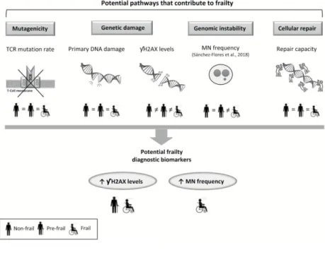
(13) Conclusions In the present study, we addressed the possible relationship between different genetic outcomes— namely mutagenicity, genetic damage, and cellular repair capacity—and frailty status by evaluating a population of older adults classified as frail, prefrail, and nonfrail according to the commonly used phenotypic criteria. No association of TCR-Mf and primary DNA damage with frailty was observed. DNA repair capacity showed a nonsignificant tendency to decrease with frailty, and the persistent levels of γH2AX increased progressively and significantly with frailty severity. Taking together our present results and the previous ones of micronucleus frequency in peripheral lymphocytes (30), the hypothesis that there is indeed a link between genomic instability, understood as fixed genetic damage, and frailty status seems to be plausible and supported by our data. Because both biomarkers, γH2AX and micronucleus rates, resulted significantly and progressively increased with frailty, they could be preliminarily proposed as tools for frailty identification or prediction (Figure 3); still, this is only an initial proposal and further validation is required to confirm the existence of this association and its specificity with frailty status and not through other age-related conditions probably associated with frailty. Besides, as γH2AX level resulted altered in both prefrail and frail groups, whereas micronucleus frequency was significantly increased only in frail individuals, a combination of both parameters could provide useful information regarding frailty severity, which would help clinicians to distinguish between prefrail and frail status and assisting them to offer personalized care. Consequently, results reported in the present study may contribute to improve health care/therapeutic strategies in older patients. Nevertheless, further investigation is necessary to prove whether the current findings are consistent and reproducible in larger sample sizes and different populations that may differ in the presence of other factors not considered in the analysis, to eventually standardize these biomarkers before they can be used in clinics, and to fully understand the influence of cognitive impairment on the results obtained.. Figure 3. Relationship between frailty and genetic outcomes analyzed in the study population. Biomarkers of mutagenicity, primary DNA damage, and cellular repair capacity do not show differences according to frailty status. MN frequency discriminates between nonfrail and frail individuals, meanwhile γH2AX levels are different in nonfrail, prefrail, and frail groups. A combination of both MN and γH2AX rates shows potential to be employed in frailty identification. MN = micronucleus; γH2AX = phosphorylated H2AX histone; TCR = T-cell receptor..
(14) Funding This study was supported by Xunta de Galicia (ED431B 2016/013, ED431C 2017/49, FrailNet network IN607C 2016/08, and ED481B 2016/190-0 to V.V.) and by the COST Action CA15132, “hCOMET,” Spanish Ministry of Economy, Industry, and Competitiveness (Ramon y Cajal Research Fellowship RYC-2015–18394 to L.L.-L.), INDITEX-UDC (to M.S.-F. and D.M.-P.), and Deputación Provincial de A Coruña (to M.S.-F.).. Conflict of Interest None reported.. References 1. 2. 3.. 4. 5. 6. 7. 8. 9. 10. 11.. 12.. 13.. 14.. 15. 16. 17.. 18.. Fried LP, Tangen CM, Walston J, et al. Frailty in older adults: evidence for a phenotype. J Gerontol A Biol Sci Med Sci. 2001;56:M146–M156. doi:10.1093/gerona/56.3.M146 Topinkova E. Aging, disability and frailty. Ann Nutr Metab. 2008;52(suppl 1):6–11. doi:10.1159/000115340 Theou O, Cann L, Blodgett J, et al. Modifications to the frailty phenotype criteria: systematic review of the current literature and investigation of 262 frailty phenotypes in the Survey of Health, Ageing, and Retirement in Europe. Ageing Res Rev. 2015;21:78–94. doi:10.1016/j.arr.2015.04.001 Zaslavsky O, Cochrane BB, Thompson HJ, et al. Frailty: a review of the first decade of research. Biol Res Nurs. 2013;15:422–432. doi:10.1177/1099800412462866 Howlett SE, Rockwood MRH, Mitnitski A, et al. Standard laboratory tests to identify older adults at increased risk of death. BMC Med. 2014;12:171. doi:10.1186/s12916-014-0171-9 Mitnitski A, Collerton J, Martin-Ruiz C, et al. Age-related frailty and its association with biological markers of ageing. BMC Med. 2015;13:161. doi:10.1186/s12916-015-0400-x Mitnitski AB, Mogilner AJ, Rockwood K. Accumulation of deficits as a proxy measure of aging. Sci World. 2001;1:323–336. doi:10.1100/tsw.2001.58 Roland KP, Theou O, Jakobi JM, et al. How do community physical and occupational therapists classify frailty? A pilot study. J Frailty Aging. 2014;3:247–250. doi:10.14283/jfa.2014.32 Dent E, Kowal P, Hoogendijk EO. Frailty measurement in research and clinical practice: a review. Eur J Intern Med. 2016;31:3–10. doi:10.1016/j.ejim.2016.03.007 Sánchez-Flores M, Marcos-Pérez D, Costa S, et al. Oxidative stress, genomic features and DNA repair in frail elderly: a systematic review. Ageing Res Rev. 2017;37:1–15. doi:10.1016/j.arr.2017.05.001 Cole J, Skopek TR. International Commission for Protection Against Environmental Mutagens and Carcinogens. Working Paper No. 3. Somatic mutant frequency, mutation rates and mutational spectra in the human population in vivo. Mutat Res. 1994;304:33–105. doi:10.1016/0027-5107(94)90320–4 Vershenya S, Biko J, Drozd V, et al. Dose response for T-cell receptor (TCR) mutants in patients repeatedly treated with 131I for thyroid cancer. Mutat Res. 2004;548:27–33. doi:10.1016/j.mrfmmm.2003.12.015 García-Lestón J, Roma-Torres J, Vilares M, et al. Genotoxic effects of occupational exposure to lead and influence of polymorphisms in genes involved in lead toxicokinetics and in DNA repair. Environ Int. 2012;43:29–36. doi:10.1016/j.envint.2012.03.001 Taooka Y, Takeichi N, Noso Y, et al. Increased T-cell receptor mutation frequency in radiation-exposed residents living near the Semipalatinsk nuclear test site. J Radiat Res. 2006;47(suppl A):A179–A181. doi:10.1269/jrr.47.A179 Duthie SJ, Ma A, Ross MA, et al. Antioxidant supplementation decreases oxidative DNA damage in human lymphocytes. Cancer Res. 1996;56:1291–1295. Siddiqui MS, Francois M, Fenech MF, et al. Persistent gammaH2AX: a promising molecular marker of DNA damage and aging. Mutat Res Rev Mutat Res. 2015;766:1–19. doi:10.1016/j.mrrev.2015.07.001 Bouquet F, Muller C, Salles B. The loss of gammaH2AX signal is a marker of DNA double strand breaks repair only at low levels of DNA damage. Cell Cycle. 2006;5:1116–1122. doi:10.4161/cc.5.10.2799 Sedelnikova OA, Horikawa I, Redon C, et al. Delayed kinetics of DNA double-strand break processing in normal and pathological aging. Aging Cell. 2008;7:89–100. doi:10.1111/j.1474-9726.2007.00354.x.
(15) 19. 20. 21.. 22. 23.. 24. 25. 26.. 27. 28.. 29.. 30.. 31. 32.. 33.. 34.. 35.. 36. 37.. 38.. 39.. 40.. 41.. Valdiglesias V, Pasaro E, Mendez J, et al. Assays to determine DNA repair ability. J Toxicol Environ Health A. 2011;74:1094–1109. doi:10.1080/15287394.2011.582320 Nestlé Nutrition Institute. A guide to completing the Mini Nutritional Assessment-Short Form (MNA®SF). 2009. http://www.mna-elderly.com/mna_forms.html. Accessed November 20, 2017. Kaiser MJ, Bauer JM, Ramsch C, et al. Validation of the Mini Nutritional Assessment Short-Form (MNA-SF): a practical tool for identification of nutritional status. J Nutr Health Aging. 2009;13:782– 788. doi:10.1007/s12603-009-0214-7 Blesa R, Pujol M, Aguilar M, et al. Clinical validity of the ‘mini-mental state’ for Spanish speaking communities. Neuropsychologia. 2001;39:1150–1157. doi:10.1016/S0028-3932(01)00055-0 Charlson ME, Pompei P, Ales KL, et al. A new method of classifying prognostic comorbidity in longitudinal studies: development and validation. J Chronic Dis. 1987;40:373–383. doi:10.1016/00219681(87)90171–8 Hutchinson TA, Thomas DC, MacGibbon B. Predicting survival in adults with end-stage renal disease: an age equivalence index. Ann Intern Med. 1982;96:417–423. doi:10.7326/0003-4819-96-4-417 Akiyama M, Kyoizumi S, Hirai Y, et al. Mutation frequency in human blood cells increases with age. Mutat Res. 1995;338:141–149. doi:10.1016/0921-8734(95)00019-3 Laffon B, Pasaro E, Mendez J. DNA damage and repair in human leukocytes exposed to styrene-7,8oxide measured by the comet assay. Toxicol Lett. 2002;126:61–68. doi:10.1016/S0378-4274(01)004325 Cebulska-Wasilewska A. Response to challenging dose of X-rays as a predictive assay for molecular epidemiology. Mutat Res. 2003;544:289–297. doi:10.1016/j.mrrev.2003.07.003 Sánchez-Flores M, Pasaro E, Bonassi S, et al. gammaH2AX assay as DNA damage biomarker for human population studies: defining experimental conditions. Toxicol Sci. 2015;144:406–413. doi:10.1093/toxsci/kfv011 Laffon B, Valdiglesias V, Pásaro E, Méndez J. The organic selenium compound selenomethionine modulates bleomycin-induced DNA damage and repair in human leukocytes. Biol Trace Elem Res. 2010;133:12–19. doi:10.1007/s12011-009-8407-9 Sánchez-Flores M, Marcos-Pérez D, Lorenzo-López L, et al. Frailty syndrome and genomic instability in older adults. Suitability of the cytome micronucleus assay as a diagnostic tool. J Gerontol A Biol Sci Med Sci. 2018. doi:10.1093/gerona/glx258 Swets JA. Measuring the accuracy of diagnostic systems. Science. 1988;240:1.285–1.293. doi:10.1126/science.3287615 Marcos-Pérez D, Sánchez-Flores M, Maseda A, et al. Frailty status in older adults is related to alterations in indoleamine 2,3-dioxygenase 1 and guanosine triphosphate cyclohydrolase I enzymatic pathways. J Am Med Dir Assoc. 2017;18:1049–1057. doi:10.1016/j.jamda.2017.06.021 Shao H, Ou Y, Wang T, et al. Differences in TCR-Vβ repertoire and effector phenotype between tumor infiltrating lymphocytes and peripheral blood lymphocytes increase with age. PLoS One. 2014;9:e102327. doi:10.1371/journal.pone.0102327 Lanza A, Robustelli della Cuna FS, Zibera C, et al. Somatic mutations at the T-cell antigen receptor in antineoplastic drug-exposed populations: comparison with sister chromatid exchange frequency. Int Arch Occup Environ Health. 1999;72:315–322. doi:10.1007/s004200050381 Collerton J, Martin-Ruiz C, Davies K, et al. Frailty and the role of inflammation, immunosenescence and cellular ageing in the very old: cross-sectional findings from the Newcastle 85+ Study. Mech Ageing Dev. 2012;133:456–466. doi:10.1016/j.mad.2012.05.005 Maynard S, Fang EF, Scheibye-Knudsen M, et al. DNA damage, DNA repair, aging, and neurodegeneration. Cold Spring Harb Perspect Med. 2015;5:a02513. doi:10.1101/cshperspect.a025130 Humphreys V, Martin RM, Ratcliffe B, et al. Age-related increases in DNA repair and antioxidant protection: a comparison of the Boyd Orr Cohort of elderly subjects with a younger population sample. Age Ageing. 2007;36:521–526. doi:10.1093/ageing/afm107 Hyland P, Duggan O, Turbitt J, et al. Nonagenarians from the Swedish NONA Immune Study have increased plasma antioxidant capacity and imilar levels of DNA damage in peripheral blood mononuclear cells compared to younger control subjects. Exp Gerontol. 2002;37:465–473. doi:10.1016/S0531-5565(01)00216-9 Mladinic M, Kopjar N, Milic M, et al. Genomic instability in a healthy elderly population: a pilot study of possible cytogenetic markers related to ageing. Mutagenesis. 2010;25:455–462. doi:10.1093/mutage/geq027 Piperakis SM, Kontogianni K, Karanastasi G, et al. The use of comet assay in measuring DNA damage and repair efficiency in child, adult, and old age populations. Cell Biol Toxicol. 2009;25:65–71. doi:10.1007/s10565-007-9046-6 Mutlu-Turkoglu U, Ilhan E, Oztezcan S, et al. Age-related increases in plasma malondialdehyde and protein carbonyl levels and lymphocyte DNA damage in elderly subjects. Clin Biochem. 2003;36:397– 400. doi:10.1016/S0009-9120(03)00035-3.
(16) 42. 43.. 44.. 45.. 46. 47.. 48. 49. 50.. Azqueta A, Collins AR. The essential comet assay: a comprehensive guide to measuring DNA damage and repair. Arch Toxicol. 2013;87:949–968. doi:10.1007/s00204-013-1070-0 Silva AR, Santos AC, Farfel JM, et al. Repair of oxidative DNA damage, cell-cycle regulation and neuronal death may influence the clinical manifestation of Alzheimer’s disease. PLoS One. 2014;9:e99897. doi:10.1371/journal.pone.0099897 Kelaiditi E, Cesari M, Canevelli M, et al.; IANA/IAGG. Cognitive frailty: rational and definition from an (I.A.N.A./I.A.G.G.) international consensus group. J Nutr Health Aging. 2013;17:726–734. doi:10.1007/s12603-013-0367-2 Schurman SH, Dunn CA, Greaves R, et al. Age-related disease association of endogenous γ-H2AX foci in mononuclear cells derived from leukapheresis. PLoS One. 2012;7:e45728. doi:10.1371/journal.pone.0045728 Collins A, Koppen G, Valdiglesias V, et al. The comet assay as a tool for human biomonitoring studies: the ComNet project. Mutat Res Rev Mutat Res. 2014;759:27–39. doi:10.1016/j.mrrev.2013.10.001 Valdiglesias V, Giunta S, Fenech M, et al. gammaH2AX as a marker of DNA double strand breaks and genomic instability in human population studies. Mutat Res. 2013;753:24–40. doi:10.1016/j.mrrev.2013.02.001 Rothkamm K, Barnard S, Moquet J, et al. DNA damage foci: meaning and significance. Environ Mol Mutagen. 2015;56:491–504. doi:10.1002/em.21944 Flach J, Bakker ST, Mohrin M, et al. Replication stress is a potent driver of functional decline in ageing haematopoietic stem cells. Nature. 2014;512:198–202. doi:10.1038/nature13619 Bieniek J, Wilczyński K, Szewieczek J. Fried frailty phenotype assessment components as applied to geriatric inpatients. Clin Interv Aging. 2016;11:453–459. doi:10.2147/CIA.S101369.
(17)
Figure

+2
Documento similar
There are other factors in the development of cancer such as genetic, environmental, as well as the role of oxidative stress and free radicals in response to damage caused by
Provided the historical territorial isolation to which CR and CB have been exposed, a solid interconnection between available genetic
Statistically significant differences in the profiles of antimicrobial susceptibility and genetic diversity (PFGE) of Trueperella pyogenes have been detected between
Here we can verify what we believe constitutes a paradox: the necessity of con- siderations of an ethical kind is imposed, but from that tradition of Economic Theory whose
Subtelomeric macrosatellite repeat D4Z4 methylation: genetic and non-genetic predictors and risk of urothelial carcinoma of the bladder 77 Chapter VI.. Lack of association
The Genetic-based community finding algorithm uses a genetic algorithm to find the best k communities in a dataset that could be represented as a graph and where any
In this study we performed a detailed genome-wide lin- kage analysis using MOD scores and 6 families affected by different CHD to test for a common genetic background among
c) 27K Sympathetic paraganglioma; Meta: Metastasis. f) Diagnosis was clinically determined. g) Diagnosis of PC.. d) B: without metastasis or M:
