Templated Hydroxyapatite Nucleation and Growth at
Physiological Conditions onto Self-assembled
Elastin-Like Nanoparticles.
By
Mohamed Hamed Misbah
Department of
Condensed Matter Physics, Crystallography and Mineralogy
G.I.R. Bioforge
A Master Thesis Submitted in Partial Fulfillment of Requirements for the Degree of M.Sc. in Molecular Nanoscience and Nanotechnology
Supervisor
Prof. J. Carlos Rodríguez-Cabello
Professor of Condensed Matter Physics, Crystallography and Mineralogy
School of Industrial Engineering
University of Valladolid
2013
University of Valladolid Department of Condensed Matter
Supervisor
Thesis title
:Templated Hydroxyapatite Nucleation and Growth at
Physiological Conditions onto Self-assembled
Elastin-Like Nanoparticles.
Researcher name:
Mohamed Hamed Misbah Elzehiri
Supervisor
:
Prof. J. Carlos Rodríguez-Cabello
Professor of Condensed Matter Physics,
Crystallography and Mineralogy
School of Industrial Engineering
University of Valladolid
Supervisor Signature University of Valladolid
Referees Committee Discussion
Thesis title:
Templated Hydroxyapatite Nucleation and Growth at
Physiological Conditions onto Self-assembled
Elastin-Like Nanoparticles.
Researcher name:
Mohamed Hamed Misbah Elzehiri
Referees Committee:
President
Prof. José Antonio de Saja
Sáez
Condensed Matter Physics,
Crystallography and Mineralogy
University of Valladolid
Vocal
Prof. Miguel Ángel
Rodríguez Pérez
Condensed Matter Physics,
Crystallography and Mineralogy
University of Valladolid
secretary Prof. Maria Luz Rodríguez
Méndez
Physical Chemistry and
Inorganic Chemistry.
University of Valladolid
Note
The present thesis is submitted to Valladolid University in partial fulfillment for the requirements of M. Sc. degree in Molecular Nanoscince and Nanotechnology. The courses during the academic years (2011-2013) are:
1er COURSE LEVEL COURSE
B0 M0 Introduction to the Master in Molecular Nanoscience and Nanotechnology.
BASIC COURSE
B1 Nanoscience Fundamentals: Concepts of nanochemistry and nanophysics. Characterization techniques
M1 Fundamentals of Nanophysics.
M2 Fundamentals of Nanochemistry.
M3 Characterization Techniques for Nanoscience.
B2 Molecular nanostructures and nanomaterials: methods of preparation, properties and applications.
M4 Preparation methods I: Supramolecular chemistry and bottom-up approach.
M5 Preparation methods II: Approximation down for nanofabrication.
M6 Molecular nanomaterials
2nd COURSE ADVANCED COURSE
B3 M7 Use of supramolecular chemistry for the preparation of nanomaterials and nanostructures.
B4 Electronics and molecular magnetism: basic concepts, major developments and applications.
M8 Introduction to molecular electronics.
M9 Unimolecular Electronics
M10 Molecular nanomagnetism
B5 M11 Current Issues in Molecular Nanoscience and Nanotechnology.
ACKNOWLEDGEMENTS
In the nameof ALLAH, the most Gracious and the most Merciful. Alhamdulillah, thanks and indebtedness are directed first and always to ALLAH for all his graves, without the power he gave me, the accomplishment of this work would have been certainly impossible.
Infinite thanks to my parents, thanks for their love and their support throughout my life. Thanks for both who tempered each other's advice to give me the right mix of keeping my feet on the ground and reaching for the stars (Be the best!).
Thank very much to my supervisor, Prof. J. Carlos Rodríguez-Cabello for all the orientation and support given and making me going in the right direction during this work. Thanks to G.I.R. Bioforge group, in this group I learned a lot of things from the first day. Thanks to Prof. Matilde Alonso Rodrigo, Prof. F. Javier Arias, Dr. Ana, Dr. Mercedes, Dr. Alessandra, Dr. Luis. My gratitude to my colleagues who finished their PhD, Dr. Laura, Dr. Menchu, Dr. Rosa, and for who making their PhD with me, María, Israel, Guillermo, Alicia, Piña, Ito, Arturo. My gratitude to Rocio, Irene and Vanesa.
Thanks to the coordination of the master, Prof. M. Luz Rodriguez Mendez.
I didn't can forget my professors, Prof. H. Doweidar and Prof. G. El-Damrawi in glass research group- Physics department – Mansoura university- Egypt, thanks to him who learned me the basis of the scientific research and what scientific research mean.
My gratitude to my friends and colleagues in my first group, Glass research group. My gratitude to my friends in the physics department at Mansoura University in Egypt.
My gratitude to my friends in Egypt and in Spain. Thanks for all of the people who learned me anything in my life.
Thanks a lot to my family that mean everything in my life that means the world to me, my parents, my uncle-aunty, my brothers and my sister. I extend my respect to my parents, my parental and maternal grandparents and all elders to me in the family. Thank you my mother, my father, my uncle and my aunty for showing love and faith in me. Really I am very lucky with this supportive family.
" No wealth is more useful than intelligence and wisdom; no solitude is more horrible than when people avoid you on account of your vanity and conceit or when you wrongly consider yourself above everybody to confide and consult; no eminence is more exalting than piety; no companion can prove more useful than politeness; no heritage is better than culture; no leader is superior to Divine Guidance; no deal is more profitable than good deeds; no profit is greater than Divine Reward; no abstinence is better than to restrain one's mind from doubts (about religion); no virtue is better than refraining from prohibited deeds; no knowledge is superior to deep thinking and prudence; no worship or prayers are more sacred than fulfillment of obligations and duties, no religious faith is loftier than feeling ashamed of doing wrong and bearing calamities patiently; no eminence is greater than to adopt humbleness; no exaltation is superior to knowledge; nothing is more respectable than forgiveness and forbearance; no support and defense are stronger than consultation."
Ali Ibn Abi Taleb
The present work was granted by the following research project:
Development of highly functional materials and systems from recombinamers having a complex molecular architecture.
Funded by: Ministry of Science and Innovation. National R + D + i 2008-2011.
MAT-2010-15310.
Entity: University of Valladolid. Period: 2011 to: 2013.
Contents
Abstract ... III
Chapter 1 ... - 1 -
Background ... - 1 -
1.1 Introduction to nanoscince ... - 1 -
1.1.1 Types of size-dependent effects on materials properties ... - 1 -
1.1.2 Manipulating nanoscience approaches ... - 3 -
1.2 Polymers material ... - 4 -
1.2.1 Synthetic polymers, for examples ... - 4 -
1.2.2 Natural biopolymers, for examples ... - 4 -
1.3 Natural amino acids ... - 5 -
1.3.1 Amino acids classified by R group ... - 7 -
1.3.2 Four levels complexity of protein structure ... - 10 -
1.4 Elastin like polymers (ELPs) ... - 19 -
1.4.1 Definition of ELP ... - 19 -
1.4.2 The molecular basis of the inverse transition temperature of ELP ……….……….. ... - 19 -
1.4.3 Molecular structure of (VPGVG) ELP ... - 21 -
1.4.4 Hydrophobic nature of ELP ... - 22 -
1.5 Self-Assembly ... - 28 -
1.5.1 Static self-assembly ... - 28 -
1.5.2 Dynamic self-assembly ... - 30 -
1.5.3 Self-assembly of recombinant polypeptides ... - 30 -
Chapter 2 ... - 33 -
Synthesis of Elastin-Like Polymer and Characterization Methods ... - 33 -
2.1 Genetically Engineered Polypeptides ... - 33 -
2.1.1 Concatemerization method ... - 33 -
2.1.2 Directional ligation method ... - 34 -
2.2 Materials and Methods ... - 36 -
II
2.2.2 Methods ... - 41 -
2.3 Characterization methods ... - 54 -
2.3.1 MALDI-TOFMS ... - 54 -
2.3.2 Fourier Transform Infrared Spectroscopy (FTIR) ... - 54 -
2.3.3 Differential Scanning Calorimetry ... - 54 -
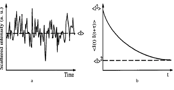
2.3.4 Light scattering ... - 55 -
2.3.5 X-ray diffraction (XRD) ... - 55 -
2.3.6 Electron microscopy ... - 55 -
Chapter 3 ... - 57 -
Properties of Recombinant Polymers (SNA15)3E50I60 and (SNA15)6E50I60 .... - 57 -
3.1 Molecular weight of (SNA15)3E50I60 and (SNA15)6E50I60 ELPs ... - 57 -
3.2 FTIR of polymer E50I60, (SNA15)3E50I60 and (SNA15)6E50I60 ELPs in solid state ... - 59 -
3.3 Transition temperature of E50I60, (SNA15)3E50I60 and (SNA15)6 E50I60 ELPs as a function of pH ... - 60 -
3.4 Dynamic Light Scattering of E50I60, (SNA15)3E50I60 and (SNA15)6E50I60 ELP nanoparticles ... - 63 -
Chapter 4 ... - 67 -
4.1 Biomimetic mineralization ... - 67 -
4.2 Biomimetic mineralization method ... - 69 -
4.3 Characterization of (SNA15)3E50I60 ELP nanoparticles and formed Nanostructured calcium phosphate: ... - 69 -
Conclusion ... - 79 -
Abstract
Studying materials in the nanosize scale lead to develop new synthetic approaches and discover a lot of new properties, and therefore manipulating to develop new materials that are used for different applications. In the nanoscale,
physics, chemistry, biology, material science and engineering converge toward the same principles and tools. In this work, nanoparticles that developed genetically have been used to form hydroxyapatites.
Polymers such as elastin-like polymers can be manipulated by the
bottom-up approach to form nanoparticles and also by top-down to form patterns in scales of nanometers on the polymer hydrogel.
The first aim of this work is to synthesis a nanoparticles and characterizing it. These nanoparticles are synthesized from amphiphilic
elastin-like copolymers that exhibit a lower critical solution temperature, (LCST), and under the effect of the environmental stimuli could show a transition from soluble phase to insoluble phase. Below this transition temperature, in aqueous solution, the polymers chains are hydrated and extended by the hydrophobic interaction. Above this transition temperature, the chains can be assembled to
form a phase separated state and adopt a dynamic, regular and nonrandom structure.
Due to the self-assembly properties of thess amphiphilic elastin-like polymers at low temperature, recombinant DNA genetic engineering has been
used to recombine it with SNA15 fragment. The SNA15 fragment is the first
fifteen amino acid from salivary protein statherin wherein the two phosphoserine amino acid residues at positions 2 and 3 have been substituted by L- aspartic acid. This fragment has a negative charge and a helical structure in
all solvents that has a high affinity to nucleate and promote the crystallization of hydroxyapatites. The amphiphilic block copolymer has been recombined with three and six fragments of SNA15. These polymers have been characterized in
IV
scattering; furthermore, have been characterized in the solid state by Fourier Transform Infrared Spectroscopy (FTIR).
The second aim of this work is using these nanoparticles as a template to form nanoparticles of calcium phosphates under physiological conditions. The
elastin-like polymers assembled to nanoparticles that are able to form calcium phosphate in SBF at 37oC. Electron microscopy used to study the formed nanoparticles of calcium phosphate by performing electron diffraction and elemental analysis over the nanoparticles. Also, the calcium phosphates formed
Chapter 1
Background
1.1 Introduction to nanoscince
Nanoscience is the since that study the phenomena and manipulation of materials at atomic, molecular and macromolecular scales, which is derived from the distance of a nanometre (nm), one-billionth of a meter, 10-9 m.
In physics and electrical engineering, nanoscience is related to the quantum behavior and the behavior of photons and electrons in the nanoscale structures.1 In chemistry, nanoscince is associated with the size of polymer molecules2, phase separated polymers3-4, colloids5 and micelles6-7 etc... In biology and biochemistry, the nanostructures of components of the cell have to
be interested structures from DNA and viruses to subcellular organelles and gap junctions that are nanostructures.8-9 The properties of materials in smaller size are different from the properties in bulk size, where the properties of materials are size-dependent. The types of size-dependent are surface effects and quantum
effects.10
1.1.1 Types of size-dependent effects on materials properties
1.1.1.1 Surface effects
Surface effects are so important because atoms at surfaces have fewer
neighbors than atoms in bulk. Because of this lower coordination and unsatisfied bonds, surface atoms are less stabilized than bulk atoms. Also the fraction of atoms at the surface are depend on the size of the particles where the smaller the particle, the larger the fraction of atoms. The ratio between the surface area and the volume is defined as dispersion. The dispersion F on the
-2
-to the radius. The -total number of a-toms N in this sphere scales linearly with volume and so the dispersion F proportional to N-1/3; Fig.1.1.10
Fig.1.1: Evolution of the dispersion F as a function of n for cubic clusters up to n=100(N=106). The structure of the first four clusters is displayed.10
1.1.1.2 Quantum effects
Fig.1.2: A bulk semiconductor has continuous condition and valence energy bands separated
by a fixed energy gap, Eg, while a semiconductor NC is characterized by discrete atomic-like
-3
-Quantum effects are dominating the behavior of matter at the Nano-scale particularly at the lower end. In a bulk semiconductor there are energy levels and so many close to each other formed a system of continuous energy called band. By contrast, the energy levels of electrons in the semiconductor
nanoparticles are far apart, as shown in Fig.1.2.1
1.1.2 Manipulating nanoscience approaches
1.1.2.1 Top-Down approach
In this approach (physics); there are smaller pieces of matter is
manipulated by photolithography and related techniques. This approach is subject to drastic limitations for dimensions smaller than 100 nm.11,12 Therefore, "there is plenty of room at the bottom for further miniaturization" is for Richard Feynman13 stated in a famous talk to the American Physical Society in 1959.
1.1.2.2 Bottom-up approach
This approach (chemistry) starts from nanoscale or subnanoscale objects to build up nanostructures.14-15 This method includes the technique of molecular synthesis15, polymer science2, colloid chemistry5 and the related areas for
-4
-1.2 Polymers material
Polymers such as elastin-like polymers (ELPs) can be manipulated by the bottom-up approach to form nanoparticles6, 16 and also by top-down to form patterns in scales of nanometers on the polymer hydrogel12.
Polymers consist of large molecules, i.e. macromolecules. According to the basic IUPAC definition (Metanomski 1991): "A polymer is a substance composed of molecules characterized by the multiple repetition of one or more species of atoms or groups of atoms (constitutional repeating units) linked to
each other in amounts sufficient to provide a set of properties that do not vary markedly with the addition of one or a few of the constitutional repeating units."17
A polymer can be a three-dimensional network or two-dimensional network or one-dimensional network.18 There are many types of polymers
including synthetic and natural polymers.
1.2.1 Synthetic polymers, for examples
Plastics.19-20
Elastomers - solids with rubber-like qualities.21-22
Fibers.23
1.2.2 Natural biopolymers, for examples
Polypeptides in proteins24-silk25, Collagen26, Keratin27-28.
Polysaccharides24(Carbohydrates)29 - Cellulose30, Starch31, Glycogen32.
Nucleic acids33 - DNA34 and RNA35.
-5
-1.3 Natural amino acids
An amino acid is an organic compound containing an amino group and an acidic group, where there are 20 natural amino acids.36 The amino acids that
constitute proteins are all -amino acids37. The ―α-carbon‖ is bonded to four
different groups: an amino group, a carboxylic acid, hydrogen, and the side chain as shown in the following schematic:
Because these are four different chemical groups, the α-carbon is chiral,
(Stryer,1988).38-39 For identifying the carbon in the amino acid, the ―first‖ carbon of the amino acid is the carboxylate carbon. The next carbon is called the
-carbon, because it is in the position - to the first carbon. The side chain is
then bonded to the -carbon. The side-chain carbons are given Greek letters in
the order , γ, δ, ε counting away from the -carbon; for example, in Lysine
amino acid as shown in the following schematic:39
The four different groups, a carboxyl group, an amino group, an R group, and a hydrogen atom, can occupy two unique spatial arrangements that are due
to the tetrahedral arrangement of the bonding orbital around the -carbon atom
-6
-There are two possible stereoisomers where the -carbon is the chiral
center. All molecules with a chiral center are optically active, that is they rotate
in a plane of polarized light. The two forms represent a class of stereoisomers called enantiomers as shown in Fig.1.3. The amino acid residues in proteins are L-stereoisomers. D-Amino acid residues have been found only in a few, generally small peptides, including some peptides of bacterial cell walls and
certain peptide antibiotics.38, 40-42
Fig.1.3: Stereoisomerism in -amino acids. (a) The two stereoisomers of alanine, L- and
D-alanine, are nonsuperimposable mirror images of each other (enantiomers). (b, c) Two
different conversions for showing the configurations in space of stereoisomers. In perspective
formulas (b) the solid wedge-shaped bonds project out of the plane of the paper, the dashed
bonds behind it. In the projection formulas (c) the horizontal bonds are assumed to project out
of the paper, the vertical bond behind. However, projection formulas are often used casually
-7 -1.3.1 Amino acids classified by R group
The following table shows the amino acids into five main classes based on the properties of their R groups (Table 1.1), in particular, their polarity, or tendency to interact with water at biological pH (near pH 7.0).
Table.1.1: properties and conventions associated with the common amino acids found in proteins:39
*A scale combining hydrophobicity and hydrophilicity of R groups; it can be used to measure the
tendency of an amino acid to seek an aqueous environment (- values) or a hydrophobic environment
(+ values), see Chapter 11. From Kyte, J. & Doolittle, R.F. (1982) A simple method for displaying the
hydropathic character of character of a protein. J.Mol. Biol. 157, 105-132.
t
Average occurrence in more than 1,150 proteins. From Doolittle, R.F. (1989) Redundancies in
protein sequences, in Prediction of protein Structure and the Principles of Protein Conformation
-8
-Fig.1.4 shows the structures of the 20 common amino acids and some of their properties are listed in Table 1.1. Within each class, there are gradations of polarity, size, and shape of the R groups.
Nonpolar, aliphatic R groups
Polar, uncharged R groups
Aromatic R groups
Positively charged R groups Negatively charged R groups
Fig.1.4: The 20 common amino acids of proteins. The structural formulas show the state of
ionization that would predominate at pH 7.0. The undashed portions are those common to all
the amino acids; the portions shaded in red are the R groups. Although the R group of
histidine is shown uncharged, its pKa (see table.1.) is such that small but significant of these
-9
-The amino acids are joined end to end through covalent linkage. -The α-carboxyl
group of the first amino acid reacts with the -amino group of the next amino
acid to generate a peptide bond and eliminate a water molecule as shown in Fig.1.5.
Fig.1.5: Formation of a peptide bond by condensation. The -amino group of one amino acid
(with R2 group) acts as a nucleophile to displace the hydroxyl group of another amino acid (with R1 group), forming a peptide bond (shaded in yellow). Amino groups are good nucleophiles, but the hydroxyl group is a poor leaving group and is not readily displaced. At
physiological pH, the reaction shown does not occur to any appreciable extent.39
Fig.1.6 shows the structure of a pentapeptide that has two different ends, termed respectively the N-terminus and the C-terminus. A polypeptide chain consists of a regularly repeating part, called the ‗main chain‘ or ‗backbone‘,
-01
-Fig.1.6: The pentapeptide serylglycyltyrosylalanylleucine, or Ser-Gly-Tyr-Ala-Leu. Peptides
are named beginning with the amino-terminal residue, which by convention is placed at th
left. The peptide bonds are shaded in yellow; the R groups are in red.39 1.3.2 Four levels complexity of protein structure
Fig.1.7: Levels of structure in proteins. The primary structure consists of a sequence of amino
acids linked together by peptide bonds and includes any disulfide bonds. The resulting
polypeptide can be coiled into units of secondary structure, such as an -helix. The helix is a
part of the tertiary structure of the folded polypeptide, which is itself one of the subunits
-00 -1.3.2.1 Protein primary structure
The primary structure of the protein is the linear sequence of amino acids that linked by the covalent bonds, mainly peptide bond and disulfide
bond.39, 43-45 The -carbons of adjacent amino acid residue are separated by
three covalent bonds, arranged as C-C-N-C. It is found from X-ray diffraction studies of crystals of the amino acids and of simple dipeptides and tripeptides
that the peptide C-N bond is somewhat shorter than the C-N bond in a simple amine, and all the atoms associated with the peptide bond are coplanar, which mean that the carbonyl oxygen and the amide nitrogen are partially shared by two pairs of electrons. Furthermore, a small electric dipole produced due to the
partial negative charge of oxygen and the nitrogen positive charge.39, 46-47
Theoretically, the planar peptide bond can take trans and cis configurations, Fig.1.8, but trans form is favored energetically than cis form. The rotation of the peptide bond CO-NH is restricted because of its partial
double bond character due to the resonance (Fig.1.9a.). But for the single bonds
that link each C atom to the N and C atoms of the peptide bonds show free
rotation; the angle of rotation around the N–C bond is phi (ɸ) and the angle of
rotation around the C–C bond from the C-atom is psi (Ψ).39, 48
Fig.1.8: The configurations of trans and cis forms of the peptide bond CO-NH with the angle
of rotation around the N–C bond is phi (ɸ) and the angle of rotation around the C–C bond
from the C-atom is psi (Ψ).48
-02
-When the polypeptide is in its fully extended conformation and all peptide groups are in the same plane, both (ɸ) and (Ψ) can be defined as 180o (Fig.1.9b).39 In principle, ɸ and Ψ can take any value between 180o and -180o, but the conformation at 0 of both ɸ and Ψ (Fig.1.9c) not permitted and this
conformation is used as a reference point for determining the angle of rotation.39
Fig.1.9: The planar peptide group. (a) Each peptide bond has some double-bond character due
to resonance and cannot rotate. (b) Three bonds separate sequential carbons in a
-03
-respectively. The peptide C-N bond is not free to rotate. Other single bonds in the backbone
may also be rotationally hindered, depending on the size and charge of the R groups. In the
conformation shown, ɸ and Ψ are 180o (or-180o). As one looks out from the carbon, the Ψ and ɸ angles increase as the carbonyl or amide nitrogen (respectively) rotate clockwise. (c)
By convention, both ɸ and Ψ are defined as 0o when the two peptide bonds flanking that carbon are in the same plane and positioned as shown. In a protein, this conformation is
prohibited by steric overlap between an -carbonyl oxygen and an -amino hydrogen atom.
To illustrate the bonds between atoms, the balls representing each atom are smaller than the
van der Waals radii for this scale. 1Ao=0.1 nm.39
1.3.2.2 Protein Secondary Structure39, 43-44, 48
The side-chains of the amino acids of the proteins interact with one another or with the solvent to form the global structure of the protein, which is called Protein Secondary Structure. These interactions are non-covalent, that are;
1- Hydrogen bond that is resulted from hydrophilic amino acid, where the
hydrogen atom that is connected covalently with an electronegative atom (donor) interact with another electronegativity atom (acceptor).
2- Electrostatic forces between net charges of opposite signs or between two dipoles.
3- Repulsion forces or van der Waals forces between electron orbitals of atoms.
4- Hydrophobic interactions, in which amino-acid residues, that engage in van der Waals interactions, have a tendency to repel with water and pack against each other.
The major two types of protein secondary structure are -helices, and
-04 -1.3.2.2.1 The -helix structure39, 43, 48
Fig.1.10. Four models of the -helix, showing different aspects of its structure. (a) Formation of a right-handed -helix. The planes of the rigid peptide bonds are parallel to the long axis of the helix, depicted here as a vertical rod. (b) Ball-and-sick model of a right-handed helix,
showing the interaction hydrogen bonds. The repeat unit is a single turn of the helix, 3.6
residues. (c) The -helix viewed from one end, looking down the longitudinal axis (derived
from PDB ID 4TNC). Note the positions of the R groups, represented by purple spheres. This
ball-and-sick model, used to emphasize the helical arrangement, gives the false impression
that the helix is hollow, because the balls do not represent the van der Waals radii of the
individual atoms. As the space-filling model (d) shows, the atoms in the center of the -helix
are very close contact.39
Fig.1.10 show a polypeptide chain arranged in a helical structure or
-helix structure. It is shown that the polypeptide backbone is tightly wound around an imaginary axis in the middle of the helix, and the R groups of the amino acids stand outward from the helical backbone. A single turn of the helix
-05
-covalently bonded to the nitrogen atom of a peptide linkage and the carbonyl oxygen atom of the fourth amino acid from this peptide linkage (Fig.10b).
Each peptide bond possesses an individual dipole moment, and these dipoles are connected through the hydrogen bonds of the helix that resulting in a
net dipole with a positive charge at the amino end and a negative charge at the carboxyl end as shown in Fig.1.11. The net dipole increases with the length of the helix.39, 48
Fig.1.11: Helix dipole. The electric dipole of a
peptide bond is transmitted along an -helical
segment through the intrachain hydrogen bonds,
resulting in an overall helix dipole. In this
illustration, the amino and carbonyl constituents
of each peptide bond are indicated by + and –
symbols, respectively. Non-hydrogen-bonded
amino and carbonyl constituents in the peptide
bonds near each end of the -helical region are
shown in red.39
1.3.2.2.2 The -Conformation
The -sheet structure39, 43, 48
A five to ten residues can form a strand of a rotation angle of -120o for ɸ and of 140 for Ψ that is called -strand. This -strand is not stable
structure and tend to interact with others -strand of another region of the
same polypeptide chain. These strands are interacted by hydrogen bonds
forming -sheet. The polypeptide chain backbone is arranged side by side by
-06
-polypeptide chains can be arranged in a -parallel sheets in which have the
same amino to carboxyl orientations or -antiparallel sheets in which have
the opposite amino to carboxyl orientations.
Fig.1.12: The -conformation of polypeptide chains. These top and side views revels the R
groups extending out from the -sheet and emphasize the pleated shape described by the
planes of the peptide bonds. (An alternative name for this structure is -pleated sheet.)
-07
-in which the am-ino-term-inal to carboxyl-term-inal orientation of adjacent cha-ins (arrows) is
inverse. (b) Parallel -sheet.39
The -turn structure39, 43
At the ends of each segment of the -sheet, there is a reverse turn or
-turn that makes the polypeptide chain -turn back upon itself. There are amino acids are in turns or loops in which the polypeptide chain reverses their
direction. These turns connect successive chains of -helix or
-conformations. -turns are the common turns that link two adjacent chains of
an antiparallel -sheet, which have a structure of 180o turn where the
carbonyl oxygen of amino acid residue form a hydrogen bond with the hydrogen atom, that bonded with the nitrogen atom, of the fourth amino acid
residue, as shown in Fig.1.13.
Fig.1.13: Structure of turns. (a) Type I and type II turns are most common; type I turns occur more than twise as frequently as type II. Type II turns always have Gly as the third
residue. Note the hydrogen bond between the peptide groups of the first and fourth residues
-08 -1.3.2.3 Tertiary structure39, 43-44, 48
Tertiary structure of protein is the three-dimensional structure of proteins. The tertiary structure is determined by the packing of the secondary structure that is combined to form what is called domains, see Fig1.7. A single domain
can have about 100-150 amino acid residues of about 25 Å diameter.
Protein structures can also be classified, by the types of structural motifs that formed the domains, into four main groups:
1- Proteins that have -helices in an antiparallel or perpendicular arrangement.
2- Proteins, that have -sheet where the -strands are arranged in antiparallel manner.
3- / Proteins; that have -sheets surrounded by -helices.
4- + Proteins that are a combination of and motifs.
The tertiary level of structure refers to the spatial arrangement of a polypeptide chain through folding and coiling to produce a compact globular
shape. The tertiary structure is essentially determined by the packing of the
secondary structures, -helices and -sheets, which combine to form one or several units called ‗domains‘. These combinations are limited in number, and
some of them are especially frequent in proteins. They represent the fundamental elements of globular polypeptide chains in terms of three-dimensional structure as well as in terms of function. On average, a single domain consists of 100–150 amino acid residues, corresponding to a globule of
about 25 Å diameters.
1.3.2.4 Quaternary structure39, 43-44, 48
When there are two or more separate polypeptide chains or subunits that are identical or different in some proteins, the structure that describes the spatial arrangement of these subunits in three-dimensional complexes is called
-09
-1.4 Elastin like polymers (ELPs)
1.4.1 Definition of ELP
The amino acids that are arranged in the following sequences, LGGVG49, GXGGX50, APGVGV51, AVGVP52, KGGVG53, LGAGGAG54 and VPGXaaG, in
which Xaa can be any of the natural or synthetic amino acids except for
Proline55, sequence, they are called ELP. The most common ELP is (VPGVG)n
that is considered a model for ELPs.56
1.4.2 The molecular basis of the inverse transition temperature of ELP
These polymers exhibit a lower critical solution temperature (LCST) and transition from soluble to insoluble in response to environmental stimuli.55 This transition temperature is depending on the chemical composition, chain length, concentration, and architecture of the ELP in the solution.57-59
In aqueous solutions, before this transition temperature, the polymer
chains are extended and hydrated by hydrophobic hydration (Fig.1.14a).60-61 This hydration around its hydrophobic moiety is in ordered caged-like or clathrate structures surrounding the moieties of the polymer that are stabilized by hydrogen bonding (Fig.1. 14b).60, 62
After the transition temperature, the chains assemble to form a phase-separated state or to form a strong shrinkage and collapse of hydrogels.60, 62-63 In this state, the polymer adopts a dynamic, regular, non-random structure, called β-spiral, formed by concatenation of adjacent type II β-turns and stabilized by
-21
-Fig.1.14: (a) schematic representation of the thermal transition of the ELPs between the
extended state (low temperature) and the folded state (high temperature); (b) Water in
clathrate-type structured state; (c) type II -turn in the VPGVG pentapeptides.65
The transition temperature, Tt, can be measured by different techniques.
The most used are turbidity measurements, also called measured of the ―cloud point‖, or calorimetric methods that measure the heat flow during the transition.
In Fig.1.15, the turbidity profile and the heat flow from a DSC measurement are plotted against the temperature.
Fig.1.15. (A) Turbidity profile as a function of temperature for a poly(VPGVG) 5mg/ml
sample dissolved in water and DSC thermogram of a 50mg/mL aqueous solution of the same
polymer (heating rate 5oC/min). (B) Photograph of aqueous solution (5mg/mL) of this poly(VPGVG) below (5oC) and above (40oC) its Tt.66
(a) (b)
-20
-1.4.3 Molecular structure of (VPGVG) ELP
Poly( GVGVP ) or alternatively poly(VPGVG), is the parent elastic protein based polymer, has special advantages that can be used as the model system for studying and developing the principles of free energy transduction.57
According to Urry‘s model for ELP transition, the chains below their transition temperature start to form type II β-turn conformation as they closer to
their transition temperature and then at the transition temperature they fold into a so called a β-spiral that is a helical arrangement of β-turns67
as shown in
Fig.1.16.
Fig.1.16: The formation of twisted filaments from folded ELP chains as suggested by Urry.
The -spiral formation from -turns is belived to be precursor of ELP aggregation.68
Urry69 showed that a polypentapeptide of (VPGVG)3 in its crystalline
-22
-these observations and on the (VPGVG) as the used elastin-like polypeptide, after that they employed circular dichroism (CD), NMR and dielectric relaxation studies to investigate the transition of unordered polypeptide chains at low temperatures to more ordered structures at temperatures above the
transition temperature.70-71 And then they suggested that this transition temperature is in accordance with their suggested phase diagram for ELP molecules67 and there is a molecular change accompanied by the phase change in solution. But the later studies showed that the molecular structure change is a
gradual process which starts at a temperature below the transition temperature.72
1.4.4 Hydrophobic nature of ELP
In general, as the mean polarity increases the transition temperature Tt
increase. As well as, if the polymer chain has chemical groups that have two different states of polarity it will exhibit two different transition temperature
values under the influence of an external stimulus.60-61 As the Gibbs free energy,
G=H-TS and as the change in Gibbs-free energy, G is zero, then H=TtS
and then Tt=Ht/St. The ratio Ht/St for a family of model proteins becomes
dominated by differences in the interactions of the water solvent with a substituent residue of the protein-based polymer, that Tt depends on the
hydrophobicity of the substituent residue as well as becomes a relative measure of the water numbers of hydrophobic hydration that change to bulk water during the transition.57, 73 So that by increasing hydrophobicity lead to decreasing the transition temperature, Tt. Lowering transition temperature Tt to drive
hydrophobic folding and assembly in the direct performance of mechanical work, or in hydrophobically shifting the properties of functional groups associated with the elastin like polymer or protein to achieve diverse energy
outputs, that is defined as the Tt-mechanism, and so this effect can be used to
-23
-Fig.1.17: The ΔTt hydrophobic paradigm for protein folding and function. The diagram
indicates the pairwise interconversions of energies that are possible using molecular machines
capable of inverse temperature transitions.57, 74
The hydrophobic paradigm, involving the inverse transition temperature, ITT, and the ΔTt mechanism, for protein folding and function and the intrinsic
capability of performing several energy interconversions allows new strategies for the development of ELP derivatives and working temperatures.
-24
-Axiom 1:
The temperature intervals for the hydrophobic folding and assembly transition of a host protein or protein based polymer with different guest substituents becomes a functional measure of their relative hydrophobicity.
Axiom 2:
Heating to raise the temperature from below, to above, the temperature interval for hydrophobic folding and assembly of macromolecules can drive contraction with the performance of mechanical work.
Axiom 3:
At a constant temperature, an energy input that changes the temperature interval for thermally-driven folding and assembly in a macromolecule can drive hydrophobic folding and self-assembly at a constant temperature.
Axiom 4:
Two or more different functional groups of a macromolecule, each of which can be acted upon by a different energy input that changes the temperature interval for hydrophobic folding and assembly, become coupled one to another by being part of the same hydrophobic folding and
self-assembling domain, that is, the energy input acting on one functional constituent alters the property of another functional constituent as an energy output.
Axiom 5:
More hydrophobic domains make more efficient the energy conversions
involving constituents undergoing conversion between more and less hydrophobic states.
The phase transition behavior and mechanical properties of ELPs depend critically on the identity of the residues Xaa and Yaa in the polypentapeptide
sequence [(Phe/Val/Ile)-Pro-Yaa-Xaa-Gly]. There are two types of substitutions:
-25
-undergo due to environmental conditions (residues like lysine, aspartic acid and glutamic acid).75 However, this substitution is not straightforward with the risk of losing functionality. ELPs can accept substitutions in either of the two Valine positions. The first position in the polypentapeptide, reported so far, only
accepts Val, Phe or Ile amino acids, as an example, APGVG rather than reversibly forming a viscoelastic coacervate, irreversibly forms a granular precipitate when the temperature is raised.76 But a change from a Val to Ile leads to some structural changes; when comparing poly(VPGVG) with
poly(IPGVG), with an extra CH2 group per pentamer but still retaining
β-branching, the transition temperature is lower in the latter case due to the increase in hydrophocity accompanied by an increase in the heat of the transition.76 This last statement is a result of the increase in the endothermic heat, necessary to drive the transition, as it is required more energy to
destructure the higher number of water structures from hydrophobic hydration. Additionally, reported so far, the last Gly does not accept any substitutions, i.e., the substitution of the fifth residue by an Ile or Ala residue, inserting extra CH2
moieties in the side chain will lead to loss of elastic properties completely.77
Alterations in the fourth residue, Xaa, modulate the values of Tt in
aqueous solution, depending on the polarity of the amino acid side chain. While Xaa accepts many isomorphic substitutions, Yaa does not accept other amino
acids than glycine or L- Ala.60, 78 This is because, according to Urry, there is a type II β-turn per pentamer that involves the glycine of the basic VPGVG in the
folded state of the polymer. The presence of bulky moieties of amino acids with L chiralty impedes the formation of the β-turn leading to non-functionality.
Although the substitution of Yaa by L-alanine is the only possibility, reported so far, the resulting polymer shows significant different mechanical and thermal
-26
-Fig.1.18: Data for developing the Tt-based Hydrophobicity. (A) Dependence of Tt on Amino
Acid Composition for poly-[fV(GVGVP),fX(GXGVP)] where fX=0.2. (X stands for the guest
amino acid residue; fX is the mole fraction of (GXGVP) pentamers, and fV is the mole
fraction of (GVGVP) pentamers, such that fX+fV=1). (B) Using the composition of the First
Basis Set poly[fV(GVGVP),fX(GXGVP)], the value of Tt is plotted versus fX, the mole
fraction of pentamers containing the residue X. Extrapolation to fX=1 gives the value of the
temperature of inverse temperature transition, Tt, for poly(GXGVP). (C) Hydrophobic scale
in terms of , the change Gibbs free energy of hydrophobic association, for aminoacid
residue (X) of chemically synthesized poly[fV(GVGVP),fX(GXGVP)], 40 mg/mL, Mw ≈ 100
kDa in 0.15 M NaCl, 0.01 M Phosphate, using the net heat of the inverse temperature
transition, ΔHt ≈ ,, per (GXGVP), determined at fX = 0.2 and extrapolated to fX = 1.77, 79
-27
-In the hydrophobicity scale of amino acids, Trp > Tyr > Phe > Leu ≈ Ile ≈
Met > Val > Ala > Gly ( Trp = Tryptophan, Tyr = Tyrosine, Phe = Phenylalanine, Leu = Leucine, Ile = Isoleucine, Met = Methionine, Val = Valine, Ala = Alanine, Gly = Glycine), when changing the 4th amino acid, Xaa
by any other residue, the temperature of the reversible aggregation transition in water (described as coacervation) is inversely dependent on the mean hydrophobicity and the heat of the transition is found to be directly proportional to the mean residue hydrophobicity.76 That is, as the polypeptide is more
hydrophobic, the value of the Tt is lower, whereas as the polypeptide is more
polar, the Tt is higher. By changing the guest residue, Xaa, it is possible to
design polymeric materials with a particular Tt or to introduce temperature
modulated switches. Accordingly, a hydrophobicity scale was proposed by Urry (Fig.1.18). In addition, when inserting a charged residue in the Xaa site, it is
possible to obtain a functional polymer. By substituting this guest residue by a glutamic acid, it is possible to obtain a polymer responding to pH. On ionization of the glutamic acid side chains, the polymer chains become less hydrophobic and thus the transition temperature shift to higher temperatures.77 Urry and
co-workers observed that when the valine in the 4th position of (VPGVG)n is
replaced with one out of five pentamers by a glutamic acid residue, the Tt shifts
-28
-1.5 Self-Assembly
Self-assembly is the process in which a well-defined, discrete supramolecular architecture is generated spontaneously from a given set components under thermodynamic equilibration.81-85 Self-assembly concept has
been coming from studying the molecular processes that is illustrated in Fig.1.19.
A self-assembly system is composed of a group of molecules or segments of macromolecules that interact with each other to change from some less
ordered state (solution, disordered aggregate, or random coil) to a final state (crystal or folded macromolecule) which is a more ordered state. These interactions are weak compared to thermal energies and non-covalent; van der Waals and coulomb interactions, hydrophobic interactions, and hydrogen bonds but relatively weak covalent bonds (coordination bonds) are also appropriate for
self-assembly.86 The self-assembly of the molecules normally is carried out in solution or at an interface for the motion of molecules, and the molecules must be mobile. In solution, thermal motion is the major part of the motion required to bring the molecules in contact.81-85 There are two types of self-assembly,
static self-assembly and dynamic self-assembly.87
1.5.1 Static self-assembly
It involves systems that not dissipate energy and are global or local equilibrium systems. In this type, the formation of the ordered structure may
-29
-Fig.1.19: (A) Aggregation occurs when there is a net attraction and an equilibrium separation
between the components. The equilibrium separation normally represents a balance between
attraction and repulsion. These two interactions are fixed in molecular self-assembly but can
be engineered independently in macroscopic self-assembly. (B and C) Schematic illustration
of the essential differences between irreversible aggregation and ordered self-assembly. (B)
Components (shown in blue) that interact with one another irreversibly form disordered
glasses (shown in green). (C) Components that can equilibrate, or adjust their positions once
in contact, can form ordered crystals if the ordered form is the lowest-energy form (shown in
red). (D) Biology provides many examples of self-assembly (here, the formation of a protein,
an asymmetric, catalytically active nanostructure); these examples will stimulate the design
-31 -1.5.2 Dynamic self-assembly
The system is dissipating energy. For example, Bacterial colonies.92 Also, Oscillating and diffusion reactions that used in biological oscillations.90, 93
The important driving force for self-assembly is the amphiphilicity. Molecules that have polar and apolar elements tend to minimize unfavorable interaction, where in the polar solvent, by the self-assembly process, the hydrophobic moieties that repel with the aqueous solution and become shielded
by the hydrophilic domains, and vice versa with nonpolar solvent.94-95 A classic example is the block copolymers that connected covalently by flexible blocks,96 and achieved a lot of structures and hierarchies97 by incorporating a large number of flexible blocks or by blending,98-100 by engineering the architectures,101 or by incorporating rod-like moieties within the blocks102-108. In the block copolymer bibliography, separation of microphase is used.3, 109. In equilibrium thermodynamics, self-organization is used for dissipative non-equilibrium structure.110 Static self-assembly, as called by Whitesides, refers to structures near thermal equilibrium.87
1.5.3 Self-assembly of recombinant polypeptides
Recombinant polypeptides, that incorporate functional sequences of the human elastin protein, are attractive as amphiphilic block copolymers with alternating hydrophobic blocks and cross-linking domains.111-113 The hydrophobic domains, that can facilitate both self-aggregation and elastomeric
functions114, are rich in valine (V), proline (P), alanine (A) and glycine (G)115 which are present in tetrapeptide, pentapeptide and hexapeptide tandem repeats, VPGG, VPGVG and VAPGVG, respectively 113, 116.
Linear AB diblock copolymers are the simple example of self-assembly
self -30
-assembling that consisted of a core made up of an insoluble block that is shielded from the solvent by a hydrated corona composed of a more soluble block.117 Other structures such as cylindrical micelles, toroids, lamellar sheets, or vesicles can also be formed based on amphiphilicity of the blocks.119
The formation of these structures depends on the length of the A and B segments relative to each other as well their organization in the macromolecule as diblock or triblock copolymer. In Fig.1.20, are represented possible nano and microstructures as a result of the self-assembly process of amphiphilic block
copolymers.
Fig.1.20: self-assembly nano- and micro-structures adopted by amphiphilic block
copolymers.119
Other structural assemblies such as those obtained from tri-blocks6, 120 and tetra-block121 assemblies have been also prepared into highly desirable structures.
-33
-Chapter 2
Synthesis of Elastin-Like Polymer and Characterization Methods
2.1 Genetically Engineered Polypeptides
A highly repetitive nature of elastin like polymers (ELPs) has to be synthesized with a better candidate by the genetic engineering. The biosynthesis of a recombinant protein can be drafted in six main steps:
1- Design and construction of the codifying gene for the protein of interest and the cloning in a vector with tight control over transcription.
2- Vector insertion in bacterial cells suitable for cloning.
3- Analysis of the constructions inserted in the bacteria, in order to obtain a positive transforming into what concerns to gene sequence and size.
4- Transformation of a bacterial strain suitable for expression with the selected positive plasmid.
5- Bio production of the recombinant polypeptide. 6- Purification of the protein.
There are two approaches in genetically engineering for polypeptides synthesizing, concatemerization and directional ligation.
2.1.1 Concatemerization method
In concatemerization, two single stranded forward and reversed sequence DNAs has to be used for synthesizing a double-stranded DNA sequence with
two well-defined identical sticky ends. Due to the same sticky ends on both sides of the gene, the ligation process resulted in a random distribution of different lengths of the gene. Then the ligated DNA sequence with different lengths are inserted into a vector DNA digested at the same cut sites as the two
-34
-Then the genes are separated based on their molecular weight that results in a range of gene sequences with different molecular weights. So this method is useful for obtaining different lengths of gene.122
Fig.2.1: Schematics of the concatenation method.123 2.1.2 Directional ligation method
The second method of genetically synthesized engineering is recursive directional ligation (RDL), that is become more common after applying this
-35
-enzymes of the insert DNA sequence, see Fig.2.2, and then this plasmid amplified in vivo in a host bacterial cell.
-36
-2.2 Materials and Methods
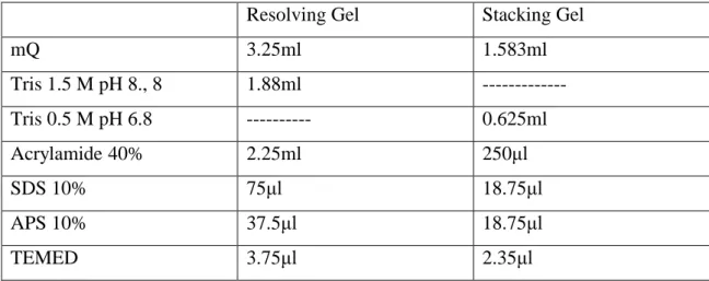
2.2.1 Materials
2.2.1.1 Chemicals Reactive
All the reactive employed in this work are listed on Table2.1 Table2.1: Reactive employed and suppliers.
REACTIVE AND ABBREVIATION BRAND
Acetone
Acrylamide
Ammonium Persulfate (APS)
Ampicilin
Bacto Agar
Bromophenol blue
Chloridric Acid
Dimethyl Sulfoxide (DMSO)
D(+) Glucose
Ethanol
Ethylenediamine tetraacetic acid (EDTA)
Glycerol Glycine Gel red Isopropanol Kanamicine Seakem Agarose Sodium Chloride
Sodium Dodecyl Sulfate (SDS)
-37 -2.2.1.2 Glass Materials
The glass materials have been washed and rinsed several times
with distilled water.
2.2.1.3 Other Materials
Other laboratory materials like tips, microtubes, conical tubes, Eppendorf tubes, etc.., are bought sterile or are sterilized on an autoclave (Autester E-75) for 20 minutes, at 120ºC and one atmosphere.
2.2.1.4 Buffers
In this work, the following buffers have been used:
► PBS (pH 7.4): 5mM pH 7.4 phosphate buffer, 140mM NaCl ► TAE: 40mM Tris-acetate, 1mM pH 8 EDTA
► SB (sonication buffer): 20mM Tris-base, 2.5 mM pH 8 EDTA. ► Washing buffer: 20mM Tris pH7.5, 0.2M NaCl.
► Running buffer: 15.1g Tris-base, 71g Glycine, 5g SDS.
All the solutions are prepared using ultrapure de-ionized water (Millipore), and when necessary are sterilized in the autoclave.
2.2.1.5 Biological Materials
2.2.1.5.1 Bacterial strains
The Escherichia coli strains used in this work have the following genotypes:
► XL1-Blue (Stratagene): endA1 supE44 hsdR17 thi1 recA1 gyrA96 relA1 lac [F‘ proAB lacIq
ZΔM15 Tn (Tetr)]. ► BL21 (DE3) (Novagen): F- ompT hsdSB (rB
mB
-) gal dcm (DE3-).
2.2.1.5.2 Culture Media
The culture media used for bacteria growth and transformations are: ► LB (Luria-Bertani):( Pronadisa)
Formula in grams per liter of distilled water:
-38 -Yeast extract: 5g/l Sodium chloride: 10g/l
Final pH: 7.0±0.2at 25oC and is sterilized on the autoclave.
► LB-agar, composition per liter: 15g of Agar in LB medium.
Sterilization on autoclave. ► TB (Terrific Broth):
Formula in grams per liter of distilled water: 20g/l Yeast extract
35g/l Tryptone
3.3g/l Ammonium Sulphate
6.5g/l potassium Dihydrogen Phosphate 7.1g/l Disodium Hydrogen Phosphate 0.5g/l Glucose
2g/l Alpha lactose
0.15g/l Magnesium Sulphate
0.35g/l Auto Induction Medium Element Mix, Then sterilizing on autoclave.
► SOC Broth (Sigma Aldrich).
2.2.1.5.3 Endonucleases
The endonucleases used for this work are:
► DpnI, EarI, EcoRI, HindIII, SapI, XbaI (NewEngland Biolabs) ► Fast EcoRI, Fast HindIII, Fast XbaI, (Fermentas)
► T4 DNA ligase (NewEngland Biolabs, Fermentas)
► Shrimp Alkaline Phosphatase (S.A.P.) (NewEngland Biolabs) ► Antarctic Alkaline Phosphatase (NewEngland Biolabs)
-39 -2.2.1.5.4 Vectors
a) Cloning Vector
Fig.2.3 represents the scheme of the pDrive cloning vector (Qiagen). In the scheme are represented the functional domains of the vector as the position of the Ear I endonuclease, also indicate the Ear I/Sap I restriction sites at position 3843bp, eliminated during mutagenesis.
-41 -b) Expressing Vector
pET10: is used for protein production which is modified from pET-9a, Fig.2.4, (Novagen Ca.No.69431-3), in Bioforge group by M. Pierna
Fig.2.4: Scheme of pET-9a-d cloning/expressing region.
2.2.1.5.5 DNA materials
1- E50I60 DNA has been recombined by Carmen Garcia-Arevalo in
Bioforge group.65
2- DNA of (DDDEEKFLRRIGRFG) 1, 3, 6 amino acids that have been
prepared in Bioforge group by A. RIBEIRO. c) Other reagents
1- QuantumPrep® Plasmid Kit (Biorad).
-40 -2.2.2 Methods
Recombinant DNA molecules are defined as either molecules that are constructed outside living cells by joining natural or synthetic DNA segments to DNA molecules that can replicate in a living cell.125
The DNA of amino acid sequences of E50I60 with DNA of amino acid
sequences of (SNA15)3 and also with amino acid sequences of (SNA15)6 have to
be recombined and transformed.
The amino acid sequence of E50I60 elastin like polymer is
[((VPGVG)2(VPGEG)(VPGVG)2)10-(VPGIV)60]
and for (SNA15)3 is
(DDDEEKFLRRIGRFG)3
and for (SNA15)6 is
(DDDEEKFLRRIGRFG)6
The DNA of E50I60, the DNA of (SNA15)3 and DNA of (SNA15)6 have
been transformed, using the following steps:
2.2.2.1 Transformation of XL1-Blue cells
An aliquot of 50µl of cells (stored at -80 oC) is thawed on ice and
-mercapthoethanol is added to a final concentration of 25mM. After mixing gently with the tip, the cells are kept on ice for 10 minutes and shaking gently each two minutes. A 10µl from the cells and 0.7µl of plasmids that have the
DNA to be colonized are mixed in pre-chill 14-mlBD Falcon polypropylene round-bottom tubes on ice for 20 minutes (A volume of ligation lower than the 10% of the initial cell volume).
A heat shock is applied to the cells in 42oC water bath for 30 seconds
50 -42
-200µl of the mixture plated in LB agar plate containing the appropriate antibiotic which are incubated for 16-20 hours at 37 oC.
A single colony is isolated from the LB agar plate and used to inoculate 5ml of LB medium containing the appropriate antibiotic and is grown at 37oC
with shaking at 225- 250rpm for 16-20 hours.
2.2.2.2 Plasmid purification
For plasmid purification, the "Quantum Prep Plasmid Mini, Midi and Maxiprep" from BioRad kits have been used following the manufactures
instructions. These methods are based on bacteria alkaline lyses126 followed by a partial purification and selective DNA adsorption to a silica gel. And the eluted DNA is stored under -20oC until using.
2.2.2.3 DNA Agarose gel electrophoresis
To separate and check the appearance and size of DNA fragments from a
plasmid; fragments resulting from an enzymatic digestion with endonucleases; it used the agarose gel electrophoresis.
Different concentrations in 1x TAE, are applied according to the sizes of the DNA fragments and the kind of gel, analytical or preparative (Table 2.2).
The gels are prepared adding in an Erlenmeyer flask the quantity of agarose and a volume of buffer according to the gel concentration and size. The agarose is melted in the microwave, after weight and hydration, until the formation of a gel. Once melted, the flask with the gel is weighted again and ultrapure
-43 -Table 2.2: Optimal resolution for linear DNA.
Fragment size in base pair (bp)
Agarose final %
1x TAE
800-10000 0.8
400-8000 1
300-7000 1.2
200-4000 1.5
100-2000 2
2.2.2.4 DNA fragment analysis
The DNA is cut at the desired position using the Ear I and Sap I endonucleases, then performing electrophoresis analysis. The samples are applied adding 0.20 volumes of 5x loading buffer [(30% (v/v) glycerol, 0.1% (w/v) SDS, 0.05% (w/v) bromophenol blue, 0.05% xilen cianol, 50mM Tris pH
8, 0.05mM EDTA)]. A fixed voltage, between 2 and 7 V/cm according to each sample, is applied.
Last, the gel is stained for 10 to 30 minutes in a Gel Red nucleic acid stain solution, and the DNA bands are visualized by exposition to UV light in a Viber Lourmat, TFX-20M transilluminator.
Picture image:
Images were taken with the Kodak "Gel Logic 100 imaging System" digital camera. Visualization and manipulation were made using the "Kodak 1D image Analysis" and "Paint Shop pro Version 9.00" (Jasc software) programs.
-44
Fig.2.5: Electrophoresis analysis for DNA of E50I60 in opened pDrive vector at 5500pb and
(SNA15)3at 165pb and (SNA15)6 at 330pb DNA cut fragments.
(SNA15)3in pDrive
(SNA15)6in pDrive
E50I60 in pDrive vector
E50I60in pDrive vector opened with
SAP I
(SNA15)6
(SNA15)3
1: E50I60 in pDrive opened (pb).
2: Marker (pb).
1: Marker (pb).
2: Cut fragment of (SNA15)3 (pb).
-45
-From electrophoresis experiment, it is found that a Sap I enzyme open the DNA of E50I60 in pDrive vector at the desired position and Ear I enzyme for
(SNA15)3 and (SNA15)6 DNA, see Fig.2.5. Making gel for obtaining more
amounts DNA of E50I60 in pDrive, and also for (SNA15)3 and (SNA15)6 DNA.
2.2.2.5 DNA fragments purification from an agarose gel
The target DNA band is first separated and visualized in an agarose gel of an appropriated concentration and stained with Gel Red nucleic acid stain solution. Secondly, the band is extracted from the gel with the help of a scalpel.
Minimum quantity of agarose should be cut during band extraction.
An approach that has been used for fragment purification is the "Pure Link Quick Gel Extraction kit" system. Following the manufacturer's instructions the DNA is purified by adsorption to silica gel membrane127, and eluted with elution buffer. And the eluted DNA is stored under -20oC until
using.
2.2.2.6 Recombination between DNA of E50I60 in pDrive vector and DNA of
(SNA15)3 and also Recombination between DNA of E50I60 in pDrive
vector and DNA of (SNA15)6
First, opening the pDrive vector recombined by the DNA of E50I60 using
Sap I enzyme then dephosphorylation of the opening plasmid that has sequence of E50I60.
The dephosphorylation has to be performed in 50l of opening DNA of
E50I60 in pDrive vector with 5.7l Buffer (NEB buffer for Antartch) and 0.5l
Antartch enzyme in an Eppendorf, then putting the Eppendorf at 37oC for 30 minutes, and then inactivate 5 minutes at 65oC.
Second, making a ligation between the DNA of (SNA15)3 with
disphosphorylated DNA of E50I60 in pDrive vector, and a ligation between the
-46
-The ligation has been performed in 6l of the disphosphorylated DNA of E50I60
in pDrive vector with 4l from DNA of (SNA15)3 and 0.8l from T4DNA ligase
using 1.2l from the buffer, Fig.2.6a, then making electrophoresis experiment for examining the ligated fragments with a gel of concentration 1.8% of
agarose. The electrophoresis analyses are shown in Fig.2.6b.
Fig.2.6: (a) Schematics of the ligated DNA of E50I60 in pDrive vector with (SNA15)3 and for
ligated DNA of E50I60 in pDrive vector with (SNA15)6 DNA. (b) Electrophoresis analysis for
ligated DNA of E50I60 with (SNA15)3 and for ligated DNA of E50I60 with (SNA15)6 DNA, that
cut with endonuclease. Ligation
(SNA15)3
(SNA15)6
E50I60in pDrive vector opened with
SAP I
Ligated DNA of E50I60in pDrive
vector with
(SNA15)3DNA
Ligated DNA of E50I60in pDrive
vector with
(SNA15)6 DNA
(a)
(b)
1, 2: Cut fragment of (SNA15)3E50I60 (pb).
3: Cut fragment of E50I60 (pb).
4, 5: Cut fragment of (SNA15)6E50I60 (pb).
-47
-Then making another gel to cut and obtain a more amount of ligated DNA of (SNA15)3E50I60 in pDrive vector and of ligated DNA of (SNA15)6 E50I60
in pDrive vector. Then these DNAs are introduced in expressing vectors for producing the proteins.
2.2.2.7 Inserting DNA of (SNA15)3E50I60 in pET10 expression vector and also
inserting DNA of (SNA15)6E50I60 in pET10 expression vector
First, cutting the fragments of the DNA of (SNA15)3E50I60 from
(SNA15)3E50I60 pDrive vector and opening pET10 expression vector using the
appropriate enzymes then put them overnight at 37oC. Then putting a 1l of
Dpn I endonuclease in the DNA of (SNA15)3E50I60 and performing the
electrophoresis experiment for cutting the DNA of (SNA15)3E50I60. Then,
making purification of cut (SNA15)3E50I60 DNA and purifying DNA from gel,
then Store the purified DNA at 4oC for immediate use or at -20oC until using.
Also the same for the DNA of (SNA15)6E50I60
For inserting the DNA of (SNA15)3E50I60 and the DNA of (SNA15)6E50I60
in the pET10 expression vector, the dephosphoralation and ligation are applied as in 2.5.6 using the appropriate enzymes and appropriate conditions.
Then making electrophoresis, of 1.8% agarose concentration, experiment
using Hin III and Xba I enzymes for cutting the plasmid of (SNA15)3E50I60pET10 and also the plasmid of (SNA15)6E50I60pET10, the
-48
Fig.2.7: Electrophoresis analysis for cut plasmid of (SNA15)3E50I60pET10 and also for cut
plasmid of (SNA15)6E50I60pET10.
1: Marker (pb).
2,3,4,5: Cut fragment of (SNA15)3E50I60 (pb).
1: Marker (pb).
2,3: Cut fragment of (SNA15)6E50I60 (pb).
Plasmid of
(SNA15)3E50I60
pET10
Plasmid of
(SNA15)6E50I60