A low-cost pedagogical environment for training on
technologies for image-guided robotic surgery
B. Rodríguez-Vila1,2, A. Gutiérrez3, M. Peral-Boiza2,3, H. Ying1, T. Gómez-Fernández2,3, E.J. Gómez1,2,4, P. Sánchez-González1,2,4
1 Biomedical Engineering and Telemedicine Centre. ETSI de Telecomunicación, Universidad Politécnica de Madrid, 28040, Madrid. Spain, {brvila, egomez, psanchez}@gbt.tfo.upm.es
2 Center for Biomedical Technology, Universidad Politécnica de Madrid, Madrid. Spain. 3 E.T.S. Ingenieros de Telecomunicación, Universidad Politécnica de Madrid, Madrid, Spain,
4 Biomedical Research Networking Center in Bioengineering, Biomaterials and Nanomedicine, Madrid. Spain
Abstract. This research presents a novel low-cost pedagogical environment ori-ented to ease the use experiential learning methods for training on image-guided robotic surgery technologies. The environment proposes a simplified surgical simulation use case: the movement of insertion and extraction of a needle, similar to an image-guided biopsy situation. The training environment is composed of a 3D-printed phantom, a CT scan of the phantom, a virtual reality environment that includes haptic information, an ad-hoc 1 degree of freedom (DOF) robotic system for insertion/extraction and a Novint Falcon, a low cost 3 DOF manipulator that allows achieving haptic feedback. A first pilot experience has been carried out in the “Surgical Simulation and Planning” course of the Bachelor of Biomedical Engineering at Universidad Politécnica de Madrid (UPM). Results of the surveys carried out (by teachers and learners) show that the new project-based methodol-ogy improves in all cases the values of student’s satisfaction obtained using the classical methodology based on master classes and practices.
Keywords: Experimental training, learn by doing, image guided surgery, ro-botic surgery.
1
Motivation
Image-guided surgery (IGS) is any surgical procedure where the surgeon uses tracked surgical instruments in conjunction with preoperative or intraoperative images and vir-tual models in order to indirectly guide the procedure. Robotic surgery or robot-assisted surgery (RAS) are terms for technological developments that use robotic systems to aid in surgical procedures. In case of RAS, instead of directly moving the surgical instru-ments the surgeon uses a tele-manipulator for remotely controlling them.
and master classes in which the knowledge that the learners have to acquire is presented. In the best scenario, guided practical sessions are carried out in which learners can ob-serve the results of applying that theoretical knowledge to a specific simulated case. On the contrary, experiential learning [1] allows the students to direct their own learning process, increasing their commitment and motivation through a team-based research methodology.
However, the high cost of real IGS and RAS environments makes them inaccessible for biomedical engineering training. Thus, this research presents a new methodology and a novel low-cost pedagogical environment for increasing learners’ motivation and autonomy, giving them an active role during their didactic process and based on expe-riential training methods. The environment proposes a simplified surgical simulation use case: the movement of insertion and extraction of a needle, similar to an image-guided biopsy situation.
2
Design of the pedagogical environment
The training environment is composed of the following systems:
- 3D-printed physical phantom from which CT medical images are obtained and serves as the basis for creating 3D virtual models. This helps students un-derstand the role of medical images, as well as their processing and analysis, in IGS and RAS.
The material of the phantom has to be suitable for the selected medical imaging modality, CT in our case. Furthermore, the 3D-printed phantom: (1) must have holes through which the instruments are inserted; (2) inside the phantom, there should be objects with different mechanical properties to understand the role of haptic information in the surgical intervention; (3) it should be filled with a vis-cous medium that simulates tissue and interstitial fluid and (4) it can have a simple geometry, since the aim is not to achieve a realistic model of the human body but to transmit the basic concepts of a simulation to engineering learners.
- A virtual reality software tool in which virtual models are presented (tissues and surgical tools). The software tool must fulfill several features to cover the needs of the final environment: (1) loading and viewing of medical images; (2) image processing, more specifically segmentation; (3) creation and visualiza-tion of virtual models; (4) user interacvisualiza-tion with the models; (5) collision detec-tion; and (6) haptic feedback.
3
Results and discussion
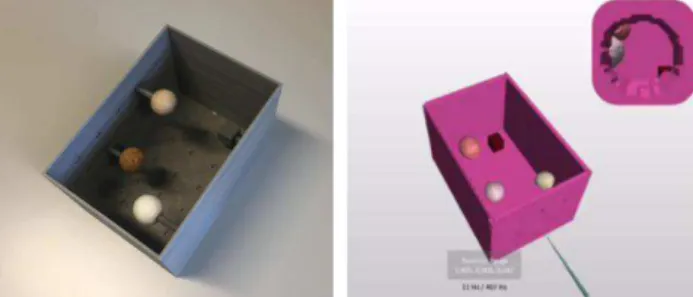
The phantom is formed by an assembled rectangular box built by 3D printing using PLA. Dimensions are 140 x 210 x140 mm (width, depth and height). In addition, inside the phantom, circular notches of 10 mm diameter are available every 30 mm, where the objects that are inserted inside the box are fitted. As objects, there are different geomet-ric shapes (cube, sphere and cylinder) and materials (PLA, cork, wood and polypropyl-ene) and (see Fig. 1, left) visible in the CT image as different Hounsfield levels.
There are different software tools that cover the needs of medical image visualization and processing, including the creation and visualization of virtual models. Some of them allow user interactions beyond a simple change of perspective in the visualization of objects. However, the authors have not found any software tools that include the above features and the collisions detection and haptic feedback. For this reason, two independent tools have been selected, one associated with the visualization and pro-cessing of medical images and another associated with the simulation of virtual reality. For acquiring the virtual models 3DSlicer [2][3] is used and the virtual reality simula-tion is developed using Chai3D [4][5], an open-source and multiplatform programming environment designed to integrate real-time tactile sensing, visualization and interac-tive simulation. This environment, developed in C ++, allows the student to understand the basic concepts related to visualization (cameras use, lights, textures, etc.), simula-tion and collision management, haptic feedback and interacsimula-tion (using keyboard, mouse or haptic devices).
Fig. 1. (Left) Phantom and (Right) visualization example of the virtual models
Fig. 2. EDUCIR Robotic system and its integration within the training environment
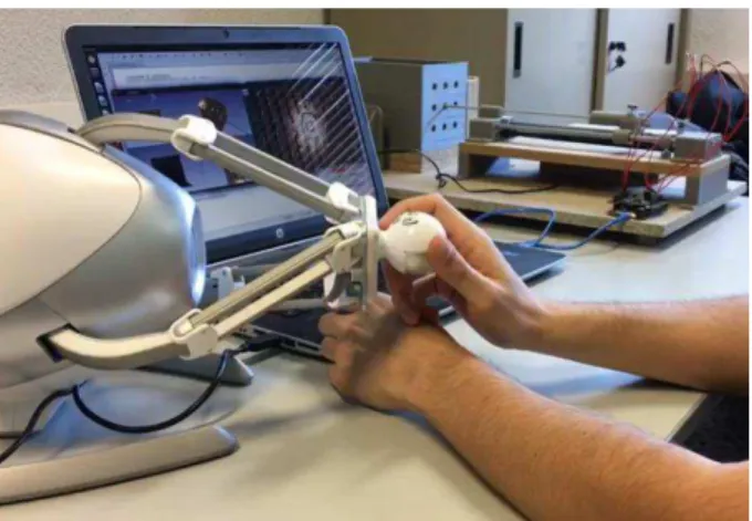
As a haptic feedback device, a low-cost commercial system, Novint Falcon (Novint Technologies, Inc.) has been selected (see Fig. 3). Novint Falcon is compatible with Chai3D and its integration with it does not require any additional development. This device has 3 DOF, so the associated Chai3D controller has been modified so that only movements on the insertion axis are transmitted.
Fig. 3. Novint Falcon, in foreground, with the robotic system and the phantom in background
A proof of concept has been carried out in the “Surgical Simulation and Planning” course of the fourth year of the Bachelor in Biomedical Engineering of the UPM. The course has been designed following the concept of project-based learning (PBL), where learners have worked in small groups (four members) over a semester in the realization of their own simulator.
movements of the haptic device. The learners had to relate the movements of the Novint Falcon with the behaviour of the motor and make the registration between the virtual world and the real one. All the groups were able to deliver an original and unique func-tional simulation application that integrated all the necessary components.
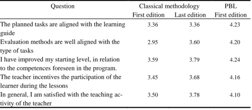
In order to have a first evaluation of the proof of concept, the survey of the teaching activity was used. Table 1 shows the average values of some of the questions analyzed. In general, the values obtained by the new methodology improve in all cases the results using the classical methodology based on master classes and practices. This improve-ment is significantly increased if we compare the first edition of the course in which each of the methodologies was applied.
Table 1.Results of the survey (max. score 5)
Question Classical methodology PBL
First edition Last edition First edition The planned tasks are aligned with the learning
guide
3.36 3.36 4.23
Evaluation methods are well aligned with the type of tasks
2.95 3.60 4.20
I have improved my starting level, in relation to the competences foreseen in the program.
3.59 3.79 4.24
The teacher incentives the participation of the learner during the lessons
3.45 3.68 4.16
In general, I am satisfied with the teaching ac-tivity of the teacher
3.50 3.78 4.10
As shown in the table, students support the change made regarding tasks and learning
outcomes, the evaluation methodology, the skills acquired and the learner’s
participa-tion in the development of the subject. In most cases, the difference is near the half a point on a scale of 1-5.
4
Conclusions
Traditional methodologies based on master classes and practices for training IGS and
RAS concepts are being replaced by others that encourage learners’ motivation and
autonomy. This work presents a new methodology and a novel low-cost pedagogical environment based on experiential learning methods for training on technologies for both surgical concepts. A proof of concept has been carried out in the Bachelor in Bio-medical Engineering of UPM. Results of the surveys carried out (by teachers and learn-ers) show that the new project-based methodology improves in all cases the values of
student’s satisfaction obtained using the classical methodology based on master classes
Acknowledgments
This work was partially supported by the innovative Educational Project EDUCIR of the Universidad Politécnica de Madrid.
References
1. Kolb AY., Kolb DA. Learning styles and learning spaces: Enhancing experiential learning in higher education. Academy of management learning & education. 2005 Jun; 4(2):193:212.
2. Fedorov A., Beichel R., Kalpathy-Cramer J., Finet J., Fillion-Robin J-C., Pujol S., Bauer C., Jennings D., Fennessy F., Sonka M., Buatti J., Aylward S.R., Miller J.V., Pieper S., Kikinis R. 3D Slicer as an Image Computing Platform for the Quantitative Imaging Network. Mag-netic Resonance Imaging. 2012 Nov; 30(9):1323-41. PMID: 22770690.
3. 3D Slicer Homepage, www.slicer.org, , last accessed 2018/01/30
4. Conti, F. and Barbagli, F. and Balaniuk, R. Halg, M. and Lu, C. and Morris, D. Sentis, L. Warren, J. and Khatib, O. Salisbury, K. The CHAI libraries. Proceedings of Eurohaptics 2003, Dublin, Ireland, 496-500.