Vol. 6, No. 2 April-June 2018 pp 96-102
Revista Mexicana de Ortodoncia
CASE REPORT
www.medigraphic.org.mx
INTRODUCTION
Biprotrusive skeletal class I is a condition in which the maxilla and the mandible are in an adequate intermaxillary relationship but both are found ahead of the skull base.1 Bimaxillary protrusion is characterized by proclination of the anterior teeth and convexity in the patient’s profi le; this can occur in any ethnic group, although it is more common in African-American and Asian patients.1 The etiology
Corrective treatment of a moderate class I bimaxillary
dentoalveolar protrussion: bimaxillary dentoalveolar
distalization with mini-screws
Tratamiento correctivo de protrusión dentoalveolar bimaxilar clase I
moderada: distalización dentoalveolar bimaxilar con miniimplantes
Francisco Shamed Méndez Ordóñez,* Gisel García García,§ Roberto Ruíz Díaz,§ Isaac Guzmán Valdivia Gómez||
* Student of the Orthodontics Specialty.
§ Professor of the Orthodontics Specialty. || Head of the Orthodontics Specialty.
Odontology Faculty, Division of Post-Graduate Studies and Research of the Odontology Faculty, UNAM.
© 2018 Universidad Nacional Autónoma de México, [Facultad de Odontología]. This is an open access article under the CC BY-NC-ND license (http://creativecommons.org/licenses/by-nc-nd/4.0/).
This article can be read in its full version in the following page: http://www.medigraphic.com/ortodoncia
RESUMEN
Existen diversas formas de tratar las clase I biprotrusivas y és-tas van a estar directamente relacionadas con la severidad del caso. La forma más común de tratamiento incluye la extracción de los primeros premolares maxilares y mandibulares, así como la retracción del segmento anterior para disminuir la biprotrusión bimaxilar. Una nueva alternativa de tratamiento para este tipo de maloclusiones puede ser la distalización bimaxilar con el uso de miniimplantes, lo cual además de garantizarnos un máximo an-claje nos permitirá mover múltiples dientes en una sola dirección con movimientos controlados. Material y métodos: Se colocaron cuatro miniimplantes como anclaje máximo (dos infracigomáticos de 10 mm y dos en el shelf mandibular de 12 mm) para realizar una distalización bimaxilar, se colocaron brackets de autoligado pasivo y apoyados en cadenas elásticas se realizó la distalización en masa de ambas arcadas. Resultados: Los miniimplantes de-mostraron ser una alternativa efi ciente para la corrección de una protrusión bimaxilar moderada, la distalización se realizó hasta conseguir una adecuada clase I molar y clase I canina en ambos lados, así como una sobremordida horizontal y vertical adecuada, los cambios estéticos faciales fueron una disminución de la bipro-quelia. Conclusiones: El tratamiento de la clase I biprotrusiva dependerá de la severidad del caso, los miniimplantes demues-tran ser una opción interesante para el tratamiento de este tipo de maloclusiones.
Key words: Bimaxillary distalization, mini-implants, bimaxillary protrusion. Palabras clave: Distalización bimaxilar, miniimplantes, protrusión bimaxilar. ABSTRACT
There are several ways to treat biprotrusive class I and they are directly related to the severity of the case. The most common form of treatment includes the extraction of maxillary and mandibular first premolars, as well as retraction of the anterior segment to reduce bimaxillary biprotrusion. A new treatment alternative for this kind of malocclusion is bimaxillary distalization with the use of mini implants, which in addition to guaranteeing a maximum anchorage will allow us to move multiple teeth in a single direction with controlled movements. Material and methods: Four mini-implants were placed for maximum anchorage (two 10 mm infra-cygomatic mini-implants and two 12 mm mini implants in the mandibular shelf) to perform a bimaxillary distalization. Passive self-ligating brackets were placed with elastic chains to perform mass distalization of both arches. Results: Mini implants proved to be an effi cient alternative for the correction of moderate bimaxillary protrusion; distalization was performed until molar class I and canine class I on both sides was obtained, as well as a normal overjet and overbite. Among the esthetic facial changes achieved was a decrease in the biprochelia. Conclusions: Treatment of biprotrusive class I will depend on the severity of the case but mini implants prove to be an interesting option for the treatment of this kind of malocclusion.
www.medigraphic.org.mx
dentoalveolar protrusion.Unfortunately, there are patients who do not want to have premolar extractions, so the protrusion reduction and improvement in facial aesthetics that would lead to the success of treatment will be compromised. Because of this it is of great importance to know other treatment alternatives that allow us to achieve our goals while respecting the patient’s decisions.
Considering that every orthodontic movement is accompanied by a reaction (Newton’s first law), it may be difficult to correct a malocclusion simply by using intraoral devices,6 especially when it is necessary to perform an en masse movement of all teeth or a group of them in both the maxilla and mandible, which will increase our demands for anchorage.
There are several articles in the literature that describe dentoalveolar distalizations of the maxilla in class II patients and of the mandible in class III patients using temporary anchorage devices (TADS) which, among their advantages, offer us maximum anchorage, little cooperation from the patient and no loss of anchorage.
Kuroda in 2005 reported the use of temporary skeletal anchorage devices for the treatment of class III adult patients, and suggested that such devices can be placed in the retromolar area or between tooth roots for direct or indirect mass distalization in the lower arch.7 In 2013 Ishida et al. reported the case of a patient with class II malocclusion who was corrected with an asymmetric distalization of the upper molars using mini-implants placed in the zygomatic arch thus distalizing the entire dentition.8 Tai et al. also reported the case of a patient with class III malocclusion who had the mandibular dentition moved distally with temporary skeletal anchorage devices.9
This article reports the clinical case of a 23-year-old skeletal class I male patient with moderate biprotrusion who was successfully corrected through bimaxillary dentoalveolar distalization using mini implants as anchorage devices.
biprotrusive skeletal class I patient with neutral growth, mesofacial biotype, convex profi le, no coincidence of dental and facial midlines; edge to edge bite, bilateral molar class I and bilateral canine class I, moderate crowding in upper and lower arch, lower incisor proclination, protrusion of upper and lower incisors
(Figures 1 to 3).
The initial treatment plan indicated the removal of the maxillary and mandibular fi rst premolars. In view of the patient’s refusal to accept extractions, it was decided to place four mini-implants (two infracigomatic and two in the mandibular shelf) to perform an en masse distalization of the upper and lower arches.
Objectives
Among the objectives established during treatment there are:
• Bimaxillary distalization with mini implants. • Eliminate crowding.
• Maintain molar and canine class I. • Achieve a normal overjet and overbite. • Coordinate arches.
• Achieve root parallelism. • Closure of spaces. • Retention.
Treatment plan
Placement of H4® fi xed appliances; 0.022 ” x 0.028” slot (1300 Alfa Dr. NE McMinnville, Oregon), with bondable tubes in fi rst and second upper and lower molars.
Placement of Dewimed® mini-implants (Blvd. Adolfo Ruíz Cortines No. 5271, Del Tlalpan, Isidro Fabela, Mexico City), two in the upper arch of 10 mm (infracigomatic) and two in the lower arch of 12 mm (mandibular shelf).
Immediate loading after mini-implant placement. Phase I: alignment and leveling.
• 0.014”, 0.016”, 0.018”, 0.014” x 0.025” 0.016” x 0.025” NiTi archwires.
www.medigraphic.org.mx
Phase II: arch coordination and space closure.• 0.017” x 0.025” NiTi; 0.017 x 0.025” stainless steel, 0.019” x 0.025” NiTi, 0.019 x 0.025” stainless steel archwires.
• Panoramic radiograph for bracket repositioning. Phase III: detailing and occlusal settlement.
• 0.019” x 0.025” SS braided archwire, ¼ medium elastics for settlement.
• Bracket removal and placement of circumferential retainers.
RESULTS
At the end of treatment, the objectives set at the beginning were fulfi lled: to maintain bilateral I molar and canine class; distalize to obtain space and thus eliminate crowding; match dental midlines; achieve a
normal overjet and overbite; obtain root parallelism
(Figures 4 to 7).
On an aesthetic level, a good smile was achieved, as well as a decrease in the lip protrusion and an improvement in the patient’s profi le.
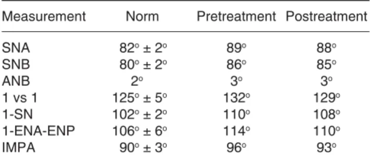
C e p h a l o m e t r i c a l l y , s k e l e t a l c l a s s I w a s preserved, bimaxillary protrusion was reduced and some cephalometric measurements were improved
(Table I).
DISCUSSION
Among the clinical characteristics of skeletal class I with bimaxillary protrusion we may fi nd a molar and canine class I, presence or not of crowding, as well as a moderate or severe lip protrusion. Treatment may have multiple options and will be directly related to the severity with which each case is diagnosed.
Figure 1.
Initial facial and intraoral photographs.
www.medigraphic.org.mx
Each patient will present unique characteristicsand therefore, the various ways of approaching and treating each particular case must be known.
Among the forms of treatment that we may fi nd to treat this kind of cases there are: extractions of fi rst
premolars, orthognathic surgery, orthognathic and orthodontic surgery or the use of mini implants to perform bimaxillary distalizations.
With the surge of mini implants, it is now possible to perform en masse or group dental movements using
Figure 2.
Initial study models.
Figure 3.
Initial radiographs and cephalometric tracing.
A. Lateral headfi lm. B. Cephalometric tracing. C. Panoramic radiograph.
A
C
www.medigraphic.org.mx
the benefits of absolute anchorage. Traditionally,distalization of a tooth or group of teeth after eruption of the second molars becomes an absolute challenge. Over the years, headgears and pendulums have been used to achieve the distalization of one or more teeth.10,11
In one way or another the use of these appliances almost always required very good cooperation from the patient, so the treatment result did not depend directly on the orthodontist. Thanks to new interest in temporary anchorage systems, we can achieve satisfactory results where patient cooperation is not so much required and the orthodontist can have better control of the case.
To achieve a successful bimaxillary distalization, three factors must be considered: 1) mini implant placement, which must be in the cortical bone and at an adequate distance from the roots. The infracigomatic crest in the maxilla,8 the mandibular shelf and/or retromolar area in the mandible12,13 appear to be the appropriate areas for the placement of mini implants. 2) The absence of third molars to take advantage of the space in the posterior areas for distalization and 3) the patient’s growth direction.
Table I. Cephalometric values pre
and post-bimaxillary distalization.
Measurement Norm Pretreatment Postreatment SNA 82o ± 2o 89o 88o SNB 80o ± 2o 86o 85o ANB 2o 3o 3o 1 vs 1 125o ± 5o 132o 129o 1-SN 102o ± 2o 110o 108o 1-ENA-ENP 106o ± 6o 114o 110o IMPA 90o ± 3o 96o 93o Figure 4. F i n a l f a c i a l a n d intraoral photographs.
www.medigraphic.org.mx
Este documento es elaborado por Medigraphic
Figure 5.
Final study models.
Figure 6.
F i n a l r a d i o g r a p h s a n d cephalometric tracing. A. Lateral headfilm. B. Cephalometric tracing. C. Panoramic radiograph.
A B
www.medigraphic.org.mx
REFERENCES
1. Chen G, Teng F, Xu TM. Distalization of the maxillary and mandibular dentitions with miniscrew anchorage in a patient with moderate Class I bimaxillary dentoalveolar protrusion. Am
J Orthod Dentofacial Orthop. 2016; 149 (3): 401-410.
2. Lamberton CM, Reichart PA, Triratananimit P. Bimaxillary protrusion as a pathologic problem in the Thai. Am J Orthod. 1980; 77 (3): 320-329.
3. Solem RC, Marasco R, Guiterrez-Pulido L, Nielsen I, Kim SH, Nelson G. Three-dimensional soft-tissue and hard-tissue changes in the treatment of bimaxillary protrusion. Am J Orthod
Dentofacial Orthop. 2013; 144 (2): 218-228.
4. Ouyang L, Zhou YH, Fu MK, Ding P. Extraction treatment of an adult patient with severe bimaxillary dentoalveolar protrusion using microscrew anchorage. Chin Med J (Engl). 2007; 120 (19): 1732-1736.
5. Aljhani A, Zawawi KH. The use of mini-implants in en masse retraction for the treatment of bimaxillary dentoalveolar protrusion. Saudi Dent J. 2010; 22 (1): 35-39.
6. Chung KR, Kim SH, Choo H, Kook YA, Cope JB. Distalization of the mandibular dentition with mini-implants to correct a Class III malocclusion with a midline deviation. Am J Orthod Dentofacial
Orthop. 2010; 137 (1): 135-146.
7. Kuroda S, Sugawara Y, Yamashita K, Mano T, Takano-Yamamoto T. Skeletal Class III oligodontia patient treated with titanium screw anchorage and orthognathic surgery. Am J
Orthod Dentofacial Orthop. 2005; 127 (6): 730-738.
8. Ishida T, Yoon HS, Ono T. Asymmetrical distalization of maxillary molars with zygomatic anchorage, improved superelastic nickel-titanium alloy wires, and open-coil springs. Am J Orthod
Dentofacial Orthop. 2013; 144 (4): 583-593.
9. Tai K, Park JH, Tatamiya M, Kojima Y. Distal movement of the mandibular dentition with temporary skeletal anchorage devices to correct a Class III malocclusion. Am J Orthod Dentofacial
Orthop. 2013; 144 (5): 715-725.
10. Toy E, Enacar A. The effects of the pendulum distalising appliance and cervical headgear on the dentofacial structures.
Aust Orthod J. 2011; 27 (1): 10-16.
11. Caprioglio A, Beretta M, Lanteri C. Maxillary molar distalization: Pendulum and Fast-Back, comparison between two approaches for Class II malocclusion. Prog Orthod. 2011; 12 (1): 8-16.
12. Anhoury PS. Retromolar miniscrew implants for Class III camoufl age treatment. J Clin Orthod. 2013; 47 (12): 706-715. 13. Poletti L, Silvera AA, Ghislanzoni LT. Dentoalveolar class III
treatment using retromolar miniscrew anchorage. Prog Orthod. 2013; 14: 7.
Mailing address:
Francisco Shamed Méndez Ordóñez
E-mail: [email protected]
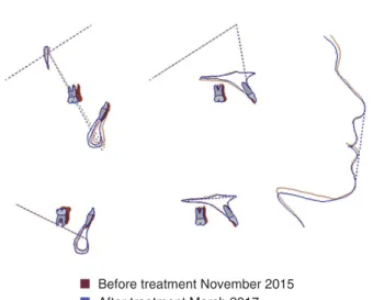
Figure 7. Superimposition.
Before treatment November 2015 After treatment March 2017