Design and Synthesis of Pironetin Analogue/Combretastatin A 4 Hybrids and Evaluation of Their Cytotoxic Activity
18
0
0
Texto completo
(2) Design and Synthesis of Pironetin AnalogueCombretastatin A-4 Hybrids and Evaluation of Their Cytotoxic Activity Sandra Torijano-Gutiérrez,a Concepción Vilanova,a Santiago Díaz-Oltra,a Juan Murga,a,* Eva Falomir,a Miguel Cardaa and J. Alberto Marcob,*. aDepart.. de Q. Inorgánica y Orgánica, Univ. Jaume I, E-12071 Castellón, Spain. bDepart.. de Q. Orgánica, Univ. de Valencia, E-46100 Burjassot, Valencia, Spain. *Authors to whom correspondence should be addressed. E-Mail addresses: [email protected], [email protected] ABSTRACT We here describe the preparation of a series of hybrid molecules containing a combretastatin A-4 moiety and a pironetin analogue fragment. The cytotoxic activities of these compounds have been measured. Relations between structure and cytotoxicity are discussed. Some of the tested compounds showed cytotoxicity values of the same order of magnitude as the parental molecules, combretastatin A4 and pironetin, and were less toxic than the latter for normal cells.. KEYWORDS Combretastatin. A-4;. pironetin. analogues;. hybrid. molecules;. tubulin-active. compounds;. cytotoxicity.. HIGHLIGHTS . Hybrid molecules containing a combretastatin A-4 moiety and a pironetin analogue fragment have been prepared and tested for cytotoxicity against a normal and two tumoral cell lines. 1.
(3) . Some of the tested compounds show cytotoxicity values of the same order of magnitude as the parental molecules, combretastatin A-4 and pironetin.. . The normal vs. tumoral cytotoxicity ratio of some of the tested compounds are higher than in the case of the parental molecules, that is, they are safer than the latter compounds.. 1. Introduction It is widely known that cancer, one leading cause of death in developed countries [1], may be induced by a plethora of both external and internal factors, including genetic mutations. Accordingly, a number of types of therapeutic attack has been investigated [2,3]. One of these involves the use of cytotoxic drugs, which exert their effect in many cases by means of inducing various mechanisms of cell death [4]. Many of such drugs owe this property to interaction with the microtubule network. Microtubules are dynamic polymers that play a central role in a number of cellular processes, most particularly cell division, as they are key constituents of the mitotic spindle. Microtubules are constituted of a protein named tubulin, the functional form of which is a heterodimer formed through non-covalent binding of two monomeric constituents, called - and -tubulin. For cell division to occur in a normal way, microtubules must be in a constant state of formation and disruption, a process named microtubule instability 5. Any molecule which influences microtubule instability will also influence the cell division process, not only of normal cells but also of tumoral ones. Therefore, it is not surprising that tubulin-binding molecules (TBM) constitute a most important class of anticancer agents. TBMs may be divided in two broad categories, those that bind to -tubulin and those that bind to -tubulin. The latter group is presently by far the most numerous and contains products which cause either disruption or stabilization of microtubules. Among the drugs that belong to this group, colchicine 6 and the combretastatins 7 (Fig. 1) exert their effects by causing disruption of microtubules. In contrast, another important representative of the same group, paclitaxel, was the first-described tubulin-binding drug that was found to stabilize microtubules 8. Even though they display opposite effects, these drugs are known to bind to -tubulin, whenever to different sites within this protein subunit 9-11.. 2.
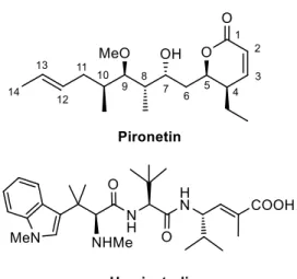
(4) Figure 1. Structures of some natural products reported to selectively bind to -tubulin. The number of products that bind to -tubulin is very small, the naturally occurring 5,6-dihydro-pyrone pironetin being the first-reported example 12, followed a short time later by the peptide-like hemiasterlin family 13 (Fig. 2). Pironetin is a potent inhibitor of tubulin assembly and has been found to arrest cell cycle progression in the G2/M phase 14.. Figure 2. Structure of two natural products reported to selectively bind to -tubulin.. 3.
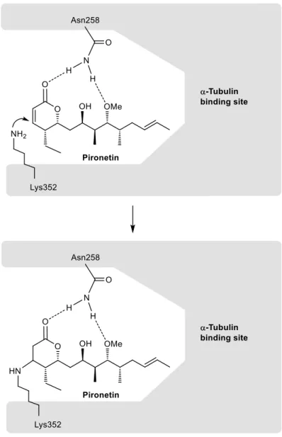
(5) Some structure-activity (SAR) studies on pironetin have been reported. These studies have shown that the presence of the conjugated C2C3 double bond and of the hydroxyl group at C-9, either free or methylated, are essential for the biological activity. The presence of a (7R)-hydroxyl group also seems to be relevant. It has been suggested that the Lys352 residue of the -tubulin chain adds in a Michael fashion to the conjugated double bond of pironetin, therefore forming a covalent bond with C-3 of the dihydropyrone ring (Fig. 3). In addition, it has been suggested that the Asn258 residue of -tubulin holds the pironetin molecule through two hydrogen bonds to the dihydropyrone carbonyl and the methoxyl oxygen atoms 14.. Figure 3. Schematic model of the covalent union of pironetin to its binding site at the -tubulin surface.. The emergence of resistances to existing drugs has led to a continuous need of developing new bioactive compounds that overcome such problems. Even though first observed in the case of antibiotics 4.
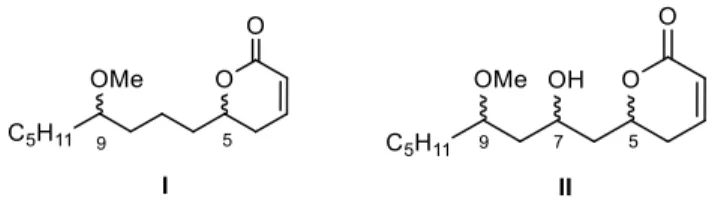
(6) [15], resistances have been reported to therapies with various types of cytotoxic agents [16]. The investigation of new compounds with such biological properties therefore constitutes an important goal in chemistry and pharmacology. 2. Concept and design of hybrid tubulin-binding ligands As a member of the up to now small group of TBMs that bind to -tubulin, pironetin constitutes a pharmacologically interesting target. Not surprisingly, an appreciable number of total syntheses of this natural compound has appeared in the literature [17]. In order to develop SAR studies based upon the pironetin framework, we designed two years ago [5a] a simplified model structure where all elements that had not yet proven to be essential for the biological activity were removed. The target structures I/II are schematically shown in Figure 4. The elements that were maintained are the conjugated dihydropyrone ring and the side chain with the methoxy group at C-9. The hydroxyl group at C-7 was removed in some substrates (I) and retained in others (II), in order to see its influence on the activity. All alkyl pendants (methyl groups at C-8 and C-10, ethyl at C-4) and the isolated C12−C13 double bond were removed. The configurations of the two/three remaining stereocentres were then varied in a systematic way. Thus, all four possible stereoisomers with general constitution I, with no hydroxyl group at C-7, were prepared. Likewise, all eight stereoisomers exhibiting general structure II, with a hydroxyl group at C-7, were synthesized [5a].. Figure 4. General structures of simplified pironetin analogues of the first generation (ref. 5a). The cytotoxic activity of these analogues and their interactions with tubulin were subsequently investigated. It was found on one hand that analogues I/II were cytotoxic in the low micromolar range, about three orders of magnitude less active than the parent molecule. On the other hand, we also found that they behave in the same way as pironetin, share the mechanism of action of the natural compound and compete for the same binding site in -tubulin. As the parent compound, they also lead to disruption of the microtubule network [5a]. With the aim at extending our project, our idea was to prepare cytotoxic TBMs with a dual ability to bind to both - and -tubulin and exert a microtubule-destabilizing effect. Since these properties are specifically exhibited by pironetin (binds to -tubulin) and combretastatin A-4 (binds to -tubulin), 5.
(7) respectively, we decided to prepare hybrid molecules such as 1-8 (Fig. 5) containing a moiety of combretastatin A-4 (itself numbered as 13 in Fig. 5) and another of the simplified pironetin type (14, ent-14 and 15), connected in turn by an ester spacer of variable length. Some of them have been prepared in both antipodal forms (1 to 4 and ent-1 to ent-4) [18].. Figure 5. Structures of the pironetin analogue/combretastatin A-4 hybrids used in this study (1-8) and of their synthetic precursors (9-15), including combretastatin A-4 (13). 3. Synthetic work Combretastatin A-4 (13) was prepared according to a literature procedure 19. Its O-alkyl derivatives 9a-12a were prepared by means of O-alkylation with commercially available -bromoesters 16-19 (Scheme 1). Saponification of esters 9a-12a gave the corresponding acids 9b-12b.. 6.
(8) Scheme 1. Synthesis of combretastatin A-4 derivatives 9a-12a and 9b-12b: (a) K 2 CO 3 , DMF, RT, 24 h (9a, 91%; 10a, 91%; 11a, 98%; 12a, 90%); (b) aq NaOH, MeOH, RT, 18 h (9b, 90%; 10b, 78%; 11b, 72%; 12b, 75%). Acronyms and abbreviations: DMF, N,N-dimethylformamide.. The synthesis of the pironetin fragments 14, ent-14 and 15 depicted in Scheme 2, was performed according to the general concept used in our previous papers [5a,b]. Thus, Brown´s asymmetric allylation 20 of the known aldehyde 16 21 afforded homoallyl alcohol 17. The required chiral allylborane was prepared through reaction of allylmagnesium bromide with the commercially available (−)-diisopinocampheylboron chloride, (−)-Ipc2 BCl. Reaction of 17 with acryloyl chloride at low temperature gave acrylate 18, which was subjected to ring-closing metathesis (RCM) 22 in the presence of Grubbs first-generation catalyst Ru-I. This furnished dihydropyrone 19, deprotection of which provided 14 in good yield. Dihydropyrone ent-14 was prepared in the same way except that (+)Ipc2 BCl was the chiral boron reagent used. Dihydropyrone 15 was not connected as such with the combretastatin A-4 fragment but as its O-silyl derivative 28 (Scheme 2), which was also prepared according to the previous methodology. Brown´s asymmetric allylation of the known aldehyde 20 23 furnished alcohol 21, which was then converted into methyl ether 22. The latter was then sequentially subjected to a two-step oxidative cleavage of the olefinic bond, followed by Brown´s asymmetric allylation to yield 23. Silylation of the hydroxyl group of 23 and repetition of the previous sequence gave alcohol 25, which was esterified with acryloyl chloride. The resulting acrylate 26 was subjected to ring-closing metathesis to 27, which was then oxidatively deprotected [24] to 28. Desilylation of 28 gave 15, which was used in the biological evaluations.. 7.
(9) Scheme 2. Synthesis of pironetin fragments 14, ent-14 and 15. (a) (−)-Ipc2 BCl, allylMgBr, Et2 O, 78 ºC, 1 h, then addition of the aldehyde, 1 h, 78 ºC (17: 76%, e.r. 96:4; 21: 90%, e.r. 94:6; 25, 70% overall yield from 24, d.r. 76:24); (b) CH2 =CHCOCl, CH2 Cl2 , iPr2 NEt, 78 ºC, 30 min (18: 94%; ent18: 90%; 26, 98%); (c) 10% cat. Ru-I, CH2 Cl2 , , 2 h (19: 81%; ent-19: 80%; 27: 83%); (d) DDQ, wet CH2 Cl2 , 30 min. (14: 69%; ent-14: 67%; 28: 84%); (e) (+)-Ipc2 BCl, allylMgBr, Et2 O, 78 ºC, 1 h, then addition of the aldehyde, 1 h, 78 ºC (ent-17: 89%, e.r. 96:4; 23: 90% overall yield from 22, d.r. 86:14); (f) NaH, THF, 0 ºC, then MeI, RT, overnight (88%); (g) 1) OsO 4 , NMO, aq tBuOH, THF, RT, overnight. 2) Pb(OAc)4 , CH2 Cl2 , 0ºC; (h) TBSOTf, 2,6-lut, 0 ºC, 1 h (80%); i) PPTS (cat.), MeOH, , overnight (85%). Acronyms and abbreviations: Ipc, isopinocampheyl; DDQ, 2,3-dichloro-5,6-dicyanop-benzoquinone; TBS, tert-butyldimethylsilyl; NMO, N-methylmorpholine N-oxide; 2,6-lut, 2,6lutidine; PPTS, pyridinium p-toluenesulfonate.. Dihydropyrones 1-8, including ent-1 to ent-4, were prepared by esterification of acids 9b-12b with dihydropyrones 14, ent-14 and 28 by means of the Yamaguchi method [25] (Scheme 3). In the case of compounds 5-8, an additional desilylation step was required.. 8.
(10) Scheme 3. Synthesis of hybrid molecules 1-4, ent-1 to ent-4, 5-8. (a) 9b-12b, 2,4,6-trichlorobenzoyl chloride, Et3 N, RT, then addition of the alcohol component, RT, 2 h (1: 34%; 2: 47%; 3: 37%; 4: 35%; ent-1: 42%; ent-2: 55%; ent-3: 37%; ent-4: 49%; 29: 25%; 30: 40%; 31: 44%; 32: 21%); (b) PPTS (cat.), MeOH, , overnight (5: 85%; 6: 95%; 7: 80%; 8: 85%). After the synthesis of the pironetin-combretastatin A-4 hybrids was completed, the compounds were investigated in relation to their cytotoxic activity towards two types of tumoral lines and one normal cell line.. 4. Biological work 4.1. Cellular effects of the compounds. Cytoxicity values. We carried out a measurement of the cytotoxicity of the synthetic combretastatin A-4 derivatives 1-4 and their enantiomers, 5-8, 9a-12a and 9b-12b. Pironetin analogues 14, ent-14 and 15 were also tested. Cytotoxicity assays were performed as described in the Experimental Section using two tumoral cells, the human colon HT-29 and the breast adenocarcinoma MCF-7 cell lines, as well as one normal cell line, the human embryonic kidney cell line, HEK-293 [26]. Cytotoxicity values, expressed as the compound concentration (µmol/L) that causes 50% inhibition of cell growth (IC 50 ), are shown in Table 1. The observed values are in most cases in the low to medium micromolar range. Compounds ent-1, 3, 7, 10a, 11a, 12a and 12b showed the highest cytotoxicities for the HT-29 cell line, with IC 50 values which were not very different (in some cases even lower) of those of combretastatin A-4 and pironetin for this particular cell line. In the case of the MCF-7 cell lines, the lowest IC 50 values were shown by compounds 7, 9b, 10a, 11a, 11b, 12a, 12b and 15. But a further aspect which is also worth mentioning is the fact that some of the synthetic compounds are much more toxic for tumoral cells than for normal ones, an obviously desirable feature. This can better appreciated with the and coefficients, obtained by dividing the IC 50 values of the normal cell line (HEK-293) by those of one or the other tumoral cell line (see footnote in the Table). The higher value of either the or the coefficient, the higher the. 9.
(11) therapeutic safety margin of the compound in the corresponding cell line. Thus, combretastatin A-4 itself shows good values of both coefficients, most particularly in the case of the MCF-7 cell line. Pironetin shows a similarly good value for the HT-29 cell line (), but much less so for the MCF-7 cell line. Among the compounds which show a high cytotoxicity (low IC50 values), ent-1, 7, 11a, 12a and 12b are particularly worth mentioning as they exhibit high both and values. Compounds 3 shows a good selectivity only in the case of the HT-29 line ( > 30) whereas compounds 11b and 15 show a good selectivity in the specific case of the MCF-7 line ( > 60). Some aspects related to the relation between the structures and the observed cytotoxicity deserve comment. In the case of two subsets of hybrid molecules [27] where there are differences between in the carbon chain length (e.g. 1-4 and 5-8), the cytotoxicity reaches a maximum for compounds having a tencarbon chain in the combretastatin A-4 segment (3, 7). The cytotoxicities of the compounds of the enantiomeric series (ent-1 to ent-4) are not too different from those of 1-4, even though the maximum cytotoxicity is found here for ent-1, the compound with the shorter carbon chain. It is difficult to propose now an explanation for this fact, as we do not yet know whether these compounds are interacting with tubulin at the pironetin site (-tubulin), at the combretastatin A-4 site (-tubulin) or at both sites simultaneously. Research in this direction is being currently performed. We have also tested the synthetic intermediates used for the preparation of the aforementioned hybrid molecules. As regards combretastatin A-4 derivatives 9-12a/b, a dependence of the cytotoxicity from the carbon chain length is also observed (Table 1). For the methyl esters (9a-12a), a marked increase in the cytotoxicity is observed in going from the compound with the three-carbon chain (9a) to those having longer chains (10a-12a), with the IC50 values of the three latter being very similar (with both tumoral cell lines) and comparable with those of combretastatin A-4 itself. As for the free acids (9b12b), the cytotoxicities vary in a rather erratic way and no relation is perceived between IC50 values and the chain length. Furthermore, marked differences also observed in some cases between the IC 50 values for the two tumoral cell lines. The cytotoxicities of the three pironetin analogues 14, ent-14 and 15 were also measured. Compound 14 and its enantiomer, which display a single stereocentre, represent the most simplified model possible for pironetin. From the results presented in Table 1, it is evident that 14 and ent-14 show a very low cytotoxicity, much below of that observed for the hybrid molecules (1 to 3, and ent-1 to ent-3) bearing these fragments (exceptions to this are 4 and ent-4, which show very high IC 50 values, perhaps because of solubility problems). Even if conclusions must be still provisory, this seems to point to 1-3 and their enantiomers owing their cytotoxicity to interactions with tubulin through the combretastatin A-4 end. As regards compound 15, it has three stereocentres and corresponds more closely to the pironetin models we have being investigating in recent years [5]. Table 1 shows that its cytotoxicity for the two tumoral 10.
(12) cells is indeed much higher than those of 14/ent-14, most particularly in the case of the MCF-7 cell line. From the hybrid molecules 5-8 having this structural fragment, the highest cytotoxicity is shown by 7, the molecule that displays the ten-carbon chain, as commented above. It is also worth noting that the IC50 value of 7 with the HT-29 cell line is much lower than the IC 50 values of both 11b and 15, its two precursor components. This suggests that a hydrolysis of 7 into 11b and 15 is not taking place within the cell.. Table 1. Cytotoxicity of pironetin analogue/combretastatin A-4 hybrids 1-8 and ent-1 to ent-4, combretastatin A-4 derivatives 9a-12a and 9b-12b, and pyrones 14, ent-14 and 15.a Compound. HT-29. MCF-7. HEK-293. b. c. CoA4 Pironetin. 1 ± 0.2 18 ± 3 52 ± 10. 25 ± 3 46 ± 6. 1. 4.2 ± 0.5 8±2 12 ± 2. 25 2.5 0.7. 2. 32 ± 5. > 300. 39 ± 4 80 ± 14. 5.9 5.7 3.2 2.5. < 0.3. 3 4 ent-1. 10 ± 1 107 ± 5. > 300 > 300. > 300 > 300. > 30 > 2.8. 3.5 ± 0.9. 17 ± 2. 114 ± 5. 32. 6.7. ent-2 ent-3. 19 ± 6 38 ± 3. > 300 69 ± 7. > 300 59 ± 10. > 15 1.6. ‒ 0.8. ent-4. 64 ± 7. > 300. 72 ± 3. 1.1. < 0.3. 5 6. 15.4 ± 0.5 44 ± 5. 31 ± 1 > 300. 22 ± 2 > 300. 1.4 > 6.8. 0.7 ‒. 7. 2.9 ± 0.2. 5.6 ± 0.1. 22 ± 2. 7.6. 3.9. 8 9a. 50.8 ± 0.6 60.1 ± 0.5. 21 ± 4 10.4 ± 0.5. 81 ± 10 121 ± 5. 1.6 2. 3.8 12. 9b. 50 ± 2. 2.9 ± 0.2. 67 ± 2. 1.4. 23. 10a 10b. 3.5 ± 0.3 16 ± 2. 9.6 ± 0.2 17 ± 4. 9.6 ± 0.3 > 300. 2.7 > 19. 1 > 18. 11a. 8±1. 3 ± 0.6. > 300. > 37. > 100. 11b 12a. 103 ± 6 5±1. 5 ± 0.9 9.6 ± 0.2. > 300 > 300. > 30 > 60. > 60 > 30. 12b. 1.9 ± 0.3. 4.4 ± 0.8. 39 ± 5. 20. 8.8. 14 ent-14. 109 ± 20 81 ± 12. > 300 109 ± 8. 206 ± 15 119 ± 16. 1.9 1.5. < 0.7 1.1. 15. 25.4 ± 0.2. 2.5 ± 0.4. > 300. > 12. > 120. ‒ ‒. IC50 values, which include those of the parent compounds combretastatin A-4 (CoA4) and pironetin, are expressed as the compound concentration (µmol/L or M) that causes 50% inhibition of cell growth. The values are the average ( s.d.) of three different measurements performed as described in the Material and Methods section. b = IC50 (HEK-293) / IC50 (HT-29). a. 11.
(13) = IC50 (HEK-293) / IC50 (MCF-7). Values of and have been rounded off to a decimal figure. Compounds with both high cytotoxicity towards one or the two tumoral cell lines and low cytotoxicity towards the normal cell line have been highlighted. c. 5. Summary and conclusions We have prepared a set of synthetic hybrid molecules containing a combretastatin A-4 moiety and a fragment structurally related to the natural product pironetin. Some of these molecules have been synthesized in both enantiomeric forms. Their cytotoxic action (IC 50 values) towards a normal (HEK293) and two tumoral (HT-29 and MCF-7) cell lines has then been measured. While most of the synthetic derivatives proved cytotoxic towards at least one of the two tumoral cell lines, some of them showed cytotoxic activities of the same order as the natural compounds pironetin and combretastatin A4. More interesting was the fact that, for some of the compounds, the normal vs. tumoral cytotoxicity ratio was markedly higher than in the case of the two aforementioned natural products, that is, they proved comparatively less cytotoxic towards normal cells (these compounds are highlighted in Table 1). This may possibly endow these compounds with pharmacological interest.. 6. Experimental 6.1. Chemistry. General procedures The general reaction conditions and the physical and spectral data of all synthetic intermediates and final compounds are described in detail in the Supporting Information. The samples of compounds used for the biological studies were purified to > 95% by means of preparative HPLC. 6.2. Biological studies. Materials and methods 6.2.1. Reagents and cell culture Cell culture media were purchased from Gibco (Grand Island, NY, USA). Fetal bovine serum (FBS) was a product of Harlan-Seralab (Belton, U.K.). Supplements and other chemicals not listed in this section were obtained from Sigma Chemicals Co. (St. Louis, Mo., USA). Plastics for cell culture were supplied by Thermo ScientificT M BioLite. All tested compounds were dissolved in DMSO at a concentration of 10 g/mL and stored at –20C until use. Cell lines were maintained in Dulbecco’s modified Eagle’s medium (DMEM) containing glucose (1 g/L), glutamine (2 mM), penicillin (50 IU/mL), streptomycin (50 µg/mL) and amphoterycin (1.25 µg/mL), supplemented with 10% FBS.. 12.
(14) 6.2.2. Cytotoxicity assays The 3-(4,5-dimethylthiazol-2-yl)-2,5-diphenyltetrazolium bromide (MTT; Sigma Chemical Co., St. Louis, MO) dye reduction assay in 96-well microplates was used, as previously described [28]. Some 5 x 10³ cells of HT-29, MCF-7 or HEK-293 cells in a total volume of 100 µL of their respective growth media were incubated with serial dilutions of the tested compounds. After 3 days of incubation (37 C, 5% CO 2 in a humid atmosphere), 10 µl of MTT (5 mg/ml in PBS) were added to each well and the plate was incubated for further 4 h (37 C). The resulting formazan was dissolved in 150 µL of 0.04 N HCl/2propanol and read at 550 nm. All determinations were carried out in triplicate.. Acknowledgments Financial support has been granted by the Spanish Government (Ministerio de Economía y Competitividad, projects CTQ2008-02800 and CTQ2011-27560), by the Consellería d’Empresa, Universitat. i. Ciencia. de. la. Generalitat. Valenciana. (projects. PROMETEO/2013/027. and. ACOMP09/113) and by the Universitat Jaume I (projects P1-1B-2008-14 and PI-1B-2011-37). S. T.-G. thanks the Generalitat Valenciana for a predoctoral fellowship of the Santiago Grisolía program. C.V. thanks the Ministerio de Economía y Competitividad for a predoctoral fellowship of the FPI program. J. A. M. thanks the COST Action CM0804 for aid in establishing cooperations with other research groups.. Supplementary Information Description of general features and reaction conditions. Spectral data and graphical NMR spectra of all new compounds.. References 1 M. Garcia, A. Jemal, E. M. Ward, M. M. Center, Y. Hao, R. L. Siegel, M. J. Thun, Global Cancer Facts & Figures 2007, American Cancer Society, Atlanta, GA, 2007. 2 (a) D. Hanahan, R. A. Weinberg, Cell 100 (2000) 57-70; (b) M. R. Stratton, P. J. Campbell, P. A. Futreal, Nature 458 (2009) 719-724; (c) D. Hanahan, R. A. Weinberg, Cell 144 (2011) 646-674. 3 (a) F. T. Boyle, G. F. Costello, Chem. Soc. Rev. 27 (1998) 251-261; (b) J. B. Gibbs, Science 287 (2000) 1969-1973.. 13.
(15) 4 (a) L. Z. Penn, Curr. Opin. Invest. Drugs 2 (2001) 684-692; (b) B. Zhou, Z.-L. Liu, Pure Appl. Chem. 77 (2005) 1887-1903; (c) H.-J. Park, H.-J. Jung, K.-T. Lee, J. Choi, Nat. Prod. Sci. 12 (2006) 175-192; (d) L. Portt, G. Norman, C. Clapp, M. Greenwood, M. T. Greenwood, Biochim. Biophys. Acta 1813 (2011) 238-259; (e) F. Torres-Andón, B. Fadeel, Acc. Chem. Res. 46 (2013) 733-742. 5. For more detailed comments on tubulin structure and function, see our previous papers in this area: (a) J. A. Marco, J. García-Pla, M. Carda, J. Murga, E. Falomir, C. Trigili, S. Notararigo, J. F. Díaz, I. Barasoain, Eur. J. Med. Chem. 46 (2011) 1630-1637; (b) M. Carda, J. Murga, S. Díaz-Oltra, J. García-Pla, J. Paños, E. Falomir, C. Trigili, J. F. Díaz, I. Barasoain, J. A. Marco, Eur. J. Org. Chem. (2013) 1116-1123; (c) M. Carda, J. Murga, J. Paños, C. A. Angulo-Pachón, J. García-Pla, S. Díaz-Oltra, J. A. Marco, C. Trigili, M. Redondo-Horcajo, J. F. Díaz, I. Barasoain, Curr. Med. Chem. 20 (2013) 1173-1182. (d) J. Paños, S. Díaz-Oltra, M. Sánchez-Peris, J. García-Pla, J. Murga, E. Falomir, M. Carda, M. Redondo-Horcajo, J. F. Díaz, I. Barasoain, J. A. Marco, Org. Biomol. Chem. 11 (2013) 58095826.. 6. J. Chen, T. Liu, X. Dong, Y. Hu, Mini-Rev. Med. Chem. 9 (2009) 1174-1190.. 7 (a) A. Cirla, J. Mann, Nat. Prod. Rep. 20 (2003) 558-564; (b) V. Srivastava, A. S. Negi, J. K. Kumar, M. M. Gupta, S. P. S. Khanuja, Bioorg. Med. Chem. 13 (2005) 5892-5908; (c) G. U. Dachs, A. J. Steele, C. Coralli, C. Kanthou, A. C. Brooks, S. P. Gunningham, M. J. Currie, A. I. Watson, B. A. Robinson, G. M. Tozer, BMC Cancer 6 (2006) 280-290; (d) G. C. Tron, T. Pirali, G. Sorba, F. Pagliai, S. Busacca, A. A. Genazzani, J. Med. Chem. 49 (2006) 3033-3044; (e) Y. Shan, J. Zhang, Z. Liu, M. Wang, Y. Dong, Curr. Med. Chem. 18 (2011) 523-538. 8 Y. Fu, S. Li, Y. Zu, G. Yang, Z. Yang, M. Luo, S. Jiang, M. Wink, T. Efferth, Curr. Med. Chem. 16 (2009) 3966-3985. 9 (a) M. A. Jordan, Curr. Med. Chem.Anticancer Drugs 2 (2002) 1-17; (b) M. Abal, J. M. Andreu, I. Barasoain, Curr. Cancer Drug Targets 3 (2003) 193-203. 10 (a) J. J. Correia, S. Lobert, Curr. Pharm. Des. 7 (2001) 1213-1228; (b) J. Jiménez-Barbero, F. Amat-Guerri, J. P. Snyder, Curr. Med. Chem.Anticancer Drugs 2 (2002) 91-122;. 14.
(16) (c) J. F. Díaz, J. M. Andreu, J. Jiménez-Barbero, Top. Curr. Chem. 286 (2009) 121-149; (d) B. Gigant, A. Cormier, A. Dorléans, R. B. G. Ravelli, M. Knossow, Top. Curr. Chem. 286 (2009) 259-278; (e) E. M. Daly, R. E. Taylor, Curr. Chem. Biol. 3 (2009) 367-379. 11 (a) V. M. Sánchez-Pedregal, C. Griesinger, Top. Curr. Chem. 286 (2009) 151-208; (b) J. H. Nettles, K. H. Downing, Top. Curr. Chem. 286 (2009) 209-257; (c) M. Botta, S. Forli, M. Magnani, F. Manetti, Top. Curr. Chem. 286 (2009) 279-328. 12 F. Sarabia, M. García-Castro, A. Sánchez-Ruiz, Curr. Bioact. Comp. 2 (2006) 269-299. [13] H. J. Anderson, J. E. Coleman, R. J. Andersen, M. Roberge, Cancer Chemother. Pharmacol. 39 (1997) 223-226. 14 (a) M. Kondoh, T. Usui, S. Kobayashi, K. Tsuchiya, K. Nishikawa, T. Nishikiori, T. Mayumi, H. Osada, Cancer Lett. 126 (1998) 29-32. (b) M. Kondoh, T. Usui, T. Nishikiori, T. Mayumi, H. Osada, Biochem. J. 340 (1999) 411-416. (c) H. Watanabe, H. Watanabe, T. Usui, M. Kondoh, H. Osada, T. Kitahara, J. Antibiot. 53 (2000) 540-545. (d) T. Usui, H. Watanabe, H. Nakayama, Y. Tada, N. Kanoh, M. Kondoh, T. Asao, K. Takio, H. Watanabe, K. Nishikawa, T. Kitahara, H. Osada, Chem. & Biol. 11 (2004) 799-806. 15 (a) L. M. Rossi, P. Rangasamy, J. Zhang, X.-Q. Qiu, G. Y. Wu, J. Pharm. Sci. 97 (2007) 10601070; (b) P. Courvalin, J. Intern. Med. 264 (2008) 4-16; (c) M. Gualtieri, F. Baneres-Roquet, P. Villain-Guillot, M. Pugniere, J.-P. Leonetti, Curr. Med. Chem. 16 (2009) 390-393. 16 M. Kavallaris, Nat. Rev. Cancer 10 (2010) 194-204. [17] (a) K. Yasui, Y. Tamura, K. Nakatani, K. Kawada, M. Ohtani, J. Org. Chem. 60 (1995) 75677574; (b) M. K. Gurjar, J. T. Henri, Jr., D. S. Bose, A. V. R. Rao, Tetrahedron Lett. 37 (1996) 66156618; (c) N. Chida, M. Yoshinaga, T. Tobe, S. Ogawa, Chem. Comm. 1997, 1043-1044; (d) H. Watanabe, H. Watanabe, M. Bando, M. Kido, T. Kitahara, Tetrahedron 55 (1999) 97559776; (e) G. E. Keck, C. E. Knutson, S. A. Wiles, Org. Lett. 3 (2001) 707-710; (f) L. C. Dias, L. G. de Oliveira, M. A. de Sousa, Org. Lett. 5 (2003) 265-268; (g) X. Shen, A. S. Wasmuth, J. Zhao, C. Zhu, S. G. Nelson, J. Am. Chem. Soc. 128 (2006) 743815.
(17) 7439; (h) D. Enders, S. Dhulut, D. Steinbusch, A. Herrbach, Chem. Eur. J. 13 (2007), 3942-3949; (i) C. Bressy, J.-P. Vors, S. Hillebrand, S. Arseniyadis, J. Cossy, Angew. Chem. Int. Ed. 47 (2008) 10137-10140; (j) M. T. Crimmins, A.-M. R. Dechert, Org. Lett. 11 (2009) 1635-1638. [18] S. Torijano-Gutiérrez, Ph.D. Thesis, University Jaume I, 2013. 19 K. Gaukroger, J. A. Hadfield, L. A. Hepworth, N. J. Lawrence, A. T. McGown. J. Org. Chem. 66 (2001) 8135-8138. 20 (a) P. V. Ramachandran, G.-M. Chen, H. C. Brown, Tetrahedron Lett. 38 (1997) 2417-2420. (b) P. V. Ramachandran, Aldrichimica Acta 35 (2002) 23-35. 21 D. Tripathi, S. K. Pandey, P. Kumar, Tetrahedron 65 (2009) 2226-2231. 22 For general reviews on metathesis, some of them with particular emphasis in RCM, see: (a) A. Fürstner, Angew. Chem. Int. Ed. 39 (2000) 3012-3043. (b) L. Jafarpour, S. P. Nolan, Adv. Organometal. Chem. 46 (2000) 181-222. (c) T. M. Trnka, R. H. Grubbs, Acc. Chem. Res. 34 (2001) 18-29. (d) R. H. Grubbs, Tetrahedron 60 (2004) 7117-7140. (e) D. Astruc, New J. Chem. 29 (2005) 42-56. (f) A. H. Hoveyda, A. R. Zhugralin, Nature 450 (2007) 243-251. (g) J. Cossy, S. Arseniyadis and C. Meyer (Eds.), Metathesis in Natural Product Synthesis, WileyVCH, Weinheim, 2010. [23] R. D. Cink, C. J. Forsyth, J. Org. Chem. 62 (1997) 5672-5673. [24] K. Horita, T. Yoshioka, T. Tanaka, Y. Oikawa, O. Yonemitsu, Tetrahedron 42 (1986) 3021-3028. [25] J. Inanaga, K. Hirata, H. Saeki, T. Katsuki, M. Yamaguchi, Bull. Chem. Soc. Jpn. 52 (1979) 19891993. [26] N. Arden, M. J. Betenbaugh, Trends Biotechnol. 22 (2004) 174-180. [27] For the interest and utility of hybrid molecules in medicine and pharmacology, see: (a) S. Hanessian, ChemMedChem 1 (2006) 1300-1330; (b) B. Meunier, Acc. Chem. Res. 41 (2008) 69-77; (c) M. Decker, Curr. Med. Chem. 18 (2011) 1464-1475. [28] S. Rodríguez-Nieto, M. A. Medina, A. R. Quesada, Anticancer Res. 21 (2001) 3457-3460.. 16.
(18) Graphical Abstracts Design and Synthesis of Pironetin Analogue-Combretastatin A-4 Hybrids and Evaluation of Their Cytotoxic Activity Sandra Torijano-Gutiérrez, Concepción Vilanova, Santiago Díaz-Oltra, Juan Murga,* Eva Falomir, Miguel Carda and J. Alberto Marco*. 17.
(19)
Figure

+2

Documento similar
This World Children’s Day, UNICEF is taking stock of the global impact of COVID-19 on children and young people, laying out what we know from the latest available data and
In this guide for teachers and education staff we unpack the concept of loneliness; what it is, the different types of loneliness, and explore some ways to support ourselves
17,18 Contrary to graphene, the band gap in ML-MDS separating the valence and conduction bands is naturally large and due to the absence of inversion symmetry in ML-MDS the
Era imposible, tanto como para W e como para Wordsworth, conservar alguna fe en ambas creencias, pero en d caso de Wordsworth el escepticiamo es todavia m b
The validity of food and nutrient intake obtained with DH-E was estimated using Pearson correlation coefficients between the DH-E conducted at the end of the study (DH-E2) and the
Our working methodology seeks to turn to good account the areas of knowledge and expertise of all the members of the research project and draw up an analysis template as
There is enough statistical evidence to reject Ho and conclude that for 60 jobs to be scheduled, at least one heuristic between A, C and D is different in terms of
The potential displacement of reduction process at less negative values in the voltammograms and the changes in cathodic charge when iridium is present in the solution at
