TítuloChanges in hypothalamic expression of the Lin28/let 7 system and related MicroRNAs during postnatal maturation and after experimental manipulations of puberty
18
0
0
Texto completo
(2) The heterochronic gene, Lin28, was first identified in the nematode Caenorhabditis elegans, where it displays tissue- and stage-specific patterns of expression. Lin28 mutations results in either advanced or delayed maturational events (1). In mammals, two Lin28-related genes, named Lin28 (also termed Lin28a) and Lin28b, have been described (2–4). Lin28 paralogs are highly conserved across evolution (5), therefore suggesting similar regulatory functions. In addition to various coding mRNAs, Lin28 proteins have been shown to bind to the terminal loops of precursors of the let-7 family of microRNAs (miRNAs), blocking their processing into mature miRNAs (6). MiRNAs are small, noncoding RNAs that operate as ubiquitous post-transcriptional regulators of gene expression (7), let-7 miRNAs being highly conserved across phyla, and widely and abundantly expressed in numerous species (8). Compelling evidence has documented the involvement of let-7 and Lin28 members in the regulation of essential events in the development of different species, including mammals. Thus, as in C. elegans, drosophila let-7 mutants display maturational alterations with clear juvenile features in their neuromuscular system, which lead to defects in adult behaviors such as flight, motility, and fertility (9). Several studies have shown an increase in the expression of let-7 miRNAs in invertebrate and vertebrate species during development in different tissues (10–13). In turn, Lin28 has been implicated in the maintenance of cellular stemness and several groups have reported the reprogramming of adult human fibroblasts to induced pluripotent stem cells by targeted expression of various factors, including Lin28 (14). Indeed, in mammals, Lin28 is widely expressed during embryonic development, but its expression becomes restricted to specific tissues in adulthood (15). Recent evidence has unveiled the central position of Lin28/Lin28b within a key regulatory network involving also c-Myc and let-7 (16–19), which targets various cellular pathways. The complexity of this regulatory system is illustrated by the fact that Lin28/Lin28b redundantly represses the synthesis of mature let-7 miRNAs, which in turn are able to suppress Lin28 levels, therefore creating a double-negative feedback loop. A second (positive) feedback loop exists between Lin28 and c-Myc: Lin28/Lin28b derepresses c-Myc by suppressing mature let-7 synthesis, whereas c-Myc transcriptionally activates both Lin28 and Lin28b expression (18, 19). While the upstream regulators of the c-Myc/Lin28/let-7 triad are likely numerous, some of them have been identified recently. For instance, it has been shown that mir-145 causes inhibition of c-Myc expression at the posttranscriptional level (20, 21). In addition, mir-132 and mir-9 have been predicted as putative miRNA repressors of Lin28 by the use of bioinformatic algorithms (see Materials and Methods and Results). A schematic summary of the regulatory interactions between the aforementioned factors is provided in Supplemental Figure 1 (please see Supplemental Figure 1 published on The Endocrine Society's Journals Online web site at http://endo.endojournals.org). In the last few years, genome-wide association studies (GWAS) have disclosed the putative link between variations in/around the LIN28b locus and changes in key developmental phenomena in humans. Thus, in 2008, Lettre and coworkers reported 12 loci strongly associated with variations in height, accounting for ∼2% changes in human height, which included LIN28b and several let-7 targets (22). In addition, in 2009, four independent studies related the age of menarche in humans with changes in or around the LIN28b locus (23–26), while two additional studies supported the involvement of LIN28b in the regulation of human growth (27, 28). The strong association of changes in the LIN28b locus and the age of menarche in humans were later confirmed by a large meta-analysis (29). In good agreement, Lin28 overexpression in mice resulted in delayed puberty and increased body size, confirming the putative role of Lin28 members in the regulation of postnatal growth and puberty (30). Admittedly, however, the few genetic studies on this topic conducted so far in humans with either isolated central precocious puberty or constitutive delay of puberty have failed to identify mutations in LIN28b gene as possible cause for these conditions (28, 31, 32). Puberty is a crucial developmental event in sexual and somatic maturation, and hence in the life-cycle of any individual (33–35). Not surprisingly, puberty is under the control of a sophisticated network of regulatory signals of central and peripheral origin that ensures its appropriate timing and appropriate response to endogenous variables and environmental cues (34). Puberty critically relies on precedent developmental events, such as brain sexual differentiation, and can be considered as the end-point of a maturational continuum that leads to reproductive competence. Recently, system biology approaches have resulted in the identification of sets of.
(3) genes/proteins that may contribute to the system-wide control of the time of puberty. This has led to the proposal that, rather than the consequence of the action of a single trigger, puberty is the end-point of the concerted and hierarchical activation/inactivation of excitatory and inhibitory networks (35, 36) whose timed regulation would require precise and multifaceted control mechanisms that are yet to be fully identified. In this context, miRNAs, whose expression is finely regulated (37), and are capable to regulate the activity of numerous target genes, appear to be specially well suited for a role in the regulation of complex biological processes such as puberty. Yet, despite the suggestive evidence from GWAS and functional genomic studies summarized above, the potential involvement of miRNA regulatory pathways in general, and of Lin28/let-7 system in particular, in the orchestrated control of the central developmental events that enable/lead to pubertal activation, has not been directly addressed. Here we report the expression profiles of key components of the above regulatory pathway, and several associated factors, in the hypothalamus during male and female postnatal/pubertal maturation, and document changes in their expression patterns in various preclinical models of altered puberty.. Materials and Methods Animals and drugs All experiments and animal protocols included in this study were reviewed and approved by the Ethics Committee of the University of Córdoba, and were conducted in accordance with European Union Normative for the use and care of experimental animals. Male and female Wistar rats of different ages were used in this study; the animals were housed in a temperature-controlled room with a standard 14-hour, 10-hour, light–dark cycle, unless otherwise stated. The day the litters were born was considered day 1 of age. All rats were provided with ad libitum access to water. Estradiol benzoate (EB) and testosterone propionate (TP) were purchased from Sigma Chemical Co. (St. Louis, Missouri). Tissue dissection Rats were euthanized by cervical dislocation, and trunk blood was extracted. The hypothalami were dissected as previously reported, taking as limits the posterior margin of the optic chiasm (rostrally) and the anterior margin of the mammillary bodies (caudally), with a dissection depth of approximately 2 mm (38), and stored at −80 ºC until further processing and assays. In addition, selected expression analyses were conducted using either the suprachiasmatic region (hereinafter referred to as the preoptic area, POA) or the medial basal hypothalamus (MBH). Dissection of these fragments was conducted as previously recommended (39). The nonhuman primate brain tissue used derived from female rhesus monkeys (Macaca mulatta) obtained through the Oregon National Primate Research Center Tissue Distribution Program. The MBH of female monkeys was dissected as previously described (40), from animals classified into different stages of development according to the criteria reported by Watanabe and Terasawa (41). Bioinformatic analysis To search for potential regulatory miRNAs of Lin28b and potential targets of miRNA of interest, we used four different algorithms: TargetScan (42)(http://www.targetscan.org/); miRanda (43) (http://www.microrna.org/microrna/home.do); PicTar (44) (http://pictar.mdc-berlin.de/); and Diana Lab (45) (http://diana.cslab.ece.ntua.gr/microT/)..
(4) Experimental design Tissue distribution and differential expression of Lin28 and Lin28b genes in rats In experiment 1, the expression of profiles of Lin28 and Lin28b mRNAs was explored in a broad panel of tissues from adult (>75-days-old, n = 3) male and female rats. The tissues were removed, snap frozen and stored at −80ºC until used for RT-PCR expression analyses. Expression of c-Myc/Lin28/let-7 system and related miRNAs in the hypothalamus during postnatal maturation In experiment 2, the expression profiles of c-Myc, Lin28, and Lin28b mRNAs, as well as let7a, let-7b, mir-145, mir-132, and mir-9 miRNAs were determined in the hypothalamus of male and female rats at different age-points during postnatal maturation: neonatal (postnatal day [PND]1), late neonatal (PND-7), infantile (PND-15), juvenile (PND-24 in females; PND-30 in males), early pubertal (PND-30 in females; PND-38 in males), pubertal (PND-35 in females; PND-45 in males), and adult (>PND-75) ages; size = 7-8 per group. Lin28b expression was also comparatively analyzed in the cortex and hypothalamus of neonatal (PND-1) and pubertal male and female rats (experiment 3), as well as in the MBH of infantile, late juvenile, early pubertal, and late pubertal female rhesus monkeys (experiment 4). In addition, in experiment 5 Lin28, Lin28b, let-7a, and let-7b expression was studied in the POA region and MBH, obtained from male and female rats at the neonatal, infantile, juvenile, and pubertal stages of postnatal maturation. Changes in hypothalamic expression of the c-Myc/Lin28/let-7 system in models of perturbed puberty To provide further evidence for the putative roles of this system in the maturational program leading to puberty, we conducted a comprehensive series of expression analyses in various preclinical models of disturbed puberty. In experiment 6, neonatal male and female rats were exposed to high doses of TP or EB as a model of disrupted brain sexual differentiation and altered puberty. Alterations of the sex steroid milieu during the critical neonatal period of sexual differentiation are known to disrupt pubertal maturation and gonadotropic function later in life (46–48). Male and female rats (n = 10 per group) were injected subcutaneously (sc) on PND-1 with olive oil alone (100 μl) (vehicle: control group) or EB (neonatal estrogenization: 100 μg/rat in females; 500 μg/rat in males) dissolved in olive oil. An additional group of females (n = 10) was sc injected on PND-1 with TP (1.25 mg/rat), as recommended (46). In experiment 7, photoperiodic manipulation was used as another model of perturbed puberty. On the basis of previous evidence showing that either changes in melatonin levels or photoperiod/day length modify the timing of puberty (49–51), a model of constant darkness (CD) during lactation, between PND-5 and -10, was used. Subsets of animals (n = 8–12) were euthanized and hypothalamic tissues collected at PND-15 (ie, immediately after completion of CD) and at puberty (PND-35 in females; PND-45 in males). Pubertal maturation was monitored by detecting vaginal opening (VO) in females and balano-preputial separation (BPS) in males. In experiment 8, the impact of undernourishing during lactation on the hypothalamic expression of the c-Myc/Lin28/let-7 system was explored using female rats bred in large litters (20 pups/dam), as model of delayed puberty (52). This model has been proposed to mimic nutritional challenges during the last trimester of human gestation (53). After weaning, the rats were reared with ad libitum access to water and food. Subsets of rats were sacrificed at PND-5, -15, and -35 (females) or -45 (males). In experiment 9, the effects of persistent caloric restriction after weaning on the expression profiles of the c-Myc/Lin28/let-7 system were studied. Immature rats were submitted to a 30% reduction in their daily food intake. Hypothalamic tissue was obtained at about the time of puberty (PND-35, females, and PND-45, males)..
(5) Qualitative final time RT-PCR Total RNA was isolated from a panel of adult rat tissue samples using the Trizol reagent (Invitrogen, CA), following the instructions of the manufacturer. Expression of Lin28, Lin28b, and S11 ribosomal protein was assayed by final-time RT-PCR (Bio-Rad Laboratories, Hercules, CA). The PCR cycling conditions were as follows: 95ºC for 5 minutes, followed by 34 cycles at 95ºC for 30 seconds, 60ºC (Lin28 and Lin28b) or 58ºC (S11) for 30 seconds, and 72ºC for 10 seconds. The primer pairs used were: Lin28-forward: 5′-cccggtggacgtctttgtg-3′; Lin28-reverse: 5′cactgcctcaccctccttga-3′; Lin28b-forward: 5′-ggatcagatgtggactgt gagaga-3′; and Lin28b-reverse: 5′ggaggtagaccgcattctttagc-3′; S11-forward: 5′-cattcagacggagcgtgcttac-3′ S11-reverse: 5′tgcatcttcatcttcgtcac-3′. PCR products were visualized by 2% agarose gel electrophoresis and their identity confirmed by direct sequencing. Quantitative real-time PCR Total RNA was extracted using the Trizol reagent (Invitrogen), and employed for measurement of c-Myc, Lin28, and Lin28b mRNAs. For assays of miRNA levels, total RNA was extracted with the Ambion® mirVana™ miRNA Isolation Kit (Ambion). For real-time PCR, we used SYBR Green qPCR Master Mix (Promega) with primer pairs for Lin28 and Lin28b as described above. The primers used for c-Myc were as follows: c-Myc-forward: 5′-gagctcctcgcgttatttgaag-3′; c-Mycreverse: 5′-gagtcgtagtcgaggtcatagttcct-3′. For data analysis, relative standard curves were constructed from serial dilutions of one reference sample cDNA and the input value of the target gene was standardized to HPRT levels in each sample. HPRT-forward: 5′-agccgaccggttctgtcat-3′; HPRT-reverse: 3′-ggtcataacctggttcatcatcac-5′. PCR was initiated by one cycle of 95ºC for 10 minutes, followed by 40 cycles of 15 seconds at 95ºC, 35 seconds at 60ºC, and 10 seconds at 72ºC, followed by one hold of 72ºC for 10 minutes. For miRNA quantification, cDNA was synthesized by using 10 ng total RNA with TaqManspecific RT primers and the TaqMan microRNA reverse transcription kit (Applied Biosystems, Foster City, CA). Thereafter, quantitative RT-PCR was performed using predesigned assays for Let-7a, Let-7b, as we studied two representative miRNAs of the let-7 family belonging to different clusters (8, 54), mir-132, mir-145, mir-9, and RNU6 (Applied Biosystems). PCR reactions were carried out as follows: 50ºC for 2 minutes, 95ºC for 10 minutes, followed by 40 cycles of 95ºC for 15 seconds and 60ºC for 1 minutes. For quantitative miRNA determination, RNU6 served as the internal reference. Real-time PCR analysis of monkey LIN28b Four hundred nanograms of total RNA were reverse transcribed (RT) using the Omni RT Kit (Qiagen, Valencia, California) in the presence of random hexamer primers (Invitrogen, Carlsbad, California), as suggested by the manufacturer. The PCR reactions were performed using an ABI Prism 7900HT Real-Time PCR system. The primers used to detect 18S rRNA were purchased as a kit (Applied Biosystems). Primers for LIN28b mRNA were as follows: sense 5′gcaaaggtggtggagaagag –3′ and antisense 5′-ggcttccctctcggtttatc-3′. To determine the relative abundance of LIN28b, we used the SYBR GreenER™ qPCR SuperMix system for the ABI PRISM® instrument (Invitrogen). The real-time PCR program used consisted of 2 minutes at 50ºC, followed by 10 minutes at 95ºC, and 40 cycles of 15 seconds at 95ºC and 1 minutes at 60ºC. For data analysis, relative standard curves were constructed from serial dilutions of one reference sample cDNA, and the input value of the target gene was standardized to 18S levels in each sample..
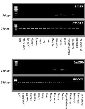
(6) Statistical analysis Expression data were analyzed using SigmaStat 3.1 (Systat Software, Inc.), expressed as percentage of the control group in each experiment, and presented as mean ± SEM. Statistical significance was determined by t test (experiments with two groups), one-way ANOVA with post hoc Tukey test (experiments with more than two groups and one variable), or two-way ANOVA with post hoc Tukey test (experiments with more than two groups and two variables). P < .05 was considered significant. Different letters above bars or asterisks indicate statistical significance.. Results Tissue distribution and differential expression of Lin28 and Lin28b genes in rats As shown in Figure 1A, Lin28 mRNA is prominently expressed in placenta, testis, ovary, and pituitary, with modest expression in the hypothalamus, and weak expression in adipose tissue, heart, lung, liver, and stomach. No expression was observed in kidney, spleen, and skeletal muscle. Similarly, Lin28b transcripts were abundantly expressed in adult rat testis and placenta, with RNA expression being also observed in the hypothalamus. In addition, modest but detectable levels of Lin28b mRNA were found in skeletal and cardiac muscle; other tissues such as the pituitary gland pancreas and stomach had very low or undetectable Lin28b mRNA levels (Figure 1B).. Figure 1. Expression profiles of Lin28 and Lin28b genes in different tissues in the adult rat. A panel of tissues from adult male and female (ovary) rats were screened for expression of Lin28 and Lin28b mRNAs, using conventional RT-PCR, as described in Materials and Methods..
(7) Bioinformatic analyses It is well established that the Lin28/let-7 tandem is subjected to a dual negative feedback regulatory loop. Lin28 and Lin28b repress the synthesis of mature let-7 miRNAs, which in turn posttranscriptionally suppress Lin28/Lin28b expression. Besides the known regulatory roles of cMyc (transcriptional activator of Lin28 (18)) and mir-145 (suppressor of c-Myc (21)) in this network, we searched for additional miRNAs and genes that may be involved in Lin28/let-7 modulation, using the four bioinformatic algorithms listed in Materials and Methods. A summary of the most salient results is shown in Supplemental Table 1. Considering the biological profiles of the potential candidates to regulate Lin28b, and their relatively large number (which precluded systematic analyses of them all), we included in our assays mir-132 and mir-9, as both are highly conserved across most vertebrates species and they are highly expressed in the rat hypothalamus, as compared with other miRNAs (for instance, mir-208 and mir-144 are not expressed in rat hypothalamus while mir-499 expression is very low; data not shown). A schematic representation of the miRNA regulatory network examined in the present study, including miRNAs predicted by TargetScan is provided in Supplemental Figure 1. We also used bioinformatic tools to identify potential target genes of the let-7 family. Besides Lin28/Lin28b, other targets include genes involved in the control of diverse cellular functions, such as membrane fusion, proteolysis, peroxisome biogenesis, endosome sorting (Fignl2, Fidgetin), chromosomal architecture (Hmga2, Arid3b), embryonic patterning, cell lineage gene regulation, cell cycle control, cellular growth and division (Arid3b, Znf512b, Ddx19b), embryonic survival and neuronal/brain maturation or central nervous system formation (Trim71, Acvr1c, Igf2bp2, Prtg), neurodegenerative disease (Pde12), signal transduction cascades or receptors (Map3k1, Pgrmc1, Adrb3, Tgfbr1, Znf512b), and tumorogenesis (N-Myc, Znf512b, Yod1, Tgfbr1, Hic2, Prtg, Igf1r). Additional putative targets of let-7 miRNAs are genes encoding TGF-beta family members (Tgfbr1, Gdf6) and proteins related to insulin-like growth factors, insulin signaling, or diabetes (Igfr1, Igf2bp2, Igf2bp1). Interestingly, many of these genes (Hmga1, Hmga2, Igf1r, Igf2bp1, Myc, Lin28a, Lin28b and Map3k3) have been identified by GWAS studies in humans as related to final height (for further details, see Supplemental Table 2). Expression of c-Myc/Lin28/let-7 and related miRNAs in the hypothalamus during postnatal maturation In male rats, very high levels of Lin28 and Lin28b mRNA were detected in the hypothalamus during the neonatal period (Figure 2). Lin28 mRNA levels declined precipitously thereafter, remaining at low values throughout adulthood (Figure 2). Lin28b mRNA abundance declined less pronouncedly, by about 50% during the infantile period, remaining at these values until early puberty. At puberty, the values declined further remaining low in adulthood (Figure 2). A similar pattern of expression of Lin28b was detected in the hypothalamus of female rats during postnatal maturation (Figure 2). Opposite patterns of Lin28b expression were found in the frontal cortex of male and female rats, with mRNA levels increasing, instead of decreasing, between the neonatal and pubertal periods (Supplemental Figure 2), thereby suggesting that the changes observed in the hypothalamus are tissue-specific. Expression levels of c-Myc mRNA in rat hypothalamus mirrored those of Lin28b in both male and female rats. In clear contrast, let-7a or let-7b, mir-132, and mir-145 miRNA levels were minimal neonatally and progressively increased during postnatal maturation in both male and female rats. These trends, however, were not detected for mir-9, whose hypothalamic levels showed the highest expression during the neonatal period and declined gradually during postnatal maturation (Figure 2)..
(8) Figure 3. Expression profiles of the components of the Lin28/let-7 axis in preoptic area (POA) and medial basal hypothalamus (MBH) regions from male and female rats and female rhesus monkeys at different stages of postnatal/pubertal maturation (A, B). *P ≤ .05 vs neonatal in each region. (a) P ≤ .05 vs MBH for each age. (b) P ≤ .05 vs infantile in each region. (c) P ≤ .05 vs juvenile in each region (two-way ANOVA with post hoc Tukey test). In addition, expression levels of LIN28b measured by qPCR in the MBH of female rhesus monkeys during postnatal development are shown in panel C. *P ≤ .05 vs juvenile group (ANOVA with post hoc Tukey test). Abbreviations: NEO, neonatal; INF, infantile; JUV or J, juvenile; PUB, pubertal; EP, early pubertal; LP, late pubertal.. Expression analyses of Lin28, Lin28b, let-7a, and let-7b during postnatal maturation were conducted in the POA or the MBH, as a means to regionalize the observed changes in our previous studies using whole hypothalamic explants (Figure 3). Overall, the expression profiles of the above targets in these two hypothalamic areas were broadly similar to those shown in Figure 2 for the hypothalamus as a whole. High expression of Lin28 and Lin28b was detected in the POA and MBH in neonatal male and female rats, and markedly declined thereafter, with virtually negligible levels of Lin28 and moderate but detectable levels of Lin28b at later stages (infantile, juvenile, pubertal) of postnatal/pubertal development (Figure 3). In clear contrast, let-7a and let-7b expression levels were low in neonatal samples from the POA and MBH, in both male and female rats, and progressively increased thereafter, with subtle differences being detected between let-7a and let-7b profiles depending on the sex and hypothalamic region. Thus, in male rats, the expression levels of let-7a and let-7b progressively rose between during postnatal maturation in both the POA and MBH, with a trend for higher expression levels of both targets (at least at some age-points) in the MBH. In females, let-7a and let-7b expression was notably higher in the MBH along postnatal maturation, with clear-cut increases during the study period. In contrast, in the POA, the rise of expression of let-7 miRNAs was more modest and only statistically significant for let-7a (Figure 3, A and B). Finally, measurement of Lin28b mRNA levels was also conducted in the MBH of female rhesus monkeys during postnatal development. These analyses revealed that, as in rats, Lin28b mRNA abundance declines from high levels during the infantile period to low values at the time of puberty (Figure 3C)..
(9) Changes in the profiles of hypothalamic expression of c-Myc/Lin28/let-7 in models of perturbed puberty Neonatal administration of EB to male rats perturbed pubertal maturation, as evidenced by decreased serum levels of both gonadotropins, LH and FSH, absent BPS and diminished testicular weights (data not shown), in agreement with previous publications (46–48). This protocol of neonatal estrogenization resulted also in detectable changes in the c-Myc/Lin28/let-7 axis at the expected time of puberty. Thus, on PND-45, neonatally estrogenized male rats displayed increased Lin28b and c-Myc mRNA levels at the hypothalamus; Lin28 levels tended also to be higher but this change did not reach statistical significance. In contrast, hypothalamic let-7a and mir-132 miRNA levels were significantly lower than in controls. Curiously, mir-9 miRNA levels were also lower in neonatally estrogenized male rats, whereas let-7b and mir-145 expression remained unchanged (Figure 4).. Figure 4. Expression profiles of the components of the Lin28/let-7 axis and related factors in the hypothalamus of pubertal male and female rats following neonatal estrogenization or androgenization. In males (upper panels), expression analyses were conducted in whole hypothalamic fragments from rats subjected to a standard protocol of neonatal estrogenization (estradiol benzoate, EB); samples were obtained from peripubertal (postnatal day, PND-45) animals. In females (lower panels), expression analyses were conducted in whole hypothalamic fragments from rats subjected to a standard protocol of neonatal estrogenization (EB) or androgenization (testosterone propionate, TP); samples were obtained from peripubertal (PND-35) animals. Expression analyses included Lin28 (males only), Lin28b, and c-Myc mRNAs, as well as let-7a, let-7b, mir-9, mir-132, and mir-145 miRNAs. *P ≤ .05; vs control (vehicle) (t Student test)..
(10) Likewise, sex steroid milieu manipulation during the neonatal period in female rats (namely, injection on PND-1 of EB or TP) resulted in hormonal (ie, perturbed gonadotropin levels) and phenotypic (ie, altered timing of VO) indices of disrupted puberty. These were associated with detectable changes in the expression patters of the c-Myc/Lin28/let-7 system in the hypothalamus at the expected time of puberty. Thus, as in EB-treated males, expression levels of Lin28b and cMyc mRNA were significantly increased in neonatally EB- and TP-treated female rats (Figure 4). In turn, neonatal treatments with EB or TP decreased the hypothalamic expression levels of let-7b and mir-145, while there was a trend for a drop of let-7a miRNA levels, especially in EB-treated females, which did not reach statistical significance. In addition, hypothalamic expression of mir-9 and mir-132 did not change with neonatal EB or TP treatments, except for a modest increase in mir-132 levels that was observed in androgenized female rats (Figure 4). Other rat models of perturbed puberty, including timed manipulation of photoperiod during postnatal maturation in both male and female rats, were also studied. Males that were submitted to CD between PND 10 and 15 displayed a significant delay in the age of BPS. Thus, at PND-45, 91.7% of control animals (reared under conventional photoperiod; 14-hour light) showed complete BPS, as compared with only 27.3% of CD males. Likewise, at PND-35 only 12.5% of CD females had vaginal opening as compared with 90% of control females. No differences were observed in terms of pubertal body weight between control and CD animals (data not shown). Analysis of CD males at PND-15 revealed a significant increase in hypothalamic Lin28b and cMyc mRNA levels. In contrast, these animals exhibited a consistent decline in let-7a, let-7b, mir132, and mir-145 miRNA abundance, while mir-9 remained unchanged (Figure 5). Surprisingly, these changes were transient, as no consistent modifications in the hypothalamic expression of the above targets were detected in CD males when studied at PND-45, the expected time of puberty (data not shown). In agreement with data for males, data for females submitted to the above CD regimen displayed increased expression levels of Lin28b and c-Myc mRNA levels in the hypothalamus at PND-15; this rise persisted on PND-35 in the case of Lin28. CD females had also significantly lower hypothalamic let-7a, let-7b, mir-132, and mir-145 miRNA levels, and no changes in mir-9. However, these changes were detected only on PND-35, instead ofPND-15, with the exception of mir-145 whose expression levels were also decreased on PND-15 (Figure 5)..
(11) Figure 5. Expression profiles of the components of the Lin28/let-7 axis and related factors in the hypothalamus of male and female rats following photoperiod manipulation (CD, constant darkness from postnatal day [PND]10–15). In males (upper panels), studies in hypothalamic samples obtained at PND-15 are shown. In females (lower panels), studies were conducted at PND-15 and PND-35. For male studies, *P ≤ .05 vs control (normal photoperiod) group (t Student test). For female studies, *P ≤ .05 vs. PND-15 group for each photoperiodic regimen. (a) P ≤ .05 vs control (normal photoperiod) group for each age. (Two-way ANOVA with post hoc Tukey test.). Models of perturbed puberty due to metabolic distress associated to different forms of undernutrition were also evaluated. First, a protocol of subnutrition during lactation, generated by breeding in large litters, was applied as a means to induce partially delayed puberty, as reported recently elsewhere Breeding in large litters caused an overt delay in the timing of puberty (at PND-45, only 22% of males reared in large litters had BPS, while 92.6% of control males did, and at PND-35, 37.5% of females from large litters had complete VO, in comparison with 90% of control females). Postnatal underfeeding of males and females resulted in increased Lin28b and cMyc mRNA levels during the neonatal and infantile periods (Figure 6). Changes in hypothalamic miRNA expression induced by postnatal subnutrition were, however, less consistent and displayed some degree of sexual dimorphism. Thus, while in males and females let-7b miRNA levels remained unchanged, let-7a modestly decreased in underfed males during the neonatal period, but increased in infantile females. Hypothalamic levels of mir-132 tended to diminish in pubertal female rats subjected to postnatal undernutrition, but did not significantly change in underfed males. Finally, hypothalamic mir-145 miRNA content showed opposite profiles, with lower levels during neonatal and infantile periods in males subjected to postnatal underfeeding and significantly increased expression in similarly treated infantile females (Figure 6)..
(12) Figure 6. Expression profiles of the components of the Lin28/let-7 axis and related factors in the hypothalamus of male and female rats following postnatal undernutrition. In males (upper panels), analyses were conducted at postnatal day (PND)–5, -15, and -45. *P ≤ .05 vs PND-5 group for each pups litter group. (a) P ≤ .05 vs. 12 pups/litter for each age. (b) P ≤ .05 vs corresponding PND-15 in each pups litter (two-way ANOVA with post hoc Tukey test). In females (lower panels), analyses were conducted at PND-5, -15, and -35. *P ≤ .05 vs PND-5 in each pups litter group. (a) P ≤ .05 vs. 12 pups/litter in each age. (b) P ≤ .05 vs PND-15 in each pups litter group (two-way ANOVA with post hoc Tukey test). P ≤ .05 was considered significant. - See more at: http://press.endocrine.org/doi/full/10.1210/en.2012-2006#sthash.lsJWWRAZ.dpuf. Discussion From a neurobiological perspective, the initiation of puberty requires an increased secretion of GnRH by a discrete neuronal population of the hypothalamus. Although it appears clear that this change is driven by the coordinated modification of transsynaptic inputs to GnRH neurons of excitatory and inhibitory nature, in conjunction with glial stimulatory signals, the mechanisms setting in motion these coordinated events remain poorly understood. Recent system biology approaches have led to the hypothesis that, rather than being the result of a single trigger, puberty is brought about by the concerted activation of genetic networks hierarchically arranged and endowed with major regulatory hubs connected not only to a plethora of subordinate genes, but also to upper-echelon genes of other regulatory networks (56). According to this concept, such coordinated changes in network activity would ensure proper timing of the pubertal process (40, 57, 58). The existence of regulatory systems able to integrate at different levels the functional output of such networks appears likely, but these systems remain to be identified. MiRNAs might be especially well suited to serve some of these functions, because of their ability to modulate gene expression at multiple hierarchical levels (7, 59). The present study begins to address this issue by providing an initial characterization of the potential roles that miRNA regulatory pathways may have in the central control of puberty. Considering recent GWAS/functional genomic data (23–26, 29), we focused our attention on the Lin28/Let-7 system. Because this system is linked to other regulators (including transcriptional factors and other miRNAs),.
(13) additional components of this putative regulatory pathway were also included in our analyses (as depicted in Supplemental Figure 1). Consistent with a role of the Lin28/let-7 system as one of the factors involved in the control of reproductive function, its various components were found to be expressed in key reproductive tissues, including the hypothalamus. Lin28 mRNA is prominently expressed only in placenta, testis, ovary, and pituitary of adult rats, with modest expression in the adult hypothalamus. Lin28b transcripts were also expressed in adult rat testis and placenta; however, contrasting with Lin28, substantial Lin28b mRNA levels were observed in the hypothalamus of adult rats, whereas no expression was detected in the ovary. Taking into account the putative relationship (as predicted by different GWAS) between Lin28b and the age at menarche (23–26) and the low/negligible expression of Lin28b in the ovary, we focused our expression analyses on the hypothalamus. Our results demonstrate that Lin28 and Lin28b (as well as c-Myc) mRNAs are abundant in the rat hypothalamus of both sexes during the neonatal period, declining strikingly during the infantile-to-juvenile transition. The fact that a similar trend was not observed in other brain areas, such as the cortex, where Lin28b mRNA levels actually increased between the neonatal and the pubertal phases of development, suggests that postnatal loss of Lin28b expression is specific to the hypothalamus. Furthermore, a similar decline in hypothalamic Lin28b mRNA levels was detected in the hypothalamus of female rhesus monkeys during postnatal development, suggesting that this maturational change is evolutionary conserved. In contrast to the decline in the hypothalamic expression of Lin28/Lin28b, let-7 (mainly, let7b), mir-132, and mir-145 miRNAs abundance was minimal in the hypothalamus of neonatal rats, increasing progressively thereafter. These results are coherent with previous studies showing that mir-145 suppresses c-Myc expression, resulting in reduced Lin28/Lin28b transcription and derepression of let-7 family maturation. Moreover, a recent study showed that seven of the eight miRNAs of the let-7 family are highly expressed in hypothalamic arcuate and paraventricular nuclei of adult rats (60). The above developmental profile is reminiscent of that found in C. elegans, in which high levels of Lin28 at the beginning of first larval stage (L1) are followed by an abrupt drop at the end of the L1 phase to allow progression to late larval stage, while let-7 levels increase progressively to reach maximum values at the adult stage (61). Moreover, overexpression of let-7 at early stages of worm development causes premature adoption of adult fates, while let-7 underexpression results in failure to terminally differentiate at the larval to adult transition. Altogether, these data suggest that the level and timing of mature let-7 miRNA expression, in a reciprocal equilibrium with Lin28, plays a vital role in defining key aspects of development in C. elegans (61). An inverse relationship between the expression levels of Lin28 and let-7 had been also reported in mammals, suggesting that Lin28 is an important regulator of let-7 biogenesis across species (8, 62–67). We also studied the changes in hypothalamic levels of mir-132 and mir9 miRNAs, two putative negative regulators of Lin28b. We observed that mir-132 levels increased gradually during postnatal/prepubertal maturation, in keeping with a potential role of this miRNA in the negative regulation of Lin28b. In contrast, mir-9 miRNA levels were the highest during the neonatal period, declining gradually thereafter. This expression pattern suggests that mir-9 does not play a major role in repressing hypothalamic Lin28/Lin28b expression during postnatal maturation. Instead, mir-9 may be primarily involved in regulating neurogenesis and maturational events at earlier, fetal stages of brain development (68–70). In order to provide further evidence for a role of the Lin28/let-7 system in the development of the hypothalamic systems leading to the initiation of puberty, we conducted a series of expression analyses using various models of perturbed puberty. One of these models uses rats in which sexual differentiation of the brain is altered by neonatal exposure to high doses of estrogen or androgens. Consistent with previous studies (46), these treatments induced persistently decreased LH and FSH levels and prevented normal puberty onset. In these animals, hypothalamic levels of c-Myc and Lin28b mRNAs were enhanced, while let-7 and mir-145 miRNA levels were decreased at the expected time of puberty, resembling, but not quite mimicking, the high Lin28/low let-7 levels detected in the hypothalamus of immature animals. The association between enhanced Lin28 expression and delayed puberty observed in rats exposed to high sex steroid levels during neonatal life is in agreement with the delay of puberty onset reported in Lin28a transgenic mice (30), and.
(14) further supports the concept that decreasing hypothalamic Lin28 expression is permissive for the correct timing of puberty. A discordant finding is the sex difference in mir-9 and mir-132 miRNA levels after neonatal estrogenization. Although in males, both mir-9 and mir-132 miRNA levels diminished with estrogenization, no such changes occurred in females rats treated with EB. In fact, mir-132 levels actually increased in females treated with TP. The physiological importance of these differences remains to be established. A trend for an increased ratio of Lin28/let-7 expression was also observed in male and female rats with delayed puberty caused by early manipulation of the photoperiodic conditions. Animals subjected to CD during a discrete postnatal window not only displayed delayed puberty onset, but also disturbed expression levels of Lin28 transcripts and several miRNAs in the hypothalamus at different stages of postnatal development. As was the case of immature rats and neonatally estrogenized/androgenized animals, Lin28b and c-Myc mRNA levels were significantly increased, whereas let-7a and let-7b miRNA levels were decreased in the hypothalamus of CD male and female rats. However, a sex dimorphism was detected for the time course of these changes, since altered Lin28/let-7 ratios were detected in infantile (but not peripubertal) males, whereas in females such alteration was only detectable around the time of puberty. As observed in rats exposed to sex steroids during neonatal life, mir-132 and mir-145 miRNA levels, but not those of mir-9, were decreased in male and female rats exposed to CD, suggesting that delayed puberty caused by either altering neonatal sex differentiation of the brain or disrupting photoperiod cues during infantile development is associated with alterations of the Lin28/let-7regulatory system, a finding that indirectly supports a tentative inhibitory role of this system on the timing of puberty. The functional relevance of these changes, and of their temporal differences, for the alteration of puberty observed after photoperiodic manipulation awaits further investigation. In contrast, altered puberty due to manipulation of the metabolic/feeding status did not result in comparable changes. Neither early subnutrition, by means of rearing large litters, nor a condition of negative energy balance around puberty, which was effective in delaying puberty onset, affected the hypothalamic Lin28/let-7 system as did early androgenization, estrogenization, and CD conditions. For instance, let-7b miRNA levels remained unchanged in the hypothalamus of male and female rats subjected to early subnutrition, whereas Let-7a levels decreased in males during the neonatal period, but increased in infantile females reared in large litters. Likewise, while mir132 levels were lower in pubertal female rats subjected to postweaning undernutrition, mir-145 miRNA levels in the hypothalamus displayed opposite profiles between males (lower levels) and females (higher levels) after postweaning underfeeding. All in all, these observations strongly suggest that, even though conditions of early metabolic stress causing pubertal alterations can perturb the expression of some of the elements of the Lin28/let-7 axis in the hypothalamus, the underlying mechanisms and cellular substrate for such alterations are likely different from those caused by early manipulations of brain sex differentiation or photoperiod. System biology approaches have allowed identification of a putative gene network operating within the female hypothalamus to control the initiation of puberty. Among the elements of this network, a set of tumor suppressor genes (TSG) was shown to be activated and to play a major role in the timing of puberty. These include enhanced at puberty protein 1 (EAP1) (40, 71), the POU gene, octamer transcription factor 2 (OCT2) (58), and thyroid transcription factor 1 (TTF1/NKX2.1) (57), which are transcription factors that are deregulated in several types of cancer (72–74). Hypothalamic TSG networks are hierarchically controlled by specific regulatory hubs; one of the predicted major hubs is p53, whose expression has been shown to increase in the hypothalamus during the pubertal transition in female rhesus monkeys (56). Importantly, p53 is a tumor suppressor that negatively regulates c-Myc, while it induces the transcription of mir-145, which in turn represses a number of oncogenes, including c-Myc itself (20, 21, 75). Given the positive regulatory loop between c-Myc and Lin28/Lin28b expression (18, 19), it is possible that the decline in c-Myc/Lin28 expression along postnatal maturation reported herein might be caused by the increase in p53 abundance (and hence, mir-145) that takes place during postnatal development. In turn, decreased levels of Lin28 binding proteins might cause the observed increase in let-7 miRNA levels. Because bioinformatic analysis predicts that EAP1, OCT-2, and TTF1 are targets of let-7 or related miRNAs (according to the computational analysis, EAP1 is only regulated by let-7e and not by several members of the let-7 family), these genes might serve as portals conveying the regulatory actions of the Lin28/let-7 axis to the neuroendocrine complex.
(15) that controls the timing of puberty. Since let-7 and other miRNAs are considered to be tumor suppressors, it is tempting to assume that they represent an additional component of the regulatory TSG network postulated to be involved in the physiological regulation of puberty. In conclusion, our study documents for the first time the changes in mir-145/c-Myc/Lin-28/let7 axis and mir-132 miRNA expression that occur in the rat hypothalamus during postnatal maturation in male and female rats. Our results show a marked decline in c-Myc/Lin28a/Lin28b mRNA levels coupled to significant increases in let-7, mir-145, and mir-132 miRNA abundance during prepubertal maturation. We also report that delayed puberty induced by either neonatal exposure to sex steroids or infantile exposure to constant darkness hampers these changes. Because the alterations in hypothalamic expression of the Lin28/let-7 system occur gradually during prepubertal maturation, our results suggest that Lin28b and its associated miRNA complex play a permissive role in the initiation of puberty. Such a role is consistent with the earlier finding of delayed puberty in Lin28-overexpressing mice (30), which exhibit delayed sexual maturation despite of enhanced body growth. Our results pave the way for functional analyses involving manipulation of these elements in vivo, including the generation of models of transient or constitutive overexpression of Lin28b, which will be required to conclusively define the precise role that the Lin28b–miRNA complex plays in the hypothalamic regulation of the pubertal process.. Abreviations BPS Balano-preputial separation; CD constant darkness; EB estradiol benzoate; GWAS genome-wide association studies; MBH medial basal hypothalamus; PND postnatal day; POA preoptic area; TP testosterone propionate; TSG tumor suppressor genes; VO vaginal opening.. Acknowledgments This work was supported by research grants BFU 2008-00984 and BFU 2011-25021 from Ministerio de Economia y Competitividad, Spain (cofunded by FEDER Program from European Union [EU]), Project P08CVI-03788 (Junta de Andalucía, Spain) (to M.T.S.), U.S. National Institutes of Health (HD025123-ARRA), U.S. National Science Foundation (IOS1121691) (to S.R.O.), and P51-OD 011092-53, supporting the operation of the Oregon National Primate Research Center, PI10/00088 FIS from Instituto de Salud Carlos III (cofunded by FEDER Program from EU), and projects IN845B-2010/187 and 10CSA916014PR (Xunta de Galicia, Spain) (to F.C.). CIBER Fisiopatología de la Obesidad y Nutrición is an initiative of Instituto de Salud Carlos III, Ministerio de Sanidad, Spain. S.S.-A. was recipient of a travel/visiting scientist fellowship from the Xunta de Galicia (IN809A: 08.02.561B.480.0 and 08.02.561A.480.1).. Disclosure Summary: The authors have nothing to disclose.. References 1. Ambros V , Horvitz HR. Heterochronic mutants of the nematode Caenorhabditis elegans. Science. 1984;226:409–416. 2. Moss EG , Tang L. Conservation of the heterochronic regulator Lin-28, its developmental expression and microRNA complementary sites. Dev Biol. 2003;258:432–442. 3. Balzer E , Moss EG. Localization of the developmental timing regulator Lin28 to mRNP complexes, Pbodies and stress granules. RNA Biol. 2007;4:16–25. 4. Guo Y , Chen Y , Ito H , Watanabe A , Ge X , Kodama T , Aburatani H. Identification and characterization of lin-28 homolog B (LIN28B) in human hepatocellular carcinoma. Gene. 2006;384:51–61. 5. Pasquinelli AE , McCoy A , Jimenez E , Salo E , Ruvkun G , Martindale MQ , Baguna J. Expression of the 22 nucleotide let-7 heterochronic RNA throughout the Metazoa: a role in life history evolution? Evol Dev. 2003;5:372–378. 6. Viswanathan SR , Daley GQ. Lin28: A microRNA regulator with a macro role. Cell. 2010;140:445–449. 7. Chekulaeva M , Filipowicz W. Mechanisms of miRNA-mediated post-transcriptional regulation in animal cells. Curr Opin Cell Biol. 2009;21:452–460. 8. Roush S , Slack FJ. The let-7 family of microRNAs. Trends Cell Biol. 2008;18:505–516. 9. Sokol NS , Xu P , Jan YN , Ambros V. Drosophila let-7 microRNA is required for remodeling of the neuromusculature during metamorphosis. Genes Dev. 2008;22:1591–1596..
(16) 10. Wulczyn FG , Smirnova L , Rybak A, et al.. Post-transcriptional regulation of the let-7 microRNA during neural cell specification. FASEB J. 2007;21:415–426 11. Xia HF , Jin XH , Song PP , Cui Y , Liu CM , Ma X. Temporal and spatial regulation of let-7a in the uterus during embryo implantation in the rat. J Reprod Dev. 2010;56:73–78. 12. Bussing I , Slack FJ , Grosshans H. let-7 microRNAs in development, stem cells and cancer. Trends Mol Med. 2008;14:400–409. 13. Schulman BR , Esquela-Kerscher A , Slack FJ. Reciprocal expression of lin-41 and the microRNAs let-7 and mir-125 during mouse embryogenesis. Dev Dyn. 2005;234:1046–1054. 14. Yamanaka S. Induction of pluripotent stem cells from mouse fibroblasts by four transcription factors. Cell Prolif. 2008;41(Suppl. 1):51–56. 15. Yang DH , Moss EG. Temporally regulated expression of Lin-28 in diverse tissues of the developing mouse. Gene Expr Patterns. 2003;3:719–726. 16. Leppert U , Henke W , Huang X , Muller JM , Dubiel W. Post-transcriptional fine-tuning of COP9 signalosome subunit biosynthesis is regulated by the c-Myc/Lin28B/let-7 pathway. J Mol Biol. 2011;409(5):710–721. 17. Nadiminty N , Tummala R , Lou W, et al.. MicroRNA let-7c suppresses androgen receptor expression and activity via regulation of Myc expression in prostate cancer cells. J Biol Chem. 2012;287(2):1527–1537. 18. Chang TC , Zeitels LR , Hwang HW, et al.. Lin-28B transactivation is necessary for Myc-mediated let-7 repression and proliferation. Proc Natl Acad Sci U S A. 2009;106:3384–3389. 19. Dangi-Garimella S , Yun J , Eves EM, et al.. Raf kinase inhibitory protein suppresses a metastasis signalling cascade involving LIN28 and let-7. EMBO J. 2009;28:347–358. 20. Sachdeva M , Mo YY. miR-145-mediated suppression of cell growth, invasion and metastasis. Am J Transl Res. 2010;2:170–180. 21. Sachdeva M , Zhu S , Wu F, et al.. p53 represses c-Myc through induction of the tumor suppressor miR145. Proc Natl Acad Sci U S A. 2009;106:3207–3212. 22. Lettre G , Jackson AU , Gieger C, et al.. Identification of ten loci associated with height highlights new biological pathways in human growth. Nat Genet. 2008;40:584–591. 23. He C , Kraft P , Chen C, et al.. Genome-wide association studies identify loci associated with age at menarche and age at natural menopause. Nat Genet. 2009;41:724–728. 24. Ong KK , Elks CE , Li S, et al.. Genetic variation in LIN28B is associated with the timing of puberty. Nat Genet. 2009;41:729–733. 25. Perry JR , Stolk L , Franceschini N, et al.. Meta-analysis of genome-wide association data identifies two loci influencing age at menarche. Nat Genet. 2009;41:648–650. 26. Sulem P , Gudbjartsson DF , Rafnar T, et al.. Genome-wide association study identifies sequence variants on 6q21 associated with age at menarche. Nat Genet. 2009;41:734–738. 27. Widen E , Ripatti S , Cousminer DL, et al.. Distinct variants at LIN28B influence growth in height from birth to adulthood. Am J Hum Genet. 2010;86:773–782. 28. Tommiska J , Wehkalampi K , Vaaralahti K , Laitinen EM , Raivio T , Dunkel L. LIN28B in constitutional delay of growth and puberty. J Clin Endocrinol Metab. 2010;95:3063–3066. 29. Elks CE , Perry JR , Sulem P, et al.. Thirty new loci for age at menarche identified by a meta-analysis of genome-wide association studies. Nat Genet. 2010;42:1077–1085. 30. Zhu H , Shah S , Shyh-Chang N, et al.. Lin28a transgenic mice manifest size and puberty phenotypes identified in human genetic association studies. Nat Genet. 2010;42:626–630. 31. Tommiska J , Sorensen K , Aksglaede L, et al.. LIN28B, LIN28A, KISS1, and KISS1R in idiopathic central precocious puberty. BMC Res Notes. 2011;4:363. 32. Silveira-Neto AP , Leal LF , Emerman AB, et al.. Absence of functional LIN28B mutations in a large cohort of patients with idiopathic central precocious puberty. Horm Res Paediatr. 2012;78:144–150. 33. Ojeda SR , Skinner MK. Puberty in the rat. In: , Neill JD, ed. The Physiology of Reproduction. San Diego: Academic Press/Elsevier, 2006:2061–2126. 34. Parent AS , Teilmann G , Juul A , Skakkebaek NE , Toppari J , Bourguignon JP. The timing of normal puberty and the age limits of sexual precocity: variations around the world, secular trends, and changes after migration. Endocr Rev. 2003;24:668–693. 35. Ojeda SR , Lomniczi A , Sandau U , Matagne V. New concepts on the control of the onset of puberty. Endocr Dev. 2010;17:44–51. 36. Ojeda SR , Lomniczi A , Mastronardi C, et al.. Minireview: the neuroendocrine regulation of puberty: is the time ripe for a systems biology approach? Endocrinology. 2006;147:1166–1174. 37. Prieto DM , Markert UR. MicroRNAs in pregnancy. J Reprod Immunol. 2011;88:106–111. 38. Sangiao-Alvarellos S , Varela L , Vazquez MJ, et al.. Influence of ghrelin and growth hormone deficiency on AMP-activated protein kinase and hypothalamic lipid metabolism. J Neuroendocrinol. 2010;22:543– 556. 39. Hiney JK , Srivastava VK , Pine MD , Les Dees W. Insulin-like growth factor-I activates KiSS-1 gene expression in the brain of the prepubertal female rat. Endocrinology. 2009;150:376–384. 40. Heger S , Mastronardi C , Dissen GA, et al.. Enhanced at puberty 1 (EAP1) is a new transcriptional regulator of the female neuroendocrine reproductive axis. J Clin Invest. 2007;117:2145–2154..
(17) 41. Watanabe G , Terasawa E. In vivo release of luteinizing hormone releasing hormone increases with puberty in the female rhesus monkey. Endocrinology. 1989;125:92–99. 42. Lewis BP , Shih IH , Jones-Rhoades MW , Bartel DP , Burge CB. Prediction of mammalian microRNA targets. Cell. 2003;115:787–798. 43. John B , Enright AJ , Aravin A , Tuschl T , Sander C , Marks DS. Human MicroRNA targets. PLoS Biol. 2004;2:e363. 44. Krek A , Grun D , Poy MN, et al.. Combinatorial microRNA target predictions. Nat Genet. 2005;37:495– 500. 45. Maragkakis M , Reczko M , Simossis VA, et al.. DIANA-microT web server: elucidating microRNA functions through target prediction. Nucleic Acids Res. 2009;37:W273–W276. 46. Navarro VM , Sanchez-Garrido MA , Castellano JM, et al.. Persistent impairment of hypothalamic KiSS1 system after exposures to estrogenic compounds at critical periods of brain sex differentiation. Endocrinology. 2009;150:2359–2367. 47. Tena-Sempere M , Navarro J , Pinilla L , Gonzalez LC , Huhtaniemi I , Aguilar E. Neonatal exposure to estrogen differentially alters estrogen receptor alpha and beta mRNA expression in rat testis during postnatal development. J Endocrinol. 2000;165:345–357. 48. Tena-Sempere M , Pinilla L , Gonzalez LC , Aguilar E. Reproductive disruption by exposure to exogenous estrogenic compounds during sex differentiation: lessons from the neonatally estrogenized male rat. Curr Top Steroid Res. 2000;3:671–678. 49. Sizonenko PC , Lang U , Rivest RW , Aubert ML. The pineal and pubertal development. Ciba Found Symp. 1985;117:208–230. 50. Ramaley JA. Entrainment of the adrenal rhythm to photoperiod prior to puberty: effects of early experience on the adrenal rhythm and puberty. Neuroendocrinology. 1976;21:225–235. 51. Leadem CA. Photoperiodic sensitivity of prepubertal female Fisher 344 rats. J Pineal Res. 1988;5:63–70. 52. Castellano JM , Bentsen AH , Sanchez-Garrido MA, et al.. Early metabolic programming of puberty onset: impact of changes in postnatal feeding and rearing conditions on the timing of puberty and development of the hypothalamic kisspeptin system. Endocrinology. 2011;152:3396–3408. 53. Boullu-Ciocca S , Dutour A , Guillaume V , Achard V , Oliver C , Grino M. Postnatal diet-induced obesity in rats upregulates systemic and adipose tissue glucocorticoid metabolism during development and in adulthood: its relationship with the metabolic syndrome. Diabetes. 2005;54:197–203. 54. Mondol V , Pasquinelli AE. Let's make it happen: the role of let-7 microRNA in development. Curr Top Dev Biol. 2012;99:1–30. 55. Castellano JM , Navarro VM , Fernandez-Fernandez R, et al.. Changes in hypothalamic KiSS-1 system and restoration of pubertal activation of the reproductive axis by kisspeptin in undernutrition. Endocrinology. 2005;146:3917–3925. 56. Roth CL , Mastronardi C , Lomniczi A, et al.. Expression of a tumor-related gene network increases in the mammalian hypothalamus at the time of female puberty. Endocrinology. 2007;148:5147–5161. 57. Mastronardi C , Smiley GG , Raber J, et al.. Deletion of the Ttf1 gene in differentiated neurons disrupts female reproduction without impairing basal ganglia function. J Neurosci. 2006;26:13167–13179. 58. Ojeda SR , Hill J , Hill DF, et al.. The Oct-2 POU domain gene in the neuroendocrine brain: a transcriptional regulator of mammalian puberty. Endocrinology. 1999;140:3774–3789. 59. Lynn FC. Meta-regulation: microRNA regulation of glucose and lipid metabolism. Trends Endocrinol Metab. 2009;20:452–459. 60. Amar L , Benoit C , Beaumont G, et al.. MicroRNA expression profiling of hypothalamic arcuate and paraventricular nuclei from single rats using Illumina sequencing technology. J Neurosci Methods. 2012;209:134–143. 61. Reinhart BJ , Slack FJ , Basson M, et al.. The 21-nucleotide let-7 RNA regulates developmental timing in Caenorhabditis elegans. Nature. 2000;403:901–906. 62. Viswanathan SR , Daley GQ , Gregory RI. Selective blockade of microRNA processing by Lin28. Science. 2008;320:97–100. 63. Newman MA , Hammond SM. Lin-28: an early embryonic sentinel that blocks Let-7 biogenesis. Int J Biochem Cell Biol. 2010;42:1330–1333. 64. Newman MA , Thomson JM , Hammond SM. Lin-28 interaction with the Let-7 precursor loop mediates regulated microRNA processing. RNA. 2008;14:1539–1549. 65. Heo I , Joo C , Cho J , Ha M , Han J , Kim VN. Lin28 mediates the terminal uridylation of let-7 precursor MicroRNA. Mol Cell. 2008;32:276–284. 66. Rybak A , Fuchs H , Smirnova L, et al.. A feedback loop comprising lin-28 and let-7 controls pre-let-7 maturation during neural stem-cell commitment. Nat Cell Biol. 2008;10:987–993. 67. Piskounova E , Polytarchou C , Thornton JE, et al.. Lin28A and Lin28B inhibit let-7 microRNA biogenesis by distinct mechanisms. Cell. 2011;147:1066–1079. 68. Denli AM , Cao X , Gage FH. miR-9 and TLX: chasing tails in neural stem cells. Nat Struct Mol Biol. 2009;16:346–347..
(18) 69. Kapsimali M , Kloosterman WP , de Bruijn E , Rosa F , Plasterk RH , Wilson SW. MicroRNAs show a wide diversity of expression profiles in the developing and mature central nervous system. Genome Biol. 2007;8:R173. 70. Krichevsky AM , King KS , Donahue CP , Khrapko K , Kosik KS. A microRNA array reveals extensive regulation of microRNAs during brain development. RNA. 2003;9:1274–1281. 71. Dissen GA , Lomniczi A , Heger S , Neff TL , Ojeda SR. Hypothalamic EAP1 (enhanced at puberty 1) is required for menstrual cyclicity in nonhuman primates. Endocrinology. 2012;153:350–361. 72. Ciarimboli G , Lancaster CS , Schlatter E, et al.. Proximal tubular secretion of creatinine by organic cation transporter OCT2 in cancer patients. Clin Cancer Res. 2012;18:1101–1108. 73. Perner S , Wagner PL , Soltermann A, et al.. TTF1 expression in non-small cell lung carcinoma: association with TTF1 gene amplification and improved survival. J Pathol. 2009;217:65–72. 74. Li R , Pei H , Watson DK , Papas TS. EAP1/Daxx interacts with ETS1 and represses transcriptional activation of ETS1 target genes. Oncogene. 2000;19:745–753. 75. Shi B , Sepp-Lorenzino L , Prisco M , Linsley P , deAngelis T , Baserga R. Micro RNA 145 targets the insulin receptor substrate-1 and inhibits the growth of colon cancer cells. J Biol Chem. 2007;282:32582– 32590..
(19)
Figure


![Figure 5. Expression profiles of the components of the Lin28/let-7 axis and related factors in the hypothalamus of male and female rats following photoperiod manipulation (CD, constant darkness from postnatal day [PND]10–15)](https://thumb-us.123doks.com/thumbv2/123dok_es/7379031.373998/11.892.152.745.125.577/expression-profiles-components-hypothalamus-following-photoperiod-manipulation-postnatal.webp)
+2

Documento similar
Diel expression of the circadian clock genes VunGI, VunELF3, VunTOC1, and VunLHY in pod and seed tissue The analysis of the circadian gene network in generative cowpea tissues
Correlation analysis between the gene expression of the inflammatory-related NFKB1 and triglyceride levels in the postprandial state at 4 h after 8 weeks of dietary intervention with
7) CB1R and CB2R have an opposite role in the regulation of CRH and GR gene expression in the PVN and mPFC of osteoarthritic mice. Therefore, the distinct role of these
Nevertheless, the ratio of corporations using patents of introduction is higher than that obtained from the analysis of the entire patent system as a whole (around 8.5% from 1820
These results correlated with our previous kinetics of arginase I expression in the hearts of both C57BL/6 and BALB/c mice (19) and indicated that during in vivo infection, arginase
When cells were cultivated in an imbalanced minimal salts medium with a high C/N ratio and octanoic acid as the sole carbon source, the expression of phaC1 during the
Late-G 1 PI3K inhibition reduced cell cycle entry in control cells and to a lesser extent in cells over- expressing WT c-Myc; c-Myc T58A expression, however, largely restored cell
Taken together, miRNA and mRNA expression analysis identified a large number of miRNAs and genes in resistance arteries that are differentially expressed upon maturation, some of
