Contents lists available atScienceDirect
Journal of Functional Foods
journal homepage:www.elsevier.com/locate/jff
Wine pomace product modulates oxidative stress and microbiota in obesity
high-fat diet-fed rats
Gisela Gerardi, Mónica Cavia-Saiz, María D. Rivero-Pérez, María L. González-SanJosé,
Pilar Muñiz
⁎Department of Biotechnology and Food Science, Faculty of Sciences, University of Burgos, Plaza Misael Bañuelos, 09001 Burgos, Spain
A R T I C L E I N F O
Keywords: High fat Obesity Polyphenols Wine pomace Wistar rats Microbiota
A B S T R A C T
Obesity is associated with inflammation and oxidative stress. Bioactive compounds can decrease obesity-related disorders by their antioxidant and anti-inflammatory actions. Wistar rats were fed with a high-fat diet during 14 weeks and received 100 mg of wine pomace product (WP)/kg body weight, from the 1st week or from the 7th week and standard diet fed rats were included. Food intake, body weight, blood pressure and plasma glucose, cholesterol and triglyceride were weekly measured. Antioxidant and lipid liver status, fat, adipocyte size, plasma interleukins and microbiota were also determined at 14th week. The results showed a significant reduction of body weight and abdominal fat area, lower blood glucose, decreased liver weight and lipids deposition with increased antioxidant status, lower adipocyte size and increased Lactobacillus spp./Bacteroides spp. ratio. Therefore, wine pomace product reduced obesity-related disorders by amelioration of inflammation and oxi-dative stress and by microbiota regulation suggesting potential preventive clinical benefits.
1. Introduction
Obesity is a multifactorial and inflammatory disease that is asso-ciated to several health disorders including hypertension, dyslipidemia, insulin resistance, type 2 diabetes, cancer and cardiovascular compli-cations. It is characterized by an excess of adipose tissue which occurs through adipocyte hypertrophy and hyperplasia (Siriwardhana et al., 2013). Adipose tissue has an active metabolism and endocrine function that control the energy balance and homeostasis through adipocyte-derived pro- and anti-inflammatory cytokines (adipokines). Therefore, the prevalence of pro-inflammatory adipokines conduces to a systemic inflammation and obesity-relates disorders (Wang et al., 2014). Other factor that also contributes to the pathogenesis of the obesity is the oxidative stress by stimulating the deposition of adipose tissue and al-tering food intake (Manna & Jain, 2015). In this sense, the combination of oxidative stress, dysregulation of adipokines and other factors asso-ciated to obesity (i.e. insulin resistance, lipotoxicity, gut microbiota alterations) are responsible of lipid accumulation in the hepatocytes leading to hepatocyte injury, fibrosis, and chronic liver disease (Polyzos, Kountouras, & Mantzoros, 2019). It is known the role of the reactive oxygen species (ROS) in the development of the obesity (Lechuga-Sancho et al., 2018; Rani, Deep, Singh, Palle, & Yadav, 2016).
Thus, antioxidant therapy could be an important strategy to deal with obesity-related disorders.
Wine pomace products, contains abundant bioactive compounds, including fiber and polyphenols such as phenolic acids, resveratrol, catechins and anthocyanidins which has been suggested to have several beneficial effects. Consumption of foods rich in these bioactive com-pounds has been accepted to decrease inflammation through their anti-inflammatory action (Maleki, Crespo, & Cabanillas, 2019; Siriwardhana et al., 2013). Dietaryfiber has potential effect on satiety and reduces the absorption of carbohydrates and triglycerides, thus leading a re-duction of cardiovascular diseases (Baboota et al., 2013; Lattimer & Haub, 2010).Del Pino-García, Gerardi et al. (2016)associated an in-creased butyrate production from the intestinal metabolism of wine pomacefibers with an increase in plasma insulin and reduction of the blood glucose in diabetic rats. In another hand, there are strong evi-dence that dietary polyphenols increase thermogenesis and energy consumption while decreasing inflammation and oxidative stress with possible weight loss and preventing obesity-related disorders (Wang et al., 2014).Wang et al. (2014)proposed that polyphenols prevent obesity through several mechanisms including reduction of food intake, decreased lipogenesis and increased lipolysis, inhibition of adipocyte differentiation and proliferation, and attenuation of pro-inflammatory
https://doi.org/10.1016/j.jff.2020.103903
Received 28 October 2019; Received in revised form 28 February 2020; Accepted 8 March 2020
⁎Corresponding author at: Plaza Misael Bañuelos, Facultad de Ciencias, Departamento de Biotecnología y Ciencia de los Alimentos, 09001 Burgos, Spain.
E-mail addresses:[email protected](G. Gerardi),[email protected](M. Cavia-Saiz),[email protected](M.D. Rivero-Pérez), [email protected](M.L. González-SanJosé),[email protected](P. Muñiz).
Available online 14 March 2020
responses and oxidative stress. There are several signaling pathways associated with the polyphenols actions in the adipose tissue including AMPK, MAPK, NF-κB, PPARα/γ and TLR2/4 (Siriwardhana et al., 2013). Some polyphenols such as resveratrol or epigallocatechin gallate (EGCG) reduce the PPARγexpression inhibiting the adipocyte diff er-entiation and proliferation. Anthocyanidins decrease fat accumulation in adipocytes through activating AMPK that converts adipocytes into lipid-oxidizing cells and contributes to the regulation of lipolysis and lipogenesis (Wei et al., 2011).
Changes in the gut microbiota are other factor involved in the de-velopment of obesity-disorders. Gut microbiota is involved in the reg-ulation of metabolic function, development of low-grade inflammation and regulation of energy balance. The microbiota composition can modifies the digestibility and absorption of dietary undigested poly-saccharides, decreases liver fatty acid oxidation by suppressing the adenosine monophosphate kinase (AMPK) and contributes in the sys-temic inflammation observed in obesity (Al-Assal et al., 2018). Diet polyphenols can alter the gut microbiota community, resulting in a greater abundance of beneficial bacteria, and a consequent increase in bioavailability (Ozdal et al., 2016). Moreover, there is a mutual re-lationship between gut microbiota and bioactive compounds (fiber, polyphenols) that increases the bioavailability of phenolic compounds and provides increased health benefits.
The present study was designed to evaluate the potential beneficial effect of a red wine pomace product (WP) against obesity-related dis-orders. Wistar rats fed with high-fat diet were used as model of obesity and WP was administered into the diet at two times: from thefirst week or from the seven week, in order to determine whether the WP prevents the obesity development or ameliorates the disorders of an established obesity.
2. Materials and methods
2.1. Chemicals and reagents
LASQCdiet®Rod14-H was obtained from Sodispan (Madrid, Spain). D12451 Rodent Diet with 45 kcal % Fat was purchased from Brogaarden (Lynge, Danmark). Cholesterol and Triglyceride Kits were acquired from BioSystems S.A. (Barcelona, Spain). 2,4-dini-trophenylhydrazine (DNFH), phosphoric acid solution (H3PO4), so-dium pyruvate, 1,1,3,3-tetramethoxypropane (TMP), 2-thiobarbituric acid (TBA), 2-vinylpyridine and cOmplete™Protease Inhibitor Cocktail were obtained from Sigma-Aldrich, Co. (St. Louis, MO, USA). Trichloroacetic acid (TCA) was purchased from Panreac Quimica S.L.U. (Barcelona, Spain). Dimethylsulphoxide (DMSO) and phosphate buf-fered saline (PBS) tablets were purchased from Merck Millipore, Co. (Darmstadt, Germany). QIAmp Mini DNA kit was obtained from Qiagen (West Sussex, UK). iQ™SYBR®Green Supermix was obtained from Bio-rad Laboratories (Hercules, CA, USA). 2.4.6-Tri(2-pyridyl)-5-triazine (TPTZ) and guanidine hydrochloride were purchased from Acros Organics. Proteinase K 20 mg/ml were acquired from ThermoFisher Scientific (Massachusetts, USA). Enzyme-Linked Inmunoabsorbent Assay (ELISA) kits for Tumor Necrosis Factor-Alpha (TNF-α), Interleukin 1 Beta (IL1β) and Interleukin 10 (IL10) were purchased from Cloud-Clone Corp. (Houston, USA).
2.2. Red wine pomace product (WP)
Red wine pomace-derived product from the vinification ofVitis vi-niferaL. cv. Tempranillo was prepared at the University of Burgos ac-cording to a previously described method (González-SanJosé, García-Lomillo, Del Pino-García, Muñiz-Rodríguez, & Rivero-Pérez, 2015). This product was obtained from seedless red wine pomace (WP) and its main characteristics and composition (dietary fiber, fat, protein, mi-nerals and phenolic classes) have been determined previously (Gerardi, Cavia-Saiz, Rivero-Pérez, González-SanJosé, & Muñiz, 2019) and are
summarized inSupplementary Table 1. 2.3. Animals
Experimentation with animals was approved by the Ethics Committee for Experimental Animal Care at the University of Burgos and was carried out in accordance with the Spanish and European laws (Royal Decree 53/2013 of the Spanish Ministry of agriculture, Food and Environment and Ministry of Economy and Competitiveness, and European Directive 2010/63/EU). Male Wistar rats (Rattus norvegicus; n =25; age, 8 weeks; weight: 312 ± 27 g) were obtained from the Animal Research and Welfare Service of Valladolid (SIBA, Valladolid, Spain). The room was maintained at 21 ± 2 °C and humidity at 40 ± 10%, with a 12:12-h light:dark cycle and free access to food (LASQCdiet®Rod14-H, Sodispan, Madrid, Spain) and water. The ani-mals were subjected to human manipulation during the weeks before the experiments for acclimatization. All procedures were designed to limit the number of animals used.
2.4. Experimental animal groups
Animals were randomly divided into two groups: control and high-fat diet (HF). Rats in the control group were fed standard chow diet (6% kcal fat) and rats in HF group received a high-fat diet (45% kcal fat). Two experimental protocols were development (Supplementary Fig. 1). The animals were randomly divides into 4 groups:
–Group 1 (SD): Rats were fed with STANDARD DIET (LASQCdiet® Rod14-H, Sodispan) from week 1 to 14 of the experiment (SD, 18–22 g/d, n = 5) and supplemented daily with 5 g of low-fat yo-gurt.
–Group 2 (HF): Rats were fed with HIGH FAT DIET (Rodent Diet with 45 kcal% Fat D12451, Research Diets) from week 1 to 14 of the experiment (HF, 18–22 g/d, n = 5) and supplemented daily with 5 g of low-fat yogurt.
–Group 3 (HF + WP1w): Rats were fed with HIGH FAT DIET (Rodent Diet with 45 kcal% Fat D12451, Research Diets) and 100 mg of WP/ kg/d from week 1 to 14 of the experiment (HF + WP, n = 5) dis-solved in 5 g of low-fat yogurt.
–Group 4 (HF + WP7w): Rats were fed with HIGH FAT DIET (Rodent Diet with 45 kcal % Fat D12451, Research Diets) from week 1 to 14 of the experiment (18–22 g/d) and received 100 mg of WP/kg/d from week 7 to 14 (HF + WP7w, n = 5) dissolved in 5 g of low-fat yogurt.
The composition of the different types of diet were evaluated and showed inSupplementary Table 2. The dosage of WP evaluated in the present work was selected by the authors according to previous studies (Del Pino-García, Rivero-Pérez, González-SanJosé, Croft, & Muñiz, 2017; Gerardi, Cavia-Saiz, Rivero-Pérez, González-SanJosé, & Muñiz, 2020). The human equivalent dose (HED) was calculated and the rat intake of 100 mg/kg/d of WP equals to 1 g/d in human.
2.5. Weekly measurements
Food and water intake was registered 3 times a week and the ani-mals were weighted once a week. Systolic blood pressure and blood glucose was also assessed weekly. For systolic blood pressure mea-surement, rats were placed into restrained cage and 10 systolic blood pressure determinations were assessed by the tail-cuffmethod using a LE 5001 Pressure Meter (PanLab Harvard Apparatus, Barcelona, Spain) and a LE 160R Pulse Transducer for Rat (PanLab Harvard Apparatus, Barcelona, Spain).
used for glucose determination using a glucometer (Ascensia Breeze2, Bayer). Then, Vacutainer tubes containing the blood samples were centrifuged at 1500 g for 10 min to recover the plasma. Plasma samples were stored at−80 °C until analysis. The volume of blood taken for each rat was never more than 1 ml as per the NC3Rs recommendations (4.4% blood volume removed) (Diehl et al., 2001). Plasma cholesterol and triglyceride were measurement in the plasma samples by kits (BioSystems S.A., Barcelona, Spain).
2.6. Final measurements
At week 15 a combined ketamine/xylazine anesthetic (80 mg ke-tamine/kg and 20 mg xylazine/kg) was administrated to rats by in-traperitoneal injection. Then, blood samples were collected in sodium/ lithium heparine-vacutainer tubes by cardiac puncture and im-mediately, rats were euthanized by intra-cardiac injection of Sodium Pentobarbital (300 mg/kg). Blood samples were centrifuged at 1500g for 10 min to recover the plasma. Plasma cholesterol and triglyceride were measurement in the plasma samples by kits following the manu-facturer‘s instructions (BioSystems S.A., Barcelona, Spain). Pancreas, liver, kidney, hearth and intestine were collect and weighted from all euthanized rats. Visceral fat tissue (thoracic, mesenteric, perirenal and gonadal) were also collected, classified and weighted. All weights are normalized with the length of the femur an expressed as mg/mm. Caecal content was collected and, as all tissues, frozen at−80 °C.
2.6.1. Liver homogenization and lipids measurement
Liver homogenates were obtained by homogenization of 500 mg of liver in cold 50 mM potassium phosphate buffer containing protease inhibitor cocktail. The homogenates were centrifuged at 10,000g for 15 min at 4 °C and the supernatant were collected. Proteins were de-termined in the homogenate by Lowry method. Lipids were extracted from homogenate by Bligh and Dyer method with some modifications and cholesterol and triglycerides were measured by kits (BioSystems S.A., Barcelona, Spain). Briefly, the homogenate were mixed with chloroform and methanol (1:1:2) and centrifuged 10 min at 3000g. The lower organic phase was extracted and the lipid extract was dried at 55 °C under nitrogen atmosphere. The resultant residue was recon-stituted in isopropanol and cholesterol and triglycerides were de-termined.
2.6.2. Retroperitoneal adipose tissue measurements
The adipocytes were observed in fresh samples by a Nikon Eclipse LV100NPOL microscope with a high-precision rotating stage and sizes measurement with the NIS Element Software. Retroperitoneal adipose area was measured with the ImageJ Sofware.
2.6.3. Interleukins measurement
The levels of Tumor Necrosis Factor-Alpha (TNF-α), Interleukin 10 (IL-10) and Interleukin 1 beta (IL-1β) were determined by ELISA kits in plasma samples.
2.6.4. FRAP determination
The total antioxidant capacity of the plasma samples were assessed by the FRAP method (Benzie & Strain, 1996). This method evaluates the reduction of Fe (III) to Fe (II) by the plasma and measured the blue-colored Fe (II)-TPTZ complex formed at 593 nm. The results are ex-pressed as mM of Fe (II).
The ferric reducing antioxidant power (FRAP) was also measure-ment in liver homogenates by the FRAP method.
2.6.5. Levels of GSH/GSSG ratio in liver
Reduced and oxidized glutathione ratio (GSH /GSSG ratio) was measured in the liver by the method described byGriffith (1980)with modifications. Briefly, the liver homogenates were mixed with DTNB, NADPH and Glutathione reductase and the levels of total GSH (reduced
and oxidized) were evaluated measuring absorbance (410 nm) at 2 min intervals for 20 min. Quantification of GSSG was performed following GSH derivatization with 2-vinylpyridine, and GSH was estimated by subtracting GSSG from total GSH. The results are expressed as GSH/ GSSG ratio.
2.6.6. Oxidative stress biomarkers
Indicators of biomolecular damage were analyzed in plasma and liver samples: carbonyl groups (CG), malondialdehyde (MDA) and 8-hydroxydeoxyguanosine levels (8OHdG).
Carbonyl groups were used to evaluate the level of protein damage in the plasma samples. A previously reported method (Levine et al., 1990) was used for the quantification of CG in the plasma. Briefly, 10 µl of plasma or liver homogenate was mixed with 500 µl of DNFH (0.2%/ HCl 2 M) and incubated 4 h at room temperature. Then, the derivatized proteins were precipitated by incubation (15 min, 4 °C) with 500 µl of 20% trichloroacetic acid (TCA) and centrifuged at 6000 × g during 3 min. The supernatants were discard and the pellets washed with ethanol:etyl acetate (1:1). The pellets were reconstituted in 6 M gua-nidine (37 °C, 30 min) and the absorbance was measured at 373 nm. The results are expressed as µmoles of CG per mg of protein.
Plasma and liver MDA levels were assessed by HPLC-DAD following a previously described method (Grotto et al., 2007) and evaluate the lipid peroxidation. Briefly, a mix of 75 µl of plasma or liver homo-genate, 25 µl of distilled water and 25 µl of 3 M NaOH were incubated at 60 °C during 30 min. Then, 125 µl of 6% H3PO4and 125 µl of 0.8% thiobarbituric acid (TBA) were added and incubated at 90 °C during 45 min. Next, 50 µl of 10% sodium dodecyl sulfate (SDS) was added following the extraction of the generated complex by incorporation of 300 µl of butanol and centrifugation at 3000 × g during 10 min. Fi-nally, 20 µl of the supernatant was injected in the HPLC-DAD system and the absorbance of the eluent was measured at 532 nm. An Agilent 1100 series HPLC system (Agilent Technologies) equipped with a diode array detector (DAD) and a Spherisorb3® ODS2 reversed phase C18 (250 mm × 4.6 mm,3 µm particle size; Waters Cromatografia, S.A., Barcelona, Spain) column were used. The mobile phase was a mixture of Milli-Q water:methanol (1:1, v/v) with a 0.6 ml/min of isocratic
flow. MDA levels were calculated by using calibration curves of stan-dard TMP and they are expressed as µM of MDA per mg of protein.
For the determination of 8OHdG in plasma samples, 50 µl of plasma was incubated with 10 µl of proteinase K (20 mg/ml) at 37 °C during 1 h. Then, 60 µl of 4% acetonitrile/ 130 mM sodium acetate/0.6 mM sulfuric acid solution was added and incubated 40 min at 37 °C. Next, the samples were centrifuged at 13,000gduring 5 min and the super-natant were injected in the HPLC-EC system. A 1260 Infinity Agilent HPLC system (Agilent Technologies, California, USA) with the electro-chemical detector DECADE II (Antec Scientific, NV Zoeterwoude, The Netherlands) and a Agilent Eclipse Plus C18 (100 mm × 4.6 mm, 3.5 µm particle size; Agilent Technologies) column were used for the analysis of 8OHdG. 8OHdG levels were calculated by using calibration curves of standard 8OHdG and they are expressed as µM of 8OHdG.
2.6.7. Microbiota analysis
Eubacterium rectale/Clostridium coccoides and Clostridium leptum, PCR conditions included afirst step at 94 °C for 5 min for initial denaturing and 40 cycles of denaturing at 94 °C for 20 s, primer annealing at 50 °C for 20 s and product elongation at 72 °C for 1 min. The proper product amplification was verified by melting curve analysis. The results were obtained using calibration curves and are expressed as log copy number per ng of DNA.
2.7. Data presentation and statistical analysis
The results were expressed as means ± standard deviation of in-dependent samples. Statistical analysis was performed using Statgraphics®Centurion XVI, version 16.2.04 (Statpoint Technologies, Inc., Warranton, VA, USA). One-way analysis of variance (ANOVA), using Fisher's least significant difference (LSD) test, was used to de-termine significant differences between the different experimental groups. A value ofp< 0.05 was applied to all analyses.
3. Results
In the study, we evaluated the effect of the supplementation with 100 mg of WP/kg/d on disorders associated with the obesity induced by high-fat-diet in Wistar rats. Rats fed with a standard diet (SD) were used a control group. Furthermore, in parallel, we evaluated other standard control group supplemented daily with WP (data not shown) and not differences with thea standard group (SD) were observed for all of the parameters studied.
3.1. Energy intake and weight gain
High-Fat Diet (HF) increased energy intake in Wistar rats (Supplementary Fig. 2A). Energy ingestion was higher in all groups of HF rats from the beginning of the experiment compared with the Standard Diet group (SD).
All groups increased their weight during the 14 weeks of experiment and the HF group always had the highest weight (Supplementary Fig. 2B). At the end of the study the body weights of rats fed with the HF diet were significantly higher (54%) than those fed with the stan-dard diet (Table 1). However, the HF rats that received the Wine
Product (HF + WP1w and HF + WP7w) showed a 23% and a 29% less increased body weight than the HF that not ingested the WP. No sig-nificant difference was observed between the HF + WP1w and HF-WP7w groups.
3.2. Systolic blood pressure
After 14 weeks of study, HF diet was able to significantly cause an elevation of systolic blood pressure (SBP) in rats of the HF group (Table 1). Furthermore, the SBP was significantly different between SD and HF groups until the 5th week (Supplementary Fig. 3). In contrast, the ingestion of wine product by HF rats (HF-WP1w and HF-WP7w) produced a significant reduction in their systolic blood pressure com-pared with the HF group (Table 1andSupplementary Fig. 3).
3.3. Blood glucose, cholesterol and triglyceride in plasma
The consumption of HF diet conduced to an increase in the levels of blood glucose of about 27% compared with the standard fed rats (Table 1). Interestingly, the WP intake reduced the blood glucose in the HF rats.
The cholesterol levels in plasma increased in HF rats compared to control group and the intake of the wine pomace product decreased the levels (Table 1). No difference was observed between groups for the triglyceride plasma levels (Table 1).
3.4. Lipid content in liver
Hepatic levels of cholesterol and triglyceride were increased in HF rats (Table 1). Both WP groups (HF-WP1w and HF-WP7w) showed a reduction of the cholesterol and triglyceride levels compared with the HF group that not receive WP. With regard to the triglyceride, the re-duction was more marked for the HF-WP7w group.
3.5. Organs weight
As observed inTable 1, the liver was the only organ whose weight of HF rats was significant higher than the SD rats. The WP intake results in a decrease of the weight of the liver and no changes were observed in
Table 1
Energy intake, weight gain, blood pressure and plasma and organ measurements at 14th week.
Control (SD) Obese (HF) Obese + Product (HF + WP1w) Obese + Product 7w(HF + WP7w) Energy Intake (kCal/d) 62.7 ± 4.7a 84.8 ± 17.0b 75.6 ± 17.4b 81.4 ± 13.9b
Weight Gain (g) 155 ± 6a 283 ± 39b 218 ± 22a 201 ± 30a
Systolic Blood Pressure (mmHg) 128 ± 5a 149 ± 4b 143 ± 14ab 136 ± 3ab Plasma
Glucose (mg/dl) 111 ± 23a 153 ± 12c 140 ± 19bc 124 ± 14ab
Cholesterol (mg/dl) 74.7 ± 4.4a 97.0 ± 12.0b 85.6 ± 10.9ab 80.7 ± 10.7a
Triglycerides (mg/dl) 165 ± 22a 168 ± 22a 188 ± 49a 179 ± 43a
Liver Cholesterol (mg/g) 1.06 ± 0.20a 2.86 ± 0.39c 2.06 ± 0.45b 2.19 ± 0.06b Liver Triglycerides (mg/g) 2.60 ± 0.21a 7.15 ± 1.10d 5.50 ± 0.58c 3.69 ± 0.43b Organs (mg/mm)
Pancreas 13.0 ± 0.6a 15.4 ± 1.4ab 17.2 ± 1.2b 15.2 ± 1.6ab
Liver 211 ± 9a 270 ± 36b 246 ± 24ab 219 ± 14a
Kidney 43.5 ± 1.4a 43.8 ± 4.1a 45.3 ± 3.0a 43.5 ± 4.7a
Hearth 21.1 ± 1.9a 23.0 ± 2.9a 23.0 ± 4.3a 19.6 ± 0.4a
Intestine 59.6 ± 7.7a 69.4 ± 5.6a 69.5 ± 3.9a 66.8 ± 6.3a
Visceral fat weight
Mesenteric Fat 212 ± 17a 462 ± 18c 388 ± 3b 385 ± 152bc
Gonadal Fat 249 ± 31a 633 ± 15c 559 ± 29b 510 ± 222bc
Thoracic Fat 26.5 ± 1.1a 71.2 ± 0.4c 59.8 ± 4.8b 37.2 ± 13.5a
Perirenal Fat 250 ± 20a 644 ± 26c 444 ± 2b 560 ± 228bc
Total Fat 738 ± 64a 1809 ± 7c 1451 ± 20b 1492 ± 625bc
1Data are mean ± standard deviation of 5 independent samples.
2Significant differences (p < 0.05) between groups are indicated with latin letters.
3SD: standard diet fed rats; HF: high fat diet fed rats; HF + WP1w: High Fat diet + wine pomace product (WP) from the 1st week fed rats; HF + WP7w: High Fat
the other organs.
3.6. Abdominal adiposity, total fat, adipocyte area and visceral fat
As observed inFig. 1(A and B), HF diet increased the retroperitoneal adipose area compared with the standard diet in about 2.3 fold. How-ever, the ingestion of WP produced a significant reduction in the fat area as observed for the HF-WP1w and HF-WP7w groups. Furthermore, the HF diet increased the area of mesenteric adipocytes (Fig. 1C and D) and the adipocyte area of both HF-WP groups was similar than the SD group. These results agree with the total mass of mesenteric tissue that was lower for the SD, HF-WP1w and HF-WP7w (212 mg/mm, 388 mg/ mm and 385 mg/mm) groups than the HF group (462 mg/mm) (Table 1). Furthermore, the weight of total visceral fat (mesenteric, gonadal, thoracic and perirenal) was higher for the HF rats and the WP intake reduced the visceral fat of rats (Table 1).
The consumption of HF diet exhibited fat tissue around the heart (Fig. 1F). The weight of the fat observed in this organ was 19 fold times more for the HF group compared with the SD group (Fig. 1E and F). The ingestion of the WP with the HF diet reduced the fat heart in a 53% and 55% for the HF-WP1w and HF-WP7w groups, respectively. However, not difference was observed in the hearth weight (Table 1). In another hand, thoracic fat was significant lower for the SD and HF-WP1w groups (Table 1).
3.7. Plasma inflammatory makers
Plasma levels of Tumor Necrosis Alpha (TNF-α) and Interleukin 1 beta (IL-1β) were increased in HF rats (Fig. 2A and B). The WP inges-tion significantly reduced the plasma levels of both of these
pro-in-flammatory cytokines.
In contrast, the Interleukin 10 (IL-10) was reduced in the plasma of
HF rats. No significant changes were observed between HF and HF-WP groups (Fig. 2C).
3.8. Biomarkers of oxidative stress
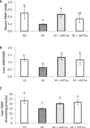
3.8.1. Antioxidant levels of plasma and liver
As observed inFig. 3A, plasma antioxidant capacity assessed by FRAP method was altered in rats fed the HF diet in comparison with the SD diet rats. The SD rats had values of 0.5 mM for FRAP while the HF rats had 0.19 mM. However, the intake of 100 mg/kg of WP by the HF-WP1w significantly increased the antioxidant capacity of the plasma a 42%.
A similar effect was observed in the redox environmental in the liver of rats fed with the WP. Both HF-WP1w and HF-WP7w increased the liver antioxidant capacity measured by FRAP and the levels of GSH/ GSSG ratio compared with the HF group (Fig. 3B and C).
3.8.2. Plasma and liver biomarkers of oxidative stress
In plasma, three types of biomarkers of oxidative stress were eval-uated: carbonyl groups (CG) as biomarker of protein damage (Fig. 4A), malondialdheyde (MDA) for lipid peroxidation asses (Fig. 4B) and 8-hydroxydeoxyguanosine (8OHdG) levels as indicator of DNA damage (Fig. 4C).
All three markers were increased for the HF while the WP reduced the plasma levels of GC, MDA and 8OHdG for both groups (HF-WP1w and HF-WP7w).
In liver, both CG and MDA levels was higher for the HF rats (Fig. 4D and 4E). The intake of WP conduced to a reduction of the CG and MDA levels in liver reaching similar levels to the SD group.
3.8.3. Microbiota changes in the caecal content
for the HF group compared with the SD group. Although the supple-mentation with the WP did not established any significant reduction with respect to the total content.
High fat diet (HF) also increased Bacteroides and decreased Lactobacillus, as showedTable 2. Not significant differences were ob-served for Eubacterium rectale/Clostridium coccoides and Clostridium leptum groups between SD and HF fed rats.
The WP intake after 7th week of HF diet ingestion (HF + WP7w) conduced to a significant reduction in the content Bacteroides and Eubacterium rectale/Clostridium coccoides.
In addition, the WP intake increased the amount of Lactobacillus spp. in the HF-WP1w and HF-WP7w groups.
4. Discussion
Many studies showed that the intake of high fat diets to model obesity in rodents conduced to several disorders in the animals that resemble the human metabolic syndrome and their cardiovascular complications (Buettner et al., 2006). In the present study, the animals fed with the high fat diet gained more weight, had higher plasma glu-cose, cholesterol and triglycerides levels, and showed elevated systolic blood pressure. These results are expected and they agree with other studies where the intake of high fat diets with lard as fat source con-duces to a pronounced obesity and insulin resistance (Buettner et al., 2006).
Several studies have evaluated the impact of polyphenols in the development of obesity (Jin, Song, Weng, & Fantus, 2018; S.Wang et al., 2014; Zhao et al., 2017). It is established that obesity is a
multifactorial condition of chronic inflammation and oxidative stress (Wang et al., 2014). Therefore, the antioxidant and anti-inflammatory effects of the wine pomace product through radical scavenging and signaling pathways modulation (SIRT1, AP1, NF-κB), demonstrated in previous works (Gerardi et al., 2019; Del Pino-García, Gerardi et al., 2016), could be an important strategy against obesity-related
in-flammation. In this aspect, the intake of the wine pomace product (WP) by the high-fat fed rats reduced the alterations of the parameters mentioned above (body weight, plasma glucose, cholesterol and tri-glycerides, and blood pressure), possible due its high content infiber and polyphenols such as resveratrol, epigallocatechin and phenolic acids (Del Pino-García, Gerardi et al., 2016; Del Pino-García, González-Sanjosé et al., 2016; Zou et al., 2018).
Surprisingly, the supplementation with 100 mg/kg of WP reduced the energy intake by the SD rats in a 19% (data not shown). However, this reduction was not accompanied by a reduction in the body weight compared with the SD group. This fact would indicate that the WP ingestion contributes to the satiation, probably by theirfiber content, but this is not sufficient to have an impact in the body weight.Manna and Jain (2015)commented that dietaryfibers increase postprandial satiety and decrease hunger in short-term studies.
Accumulation of excessive fat in adipose tissue and other organs leads to metabolic changes that increase the risk of morbidity in obese individuals. One of the organs that are affected by the deposition of fat is the liver and this is associated with a major risk of fatty liver disease (Aoun et al., 2010). This disease is characterized by an accumulation of lipids in the hepatocytes known as hepatic steatosis and posterior he-patocyte injury, inflammation and fibrosis (cirrhosis) in which Fig. 2.Inflammatory Markers. Plasma levels of Tumor Necrosis Alpha (A),
Interleukin 1 beta (B) and Interleukin 10 (C) determined by ELISA assays by triplicate. Significant differences (p < 0.05) between groups (n = 5) are in-dicated with latin letters. SD: standard diet fed rats; HF: high fat diet fed rats; HF + WP1w: High Fat diet + wine pomace product (WP) from the 1st week fed rats; HF + WP7w: High Fat diet + wine pomace product (WP) from the 7th week fed rats; TNF-α: Tumor Necrosis Alpha; IL-1β: Interleukin 1 beta; IL-10: Interleukin 10.
oxidative stress plays an important role. In our study, the liver weight and their cholesterol and triglycerides levels were increased by the high fat diet. Nerveless, the ingestion of the WP reduced the liver weight and the levels of lipids in this organ. The content of polyphenols andfiber in the WP explains this protective effect of the WP in the liver metabolism. Wine pomace product (WP) act on lipid metabolism by modulating desaturase activity (gene expression and activity) and signaling path-ways of lipid synthesis and degradation (Aoun et al., 2010; Feillet-Coudray et al., 2009) including activation of sirtuin-1 deacetylase (SIRT1) as we saw in previous studies in culture cells (Gerardi et al., 2019). Furthermore, in previous works of our group we demonstrated that the WP modulates the Keap1-Nrf2-ARE system, an important an-tioxidant signaling pathway that regulates the lipid metabolism-related genes (Gerardi et al., 2019; Tu, Wang, Li, Liu, & Sha, 2019). In addition, polyphenols presents in WP can also inhibit the absorption of fatty acids (Xu et al., 2015) and this contribute to the reduction in the lipids levels (plasma and liver) on the body observed for the rats that intake the WP. On the other hand, it is known that short-chain fatty acids (SCFAs) from
fiber microbiota metabolism act as signaling molecules regulating dif-ferent biological processes (Koh, De Vadder, Kovatcheva-Datchary, &
Bäckhed, 2016), the expression of different genes involved in the pro-duction of pro-inflammatory agents (Johnstone, 2002; Koh et al., 2016; Brooks et al., 2017) and modulates glucose and lipid metabolism through microbiota composition regulation (Makki, Deehan, Walter, & Bäckhed, 2018; Bäckhed et al., 2004). In previous studies, we observed that the intake of WP by Wistar rats leads to the increase of the butyric acids levels and the decrease of acetic acid levels in fecal samples (Del Pino-García et al., 2017) that could explain the results obtained. In this regards, the decrease in glucose, cholesterol and triglycerides levels in plasma was more marked in the WP rats at the seven week when they already showed altered parameters associated with the established obesity. These results suggest a potential effect of WP in the ameli-oration of the metabolic disorders of obesity more than in the preven-tion of its development.
Besides the accumulation of fat in liver, the increased abdominal fat observed for the high fat diet fed rats also are an important factor of risk in obesity. The adipose tissue acts as an endocrine organ that produce pro-inflammatory cytokines and, more precisely, adipokines produced principally in the abdominal fat, that regulate energy homeostasis,
in-flammation and insulin resistance (Seymour et al., 2009). Therefore, a Fig. 4.Biomarkers of oxidative stress in plasma and liver samples. Plasma carbonyl groups (A), malondialdehyde (B) and 8-hydroxydeoxyguanosine levels (C). Liver carbonyl groups (D) and malondialdehyde (E). All measurements were performed in triplicate. Significant differences (p < 0.05) between groups (n = 5) are indicated with latin letters. MDA: malondialdehyde; 8OHdG: 8-hydroxyguanosine; SD: standard diet fed rats; HF: high fat diet fed rats; HF + WP1w: High Fat diet + wine pomace product (WP) from the 1st week fed rats; HF + WP7w: High Fat diet + wine pomace product (WP) from the 7th week fed rats.
Table 2
Microbiota composition in caecal samples of rats fed with SD, HF and HF + WP assessed by qPCR.
Control (SD) Obese (HF) Obese + Product (HF + WP1w) Obese + Product 7w (HF + WP7w)
All bacteria 9.85 ± 0.15a 10.06 ± 0.10b 10.06 ± 0.03b 9.99 ± 0.10ab
Bacteroidesspp. 10.19 ± 0.06a 10.78 ± 0.26c 10.83 ± 0.09c 10.52 ± 0.10b
E. rectale/C. coccoides 9.65 ± 0.13a 9.71 ± 0.09b 9.87 ± 0.09b 9.54 ± 0.06a
C. leptum 10.02 ± 0.11a 10.01 ± 0.12a 10.01 ± 0.10a 10.00 ± 0.16a
Lactobacillusspp. 10.54 ± 0.26b 8.94 ± 0.45a 10.58 ± 0.22b 9.88 ± 0.30b
1Results are expressed as log
10copy number/ng DNA.
2Data are mean ± standard deviation of 5 independent samples.
3Significant differences (p < 0.05) between groups are indicated with latin letters.
4SD: standard diet fed rats; HF: high fat diet fed rats; HF + WP1w: High Fat diet + wine pomace product (WP) from the 1st week fed rats; HF + WP7w: High Fat
reduction of the abdominal fat contributes in the amelioration of the obesity complications by decreasing the adipokines and their
pro-in-flammatory effects. In the current study, wine pomace ingestion was associated with significantly reduced abdominal fat. Previous studies demonstrated that diet enriched with anthocyanin reduced glucose, insulin and triglyceride and increased liver and fat tissue expression of PPAR-γ, PPAR-α and lipoprotein lipase (Park, Lee, & Ma, 2005). Seymour et al. (2009) associated the increased liver PPAR-αmRNA with reduced liver lipid content, enhanced hepatic acyl-coenzyme A oxidase activity, and reduced fatty acid synthase activity. In our study, the effect of the WP in the reduction of the abdominal fat area may be a consequence of these PPAR-α-related mechanisms. Remarkably, the WP ingestion attenuates the fat abdominal accumulation when it was ad-ministrated both from the first week and from the seven week, sug-gesting a beneficial effect in both the development and amelioration of the obesity-related abdominal fat.
The size of the adipose cells, the adipocytes, is another important aspect of the inflammatory process in the obesity. Adipocytes are re-sponsible of the production of several cytokines such as TNF-αand IL-6 that regulate the synthesis of fatty acids and their oxidation, and modulates the insulin sensitivity. Therefore, the higher adipocyte size observed for the high fat diet will contribute with the inflammatory process while the reduced size observed for the WP intake could im-prove this pathology. Both, polyphenols and dietaryfiber metabolites (SCFAs) have demonstrated anti-lipolysis action reducing adipocyte size. In addition, the PPAR-αactivation by certain polyphenols reduces adipocyte size conducing to a less secretion of inflammatory cytokines and increases the inhibition of NF-κB transcription factor (Seymour et al., 2009). In previous studies we observed a downregulation of the NF-κB pathway by the WP (Gerardi et al., 2019). In this sense, con-sidering that NF-κB increases the generation of adipokines by the adi-pose tissue, the WP inhibition of this pro-inflammatory pathway could contribute to the reduction of the adipocyte pro-inflammatory action. The observed increase of the pro-inflammatory cytokines TNF-αand IL-1βin plasma of the HF rats, possibly induced by the NF-κB, supports the existence of an inflammatory state in the obese rats that is prevented by the WP intake. Thus, the inhibitory effect of the WP in the expression and activation of NF-κB observed in previous studies (Gerardi et al., 2019), may be responsible of the reduced levels of these
pro-in-flammatory cytokines in the WP fed rats. Furthermore, it had been demonstrated that the microbial metabolism of the dietaryfiber leads to the release of the ferulic acid present in different sources offiber and polyphenols such as the wine pomace and this ferulic acid have anti-inflammatory action reducing the production of pro-inflammatory cy-tokines (Sadar, Vyawahare, & Bodhankar, 2016). In contrast, inter-leukine-10 (IL-10), an anti-inflammatory cytokine responsible of the limitation and termination of the inflammatory response (Febbraio, 2014), was reduced in the plasma of HF rats. These agrees with other works in which the IL-10 was reduced in serum of obese individuals playing an important role in the obesity-related inflammation of several organs (Kondo, 2018). With regard to the adipocytes, it has been de-monstrated that some polyphenols have a role in the differentiation and proliferation of the adipose cells. In particularly, an anti-obesity action has been observed for epigallocatechin gallate (EGCG), a type of polyphenol of the WP. Several studies suggested that EGCG inhibits pre-adipocyte differentiation, decreases adipocyte proliferation, induces adipocyte apoptosis, suppress lipogenesis, and promotes lipolysis and beta (β)-oxidation (Wang et al., 2014). The exposure of high fat diet fed mice to polyphenol extract decreased adipocyte proliferation, diff er-entiation and size through the decreasing expression of PPARγ, SREBP-1 and FAS transcription factors (Trindade et al., 2019). Thus, the modulatory effect of the WP polyphenols could contribute to the re-duction in the size of the rat WP fed adipocytes.
Obesity is considered a risk factor for cardiovascular disease and heart failure. Accumulation of lipids in the cardiac tissue results in cardiac dysfunction as consequence of lipotocixity where the lipids
cause oxidative stress, inflammation, mitochondrial dysfunction and endoplasmic reticulum stress, apoptosis, and abnormal myofibrillar function (Manley, 2013). In the current study, fat tissue was increased in the hearth of rats fed with the high fat diet. Excessive epicardial fat is common in obesity and has a strong relationship with visceral adip-osity. This agrees with our results in which the total visceral fat mass was increased by the high fat diet. In contrast, the WP intake prevents the risk of cardiac dysfunction by reducing the fat deposit around the hearth along with the reduction in the systolic blood pressure. Many studies suggested that resveratrol, a stilbene present in the WP, has anti-obesity action and reduction of the adipose tissue by down-regulation of PPARγthrough indirect activation of SIRT1 and FOXO1 (Costa et al., 2011).
The excessive accumulation of fat in the organs stimulates pro-oxidant and pro-inflammatory states. Obesity induces systemic oxida-tive stress through several mechanisms including superoxide produc-tion from NADPH oxidases, oxidative phosphorylaproduc-tion, protein kinase C (PKC) activation, decreased antioxidant defenses, postprandial ROS generation, chronic inflammation and polyol and hexosamine pathways induction (Feillet-Coudray et al., 2009; Manna & Jain, 2015). Ferric reducing antioxidant power (FRAP) has been used to evaluates the ra-dical quenching capacity by the plasma antioxidants. In agree with others studies, we reported lower plasma levels of FRAP in obese rats (HF) compared with standard fed rats (SD) (Vincent & Taylor, 2006). This increased oxidative stress in obese rats is a consequence of its as-sociated plasma free fatty acids elevation that promotes the generation of superoxide radical through PKC activation of the endothelial NADPH oxidase, and the decreased expression and activities of antioxidant enzymes by obesity (Manna & Jain, 2015). ROS reduction and induc-tion of the antioxidant systems are related with the increase of the antioxidant capacity of the plasma by polyphenols. The increased an-tioxidant capacity of the plasma (TAC) of WP-fed rats agrees with several studies that found significant elevations in plasma TAC after consumption of polyphenol-rich foods such as fruits and vegetables, tea, and red wine (Wang, Chun, & Song, 2013). Several studies demon-strated that high fat diets in rats modified the hepatic mitochondrial capacity. Excessive energy substrate causes mitochondrial dysfunction increasing ROS production and altered redox homeostasis ( Feillet-Coudray et al., 2009). Considering that glutathione represents an an-tioxidant system that provides reducing equivalents for the reduction of H2O2and lipid hydroperoxides by glutathione peroxidase in liver, the observed reduction of glutathione in liver of HF rats reflects a pro-oxidant environment in this organ as consequence of the HF diet. This also agrees with the lower FRAP levels observed in the liver of the HF rats. In contrast, this pro-oxidant environment was reduced by the WP ingestion as result of the antioxidant and anti-obesity effects of the WP polyphenols. This antioxidant effect can be attributed to the presence of anthocyanins, flavanols and flavonols, including catechin and quer-cetin, which may have different mechanisms of action against glu-tathione. First, the antioxidant capacity of polyphenols reduce the amount of endogenous antioxidants such as GSH, used to offset the pro-oxidant environmental. In addition, some polyphenols present in grape skins can increase the expression of γ-glutamyl-cysteine synthetase, whose function is to synthesize glutathione. Similar results were ob-tained in the investigations ofHsu and Yen (2007)in which gallic acid administered to obese rats demonstrates a reduction in GSSG levels, and increases the GSH/GSSG ratio (Hsu & Yen, 2007).
and lipids is a consequence of the lower antioxidant environment ob-served in the liver of HF rats. Mitochondria and NADPH oxidase sys-tems are the principal source of ROS. However,Feillet-Coudray et al. (2009)showed that the increased oxidative stress in liver from obesity rats mainly results from the increased mitochondrial ROS production. It is to be expected that a higher production of ROS have deleterious ef-fects by irreversible oxidation of lipids, proteins and DNA. The intake of WP conduced to a reduction of both lipid and protein damage. In agree with other authors (Feillet-Coudray et al., 2009), polyphenols supple-mentation prevented the lipid and protein oxidation induced in liver by the HF diet, probably by acting directly as ROS scavengers or by sti-mulating endogenous antioxidant systems. These results showed a protective effect against oxidative damage in the liver of obese rats.
The diet plays an important role in the composition of the gut mi-crobiota (Flint, Scott, Louis, Duncan, & Abstract, 2012). There is a close relationship between the host and the microorganisms that colonize it, which can be both beneficial and harmful depending on the majority groups that composite it and their metabolic activity. The gut micro-biota provides essential nutrients and prevents the development of pathogens, which maintains the immunity of the intestinal mucosa and the metabolism of fats. Several studies show that high fat diet causes changes in the composition of the microbiota facilitating the develop-ment of harmful microorganisms that produce endotoxins and alter the metabolism of beneficial bacteria such asBifidobacteria (Yang et al., 2019). These changes can be associated with an increase in bacteria that degrade indigestible polysaccharides allowing their intestinal ab-sorption and conducing to the generation of energy from substances that normally are eliminated by the feces. Thus, there is a specific mi-crobiota that is capable of obtaining more energy from the same daily caloric intake (Tinahones, 2017). The results showed thatBacteroides, a group of highly pathogenic bacteria and related resistance to anti-biotics, were incremented by the high fat diet. The modulation of mi-crobiota by our WP its agree with other studies in which observed that some polyphenols present in the WP, significantly reducedBacteroides spp. growth and enhanced the total bacterial number (Ozdal et al., 2016). Similar results were found in other studies where a decrease of Bacteroideswas observed in obese rats supplemented with sinapic acid and resveratrol (Yang et al., 2019). Dietaryfiber also produces shifts in the microbiota composition by allowing the proliferation of microbial species that are able to utilize this fiber as substrates (primary de-graders) such asFirmicutesor those species that benefit from the car-bohydrate breakdown products (secondary degraders) (Makki et al., 2018). The change in the population ofBacteroides due to the effect wine pomace bioactive compounds reduces the risk of inflammation and resistance to antibiotics; therefore it can contribute to an im-provement in the intestinal health of individuals. In contrast, Lactoba-cillus spp., a group of beneficial bacteria, was reduced in the HF rats while the WP ingestion incremented the amount of this bacteria. Si-milar studies reveal positive effects of the supplement with grape, since they increased the populations of beneficial bacteria ofLactobacillusand Bifidobacterium(Requena et al., 2010). Some of wine pomace bioactive compounds including fiber or polyphenols such asflavanols, antho-cyanins and stilbens, promoted the growth of Lactobacillus inducing changes in the gut microbiota and positively selection for beneficial microbes (Nicolucci et al., 2017; Ozdal et al., 2016; Zou et al., 2018). Furthermore, the acidification of the intestinal environment by SCFAs, as product of microbiotafiber fermentation, increase the absorption of ions with antimicrobial action (Baye, Guyot, & Mouquet-Rivier, 2017; Makki et al., 2018) that could contributing to the reduction of some species such asBacteriodes. In general, there has been a decrease in the groups of harmful bacteria and an increase in those that have a
bene-ficial effect on the organism, which suggests a possible prebiotic effect of the product derived from the red wine pomace.
In summary, the positive effects of the wine pomace product on the obesity, obesity-induced inflammation, and metabolic syndrome are illustrated by the significantly reduced body weight, abdominal fat
area, lower blood glucose, decreased liver weight and lipids deposition, and lower adipocyte size. These anti-obesity effects are associated with a reduction of the oxidative stress in the organism and an improvement of the healthy/harmful microbiota composition. As rich source of polyphenols andfiber, wine pomace product introduced in the diet may modify and reduce several risk factors of obesity complications. Further studies are needed in human subjects with obesity that evaluates the clinical benefits.
Ethics statement.
The authors of the manuscript “Wine pomace product modulates oxidative stress and microbiota in obesity-high fat diet-fed rats”declare that the experimentation with live animals was approved by the Ethics Committee for Experimental Animal Care at the University of Burgos and was carried out in accordance with the Spanish and European laws (Royal Decree 53/2013 of the Spanish Ministry of agriculture, Food and Environment and Ministry of Economy and Competitiveness, and European Directive 2010/63/EU) and following the ARRIVE guidelines.
CRediT authorship contribution statement
Gisela Gerardi:Conceptualization, Methodology, Formal analysis, Investigation, Writing - original draft. Mónica Cavia-Saiz:
Conceptualization, Methodology, Investigation. María D. Rivero-Pérez: Methodology, Investigation. María L. González-SanJosé:
Conceptualization, Project administration, Funding acquisition.Pilar Muñiz: Conceptualization, Formal analysis, Writing - original draft, Project administration, Funding acquisition.
Declaration of Competing Interest
The authors declared that there is no conflict of interest.
Appendix A. Supplementary material
Supplementary data to this article can be found online athttps:// doi.org/10.1016/j.jff.2020.103903.
References
Al-Assal, K., Martinez, A. C., Torrinhas, R. S., Cardinelli, C., Waitzberg, D., & Laboratory (2018). Gut microbiota and obesity.Clinical Nutrition Experimental, 20, 60–64. https://doi.org/10.1684/mtp.2018.0647.
Aoun, M., Michel, F., Fouret, G., Casas, F., Jullien, M., Wrutniak-Cabello, C., et al. (2010). A polyphenol extract modifies quantity but not quality of liver fatty acid content in high-fat-high-sucrose diet-fed rats: Possible implication of the sirtuin pathway.British Journal of Nutrition, 104(12), 1760–1770.https://doi.org/10.1017/
S0007114510002850.
Baboota, R. K., Bishnoi, M., Ambalam, P., Kondepudi, K. K., Sarma, S. M., Boparai, R. K., et al. (2013). Functional food ingredients for the management of obesity and asso-ciated co-morbidities–A review.Journal of Functional Foods, 5(3), 997–1012. Bäckhed, F., Ding, H., Wang, T., Hooper, L. V., Koh, G. Y., Nagy, A., et al. (2004). The gut
microbiota as an environmental factor that regulates fat storage.Proceedings of the National Academy of Sciences, 101(44), 15718–15723.
Baye, K., Guyot, J. P., & Mouquet-Rivier, C. (2017). The unresolved role of dietaryfibers on mineral absorption.Critical Reviews in Food Science and Nutrition, 57(5), 949–957. Benzie, I. F. F., & Strain, J. J. (1996). The Ferric Reducing Ability of Plasma (FRAP) as a
measure of‘“Antioxidant Power”’: The FRAP assay.Analytical Biochemistry, 239, 70–76.
Brooks, L., Viardot, A., Tsakmaki, A., Stolarczyk, E., Howard, J. K., Cani, P. D., et al. (2017). Fermentable carbohydrate stimulates FFAR2-dependent colonic PYY cell expansion to increase satiety.Molecular metabolism, 6, 48–60.
Buettner, R., Parhofer, K. G., Woenckhaus, M., Wrede, C. E., Kunz-Schughart, L. A., Schölmerich, J., et al. (2006). Defining high-fat-diet rat models: Metabolic and mo-lecular effects of different fat types.Journal of Molecular Endocrinology, 36(3), 485–501.https://doi.org/10.1677/jme.1.01909.
Costa, C. D. S., Rohden, F., Hammes, T. O., Margis, R., Bortolotto, J. W., Padoin, A. V., et al. (2011). Resveratrol upregulated SIRT1, FOXO1, and adiponectin and down-regulated PPARγ1-3 mRNA expression in human visceral adipocytes.Obesity Surgery, 21(3), 356–361.https://doi.org/10.1007/s11695-010-0251-7.
hyperglycaemia-induced endothelial dysfunction and oxidative damage in endothelial cells.Journal of Functional Foods, 22, 431–445.https://doi.org/10.1016/j.jff.2016.02.001. Del Pino-García, R., González-Sanjosé, M. L., Rivero-Pérez, M. D., García-Lomillo, J., &
Muñiz, P. (2016). Total antioxidant capacity of new natural powdered seasonings after gastrointestinal and colonic digestion.Food Chemistry, 211, 707–714.https:// doi.org/10.1016/j.foodchem.2016.05.127.
Del Pino-García, R., Rivero-Pérez, M. D., González-SanJosé, M. L., Croft, K. D., & Muñiz, P. (2017). Antihypertensive and antioxidant effects of supplementation with red wine pomace in spontaneously hypertensive rats.Food & Function, 8(7), 2444–2454. https://doi.org/10.1039/C7FO00390K.
Diehl, K.-H., Hull, R., Morton, D., Pfister, R., Rabemampianina, Y., Smith, D., et al. (2001). A good practice guide to the administration of substances and removal of blood, including routes and volumes.Journal of Applied Toxicology, 21, 15–23. Febbraio, M. A. (2014). Role of interleukins in obesity: Implications for metabolic disease.
Trends in Endocrinology & Metabolism, 25(6), 312–319.https://doi.org/10.1016/j. tem.2014.02.004.
Feillet-Coudray, C., Sutra, T., Fouret, G., Ramos, J., Wrutniak-Cabello, C., Cabello, G., et al. (2009). Oxidative stress in rats fed a high-fat high-sucrose diet and preventive effect of polyphenols: Involvement of mitochondrial and NAD(P)H oxidase systems. Free Radical Biology and Medicine, 46(5), 624–632.https://doi.org/10.1016/j. freeradbiomed.2008.11.020.
Flint, H. J., Scott, K. P., Louis, P., Duncan, S. H., & Abstract (2012). Role of the gut microbiota in nutrition and health.Gastroenterology & Hepatology, 9, 577–589. https://doi.org/10.1136/bmj.k2179.
Gerardi, G., Cavia-Saiz, M., Rivero-Pérez, M. D., González-SanJosé, M. L., & Muñiz, P. (2019). Modulation of Akt-p38-MAPK/Nrf2/SIRT1 and NF-κB pathways by wine pomace product in hyperglycemic endothelial cell line.Journal of Functional Foods, 58(May), 255–265.https://doi.org/10.1016/j.jff.2019.05.003.
Gerardi, G., Cavia-Saiz, M., Rivero-Pérez, M. D., González-SanJosé, M. L., & Muñiz, P. (2020). Dose-response effect on polyphenols bioavailability after intake of white and red wine pomace products in Wistar rats.Food & Function.https://doi.org/10.1039/ C9FO01743G.
González-SanJosé, M. L., García-Lomillo, J., Del Pino-García, R., Muñiz-Rodríguez, P., & Rivero-Pérez, M. D. (2015). Sazonador de origen vegetal con propiedades con-servantes, sustitutivo de la sal, y procedimiento de obtención del mismo. Griffith, O. W. (1980). Determination of glutathione and glutathione disulfide using
glutathione reductase and 2-vinylpyridine.Analytical Biochemistry, 106(1), 207–212. Grotto, D., Santa Maria, L. D., Boeira, S., Valentini, J., Charão, M. F., Moro, A. M., et al. (2007). Rapid quantification of malondialdehyde in plasma by high performance li-quid chromatography–visible detection.Journal of Pharmaceutical and Biomedical Analysis, 43(2), 619–624.
Hernández-Hernández, O., Marín-Manzano, M. C., Rubio, L. A., Moreno, F. J., Sanz, M. L., & Clemente, A. (2012). Monomer and linkage type of galacto-oligosaccharides affect their resistance to ileal digestion and prebiotic properties in rats.The Journal of Nutrition, 142(7), 1232–1239.
Hsu, C. L., & Yen, G. C. (2007). Effect of gallic acid on high fat diet-induced dyslipi-daemia, hepatosteatosis and oxidative stress in rats.British Journal of Nutrition, 98(4), 727–735.https://doi.org/10.1017/S000711450774686X.
Jin, T., Song, Z., Weng, J., & Fantus, I. G. (2018). Curcumin and other dietary poly-phenols: Potential mechanisms of metabolic actions and therapy for diabetes and obesity.American Journal of Physiology-Endocrinology and Metabolism, 314(3), E201–E205.https://doi.org/10.1152/ajpendo.00285.2017.
Johnstone, R. W. (2002). Histone-deacetylase inhibitors: Novel drugs for the treatment of cancer.Nature Reviews Drug Discovery, 1(4), 287–299.
Koh, A., De Vadder, F., Kovatcheva-Datchary, P., & Bäckhed, F. (2016). From dietaryfiber to host physiology: Short-chain fatty acids as key bacterial metabolites.Cell, 165(6), 1332–1345.
Kondo, H. (2018). Interleukin 10 treatment ameliorates high-fat diet–induced in-flammatory atrial remodeling andfibrillation.Circulation: Arrhythmia and Electrophysiology.https://doi.org/10.1161/CIRCEP.117.006040.
Lattimer, J. M., & Haub, M. D. (2010). Effects of dietaryfiber and its components on metabolic health.Nutrients, 2(12), 1266–1289.
Lechuga-Sancho, A. M., Gallego-Andujar, D., Ruiz-Ocaña, P., Visiedo, F. M., Saez-Benito, A., Schwarz, M., et al. (2018). Obesity induced alterations in redox homeostasis and oxidative stress are present from an early age.PLoS ONE, 13(1), 1–17.https://doi. org/10.1371/journal.pone.0191547.
Levine, R. L., Garland, D., Oliver, C. N., Amici, A., Climent, I., Lenz, A. G., et al. (1990). Determination of carbonyl content in oxidatively modified proteins.Methods in Enzymology, 186, 464–478.
Makki, K., Deehan, E. C., Walter, J., & Bäckhed, F. (2018). The impact of dietaryfiber on gut microbiota in host health and disease.Cell Host & Microbe, 23(6), 705–715. Maleki, S. J., Crespo, J. F., & Cabanillas, B. (2019). Anti-inflammatory effects of
flavonoids.Food Chemistry, 299, 125124.https://doi.org/10.1016/j.foodchem.2019. 125124.
Manley, G. (2013). Mechanisms of heart failure in obesity.Imo, 71(2), 233–236.https:// doi.org/10.1038/mp.2011.182.doi.
Manna, P., & Jain, S. K. (2015). Obesity, oxidative stress, adipose tissue dysfunction, and the associated health risks: Causes and therapeutic strategies.Metabolic Syndrome and Related Disorders, 13(10), 423–444.https://doi.org/10.1089/met.2015.0095. Nicolucci, A. C., Hume, M. P., Martínez, I., Mayengbam, S., Walter, J., & Reimer, R. A.
(2017). Prebiotics reduce body fat and alter intestinal microbiota in children who are overweight or with obesity.Gastroenterology, 153(3), 711–722.
Ozdal, T., Sela, D. A., Xiao, J., Boyacioglu, D., Chen, F., & Capanoglu, E. (2016). The reciprocal interactions between polyphenols and gut microbiota and effects on bioaccessibility.Nutrients, 8(2), 1–36.https://doi.org/10.3390/nu8020078. Park, M., Lee, W., & Ma, J. (2005). Effects of dietary mulberry, Korean red ginseng, and
banaba on glucose homeostasis in relation with PPAR-alpha, PPAR-gamma, and LPL mRNA expressions.Life Sciences, 77, 3344–3354.
Polyzos, S. A., Kountouras, J., & Mantzoros, C. S. (2019). Obesity and nonalcoholic fatty liver disease: From pathophysiology to therapeutics.Metabolism Clinical and Experimental, 92, 82–97.https://doi.org/10.1016/j.metabol.2018.11.014. Rani, V., Deep, G., Singh, R. K., Palle, K., & Yadav, U. C. S. (2016). Oxidative stress and
metabolic disorders: Pathogenesis and therapeutic strategies.Life Sciences, 148, 183–193.https://doi.org/10.1016/j.lfs.2016.02.002.
Requena, T., Tabasco, R., Monagas, M., Pozo-Bayón, M.Á., Sánchez-Patán, F., Martín-Álvarez, P. J., et al. (2010). Polifenoles del vino y microbiota humana: modulación y metabolismo.Avances En Alimentación Nutrición y Dietética,83–90.
Sadar, S. S., Vyawahare, N. S., & Bodhankar, S. L. (2016). Ferulic acid ameliorates TNBS-induced ulcerative colitis through modulation of cytokines, oxidative stress, iNOs, COX-2, and apoptosis in laboratory rats.EXCLI journal, 15, 482.
Seymour, E. M., Lewis, S. K., Urcuyo-llanes, D. E., Tanone, I. I., Kirakosyan, A., Kaufman, P. B., et al. (2009). Regular tart cherry intake alters abdominal adiposity, adipose gene transcription, and inflammation in obesity-prone rats fed a high fat diet.Journal of Medicinal Food, 12(5), 935–942.
Siriwardhana, N., Kalupahana, N. S., Cekanova, M., LeMieux, M., Greer, B., & Moustaid-Moussa, N. (2013). Modulation of adipose tissue inflammation by bioactive food compounds.Journal of Nutritional Biochemistry, 24(4), 613–623.https://doi.org/10. 1016/j.jnutbio.2012.12.013.
Tinahones, F. J. (2017). La importancia de la microbiota en la obesidad.Revista Española Endocrinología Pediátrica, 8, 15–20.https://doi.org/10.3266/
RevEspEndocrinolPediatr.pre2017.Apr.394.
Trindade, P. L., dos Ramos Soares, E., Monteiro, E. B., Resende, Â. C., Moura-Nunes, N., Souza-Mello, V., et al. (2019). Antiadipogenic effects of açai seed extract on high fat diet-fed mice and 3T3-L1 adipocytes: A potential mechanism of action.Current Developments in Nutrition, 3.https://doi.org/10.1016/j.lfs.2019.04.051.
Tu, W., Wang, H., Li, S., Liu, Q., & Sha, H. (2019). The anti-inflammatory and anti-oxidant mechanisms of the keap1/Nrf2/ARE signaling pathway in chronic diseases.Aging and Disease, 10(3), 637–651.https://doi.org/10.14336/ad.2018.0513.
Vincent, H. K., & Taylor, A. G. (2006). Biomarkers and potential mechanisms of obesity-induced oxidant stress in humans.International Journal of Obesity, 30(3), 400–418. https://doi.org/10.1038/sj.ijo.0803177.
Wang, Y., Chun, O. K., & Song, W. O. (2013). Plasma and dietary antioxidant status as cardiovascular disease risk factors: A review of human studies.Nutrients, 5(8), 2969–3004.https://doi.org/10.3390/nu5082969.
Wang, S., Moustaid-Moussa, N., Chen, L., Mo, H., Shastri, A., Su, R., et al. (2014). Novel insights of dietary polyphenols and obesity.Journal of Nutritional Biochemistry, 25(1), 1–18.https://doi.org/10.1016/j.jnutbio.2013.09.001.
Wei, X., Wang, D., Yang, Y., Xia, M., Li, D., Li, G., et al. (2011). Cyanidin-3-O-β-glucoside improves obesity and triglyceride metabolism in KK-Ay mice by regulating lipopro-tein lipase activity.Journal of the Science of Food and Agriculture, 91(6), 1006–1013. https://doi.org/10.1002/jsfa.4275.
Xu, Y., Zhang, M., Wu, T., Dai, S., Xu, J., & Zhou, Z. (2015). The anti-obesity effect of green tea polysaccharide, polyphenols and caffeine in rats fed with high-fat diet.Food and Function, 6(1), 296–303.
Yang, C., Deng, Q., Xu, J., Wang, X., Hu, C., Tang, H., et al. (2019). Sinapic acid and resveratrol alleviate oxidative stress with modulation of gut microbiota in high-fat diet-fed rats.Food Research International, 116(September 2018), 1202–1211.https:// doi.org/10.1016/j.foodres.2018.10.003.
Zhao, Y., Chen, B., Shen, J., Wan, L., Zhu, Y., Yi, T., et al. (2017). The beneficial effects of quercetin, curcumin, and resveratrol in obesity.Oxidative Medicine and Cellular Longevity, 2017, 1–8.https://doi.org/10.1155/2017/1459497.