Biomaterials and abdominal wall
409
0
0
Texto completo
(2)
(3)
(4)
(5)
(6)
(7) DEDICATION. To Laura, Sofía and Rosamary. Thanks for all the patience and support given along this journey and endeavor. I am sure all of us enjoyed, suffered, and learned a lot; not only science but about each other. Thanks!!! To my parents Carlos and Rosa María, as well as my siblings. The example set throughout these last 51 years kept the eagerness to accomplish what was started. To God for enlightening the path to follow and always putting good friends and people around me..
(8)
(9) ACKNOWLEDGEMENTS Special thanks go to all who made the earning of this possible. All contributed in a special and particular way. For all of those contributions and support, I thank you. María Teresa González Garza y Barrón PhD – beacon, mentor, and guide through the initial part of this journey. Ciro Ángel Rodríguez González PhD – friend and mentor who has always supported my different endeavors. Eduardo Alejandro Flores Villaba MD, MSc – friend, colleague, associate, mentor, and Devil´s advocate for almost everything including patient care. Alex Elías Zúñiga PhD – eager, inquisitive, mentor willing to support in every possible way research and entrepreneurial activities related to technology and research. Montserrat Guraieb Trueba MD – former student, then resident, now colleague and friend; always willing to learn, help, and assist in all projects related to surgical research and patient care. Julieta Rodríguez de Ita MD, PhD – supporting, cheerful, inspiring, and trustworthy Mirna González González PhD – instrumental, confident, and reassuring all the time in the last and toughest part Marco Antonio Rito Palomares PhD – insightful and solution proposer as well as comforter To my School of Medicine & Healthcare Sciences and its Directors here at Tecnológico de Monterrey. Thanks for giving me the opportunity of this personal achievement and allowing the use of the facilities..
(10) To the School of Engineering & Sciences and its Directors at Tecnológico de Monterrey. Always allowing the use of all facilities at the Main Campus and PITT. To the Department of Biology and Clinical Laboratory Sciences and the Department of Mechanical Engineering at The University of Texas Rio Grande Valley for the collaboration; particularly Karen Lozano PhD. To the School of Materials & Engineering at Purdue for the collaboration; particularly Ernesto Marinero PhD..
(11) ABSTRACT Becoming a surgeon does not only require skills, but also scientific knowledge regarding the different areas in which interaction takes place. Performing a midline laparotomy incision and the subsequent abdominal wall closure are part of the skills and knowledge that a surgery resident needs to begin mastering at an early stage. All elements that lead to a faulty wound healing process will contribute to an incisional hernia formation. Understanding and knowing the behavior of suture and abdominal wall reconstruction biomaterials, especially after manipulation, is fundamental for selecting the adequate material for the particular tissue and surgical technique. Several suture biomaterials were tested and studied to find changes in biomechanical properties. This study was performed after having the biomaterial undergo a cyclic tension test. A tensile strength test and mathematical models were developed and used. Posteriorly, the study of biomechanical properties of commonly used sutures for abdominal wall fascial closure was made. A swine mode was used to simulate the human abdominal wall. The biomechanical analysis was med in used sutures by general surgery residents with different levels of experience. No statistical difference was found comparing the material or the level of experience. Polydioxanone was identified as the most suitable suture for abdominal wall closure. Several factors have been identified related to the difficulties of surgical skill acquisition. Among them economical and patient safety lead the list. Simulation has become a major element in the active learning process and skill development area. The advantages foreseen by including simulation in a surgical training program are dependent on the validation as a learning instrument of the simulator been used. A high-fidelity simulator validation study was performed confirming that the available learning tool at our Institution complies with construction, programming and measuring capabilities of a valid simulator. Conclusions and perspectives regarding mesh used in abdominal wall reconstruction are presented. Sutures biomaterials and skill development by general surgery specialists are also included..
(12)
(13) RESUMEN La formación de especialistas en cirugía general demanda que no solo sean operadores; requiere que el personal en entrenamiento tenga un conocimiento científico, y lo aplique, en las diferentes áreas que le generan impacto en su desempeño. Realizar incisiones de la línea media y la consecuente sutura, al tiempo de una laparotomía, forma parte de las habilidades y destrezas que debe dominar desde temprano un cirujano general en formación. Todos aquellos factores que conducen a una cicatrización inadecuada de la herida tendrán como consecuencia la aparición de una hernia incisional. Estudiar y conocer el comportamiento de los biomateriales de sutura utilizados en el cierre de la pared abdominal, particularmente posterior a su manipulación, es fundamental para la adecuada selección del biomaterial a utilizar; así mismo, es importante elegir la técnica de sutura más apropiada para cada tipo de tejido. Se realizó un estudio para determinar los cambios biomecánicos que sufren diversos materiales de sutura, posterior a ser sometidos a una prueba de tensión cíclica. Se llevaron a cabo mediciones y modelos matemáticos que permitieron predecir las modificaciones en las propiedades de los biomateriales utilizados, incluyendo la fuerza tensil. Posteriormente, surge la inquietud de estudiar los cambios en las características biomecánicas de las suturas más comúnmente utilizados para el cierre de fascia de la pared abdominal. Se llevó a cabo un estudio en el que se simuló en modelos porcinos el cierre de la pared abdominal. Se realizó el análisis biomecánico de las suturas utilizadas de acuerdo con el grado de experiencia de los residentes de Cirugía General que participaron en el estudio; obteniendo resultados no estadísticamente significativos, posterior al análisis comparativo de acuerdo con el tipo de biomaterial y el grado de experiencia. Sin embargo, se observó que si existen cambios en las propiedades de los biomateriales utilizados cuando se comparan con los controles. Así mismo, se identificó un material que, por su comportamiento, probablemente sea el mejor para el cierre de la fascia de la pared abdominal. Se han propuesto diversos factores que dificultan la adquisición de habilidades en cirugía. Dentro de los principales, se encuentran los económicos y de seguridad para los pacientes; dejando un lugar importante a la simulación para lograr la adquisición de las destrezas y habilidades necesarias. Esta ventaja de los simuladores es dependiente de que el equipo a utilizar cuente con una validación como instrumento didáctico. Se realizó la validación de un simulador de alta fidelidad disponible en la Institución; comprobando que es un instrumento didáctico. Por último, se presentan las conclusiones y perspectivas a futuro relacionadas a las mallas para la reparación de hernias de la pared abdominal. Lo mismo sucede para los biomateriales de sutura y la adquisición de habilidades por especialistas en Cirugía General..
(14)
(15) TABLE OF CONTENTS. Page. I. INTRODUCTION ..............................................................................................23 I.1. Introduction ........................................................................................25 I.2. References ........................................................................................36. II. PAST, PRESENT AND FUTURE OF SURGICAL MESHES: A REVIEW…… 43 II.1. Abstract…………………………………………………………………… 45 II.2. Introduction ........................................................................................46 II.3. History ...............................................................................................48 II.4. Current Research on Surgical Meshes .............................................51 II.4.1. Elasticity and Tensile Strength ……………………………………... 54 II.4.2. Pore Size ………………………………………………………..……. 55 II.4.3. Weight (Density) ………………………………………………..……. 56 II.4.4. Constitution ……………………………………………………..……. 56 II.4.5. Material Absorption ……..……………………………………..……. 56 II.4.6. Commercially Avaliable Surgical Meshes ……..……...……..……. 57 II.4.6.1. First Generation Meshes………………………………………….. 57 II.4.6.2. Second Generation Meshes………………………………………. 58 II.4.6.3. Third Generation Meshes…………………………………………..63. II.4.7. Manufacturing Processes for Surgical Meshes ……………..……. 64 II.4.7.1. The Extrusion Process ………..………………………………….. 64 II.4.7.2. The Knitting Process ………..…………………………...……….. 65. II.5. Future Perspectives ..........................................................................68 II.5.1. Coatings ……………………………………………..…………..……. 68 II.5.2. Nanofibers …………………………………………..…………..……. 69. II.6. Conclusions ......................................................................................76.
(16) II.6.1. Acknowledgements ………………………………..…………..……. 76 II.6.2. Author Contributions ………………………………..…………..……. 77 II.6.3. Conflicts of Interest ………………………………..…..………..……. 77. II.7. References .......................................................................................78. III. STRESS-SOFTENING AND RESIDUAL STRAIN EFFECTS IN SUTURE MATERIALS ....................................................................................87 III.1. Abstract ............................................................................................89 III.2. Introduction .......................................................................................90 III.3. Experimental Work ...........................................................................92 III.3.1. Suture Materials ……………………………………………………... 92 III.3.2. Uniaxial Tensile Tests ………………………………………....……. 92. III.4. Basic Concepts on Finite Deformations ...........................................95 III.5. A Nonmonotonous Stress-Softening Material Model .......................96 III.5.1. A Nonmonotonous Amended Averaged Stretch Material Model …………………………………….………... 99. III.6. Comparison with Suture Experimental Data ..................................103 III.7. Conclusions ...................................................................................109 III.7.1. Conflict of Interests ………………………………………………... 110 III.7.2. Acknowledgements ………………………………………....……. 110. III.8. References ....................................................................................111 IV. EVALUATION OF A SURGICAL SIMULATOR REGARDING ITS PERFORMANCE WHEN USED BY RESIDENTS WITH DIFFERENT LEVELS OF EXPERIENCE ...............................................................................115 IV.1. Abstract ..........................................................................................117 IV.2. Introduction .....................................................................................118 IV.3. Material and Methods .....................................................................120.
(17) IV.4. Results ...........................................................................................122 IV.5. Discussion ......................................................................................123 IV.6. Conclusion .....................................................................................126 IV.7. References .....................................................................................127. V. EFFECT OF SURGICAL EXPERTISE LEVEL ON BIOMECHANICAL PROPERTIES OF COMMONLY USED SUTURE MATERIALS AFTER ABDOMINAL WALL FASCIAL CLOSURE ........................................................129 V.1. Abstract ...........................................................................................131 V.2. Introduction ......................................................................................132 V.3. Material and Methods ......................................................................134 V.3.1. Models ………………………………………………………………...134 V.3.2. Sutures and Experience Levels ……………………………..……. 134 V.3.3. Suturing Technique …………………….……………………..……. 135 V.3.4. Mechanical Testing …………………………………………………. 135 V.3.5. Statistical Analysis ……..……………………………………..……. 136. V.4. Results ............................................................................................137 V.5. Discussion ......................................................................................142 V.6. Conclusion ......................................................................................146 V.6.1. Highlights ……………………………………………………….…... 146 V.6.2. Funding ………………………………………………………..……. 147 V.6.3. Acknowledgements …………………….……………………..……. 147. V.7. References .....................................................................................148. VI. CONCLUSIONS AND PERSPECTIVES ......................................................151 VI.1. Mesh for Hernia Repair ..................................................................153 VI.2. Sutures ...........................................................................................156.
(18) VI.3. Education .......................................................................................158 VI.4. References .....................................................................................162. APPENDIX 1. Other Published Articles & Patents. APPENDIX 2. Conference Presentations & Meeting Participations. VITA.
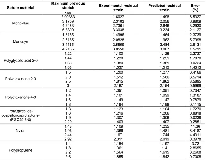
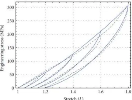
(19) LIST OF FIGURES Figure 1. Types of abdominal wall hernias. (p. 25) Figure 2. Schematic of: (a) woven; and (b) warp knitted structures. (p. 66) Figure 3. Experimental data collected from uniaxial tension cyclic loading unloading tests for Monosyn sutures. (p.93) Figure 4. Engineering stress-stretch data for MonoPlus sutures compared with theoretical predictions of the nonmonotonous amended average-stretch, full-network model for which 𝜇 = 100MPa, 𝑁 = 20, 𝑏 = 0.45, and 𝐶 = 0.0065MPa. The dashed black lines represent theoretical predictions, and the blue solid lines describe experimental data. (p. 104) Figure 5. Engineering stress-stretch data for Monosyn sutures compared with theoretical predictions of the nonmonotonous amended average-stretch, full-network model for which 𝜇 = 92MPa, 𝑁 = 20, 𝑏 = 0.85, and 𝐶 = 0.0045MPa. The dashed black lines represent theoretical predictions, and the blue solid lines describe experimental data. (p. 104) Figure 6. Engineering stress-stretch data for polyglycolic acid sutures compared with theoretical predictions of the nonmonotonous amended average-stretch, full-network model for which 𝜇 = 385MPa, 𝑁 = 70.5, 𝑏 = 1.3, and 𝐶 = 0.001MPa. The dashed black lines represent theoretical predictions, and the blue solid lines describe experimental data. (p.106) Figure 7. Engineering stress-stretch data for polydioxanone 2–0 compared with theoretical predictions of the nonmonotonous amended average-stretch, full-network model for which 𝜇 = 126MPa, 𝑁 = 6, 𝑏 = 0.65, and 𝐶 = 0.0035MPa. The dashed black lines represent theoretical predictions, and the blue solid lines describe experimental data. (p. 106) Figure 8. Engineering stress-stretch data for polydioxanone 4.0 compared with theoretical predictions of the nonmonotonous amended average-stretch, full-network model for which 𝜇 = 148MPa, 𝑁 = 1.95, 𝑏 = 0.445, and 𝐶 = 0.0115MPa. The dashed black lines represent theoretical predictions, and the blue solid lines describe experimental data. (p.107) Figure 9. Engineering stress-stretch data for PGC25 3–0 sutures compared with theoretical predictions of the nonmonotonous amended average-stretch, full-network model for which 𝜇 = 90MPa, 𝑁 = 2.35, 𝑏 = 0.75, and 𝐶 = 0.012MPa. The dashed black lines represent theoretical predictions, and the blue solid lines describe experimental data. (p.107) Figure 10. Engineering stress-stretch data for nylon sutures compared with theoretical predictions of the nonmonotonous amended average-stretch, full-network model for which 𝜇 = 155MPa, 𝑁 = 20.5, 𝑏 = 1, and 𝐶 = 0.0035MPa. The dashed black lines represent theoretical predictions, and the blue solid lines describe experimental data. (p.108) Figure 11. Engineering stress-stretch data for polypropylene sutures compared with theoretical predictions of the nonmonotonous amended average-stretch, full-network model for which 𝜇 = 200MPa, 𝑁 = 30.5, 𝑏 = 0.65, and 𝐶 = 0.00265MPa. The dashed black lines represent theoretical predictions, and the blue solid lines describe experimental data. (p.108) Figure 12. Total time lapsed (seconds) prior to rupture of the suturing material during the biomechanical testing. Data points are expressed as medians, bars represent 25 and 75% quartiles. (p. 138).
(20) Figure 13. Median maximum extension prior to rupture of sutures following their use by surgeons with different level of expertise and non-used control sutures. Data points are expressed as medians, bars represent 25 and 75% quartiles. (p.138) Figure 14. Tensile strength (Ftmax) withheld by sutures prior to rupture, following their use by surgeons with different level of expertise and non-used control sutures. Data points are expressed as medians, bars represent 25 and 75% quartiles. (p.139) Figure 15. Maximum stress exerted by the suturing material following their use by surgeons with different level of expertise (novice, intermediate and expert) and non-used control sutures. Measurements where obtained in MegaPascals (MPa). Data points are expressed as medians, bars represent 25 and 75% quartiles. (p.139) Figure 16. Force vs deformation curves expressed as (N) and (mm/mm) respectively. Each curve represents average suture behavior among test groups of all materials (A: Vicryl®, B: Prolene® and C: PDS®). Next to the different experience level groups is expressed the average maximum tension force for each one. (p.141).
(21) LIST OF TABLES Table 1. Hospital Discharge Registry 2015. (p.26) Table 2. Suture Material Classification. (p. 29) Table 3. Classification of commercially available first generation surgical meshes. (p. 59-60) Table 4. Classification of commercially available second generation surgical meshes. (p.62) Table 5. Classification of commercially available third generation surgical meshes. (p.63) Table 6. Classification of commercially available surgical meshes. (p.66) Table 7. Material properties of surgical mesh coatings. (p.70) Table 8. Examples of surgical mesh coating parameters. (p. 71) Table 9. Nanofiber based surgical meshes. (p.73) Table 10. Aspects related to hernia meshes compared in recently published reviews. (p.75) Table 11. Comparison between experimental and predicted residual strain deformations of the selected suture materials. (p.94) Table 12. Tasks made using the virtual simulator Surgical SIM®. (p. 121) Table 13. Ranges, averages, and correlations of time (seconds) per task, level of experience, and totals. (p. 123) Table 14. Ranges, averages, and correlations of the trajectory (cm) per task, level of experience, and totals. (p. 124) Table 15. Ranges, averages, and correlations of errors per task, level of experience, and totals. (p. 124) Table 16. Abdominal fascial closures performed by different experience level residents. (p. 137) Table 17. Kruskal-Wallis. (p. 140) Table 18. Mann-Whitney U test for maximum tensile force. (p. 141).
(22)
(23) I. INTRODUCTION.
(24)
(25) J. A. Díaz-Elizondo INTRODUCTION. I.1. INTRODUCTION The protrusion of a tissue or an organ through a “hole” or defect and surroundings, is known as a hernia. Usually this is referred to the described situation compromising the abdominal wall, especially at the inguinal area. These sites most commonly include the inguinal, femoral, and umbilical areas, linea alba, lower portion of the semilunar line, and sites of prior incisions. Primary hernias are classified according to the site in where they arise (Figure 1).. Figure 1. Types of abdominal wall hernias. (From Dorland's illustrated medical dictionary, ed 31, Philadelphia, 2007, WB Saunders, Plate 21.). 25.
(26) J. A. Díaz-Elizondo INTRODUCTION. The innermost musculoaponeurotic layer has the orifice of the hernia, while the sac has peritoneal lining and protrudes from the orifice. A large variability exists in the relationship between the hernia defect and the hernia sac. In the United States of America, abdominal wall hernias were associated with 4.7 million ambulatory care visits in 2004. Inguinal hernia repair accounted for 600,000 surgical procedures in 2004. Abdominal wall hernias represented 372,000 hospitalizations (more than a 24-hour period hospital stay) in 2004. They also generated more than 3.7 million prescriptions in 2004 [1-4]. More than 1600 deaths were related to abdominal wall hernias in 2007 [4, 5]. In the 2015 census in Mexico, the overall reported hospital hernia-related discharge diagnosis can be appreciated in Table 1. Patients treated for complaints related to Inguinal hernias were the most common; probably, the other hernia types, including incisional hernias, were either misclassified or underreported. Table 1. Hospital Discharge Registry 2015 ICD-10. Hernia Type. Year 2015. K40 – K40.9. Inguinal. 294962. K41 – K41.9. Femoral. 12009. K42 – K42.9. Umbilical. 141957. K43 – K43.9. Ventral. 47387. K44 – K44.9. Diaphragmatic. 9572. K45 – K45.9. Other abdominal hernias. 2135. K46 – K46.9. Other non-specified. 13463. Total. 521485. http://www.dgis.salud.gob.mx/contenidos/basesdedatos/da_egresoshosp_gobmx.html. 26.
(27) J. A. Díaz-Elizondo INTRODUCTION. Midline incision for a laparotomy is the most commonly performed surgical incision, as it provides adequate exposure. It is simple and quick to perform and presents with low risk of bleeding. The main challenge of performing this type of surgical incision is the correct identification of surgical technique and suture material to perform fascial closure [6]. In an incisional hernia, a prior abdominal incision has been made and sutured. Usually, a Surgical Site Infection (SSI) or a seroma will be involved in incisional hernia development.. The healing process is compromised and eventually, the. strength of the abdominal wall diminished, and a hernia will develop. Adequate closure and wound edges stabilization by sutures, are critical events that can influence the success in any surgical procedure. However, the presence of foreign materials in the surgical wound, enhances significantly the susceptibility of the host tissue to infection [7, 8]. Therefore, the final consequence of placing a suture can be postoperative infection resulting in wound healing compromise [9]. Incisional hernia continuous to be the most common long-term complication after a midline laparotomy. The incidence varies between 10 and 13% of all laparotomies and 3 and 8% of laparoscopy port incisions. Its incidence increases up to 23 to 40% when SSI is present during the healing process. Usually, incisional hernias develop during the first 3 years after the surgical intervention, even though 50% are clinically evident during the first year after surgery. Mortality rate of 0.24% has been reported when an incisional hernia develops [10]. Complications related with incisional hernia occurrence are: pain, diminished quality of life and other complications that can put life at risk, including incarceration (6 – 15%) or bowel strangulation (2%) [11]. Wound failure risk factors can be classified as patient or surgeon related. Diabetes mellitus, vascular disease, and smoking; cause a decrease in blood supply to the wound, hence, are factors that predispose the development of incisional 27.
(28) J. A. Díaz-Elizondo INTRODUCTION. hernias [12]. Male sex, advanced age, emergency operation, past medical history (e.g. prior abdominal surgery), comorbidities (e.g. connective tissue disorders), and health related habits such as smoking or overweight, are included among the patient related factors and most of the time cannot be standardized or modified. Meanwhile surgeon related factors can be modified or standardized with relative ease. These include: suturing technique, suture material selection, and surgeon expertise; therefore, providing an opportunity to decrease the risk of surgical site occurrences [6, 11]. As it would be expected, after a surgical incision is made, tissue requires approximation. The main function of suture materials is surgical wound closure. Suture failure after a surgical procedure, entails serious complications with the subsequent increase on surgical morbidity and mortality [13]. The word “suture” describes any type of filamentous biomaterial used to ligate a blood vessel or a conduit, as well as the materials used for tissue approximation [14, 15]. A suture acts by creating a loop and the fixation of a safety perimeter along its geometry with the help of a surgical knot. This loop can approximate adjacent structures through suture transfixion within the loop Suture security is the ability of the knot and the material to hold tissue approximation along the healing process without sliding of the knot or rupture of the suture [16]. A surgical suture consists of a fiber or fibrous structure with a metallic needle attached to one end. They can be classified as: 1) absorbable or non-absorbable, 2) monofilament or braided (multifilament), and 3) natural or biosynthetic polymers (Table 2). The main property requirements of suture materials include mechanical, physical, handling, biocompatibility and antimicrobial nature. Suture material selection is of great impact in wound healing process [15]. In the modern age, technical advances have led to a unique position where we can choose specific suture materials for each type of tissue or situation [14].. 28.
(29) J. A. Díaz-Elizondo INTRODUCTION. Table 2. Suture Material Classification Suture Material Classification Natural Absorbable. Natural Non-Absorbable. •. Catgut (simple // chromic). •. Silk. •. Collagen (simple // chromic). •. Linen. •. Cotton. Syntethic Absorbable. Syntethic Non-Absorbable. •. Polyglecaprone 25 (Monocryl). •. Polyamide. •. Polydioxanone (PDS). •. Stainless Steel. •. Polyglycolic Acid (Dexon). •. Polipropilene. •. Polyglactin 910 (Vicryl). •. Polyester (mono or multifilament). The ideal suture characteristics probably should include: 1. Must not present any allergic, carcinogenic, capillary or electrostatic reaction. 2. Should be easy to sterilize. 3. Must not produce any magnetic field around it, like a wire cable. 4. Should be easy to handle. 5. Ideally should cause the least possible tissue reaction during the healing process and after fulfilling its purpose. 6. Must not favor bacterial growth around it. 7. Should hold tissue in a secure fashion throughout the wound healing process. 8. Must retain adequate tensile strength according to its transverse diameter. None of the suture materials available today possess every one of the ideal suture characteristics.. It is the surgeon´s job to calculate and measure the. advantages and disadvantages of every available suture material and to choose the better fit for the surgical procedure [14, 17].. 29.
(30) J. A. Díaz-Elizondo INTRODUCTION. It is extremely important that the surgical strain does not break during its use; once the suture line is created and the knot is tied, suture tensile strength is of less importance. However, the suture has to have enough force to keep the tissue together along the healing process [18]. Due to the diverse amount of tissues, surgeons require suture materials with different and specific physical and mechanical properties in order to achieve better outcomes [19]. Surgeons have multiple ways to assess the biomechanical properties of suture materials [20]. The mechanical security of a suture material is defined as its ability of function without presenting significant variation on its performance [16]. Surgical sutures possess multiple biomechanical characteristics. Some of them are: •. Tensile force: amount of weight required to break the suture material, divided by the transverse diameter of the suture. The transverse area of the suture is measure by the size of the braids.. •. Smoothness: depend on the molecular characteristics of the suture or due to a specific treatment on its surface; which helps to reduce trauma to the tissue while the suture goes thru the tissue. Its relationship with the force of the knot is expressed as friction coefficient.. •. Memory and elasticity: both related; the first one, is defined as the capacity of the suture to return to its original shape after manipulation. The latter is defined as the property of the suture to lengthen when the tissue is inflamed and after a while return to its original length when tensile force disappears.. •. Inert and biocompatible: this means that the suture does not react chemically with the environment and lacks pyrogenic and antigenic properties and possibly is capable of counteract bacterial colonization [21].. 30.
(31) J. A. Díaz-Elizondo INTRODUCTION. In material science, the mechanical fatigue, is a term used to refer to the failure of a material that happens in structures that are put under fluctuant and recurrent stress [22]. The work to break of a suture material is defined as the amount of energy that is required to break it. It is represented by the area under the tension-deformity curve. It measures the capacity of a material to resist sudden impacts of a determined amount of energy. The material will break if the amount of sudden energy applied exceeds the work to rupture [19]. When a suture material is stretched and then released with a new length, the tension drops in a gradual fashion; this phenomenon is known as stress – relaxation. Similarly, when equal forces are repeatedly applied to the material, this will immediately increase its length and will continue to elongate until a point of equilibrium is reached. This phenomenon is known as approximation. Part of this elongation, irreversible elongation, persists even when the stimulus is withdrawn [23]. Klink, et al., identified three phases in the process of suture relaxation. The initial phase where rapid loss of tension lasts only a minute and it can be secondary to the damage due to the cut of the tissue. The percentage of the tension lost is intimately related with the content of collagen in the tissue. The second phase, is characterized by the slow decrease in suture tension, reflecting a plastic deformation specific for the tissue. Phase three, is characterized by a plateau, which represents structural stability left in the tissue. The ratio of the residual tension and the initial tension during the first phase is strongly related with the content of collagen in the tissue [13]. The final solution for the selection of suture materials with the appropriate mechanical characteristics for the tissue where it’s going to be used, must be choosing the suture which tension-deformation curve is equivalent, or the closest one, to the tension-deformation curve of the tissue that will be sutured. As previously mentioned, other properties such as the reaction of the tissue to the suture material, 31.
(32) J. A. Díaz-Elizondo INTRODUCTION. the condition and characteristics of the wound, knot security, the degree of suture absorption, and the biomechanical properties of the material, must been taken into consideration for the final selection of the material that will be used [19]. Hernia repair is one of the most common operations performed by general surgeons. Despite the frequency of this procedure, no surgeon has ideal results. One of the most important aspects for an adequate hernia repair is to have a tension free repair. To achieve such goal, the use of a mesh is crucial [24-26]. Mesh is being used to replace or reinforce the abdominal wall. To understand which properties are desirable in suture and mesh, it is important to understand some of the characteristics of the tissue the suture is holding and the mesh is replacing or reinforcing [27-29]. Mathematical models have been developed to determine the different strengths and forces of the abdominal.. Some groups have also determined. elasticity. Accordingly, the force of the anterior abdominal wall has been calculated in 16 N/cm. The average male abdominal wall elasticity at 16 N/cm was 23 (±7%) in the vertical axis and 15 (±5%) in the horizontal axis. The average elasticity of the female anterior abdominal wall at 16 N/cm was 32 (±7%) in the vertical axis and 17 (±5%) in the horizontal axis [27, 28]. Intra-abdominal pressure has been measured in different activities. Documentation up to 252 mm Hg occurs while lifting, coughing, or jumping. Correlation to forces of 27 N/cm has been made [29]. Basic principles in hernia repair include hernia sac reduction and removal when possible and tissue reinforcement and/or replacement. The latter is achieved by incorporating a mesh serving as a scaffold for tissue ingrowth and fibrosis. Biocompatibility degree of host-tissue response is one of the most important issues. regarding. success. of. an. implant.. Grafts. suffer. incorporation. (neovascularization and collagen remodeling throughout the implant), encapsulation (fibrotic material being deposited at the edges and around the implant), and degradation/reabsorption not leaving a trace or evidence of the mesh or implant [30].. 32.
(33) J. A. Díaz-Elizondo INTRODUCTION. Once aware of the arising problems due to initial mesh placement (1905 – 1955) in hernia repair, a next generation of mesh products (“second generation mesh”) began to develop. These second-generation products have a barrier. The barrier is aimed to prevent adhesion formation, hence avoiding complications related to mesh use. Barriers are made of collagen, methylcellulose, omega-fatty acids, or some have a combination of materials or a dual surface (polypropylene in the parietal side and expanded polytetrafluoroethylene in the visceral side). The principle is to have less inflammation in the visceral side and an adequate tissue in growth in the parietal side of the mesh. The barrier is a retardant and helps in trying to avoid different structures to adhere to the implanted mesh. Use of synthetic mesh products is thought to be contraindicated in contaminated or infected operative fields. An infection resistant mesh implant, which also supports repairs in contaminated fields, is needed. Ideally a biological mesh that resists infection and reinforces at the same time of a hernia repair, would probably be the best option in all hernia repairs [5, 31-34]. Third generations of mesh products have also appeared.. This third. generation of products is referred as biological meshes. These are being used in contaminated operative fields. Experience has been gained but yet to be compared to the outcome of a first- or second-generation mesh [35-41]. Performance and comparison among the three generations is important. Prosthetic material in excess can be associated with a higher rate of complications. Some of these complications might include adhesion formation, intense scarring and inflammation, less mesh flexibility, decreased or loss of abdominal wall compliance, increased mesh contraction, hence hernia recurrence. Chronic inflammation may lead to increased pain and mesh degradation through oxidation changing the material´s properties [42-46]. The same principles behind intestinal adhesions (inflammation, foreign body reaction, infection, host rejection to foreign material), are some of the same issues associated with problems seen with the use of mesh in hernia repair [47-52]. 33.
(34) J. A. Díaz-Elizondo INTRODUCTION. Sensations of the implanted mesh, movement limitation, pain and/or neuropraxia are some of the other common issues arising concern while using synthetic or biological mesh for reinforcement or replacement. Surgical experience consists in surgical skills as well as in an adequate judgement clinical/surgical [53]. Ericsson has contributed to elucidate surgical skills acquisition. The performance of an expert represents the maximum level of an ability acquisition and resulting in a gradual improvement in performance thru a wide experience on a determined area. In accordance to Ericsson, most of the professionals manage to achieve a stable level in performance in certain skill and keep this status for the rest of their careers. In surgery, experts have been described as experienced surgeons with consistently better outcomes than those not considered experts. Case volume by itself is not the only element that dictates the level of surgical skills among surgeons, since variations in performance have been proven among surgeons with high case volume of patients. Intentional practice is a critical process in the development of an expert. Ericsson points out that intentional practice hours are a heavier factor when compared only with surgical time [54, 55]. Surgical expertise and experience, along with scientific knowledge regarding suture and mesh biomaterial properties are fundamental aspects related to abdominal wall hernia prevention and repair. The aim of this work is to approache some of these issues and to: 1) Analyze the existent commercial surgical meshes, their properties, manufacturing procedures, and observed biological responses and the requirements for an ideal surgical mesh and potential manufacturing procedures. 2) Understand the stress-softening and residual strain effects in suture materials such as polyglycolic acid, polydioxanone, nylon, and polypropylene commonly used in surgical procedures when subjected to loading and unloading cycles by developing a phenomenological non-. 34.
(35) J. A. Díaz-Elizondo INTRODUCTION. monotonous stress-softening hyperelastic material model that depends on the amount of strain and permanent set effects. 3) Validate of a minimally invasive surgery (Surgical SIM® - METI, Sarasota, Fl) for skill acquisition. 4) Determine the impact of surgical trainee experience level on changes in biomechanical properties of different suture materials used for abdominal wall fascial closure in a porcine model. In the upcoming chapters, research papers approaching these particular issues are presented. The structure of the upcoming chapters is as follows: Chapter 2: Past, Present, and Future of Surgical Meshes: A Review (Published 2017) Chapter 3: Stress-Softening and Residual Strain Effects in Suture Materials (Published 2013) Chapter 4: Evaluation of a Surgery Simulator While Being Used by Residents with Different Levels of Experience (Published 2012) Chapter 5: Effect of Surgical Expertise Level on Biomechanical Properties of Commonly Used Suture Materials After Abdominal Wall Fascial Closure (Submitted 2019) Chapter 6: Conclusions and Perspectives Appendix 1: Other Published Articles and Patents Appendix 2: Conference and Meeting Participations. 35.
(36) J. A. Díaz-Elizondo INTRODUCTION. I.2 REFERENCES 1.. Everhart JE, Ruhl CE. Burden of digestive diseases in the United States part I: overall and upper gastrointestinal diseases. Gastroenterology. 2009;136(2):376-86. doi: 10.1053/j.gastro.2008.12.015. PubMed PMID: 19124023.. 2.. Everhart JE, Ruhl CE. Burden of digestive diseases in the United States part II: lower gastrointestinal diseases. Gastroenterology. 2009;136(3):741-54. doi: 10.1053/j.gastro.2009.01.015. PubMed PMID: 19166855.. 3.. Everhart JE, Ruhl CE. Burden of digestive diseases in the United States Part III: Liver, biliary tract, and pancreas. Gastroenterology. 2009;136(4):1134-44. doi: 10.1053/j.gastro.2009.02.038. PubMed PMID: 19245868.. 4.. [cited 2013 October 02, 2013]. http://digestive.niddk.nih.gov/statistics/statistics.aspx#2.. 5.. Sandor M, Xu H, Connor J, Lombardi J, Harper JR, Silverman RP, et al. Host response to implanted porcine-derived biologic materials in a primate model of abdominal wall repair. Tissue Eng Part A. 2008;14(12):2021-31. doi: 10.1089/ten.tea.2007.0317. PubMed PMID: 18657025.. 6.. H. Knaebel MK, S. Sauerland, M. Diener, M. Buchler, C. Seiler, and the INSECT Study Group of the Study Centre of the German Surgical Society. Interrupted or continuous slowly absorbable sutures - Design of a multi-centre randomised trial to evaluate abdominal closure techniques INSECT - Trial [ ISRCTN24023541 ]. BMC Surgery. 2005;5(3).. 7.. B. Blomstedt BO, A. Bergstrand. Suture material and bacterial transport. An experimental study. Acta Chirurgica Scandinavica. 1977;143:71 - 3.. 8.. B. Osterberg BB. Effect of suture materials on bacterial survival in infected wounds. An experimental study. Acta Chirurgica Scandinavica. 1979;145:431 4.. 9.. K.N. Leknes KAS, O.E. Boe, U.M.E. Wikesjo. Tissue reactions to sutures in the presence and absence of anti-infective therapy. Journal of Clinical Periodontology. 2005;32:130-8.. Available. from:. 10. Guías de Práctica Clínica para Hernias de la Pared Abdominal [Internet]. AMH. 2009. 11. C. Seiler TB, M. Diener, A. Papyan, H. Golcher, C. Seidlemayer, A. Franck, M. Kieser, M. Buchler, H. Knaebel. Interrupted or Continuous Slowly Absorbable Sutures For Closure of Primary Elective Midline Abdominal Incisions. A 36.
(37) J. A. Díaz-Elizondo INTRODUCTION. Multicenter Randomized Trial (INSECT: ISRCTN24023541). Annals of Surgery. 2009;249(4):576-82. 12. Hesselink VJ, Luijendijk RW, de Wilt JH, Heide R, Jeekel J. An evaluation of risk factors in incisional hernia recurrence. Surg Gynecol Obstet. 1993;176(3):22834. PubMed PMID: 8438193. 13. C. D. Klink MB, H. P. Alizai, A. Lambertz, K. T. Vontrotha, E. Junker, et al. Tension of knotted surgical sutures shows tissue specific rapid loss in a rodent model. BMC Surgery. 2011;11(36). 14. S. Jain DS. Basic Surgical Skills and Techniques. Tunbridge Wells, U.K.: Anshan Ltd; 2009. 108 p. 15. S. Saxena AR, A. Kapil, G. Pavon-Djavid, D. Letourneur, B. Gupta, A. MeddahiPellé. Development of a New Polypropylene-Based Suture: Plasma Grafting, Surface Treatment, Characterization, and Biocompatibility Studies. Macromolecular Bioscience. 2011;11:373-82. 16. J. Thacker GR, J. Moore, J. Kauzlarich, L. Kurtz, M. Edgerton, R. Edlich. Mechanical Performance of Surgical Sutures. The American Journal of Surgery. 1975;130:374-80. 17. J. C. Kim YKL, B. S. Lim, S. H. Rhee, H. C. Yang. Comparison of tensile and knot security properties of surgical sutures. Journal of Materials Science. 2007;18:2363 - 9. 18. S. Freudenberg SR, M. Kaess, C. Weiss, A. Dorn-Beinecke, S. Post. Biodegradation of Absorbable Sutures in Body Fluids and pH Buffers. European Surgical Research. 2004;36:376-85. 19. Chu CC. Mechanical Properties of Suture Materials. Annals of Surgery. 1981;193(3). 20. D. Greenwald SS, P. Albear, L. Gottlieb. Mechanical Comparison of 10 Suture Materials before and after in Vivo Incubation. Journal of Surgical Research. 1994;56:372 - 7. 21. Farid Saleh BP, Danielle Lodi, Khalid Al-Sebeih. An innovative method to evaluate the suture compliance in sealing the surgical wound lips. International Journal of Medical Sciences 2008;5(6):354-60. 22. C. Wangsgard DC, L. Griffin. Fatigue testing of three peristernal median sternotomy closure techniques. Journal of Cardiothoracic Surgery. 2008;3(52).. 37.
(38) J. A. Díaz-Elizondo INTRODUCTION. 23. Holmlund DE. Physical Properties of Surgical Suture Materials: Stress - Strain Relationship, Stress - Relaxation and Irreversible Elongation. Annals of Surgery.184(2). 24. USHER FC, OCHSNER J, TUTTLE LL. Use of marlex mesh in the repair of incisional hernias. Am Surg. 1958;24(12):969-74. PubMed PMID: 13606360. 25. Burger JW, Luijendijk RW, Hop WC, Halm JA, Verdaasdonk EG, Jeekel J. Longterm follow-up of a randomized controlled trial of suture versus mesh repair of incisional hernia. Ann Surg. 2004;240(4):578-83; discussion 83-5. PubMed PMID: 15383785; PubMed Central PMCID: PMCPMC1356459. 26. Luijendijk RW, Hop WC, van den Tol MP, de Lange DC, Braaksma MM, IJzermans JN, et al. A comparison of suture repair with mesh repair for incisional hernia. N Engl J Med. 2000;343(6):392-8. doi: 10.1056/NEJM200008103430603. PubMed PMID: 10933738. 27. Klinge U, Klosterhalfen B, Conze J, Limberg W, Obolenski B, Ottinger AP, et al. Modified mesh for hernia repair that is adapted to the physiology of the abdominal wall. Eur J Surg. 1998;164(12):951-60. doi: 10.1080/110241598750005138. PubMed PMID: 10029391. 28. Junge K, Klinge U, Prescher A, Giboni P, Niewiera M, Schumpelick V. Elasticity of the anterior abdominal wall and impact for reparation of incisional hernias using mesh implants. Hernia. 2001;5(3):113-8. PubMed PMID: 11759794. 29. Cobb WS, Burns JM, Kercher KW, Matthews BD, James Norton H, Todd Heniford B. Normal intraabdominal pressure in healthy adults. J Surg Res. 2005;129(2):231-5. doi: 10.1016/j.jss.2005.06.015. PubMed PMID: 16140336. 30. Novitsky YW. Biology of biological meshes used in hernia repair. Surg Clin North Am. 2013;93(5):1211-5. doi: 10.1016/j.suc.2013.06.014. PubMed PMID: 24035083. 31. Jansen PL, Mertens Pr P, Klinge U, Schumpelick V. The biology of hernia formation. Surgery. 2004;136(1):1-4. doi: 10.1016/j.surg.2004.01.004. PubMed PMID: 15232531. 32. Jin J, Rosen MJ, Blatnik J, McGee MF, Williams CP, Marks J, et al. Use of acellular dermal matrix for complicated ventral hernia repair: does technique affect outcomes? J Am Coll Surg. 2007;205(5):654-60. doi: 10.1016/j.jamcollsurg.2007.06.012. PubMed PMID: 17964441. 33. Marreco PR, da Luz Moreira P, Genari SC, Moraes AM. Effects of different sterilization methods on the morphology, mechanical properties, and cytotoxicity of chitosan membranes used as wound dressings. J Biomed Mater Res B Appl 38.
(39) J. A. Díaz-Elizondo INTRODUCTION. Biomater. 2004;71(2):268-77. doi: 10.1002/jbm.b.30081. PubMed PMID: 15455369. 34. Rueter A, Schleicher JB. Elimination of toxicity from polyvinyl trays after sterilization with ethylene oxide. Appl Microbiol. 1969;18(6):1057-9. PubMed PMID: 5392607; PubMed Central PMCID: PMCPMC378192. 35. Franklin ME, Gonzalez JJ, Glass JL. Use of porcine small intestinal submucosa as a prosthetic device for laparoscopic repair of hernias in contaminated fields: 2-year follow-up. Hernia. 2004;8(3):186-9. doi: 10.1007/s10029-004-0208-7. PubMed PMID: 14991410. 36. Franklin ME, Gonzalez JJ, Michaelson RP, Glass JL, Chock DA. Preliminary experience with new bioactive prosthetic material for repair of hernias in infected fields. Hernia. 2002;6(4):171-4. doi: 10.1007/s10029-002-0078-9. PubMed PMID: 12424595. 37. Helton WS, Fisichella PM, Berger R, Horgan S, Espat NJ, Abcarian H. Shortterm outcomes with small intestinal submucosa for ventral abdominal hernia. Arch Surg. 2005;140(6):549-60; discussion 60-2. doi: 10.1001/archsurg.140.6.549. PubMed PMID: 15967902. 38. Ueno T, Pickett LC, de la Fuente SG, Lawson DC, Pappas TN. Clinical application of porcine small intestinal submucosa in the management of infected or potentially contaminated abdominal defects. J Gastrointest Surg. 2004;8(1):109-12. PubMed PMID: 14746842. 39. Catena F, Ansaloni L, Gazzotti F, Gagliardi S, Di Saverio S, D'Alessandro L, et al. Use of porcine dermal collagen graft (Permacol) for hernia repair in contaminated fields. Hernia. 2007;11(1):57-60. doi: 10.1007/s10029-006-01716. PubMed PMID: 17119853. 40. Schuster R, Singh J, Safadi BY, Wren SM. The use of acellular dermal matrix for contaminated abdominal wall defects: wound status predicts success. Am J Surg. 2006;192(5):594-7. doi: 10.1016/j.amjsurg.2006.08.017. PubMed PMID: 17071190. 41. Diaz JJ, Conquest AM, Ferzoco SJ, Vargo D, Miller P, Wu YC, et al. Multiinstitutional experience using human acellular dermal matrix for ventral hernia repair in a compromised surgical field. Arch Surg. 2009;144(3):209-15. doi: 10.1001/archsurg.2009.12. PubMed PMID: 19289658. 42. Klinge U, Klosterhalfen B, Müller M, Schumpelick V. Foreign body reaction to meshes used for the repair of abdominal wall hernias. Eur J Surg. 1999;165(7):665-73. doi: 10.1080/11024159950189726. PubMed PMID: 10452261. 39.
(40) J. A. Díaz-Elizondo INTRODUCTION. 43. LeBlanc KA, Whitaker JM. Management of chronic postoperative pain following incisional hernia repair with Composix mesh: a report of two cases. Hernia. 2002;6(4):194-7. doi: 10.1007/s10029-002-0075-z. PubMed PMID: 12424601. 44. Klosterhalfen B, Klinge U, Hermanns B, Schumpelick V. [Pathology of traditional surgical nets for hernia repair after long-term implantation in humans]. Chirurg. 2000;71(1):43-51. PubMed PMID: 10663001. 45. Coda A, Bendavid R, Botto-Micca F, Bossotti M, Bona A. Structural alterations of prosthetic meshes in humans. Hernia. 2003;7(1):29-34. doi: 10.1007/s10029002-0089-6. PubMed PMID: 12612795. 46. Costello CR, Bachman SL, Ramshaw BJ, Grant SA. Materials characterization of explanted polypropylene hernia meshes. J Biomed Mater Res B Appl Biomater. 2007;83(1):44-9. doi: 10.1002/jbm.b.30764. PubMed PMID: 17285608. 47. Amid PK, Shulman AG, Lichtenstein IL, Hakakha M. Biomaterials for abdominal wall hernia surgery and principles of their applications. Langenbecks Arch Chir. 1994;379(3):168-71. PubMed PMID: 8052058. 48. Costa D, Tomás A, Lacueva J, de Asís Pérez F, Oliver I, Arroyo A, et al. Late enterocutaneous fistula as a complication after umbilical hernioplasty. Hernia. 2004;8(3):271-2. doi: 10.1007/s10029-004-0205-x. PubMed PMID: 14986173. 49. DeGuzman LJ, Nyhus LM, Yared G, Schlesinger PK. Colocutaneous fistula formation following polypropylene mesh placement for repair of a ventral hernia: diagnosis by colonoscopy. Endoscopy. 1995;27(6):459-61. doi: 10.1055/s2007-1005743. PubMed PMID: 8549447. 50. Fernández Lobato R, Martínez Santos C, Ortega Deballon P, Fradejas López JM, Marín Lucas FJ, Moreno Azcoita M. Colocutaneous fistula due to polypropylene mesh. Hernia. 2001;5(2):107-9. PubMed PMID: 11505647. 51. Losanoff JE, Richman BW, Jones JW. Entero-colocutaneous fistula: a late consequence of polypropylene mesh abdominal wall repair: case report and review of the literature. Hernia. 2002;6(3):144-7. doi: 10.1007/s10029-0020067-z. PubMed PMID: 12209305. 52. Ott V, Groebli Y, Schneider R. Late intestinal fistula formation after incisional hernia using intraperitoneal mesh. Hernia. 2005;9(1):103-4. doi: 10.1007/s10029-004-0271-0. PubMed PMID: 15616763. 53. V. Palter TG, A. Harvey, H. M. MacRae. Ex Vivo Technical Skills Training Transfers to the Operating Room and Enhances Cognitive Learning: A Randomized Controlled Trial. Annals of Surgery. 2011;253(5):886 - 9. 40.
(41) J. A. Díaz-Elizondo INTRODUCTION. 54. R. K. Reznick HM. Teaching Surgical Skills - Changes in the Wind. The New England Journal of Medicine. 2006;355(25):2664 - 9. 55. Ericsson KA. The acquisition of expert performance: an introduction to some of the issues. Associates LE, editor. Mahwah, N.J.1996.. 41.
(42)
(43) II. PAST, PRESENT AND FUTURE OF SURGICAL MESHES: A REVIEW. PUBLISHED AS: Baylon, K.; Rodriguez-Camarillo, P; Elías-Zúñiga A; Díaz-Elizondo, J. A.; Gilkerson, R; Lozano, K., “Past, Present and Future of Surgical Meshes: A Review”, Membranes, vol. 7, no. 47, 2017.
(44)
(45) J. A. Díaz-Elizondo PAST, PRESENT AND FUTURE OF SURGICAL MESHES: A REVIEW. II.1. ABSTRACT Surgical meshes, in particular those used to repair hernias, have been in use since 1891. Since then, research in the area has expanded, given the vast number of post-surgery complications such as infection, fibrosis, adhesions, mesh rejection, and hernia recurrence. Researchers have focused on the analysis and implementation of a wide range of materials: meshes with different fiber size and porosity, a variety of manufacturing methods, and certainly a variety of surgical and implantation procedures. Currently, surface modification methods and development of nanofiber based systems are actively being explored as areas of opportunity to retain material strength and increase biocompatibility of available meshes. This review summarizes the history of surgical meshes and presents an overview of commercial surgical meshes, their properties, manufacturing methods, and observed biological response, as well as the requirements for an ideal surgical mesh and potential manufacturing methods. Keywords: surgical biocompatibility. mesh;. hernia. repair;. 45. abdominal. wall. reconstruction;.
(46) J. A. Díaz-Elizondo PAST, PRESENT AND FUTURE OF SURGICAL MESHES: A REVIEW. II.2. INTRODUCTION A hernia is defined as a protrusion or projection (prolapse) of an organ through the wall of the cavity where it is normally contained [1]. There are many types of hernia, mostly classified according to the physical location, with the abdominal wall being the most susceptible site. Specifically, reports show that the most frequently seen hernia is the inguinal hernia (70–75% of cases), followed by femoral (6–17%) and umbilical (3–8.5%) hernias [2]. Hernias are also found in other sites such as the ventral or epigastric hernia, located between the chest cavity and the umbilicus. Hernias can be uncomfortable and are sometimes accompanied by severe pain, which worsens during bowel movements, urination, heavy lifting, or straining [3]. Occasionally, a hernia can become strangulated, which occurs when the protruding tissue swells and becomes incarcerated. Strangulation will interrupt blood supply and can lead to infection, necrosis, and potentially life-threatening conditions [4]. Hernia repair is one of the most common surgical procedures performed globally. It is estimated that there are over 20 million hernia repair procedures per year worldwide [5]. The number of procedures has been increasing and is predicted to further increase due to several risk factors such as obesity and prior abdominal surgeries [6]. Hernia repairs provide an important revenue stream for hospitals, estimated at $48 billion/year in the United States [7]. The use of hernia mesh products to surgically repair or reconstruct anatomical defects has been widely adopted: in fact, more than 80% of hernia repairs performed in United Sates use mesh products [8]. The surgical mesh firmly reinforces the weakened area and provides tension-free repair that facilitates the incorporation of fibrocollagenous tissue [9]. However, there are many types of meshes and there is a strong controversy regarding optimum performance and success of surgical procedures. Researchers have investigated metals, composites, polymers and biodegradable biomaterials in their quest to attain the ideal surgical. 46.
(47) J. A. Díaz-Elizondo PAST, PRESENT AND FUTURE OF SURGICAL MESHES: A REVIEW. mesh and implantation procedure [10]. The sought-after characteristics are inertness, resistance to infection, the ability to maintain adequate long-term tensile strength to prevent early recurrence, rapid incorporation into the host tissue, adequate flexibility to avoid fragmentation, non-carcinogenic response and the capability to maintain or restore the natural respiratory movements of the abdominal wall [9]. Currently, utilized surgical meshes exhibit many but not all of the desired characteristics [8]. Therefore, current research efforts focus on providing potential solutions that range from the utilization of novel materials to new designs that could ameliorate existent shortcomings [11]. The aim of this review is to illustrate the current research in surgical meshes used for hernia repair. This review provides a perspective of existent commercial surgical meshes, their properties, manufacturing procedures, and observed biological responses. Furthermore, the article seeks to establish the requirements for an ideal surgical mesh and potential manufacturing procedures.. 47.
(48) J. A. Díaz-Elizondo PAST, PRESENT AND FUTURE OF SURGICAL MESHES: A REVIEW. II.3. HISTORY In 1890, Theodor Billroth suggested that the ideal way to repair hernias was to use a prosthetic material to close the hernia defect [12]. Many materials were used, but all failed due to infections, rejections, and recurrences [13]. Surgeons concluded that the main problem was built upon the multifilament suture material, which has been proven unsuitable in many other surgical procedures [14]. Surgeons became disenchanted with the popular cotton and silk sutures because of the frequently observed rejection syndrome and resultant endless recurring infections. The use of such sutures to secure mesh in place undoubtedly contributed to aggravate the existing bias against the surgical meshes [15]. In 1955, Dr. Francis Usher focused his attention on the materials that could solve existing problems. Nylon, Orlon, Dacron and Teflon were studied and were observed to have a variety of shortcomings such as: foreign body reaction, sepsis, rigidity, fragmentation, loss of tensile strength and encapsulation [16]. All of these precluded the acceptance of polymeric materials. After reading an article about a new polyolefin material (Marlex), which demonstrated remarkable properties, Usher started to develop a woven mesh [17]. Two years later, the Marlex prostheses were implemented. These were made of large pores, which facilitated incorporation despite infections. The growth of tissue through its interstices was the main difference when compared to previous materials. After a few days of surgical incorporation, fibroblast activity was noticed to increase, more collagen was induced without giant cells, and the whole system gained strength [18]. Despite the numerous advantages of the woven and knitted polyethylene mesh, Usher continued the search for better systems. He soon found that knitted polypropylene had many more advantages: it could be autoclaved, had firm borders coupled with two-way stretching, and could be rapidly incorporated. Finally, in 1958, Usher published his surgical technique using a polypropylene mesh, and 30 years later the Lichtenstein repair (known today as “tension-free” mesh technique) was popularized for hernia repair [18]. Even when the benefits of meshes were accepted, the recollection of evidence-based cases was required to statistically quantify their advantages. In 48.
(49) J. A. Díaz-Elizondo PAST, PRESENT AND FUTURE OF SURGICAL MESHES: A REVIEW. 2002, the European Union Hernia Trialists Collaboration, a group of surgical trialists who have participated in randomized trials of open mesh or laparoscopic groin hernia repair, analyzed 58 randomized controlled trials and concluded that the use of surgical meshes was superior to other techniques [19]. In particular, they noted fewer recurrences and less postoperative pain with mesh repair. These results were supported by other studies that demonstrated that hernia repair using surgical meshes reduced the risk of hernia recurrence compared to hernia reconstruction through other methods, in 2.7% vs. 8.2% in ventral hernia repair cases and by 50– 75% of improvement through surgical meshes in inguinal repair [8]. Today, many surgeons agree that use of a prosthetic mesh is the preferred way to repair hernias. It should be emphasized that in the past, the success of repair was evaluated based on the strength and permanency of the mesh itself, not on the degree of scar tissue or other factors, which subsequently develop in and around the mesh [20]. The biocompatibility of the material has proven to be a strong contributor in the rejection of the prosthesis due to scar tissue developed by the immunological system. When a surgical mesh is implanted and lacks appropriate biocompatibility (either due to the material that it is made of or its structural design), the body responds by encapsulating the foreign system leading to the formation of a stiff scar which consequently results in poor tissue incorporation, causing hernia recurrence or infection of the mesh. A large percentage of meshes then have to be removed: approximately 69% of the explanted meshes are due to prosthesis infection [21]. Although the only treatment is surgery, there are new surgical procedures that ameliorate postoperative side effects such as the laparoscopic approach. Open surgery repair is performed by making an incision in the abdomen to identify and dissect the hernia sac through the subcutaneous tissues and fascia. Once the hernia sac is dissected away from any adjacent structures and examined for contents (intestine or any other tissues), these are inserted back into the peritoneal space, and hernia repair is carried out. Repair can be executed in two ways: (1) primary repair and (2) patch or mesh. The first involves sewing the tissue of the abdominal wall using sutures, while the second technique relies in the placement of a mesh to 49.
(50) J. A. Díaz-Elizondo PAST, PRESENT AND FUTURE OF SURGICAL MESHES: A REVIEW. cover the hernia defect and reinforce surrounding tissue, fixing it with fibrin glue, staples or sutures. In the case of a laparoscopic procedure, the surgeon starts by making several small incisions in the abdominal wall surrounding the hernia sac, in order to introduce surgical instruments and a laparoscope. In one of the incisions, carbon dioxide gas is introduced into the abdomen. The mesh or patch is then introduced, unrolled and fixed with staples or tacks. The procedure then continues with the release of the gas from the abdomen and closure of cutaneous incisions with sutures [22].. 50.
(51) J. A. Díaz-Elizondo PAST, PRESENT AND FUTURE OF SURGICAL MESHES: A REVIEW. II.4. CURRENT RESEARCH ON SURGICAL MESHES Most surgical meshes used currently are chemically and physically inert, nontoxic, stable and non-immunogenic. However, none of them are biologically inert, a property related to the mesh physiology and its role into the hernia repair process [23]. Implantation of any prosthetic material is quickly followed by an extraordinarily complex series of events that mark the initiation of the healing process [14]. As for the physiology of abdominal mesh implantation, perhaps the greatest concern, and hence the area that most research focuses on, is inflammation and wound healing [24]. The passive substrate of the biomaterials in conjunction with devitalized tissues can actively contribute to bacterial growth, resulting in infection, which delays the wound healing process [25]. The introduction of a foreign material into the body triggers a healing response characterized by one of three stereotypical reactions: (1) destruction or lysis, (2) inclusion or tolerance, and (3) rejection or removal. When an implant is introduced into the body, the immune system recognizes it as a foreign material and therefore attempts to destroy it [26]; immunosuppressive drugs must be administered to prevent the body from attacking it [27]. The rejection of an implant is primarily driven by the immune response of the T lymphocytes (T cells). The T cells are stimulated by the presence of an antigenic determinant on the foreign material. T cells are reproduced faster than the time required for immunosuppressants to interfere with its proliferation, therefore resulting in rejection of the implant given the large number of T cells attacking the foreign material [28]. Inflammation is the reaction of vascularized living tissue to injury and is the primary biological reaction to implanted medical devices. In the case of implanted meshes, the inflammatory response is presented in four stages that are related both temporally and hierarchically [29]. Immediately after implantation, prosthetics adsorb proteins, which create a coagulum around it [30]. Coagula are composed of albumin, fibrinogen, plasminogen, complement factors and immunoglobulins [31]. Platelets adhere to the proteins releasing a host of chemoattractants that invite other cells 51.
(52) J. A. Díaz-Elizondo PAST, PRESENT AND FUTURE OF SURGICAL MESHES: A REVIEW. such as polymorphonucleocytes (PMNs), fibroblasts, smooth muscle cells and macrophages to the area in a different sequence [32]. The chemotaxis process is defined as the movement of cells towards a preferred migration site triggered by a chemical stimulus [33]. The attraction of PMNs, also known as neutrophils, to the wound site is attributed to chemotaxis, and is observed as the first stage of biological response to the injured site. During the first stage or acute phase of inflammation, neutrophils phagocytize microorganisms. The neutrophil may also degenerate and die during this process, releasing its cytoplasmic and granular components near or over the surface of the prosthesis, which may also mediate the subsequent inflammatory response [34]. When the acute inflammatory response is unable to eliminate the injurious agent or restore injured tissue to its normal physiological state, the condition could progress into a state of chronic inflammation, known as second stage of inflammation. In this stage, monocytes that have migrated to the wound site during the acute inflammatory response rapidly differentiate into macrophages. In addition to macrophages, other primary cellular components such as plasma cells and lymphocytes actively contribute to the inflammatory process. Macrophages increasingly populate the area to consume foreign bodies as well as dead organisms and tissue [14]. In most of the cases where chronic inflammation is related to a medical device or biomaterial, the inflammation process will lead to an immune response or foreign body reaction, corresponding to the third stage of inflammation, where chronic inflammation macrophages fuse into a foreign body giant cell as a response to the presence of large foreign bodies [35]. Foreign body reaction is a complex defense reaction involving: foreign body giant cells, macrophages, fibroblast, and capillaries in varying amounts depending upon the form and topography of the implanted material [36]. The fourth stage of inflammation occurs in the wound healing phase and is characterized by the replacement of damaged tissue with various cells that 52.
(53) J. A. Díaz-Elizondo PAST, PRESENT AND FUTURE OF SURGICAL MESHES: A REVIEW. specialize in secreting extracellular matrix materials to form a scar [14]. Wound healing and scar formation follow the initiation of inflammation, but their progression and the magnitude of scarring can be affected by the degree of persistent inflammatory activity as well as the severity of the primary injury [37]. Fibroblasts are cells that mediate the wound healing phase. These cells enter the wound site two to five days after the injury occurs, typically once the inflammatory phase has ended. Fibroblasts proliferate at the wound site, reaching peak levels after one to two weeks. The main function of fibroblasts is to synthesize extracellular matrix and collagen to maintain the structural integrity of connective tissues; at the end of the first week, these are the only cells in charge of collagen deposition. Cells involved in the regulation of inflammation, angiogenesis (formation of new blood vessels from preexisting vasculature) and further connective tissue reconstruction attach to, proliferate, and differentiate on the collagen matrix laid down by fibroblasts [26]. From a histological standpoint, the interaction between prosthesis and organism is characterized by three main aspects: size of tissue reaction; cell density; and fibroblastic activity. As mentioned, fibroblastic activity peaks one to two weeks post-wounding, usually on the 8th day for the intraperitoneal plane and on the 10th day for the extraperitoneal plane. The optimum quantity of fibroblasts needed for a successful integration of the mesh is achieved approximately two weeks after wounding. Further accumulation of fibroblasts will cause an inflammatory phase with increased fibrosis and faster prosthesis integration associated with paresthesia and pain. Furthermore, the inflammatory process could cause contraction and shrinkage of the mesh, resulting in adhesions and fistulas, leading to prosthesis rejection and eventually explantation [25]. The wound repair process described above creates a mesh integration due to the conformational changes of the proteins. This integration is progressive, starting from the prosthesis implantation that is accompanied by the foreign body reaction followed by the inclusion of the prosthesis, which occurs within the first two 53.
(54) J. A. Díaz-Elizondo PAST, PRESENT AND FUTURE OF SURGICAL MESHES: A REVIEW. weeks. The process is finalized as the overall strength increases gradually, which last about 12 weeks and results in a relatively less elastic tissue that has only 70– 80% of the strength of the native connective tissue [32]. Although integration and collagen deposition that result from the inflammatory response provide long-term strength, as pointed out, an aggressive integration could also be harmful to the tissue that surrounds the wound site causing a severe body reaction, inflammation, fibrosis, infection, and mesh rejection [23]. The fibrotic reaction generated by the body when a prosthetic material is introduced, such as in the case of surgical meshes for a hernia repair, is governed by the chemical nature of the material implanted and its physical characteristics. The integration and overall healing process of implantable surgical meshes is highly dependent upon the intrinsic mesh characteristics such as, the primary material, filament structure, tailored coatings, and pore size. Research in abdominal wall repair has provided valuable information on the parameters, properties, and design of the meshes that influence the immune reaction of the body to the prosthesis as well as the optimal parameters to reduce fibrosis [38 ,39]. These factors are discussed below.. II.4.1. Elasticity and Tensile Strength A deterioration of the tensile strength of the mesh or a strained mesh could potentially lead to hernia recurrence or a poor functional result. Hence, materials employed in surgical meshes must possess the minimum mechanical properties necessary to withstand the stresses placed on the abdominal wall. The maximum intra-abdominal pressure generated in a healthy adult occurs when coughing or jumping and is estimated to be approximately 170 mmHg. Given this information, the mesh used to repair abdominal hernias must withstand at least 180 mmHg (20 kPa) before failing [38]. The tension placed on the abdominal wall can be calculated using Laplace’s law relating the tension, pressure, thickness, and diameter of the abdominal wall. 54.
Figure
![Table 3. Classification of commercially available first generation surgical meshes [38]](https://thumb-us.123doks.com/thumbv2/123dok_es/1999334.500058/59.1188.129.1065.148.785/table-classification-commercially-available-generation-surgical-meshes.webp)
![Table 3. Classification of commercially available first generation surgical meshes (cont.) [38]](https://thumb-us.123doks.com/thumbv2/123dok_es/1999334.500058/60.1188.127.1068.262.674/table-classification-commercially-available-generation-surgical-meshes-cont.webp)
![Table 4. Classification of commercially available second generation surgical meshes [38]](https://thumb-us.123doks.com/thumbv2/123dok_es/1999334.500058/62.1188.130.1062.156.728/table-classification-commercially-available-second-generation-surgical-meshes.webp)

+7


Documento similar
This chapter is organised as follows: section 1.1 discusses the importance and the fast and amazing development of surface science and heterogeneous catalysis; section 1.2
To Patinkin, this point makes a crucial difference between both authors. For in his opinion, one of the most decisive innovations of the General Theory was precisely to provide
Additionally, the strain gauges on the specimens used for the determination of the effect of strain hardening and/or the induction of bending residual stress field by the CCM
50 The goal is to help people to reach an optimum level in the dimensions of psychological well- being: environmental mastery, personal growth, purpose in life,
Besides duplicated protein-coding genes and tandemly re- peated genes, eukaryotic cells contain multiple copies of other DNA sequences in the genome, generally referred to
While Russian nostalgia for the late-socialism of the Brezhnev era began only after the clear-cut rupture of 1991, nostalgia for the 1970s seems to have emerged in Algeria
In chapter 3, the overview and challenges of integrative –omics analysis and the main statistical methods that have been used for data integration is described and in chapter
In Chapter 1, the main subject of the thesis is introduced, as are the objectives, hypotheses, and methods used to develop it, as well as the overall structure of the
