WASP 1, a canonical Wnt signaling potentiator, rescues hippocampal synaptic impairments induced by A beta oligomers
12
0
0
Texto completo
(2) J.Y. Vargas et al. / Experimental Neurology 264 (2015) 14–25. Moreover, several studies have shown a relationship between the loss of Wnt/β-catenin signaling and an increase in Aβ-related neurotoxicity (De Ferrari et al., 2003; Alvarez et al., 2004; Inestrosa and Arenas, 2010). Specifically, loss of Wnt/β-catenin signaling increases neuronal vulnerability to Aβ-induced apoptosis (Zhang et al., 1998). Furthermore, Aβ exposure increases Dkk-1 expression (Caricasole et al., 2004; Purro et al., 2012), causing a reduction in the number of synapses and the size of pre- and post-synaptic compartments (Purro et al., 2012). However, the activation of Wnt/β-catenin signaling has been shown to protect against Aβ-induced cytotoxic effects (De Ferrari et al., 2003; Inestrosa et al., 2012; Inestrosa and Varela-Nallar, 2014). Indeed, incubation with the canonical ligand Wnt-3a prevents neuronal cell death induced by Aβ exposure (Alvarez et al., 2004). Additionally, treatment with lithium, a pharmacological activator of Wnt/β-catenin signaling, rescues memory loss and reduces Aβ deposition in the brain of a transgenic mouse model of AD (Toledo and Inestrosa, 2010). Moreover, treatment with lithium also rescues LTP deficits in a transgenic mouse conditionally overexpressing glycogen synthase kinase-3β (GSK-3β), a key enzyme that blocks Wnt/β-catenin signaling (Hooper et al., 2007). These data suggest that direct stimulation of Wnt/β-catenin signaling or the inhibition of endogenous antagonists of this pathway could be a therapeutic approach to AD. Here, we studied the beneficial effects of WASP-1, a synthetic molecule that potentiates Wnt/β-catenin signaling (Beaumont et al., 2007; Vargas et al., 2014), on AD-related synapse damage. Through in vitro and in vivo approaches, we found that WASP-1 treatment effectively protects against Aβ-induced synaptic dysfunction. Material and methods Reagents WASP-1 (2-(2,7-diethoxy-9H-fluoren-9-ylidene) hydrazinecarboximidamide) was obtained from Chemdiv, Inc. Synthetic Aβ1–42 peptide corresponding to the human Aβ wild-type peptide was obtained from Genemed Synthesis, Inc. Actinomycin-D and cycloheximide were purchased from Sigma-Aldrich, Inc. The polyclonal antibodies used are as follows: rabbit anti-phospho-β-catenin (Ser33/37/Thr41), mouse anti-β-actin and rabbit anti-synaptophysin (Cell Signaling Technology, Inc.), mouse anti-β-catenin (Santa Cruz Biotechnology, Inc.) and mouse anti-tau epitope paired-helical filament-1 (PHF-1) (gift from Dr. Peter Davis, from the Albert Einstein College of Medicine, Bronx, New York). The monoclonal antibodies used were: mouse anti-PSD-95 and mouse anti-GluN2B, which were purchased from Neuromab, Davis/ NIH NeuroMab Facility. Conditioned medium preparation HEK-293 cells were transiently transfected, using calcium phosphate precipitation, with equal amounts of empty vector pcDNA (control) or pcDNA containing sequences encoding Wnt-3a. Cells were grown to 85% confluence and maintained in Neurobasal medium supplemented with 100 U/ml penicillin and 100 μg/ml streptomycin for 60 h. Conditioned media were prepared as described previously (Farias et al., 2007). Primary neuronal cell culture Hippocampal neurons were obtained from Sprague–Dawley rats on embryonic day 18. Hippocampi were dissected, and primary cultures were prepared as described previously (Farias et al., 2007) and maintained in Dulbecco's modified Eagle's medium (DMEM) supplemented with 10% horse serum for 2 h. The culture medium was replaced with Neurobasal medium supplemented with B27, 100 μg/ml streptomycin, and 100 U/ml penicillin. On day 3, cells were treated with 2 μM AraC for 24 h to reduce the number of proliferating non-neuronal cells.. 15. Experiments were performed on day 18 in the presence or absence of WASP-1 and Wnt-3a-conditioned media. Chronic treatments Double transgenic APPswe/PS1dE9 male mice (known as APP/PS1 in this study) were purchased from Jackson Laboratory. Wild-type (WT) C57BL/6 male mice were obtained from a colony of the Animal House Facility of P. Universidad Católica de Chile. Seven-month old mice received bilateral intra-hippocampal infusion of 5 μM WASP-1 or artificial cerebrospinal fluid (ACSF) for 21 days. Chronic delivery was achieved via an Alzet osmotic mini-pump (Alzet 1004, Durect Co.). The assembly and pre-incubation of the pumps were performed as described previously (Vargas et al., 2014), following the manufacturer's instructions. Pumps were filled with WASP-1 or ACSF and then connected to a double brain infusion cannula (Plastics One Inc.). The infusion system was incubated in sterile saline at 37 °C for 48 h before surgery. Surgical procedures For the implantation of the infusion system, mice were anesthetized by isoflurane inhalation (1.5–2.5%) and placed in a small animal stereotaxic frame (Stoelting Co.). The brain infusion cannula was inserted in the hippocampus according to the following coordinates: − 2.46 mm anterior to the bregma, 1.0 mm lateral, and 2.0 mm ventral to the skull surface. The cannula was fixed to the skull surface using binary dental cement. The mini-pump was inserted beneath the skin at the dorsum of the animal and the wound was closed using cyanoacrylate (Vargas et al., 2014). Mice were allowed to recover for 1 week before testing. Slice preparation Slices from 7-month old WT mice were prepared as described previously (Cerpa et al., 2008; Bonansco et al., 2011). Briefly, animals were decapitated, and the brain was submerged in cold ACSF bubbled with carbogen (95% O2, 5% CO2). Coronal slices from the hippocampus (250 μm) were cut using a Vibroslice microtome (World Precision Instruments Inc.) and incubated in gassed ACSF for 1 h at room temperature (24 °C). Electrophysiological recordings Slices were transferred into a chamber and superfused (3 ml/min) with gassed ACSF, unless otherwise noted. In all experiments picrotoxin (10 μM) was added to suppress inhibitory γ-aminobutyric acid (GABA), type-A transmission. Schaeffer collateral fibers were activated through bipolar cathodic stimulation, generated by a stimulator (Master 8, AMPI) connected to an isolation unit (Isoflex, AMPI). Electric pulses (50 μs, 0.3 Hz, 20–100 μA) were applied to Schaeffer collateral fibers. Recording of fEPSP was performed using a glass pipette (2–4 MΩ, filled with ACSF) with a bipolar concentric electrode (platinum/iridium, 125 μm outer diameter, FHC Inc.) placed in the middle of CA1 stratum radiatum and connected to an AC amplifier (Grass Inc.) with 10,000 × gain, 3.0 kHz low pass filter, and 0.30 Hz high pass filter. To evaluate WASP-1 regulation on fEPSP amplitude, we estimated changes in the paired pulse facilitation which are considered to be of presynaptic origin (Clark et al., 1994). The paired pulse facilitation index was calculated by (R2 − R1)/R1, where R1 and R2 are the peak amplitudes of the first and second postsynaptic responses, respectively. To elicit LTP we used a high frequency stimulation (HFS) protocol (Bozdagi et al., 2010), consisting of two trains of 500 ms, 100 Hz, separated by 1 s. Analysis of fEPSPs was performed with Clampfit 10 (pCLAMP software, Molecular Devices)..
(3) 16. J.Y. Vargas et al. / Experimental Neurology 264 (2015) 14–25. Acute treatments. Statistical analysis. Following stabilization, hippocampal slices were incubated with indicated treatments dissolved in ACSF. During 4 h incubation, slices were maintained at room temperature and continually gassed.. Data are expressed as the mean ± S.E. of the values from the number of experiments indicated in the corresponding figures. Statistical analysis was performed using Student's two-tailed t-test after normal distribution of the data was verified using the Shapiro–Wilk normality test (SigmaPlot 12.0). Differences were considered significant at *p b 0.05 or **p b 0.01.. Western blot Cultured hippocampal neurons or hippocampi from chronic and acute treatments were homogenized in RIPA buffer. Samples were centrifuged at 14,000 rpm at 4 °C for 5 min. The protein concentrations of the supernatants were determined using the BCA Protein Assay Kit (Pierce Chemical Co.). Equal amounts of protein for each sample were resolved using 10% SDS-PAGE. Samples were transferred to PVDF membranes, and immunoblotting was conducted. Immunoreactivity was visualized using a chemiluminescent substrate (Thermo Fisher Scientific Inc.), and optical densities were quantified using NIH Image J software.. Amyloid species preparation Aβ peptide stock solution was prepared by dissolving freeze-dried aliquots of Aβ in dimethyl sulfoxide (DMSO) at 15 mg/ml. Aβ oligomers were prepared by dissolving Aβ peptide in 0.5% PBS to a final concentration of 50 μM and then subjecting the solution to a basic shock by adding 2 N NaOH to reach pH 12 and neutralizing with 1 N HCl. The mixture was incubated at room temperature under constant agitation for 1 h to obtain the Aβ oligomers. Aβ oligomer formation has been previously visualized by electron microscopy (Dinamarca et al., 2010).. Amyloid aggregation assay An aliquot of Aβ peptide stock solution (15 μg/μl) was added to the aqueous PBS buffer pH 7.4. A 100 μM solution was prepared from the Aβ stock solution added to a buffer containing 0.5 μM or 5 μM WASP-1. Samples were stirred continuously at room temperature at 1300 rpm, and sterile aliquots were taken at different time points for analysis. Aliquots were taken to measure Aβ fibril formation by Thioflavin-T (Th-T) fluorescence.. Results WASP-1 potentiates Wnt/β-catenin signaling and enhances synaptic plasticity WASP-1 was identified as a transcriptional activator of β-catenindependent TopFlash reporter (Korinek et al., 1997), which in the presence of the Wnt-3a causes a synergistic increase in reporter activation (Beaumont et al., 2007). Interestingly, it appears that WASP-1 exerts its effect by acting downstream of Frizzled, the receptor for Wnt ligand (Beaumont et al., 2007). Here, we tested the effect of WASP-1 on the stability of β-catenin, a key protein in the canonical Wnt pathway. Using primary cultures of hippocampal neurons (18 days in vitro), we tested the ability of WASP-1 treatment to increase β-catenin levels in the presence or absence of Wnt-3a (Fig. 1). We found that only in the presence of Wnt-3a did WASP-1 induce a significant increase in β-catenin compared to the control (Fig. 1). This increase in β-catenin levels could be a dose-dependent effect because the expression of β-catenin was higher when cells were incubated with 5 μM WASP-1 than with 1 μM WASP-1 (Fig. 1). Because WASP-1 alone was not able to induce significant changes in β-catenin levels (Fig. 1), we concluded that Wnt-3a is required for the effect of WASP-1. Although we found that the levels of β-catenin were higher in the presence of Wnt-3a plus 5 μM WASP-1 than in the presence of Wnt-3a alone (Fig. 1A), this enhancement of the Wnt/βcatenin signaling was not synergistic, unlike what was previously described using a TopFlash reporter (Beaumont et al., 2007). Our findings suggest that WASP-1 potentiates Wnt/β-catenin signaling in. Th-T based fluorimetric assay The obtained Aβ aggregates were characterized by Th-T fluorimetric assay (LeVine, 1993). Th-T binds specifically to amyloid fibrils, thereby producing a shift in the emission spectrum and an increase in the fluorescence signal, proportional to the amount of amyloid fibril formed. Following the incubation of Aβ alone or the co-incubation of Aβ with WASP-1, 50 mM sodium phosphate buffer pH 6.0 and 0.1 mM Th-T in a final volume of 2 ml were added. Fluorescence was monitored at 450 nm excitation and 485 nm emission using a JASCO FP-6200 Spectrofluorometer, as described previously (Dinamarca et al., 2010).. Electron microscopy Fresh aliquots of samples were placed on Formvar/carbon-coated 300-mesh copper grids for 5 min. Excess of sample was removed, washed with distilled water 1 min and stained with 2% aqueous uranyl acetate for 2 min, followed by removal of the excess of staining solution with filter paper and air-drying. Representative images for each aggregation assay were obtained using a Philips Tecnai 12 electron microscope operated at 80 kV. Photographs were taken at original magnifications of 20,500×.. Fig. 1. WASP-1 potentiates canonical Wnt signaling activation. Western blot analysis of βcatenin levels in hippocampal cultured neurons treated with Wnt-3a or control vector conditioned medium alone or in the presence of 1 μM or 5 μM WASP-1. Top: immunoblotting of β-catenin in a representative experiment. Bottom: densitometric analysis of the relative levels of β-catenin after normalization to β-actin. The graph shows the fold increase versus control (black bar). The data are presented as the mean ± SE from four experiments..
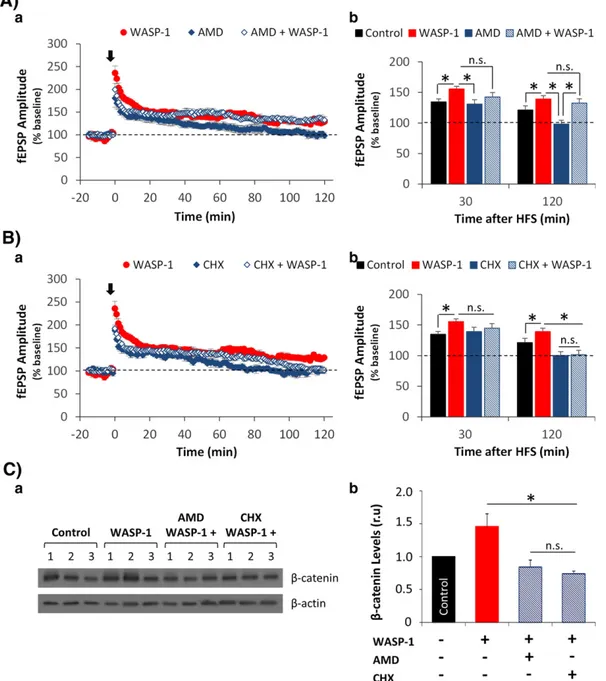
(4) J.Y. Vargas et al. / Experimental Neurology 264 (2015) 14–25. hippocampal neurons exposed to Wnt-3a. Considering these results, we used 5 μM WASP-1 to perform both electrophysiological recordings and in vivo experiments, as presented below. For these experiments, we used WASP-1 alone because endogenous expression and release of Wnt-3a in the rodent hippocampus has already been demonstrated (Chen et al., 2006; Cerpa et al., 2008). As has been shown for several Wnt ligands, WASP-1 can modulate excitatory synaptic transmission by increasing the synaptic strength of CA3–CA1 hippocampal synapses (Beaumont et al., 2007). Here we show that the perfusion of 5 μM WASP-1 alone is enough to significantly increase the amplitude of field excitatory postsynaptic potentials (fEPSPs) in hippocampal slices from adult WT mice (Fig. 2A-b). Using a paired pulse test, we addressed whether WASP-1 exerts its effect through a presynaptic mechanism as has been described for canonical Wnt ligands (Cerpa et al., 2008). In Fig. 2A-a, superimposed field responses showed that WASP-1 elicits larger synaptic responses than those observed in control slices. This increase in fEPSP amplitude was concomitant with a decrease in paired-pulse facilitation (Fig. 2A-c). Because changes in paired-pulse facilitation are considered to be of presynaptic origin (Clark et al., 1994; Kuhnt and Voronin, 1994), this result suggests that WASP-1 acts at the presynaptic terminal. Moreover, we found that WASP-1 perfusion does not change the fiber volley amplitude in comparison to control conditions (Figs. 2A-d, -e), indicating that WASP-1 does not modify neither the number nor the excitability of activated presynaptic fibers and suggesting that the effect of WASP1 on paired-pulse facilitation is due to a change in neurotransmitter release probability.. 17. A previous study has presented evidence that WASP-1 also enhances hippocampal LTP magnitude by facilitating LTP-maintenance for at least 2 h after LTP-induction (Beaumont et al., 2007), a finding that is consistent with our results (Fig. 2B). As Fig. 2B-a shows, a high frequency stimulation (HFS) protocol evoked larger fEPSP responses in WASP-1perfused slices than in control slices. Indeed, the average LTP magnitude was significantly enhanced in WASP-1-perfused slices (148.5 ± 4.9%) compared to control slices (130.1 ± 5.6%). Moreover, WASP-1dependent potentiation of synaptic responses was maintained for over 120 min after HFS (Fig. 2B-b). Considering that a) maintaining LTP for more than 1–2 h after induction, a phase known as late LTP (l-LTP), requires local dendritic protein synthesis as well as gene transcription (Citri and Malenka, 2008) and b) the activation of Wnt/β-catenin signaling can regulate the expression of genes that are involved in synaptic plasticity (Arrazola et al., 2009); we hypothesized that the WASP-1 effects on hippocampal LTP could be mediated by translation and/or transcription. Therefore, we tested the effects of WASP-1 on LTP magnitude in the presence of the gene transcription inhibitor actinomycin-D (AMD, 25 μM) or the protein synthesis inhibitor cycloheximide (CHX, 40 μg/ml). We found that perfusion of AMD alone blocked LTP-maintenance without affecting LTP-induction (Fig. 3A-a), although the LTP magnitude was significantly reduced compared to that in WASP-1 perfused slices (Fig. 3A-b). However, the co-perfusion of AMD plus WASP-1 did not affect either LTP-induction or LTP-maintenance because the HFS-evoked fEPSP amplitude was significantly different from baseline (dotted line) at all of the assayed time points (Fig. 3A-a). Additionally, we found that. Fig. 2. WASP-1 enhances hippocampal synaptic transmission and plasticity. (A) Excitatory synaptic transmission in mouse hippocampal slices (n = 6) treated with 5 μM WASP-1. (a) Average traces (60 recordings) of fEPSPs evoked by the paired pulse protocol before (control) and after 30 min of continuous perfusion with WASP-1. (b) Average values of normalized fEPSP amplitudes for the first response in the control and WASP-1-treated slices. (c) Facilitation index measured before and after WASP-1 application. (d) Average traces of fiber volley in controls and after WASP-1 treatment. (e) Normalized fiber volley amplitude in controls and after WASP-1 treatment. (B) Changes in LTP magnitude in mouse hippocampal slices perfused with WASP-1 (n = 7) or vehicle (n = 8). (a) Representative traces of fEPSP before (dotted lines) and after (filled lines) HFS. (b) Comparison of mean fEPSP amplitude between WASP-1 and control slices at 30, 60 and 120 min after HFS. The graph shows the percentage increase in fEPSP amplitude compared to baseline (dotted line)..
(5) 18. J.Y. Vargas et al. / Experimental Neurology 264 (2015) 14–25. Fig. 3. WASP-1 enhances hippocampal LTP through a mechanism that requires protein translation and the activation of the NMDA receptor. (A) Effect of gene transcription inhibitor (AMD) perfusion on WASP-1-enhanced LTP. (a) Time course of normalized fEPSP amplitude before and after HFS (black arrow). (b) Comparison of mean fEPSP amplitude at 30 and 120 min after HFS, among control (n = 8), WASP-1 (n = 7), AMD (n = 5) and AMD plus WASP-1 (n = 4) treated slices, showing the percent increase versus baseline (dotted line). (B) Effect of protein translator inhibitor (CH) perfusion on WASP-1-enhanced LTP. (a) Time course of normalized fEPSP amplitude before and after HFS (black arrow). (b) Comparison of mean fEPSP amplitude at 30 and 120 min after HFS, among control (n = 8), WASP-1 (n = 7), CH (n = 4) and CH plus WASP-1 (n = 5) treated slices, showing the percent increase versus baseline (dotted line). (C) Western blot analysis of β-catenin levels in hippocampal slices treated with control, WASP-1, AMD plus WASP-1 and CH plus WASP-1. (a) β-catenin immunoblotting. (b) Densitometric analysis of the β-catenin relative levels after normalization to β-actin. The graph shows the fold increase versus control (gray bar). The data are presented as the mean ± SE from n = 3 per experimental group.. LTP-induction was not affected by the perfusion of either CHX alone or CHX plus WASP-1; instead, LTP-maintenance was significantly reduced compared to perfusion of WASP-1 alone (Fig. 3B-a). Thus, CHX alone and CHX plus WASP-1 blocked LTP-maintenance, as observed by the fEPSP amplitude returning to basal levels (indicated by the dotted line) at 120 min after HFS application (Fig. 3B-b). The finding that the co-perfusion of CHX plus WASP-1 blocked l-LTP indicates that protein translation is necessary for the effect of WASP-1 on maintaining LTP. Together, these results suggest that WASP-1 induces an increase in hippocampal LTP magnitude in a manner dependent on local protein synthesis but that it does not involve gene transcription. In light of these results and considering the categorization of LTP into LTP1, LTP2. and LTP3 according to its persistence and mechanism (Racine et al., 1983; Raymond and Redman, 2006), WASP-1 appears to elicit LTP2, which only requires local protein synthesis from pre-existing mRNA. Additionally, we tested the effect of transcriptional and translational inhibitors on the levels of β-catenin (Fig. 3C). To compare the effects found on hippocampal LTP with β-catenin levels, we treated hippocampal slices acutely with WASP-1, WASP-1 plus AMD or WASP-1 plus CHX. We found that β-catenin levels increased with WASP-1 treatment, but this increase was completely blocked by both AMD and CHX treatments (Fig. 3C-b). This result indicates that WASP-1-induced increase of β-catenin levels depends on gene transcription and protein translation..
(6) J.Y. Vargas et al. / Experimental Neurology 264 (2015) 14–25. WASP-1 prevents Aβ-induced hippocampal synaptic dysfunction Several studies have shown that the activation of Wnt signaling has a neuroprotective effect against Aβ-induced synaptic damage (De Ferrari et al., 2003; Alvarez et al., 2004; Cerpa et al., 2010; Shruster et al., 2011). Because the administration of WASP-1 has been shown to enhance excitatory synaptic transmission and facilitate hippocampal LTP (Beaumont et al., 2007), as we also showed in Fig. 2, we hypothesized that WASP-1 perfusion would also protect against synaptic impairment of excitatory transmission induced by Aβ oligomers. To address this issue, we tested the effects of perfusing hippocampal slices with Aβ oligomers alone or Aβ oligomers plus WASP-1 on synaptic transmission (Fig. 4A) and LTP (Fig. 4B). We found that the perfusion of Aβ oligomers alone caused a large reduction in fEPSP amplitude compared to control conditions (Fig. 4A-b), since 10 min after Aβ perfusion began (Fig. 4Aa). Indeed, the average fEPSP amplitude of Aβ perfused slices (62.3 ± 6.4%) was significantly diminished compared to that of control slices (101.3 ± 1.9%). Moreover, Aβ oligomers also caused a strong inhibition of hippocampal LTP (Fig. 4B). After a brief post-tetanic potentiation evoked by an HFS protocol, the fEPSP amplitude of Aβ-perfused slices returned to basal levels (dotted line) in less than 20 min (Fig. 4B-a). These results are consistent with previous reports indicating that Aβ oligomers are able to induce a decrease in hippocampal excitatory. 19. synaptic transmission and LTP (Walsh et al., 2002; Wang et al., 2004; Shankar et al., 2008; Li et al., 2011). Interestingly, we found that slices perfused with Aβ oligomers plus WASP-1 exhibited an increase in fEPSP amplitude compared to baseline (dotted line), reaching a plateau at 15 min after co-perfusion began (Fig. 4A-a). Comparison of the average fEPSP amplitude of the Aβ alone and Aβ plus WASP-1 treatments reveals that WASP-1 prevented the Aβ-induced impairment of excitatory synaptic transmission (Fig. 4A-b). In fact, the average fEPSP amplitude of Aβ plus WASP-1-perfused slices (128.7 ± 2.8%) was significantly larger than that of Aβ-perfused slices (62.3 ± 6.4%). Furthermore, in contrast to what was observed in Aβ-perfused slices, we found that HFS application was capable of inducing LTP in hippocampal slices coperfused with Aβ oligomers plus WASP-1 (Fig. 4B-a), indicating that WASP-1 can overcome Aβ-induced LTP inhibition. Remarkably, the WASP-1-induced protecting effect against Aβ-impaired LTP was maintained for at least 1 h after the application of the HFS protocol (Fig. 4B-b). Indeed, the LTP magnitude at 30 or 60 min after HFS was higher in Aβ plus WASP-1-perfused slices (129.5 ± 7.8% and 128.8 ± 7.2%, respectively) than in Aβ-perfused slices (110.8 ± 6.6% and 104.6 ± 7.4%, respectively). Although we found significant differences in LTP magnitude in slices perfused with WASP-1 alone and those perfused with Aβ plus WASP-1 at 30 min after HFS, these differences were no longer evident at 60 min after HFS (Fig. 4B-b). These results. Fig. 4. WASP-1 prevents Aβ-induced synaptic impairment of glutamatergic synapses in mouse hippocampal slices. (A) fEPSP recordings before (control) and after 30 min of continued perfusion with either 50 μM Aβ alone or 50 μM Aβ in the presence of 5 μM WASP-1. (a) Time course of the effect of Aβ (black and white circles) or Aβ plus WASP-1 (red and white circles) on fEPSP peak amplitudes. The black bar indicates the perfusion duration of both treatments, and the dotted line indicates the baseline level. (b) Average values of normalized fEPSP amplitude measured before (control) and after a 15 min perfusion of Aβ alone (n = 4) or Aβ plus WASP-1 (n = 5). (B) fEPSP recordings of hippocampal slices before (baseline) and after the application of the HFS protocol. (a) Time course of normalized fEPSP amplitude (from 15 min before HFS (arrow) until 65 min after), in slices perfused either with 5 μM WASP-1 alone, 50 μM Aβ alone and 50 μM Aβ in the presence of 5 μM WASP-1. The graph shows the percent increase in fEPSP amplitude in slices treated with WASP-1 (red circles), Aβ (black and white circles) or Aβ plus WASP-1 (red and white circles) compared to baseline (dotted line). (b) Comparison of mean fEPSP amplitude among slices treated with control (n = 8), WASP-1 (n = 7), Aβ (n = 5) and Aβ plus WASP-1 (n = 5) at 30 or 60 min after HFS, showing the percent increases from baseline (dotted line)..
(7) 20. J.Y. Vargas et al. / Experimental Neurology 264 (2015) 14–25. indicate that WASP-1 treatment significantly prevented LTP decrease caused by Aβ oligomer exposure. Together, our findings suggest that the potentiation of canonical Wnt/β-catenin signaling by WASP-1 can successfully overcome Aβinduced impairments of both excitatory synaptic transmission and hippocampal synaptic plasticity.. WASP-1 rescues the loss of synaptic proteins and β-catenin in APP/PS1 mice Several in vitro studies have shown that the activation of Wnt/βcatenin signaling has a neuroprotective effect against Aβ-induced synaptic damage (De Ferrari et al., 2003; Alvarez et al., 2004; Shruster et al., 2011). Here we found that the activation of Wnt/β-catenin signaling by WASP-1 rescued synaptic transmission and LTP deficits in Aβperfused hippocampal slices (Fig. 4). Next, we evaluated whether WASP-1 can also rescue Aβ-induced synaptic impairments in vivo. To evaluate the effect of WASP-1 in vivo, we used double transgenic APP/PS1 mice (APPswe/PS1dE9: carrying the Swedish (K595N/ M596L) mutation of the amyloid precursor protein (APP) and the exon 9 deletion of the presenilin-1 (PS1) gene), which show the Aβ pathology of AD (Garcia-Alloza et al., 2006). The administration of WASP-1 using an osmotic pump system (see Materials and methods section for details) allowed chronic intra-hippocampal infusion of WASP-1 into APP/PS1 and WT mice. APP/PS1 animals display reduced levels of several synaptic proteins in both the cortex and the hippocampus (Inestrosa et al., 2011). Therefore, we tested whether WASP-1 treatment can reverse the loss of synaptic proteins in the hippocampus of APP/PS1 mice. We found that chronic administration of WASP-1 to adult APP/PS1 mice significantly increased hippocampal levels of the presynaptic protein SYP (synaptophysin) and the postsynaptic proteins GluN2B (N-methyl-Daspartate (NMDA) receptor subunit 2B) and PSD-95 (postsynaptic density protein-95) compared to control APP/PS1 mice (Fig. 5A). Although SYP levels in the hippocampus of APP/PS1 mice are not significantly reduced compared to WT mice (Inestrosa et al., 2011), as Fig. 5A-b shows, WASP-1 treatment increases the SYP levels in APP/PS1 mice above those in the WT. This change in the level of a presynaptic protein is consistent with the results shown in Fig. 2A, indicating that WASP1 can act through a presynaptic mechanism. However, we found that WASP-1 treatment also rescued the decreased levels of GluN2B and PSD-95 in APP/PS1 mice (Fig. 5A). We hypothesized that the WASP-1-induced increases in postsynaptic protein levels could be caused by the activation of non-canonical Wnt signaling pathways, which have been shown to modulate the structure of the postsynaptic compartment (Farias et al., 2009; Varela-Nallar et al., 2010), and could be activated by low concentrations of the canonical ligand Wnt-3a (Nalesso et al., 2011). Given that WASP-1 treatment raised the levels of both presynaptic and postsynaptic proteins, it is possible that the chronic administration of WASP-1 has a dual effect on the hippocampal synaptic structure of APP/PS1 mice. Compared to healthy brains, a significant reduction in β-catenin levels has been reported in AD patients (Zhang et al., 1998). Moreover, APP/PS1 mice also show lower hippocampal β-catenin levels than WT mice (Toledo and Inestrosa, 2010). Here, we tested whether WASP-1 treatment can rescue the loss of β-catenin in the hippocampus of APP/ PS1 mice. Interestingly, we found that APP/PS1 mice that were chronically treated with WASP-1 showed a significant increase in hippocampal β-catenin levels compared to control APP/PS1 mice (Fig. 5B). As Fig. 5B-b shows, WASP-1 was able to rescue the decrease in β-catenin levels observed in APP/PS1 mice to levels near those observed for WT mice. Our results show that in vivo administration of WASP-1 increases synaptic protein levels and reverses β-catenin loss in the hippocampus of adult APP/PS1 mice.. Fig. 5. WASP-1 rescues loss of synaptic proteins and β-catenin in the hippocampus of APP/ PS1 double transgenic mice. Western blot analysis of hippocampal homogenates from WT control mice (n = 3), APP/PS1 control mice (n = 4) and APP/PS1 mice treated with WASP1 (n = 3) to detect presynaptic (SYP) and postsynaptic (PSD-95 and GluN2B) proteins (A) or β-catenin levels (B). (a) Immunoblotting of indicated proteins. (b) Densitometric analysis of the relative levels of the indicated proteins after normalization to β-actin. The graphs show the fold increase versus the WT control (black bar), and significant differences among groups are also indicated.. WASP-1 reduces tau phosphorylation and blocks Aβ aggregation The main pathological hallmarks in the AD brain are senile plaques and neurofibrillary tangles (Sorrentino and Bonavita, 2007). Senile plaques are primarily formed by extracellular deposits of Aβ peptide, whereas neurofibrillary tangles are composed of intracellular aggregates of hyper-phosphorylated tau protein (Mayeux and Stern, 2012). According to the amyloid hypothesis, Aβ aggregation is the key driver of AD pathogenesis, whereas neurofibrillary tangle formation is proposed to result from an imbalance between both Aβ production and clearance (Hardy and Selkoe, 2002). We asked whether WASP-1 can down regulate both hyper-phosphorylated tau levels and Aβ aggregation. First, we tested whether in vivo administration of WASP-1 to the brains of APP/PS1 mice can reduce the hippocampal levels of phosphorylated tau. The phosphorylation of tau, in particular the PHF-1 epitope.
(8) J.Y. Vargas et al. / Experimental Neurology 264 (2015) 14–25. 21. Fig. 6. WASP-1 reduces tau-phosphorylation in the hippocampus of APP/PS1 mice and blocks Aβ aggregation. (A) Western blot analysis of PHF-1 in hippocampal homogenates from APP/ PS1 mice treated with or without WASP-1. (a) PHF-1 immunoblotting. (b) Densitometric analysis of the PHF-1 relative levels after normalization to β-actin. The graph shows the fold decrease in PHF-1 levels between APP/PS1 mice treated with WASP-1 (red bar) and APP/PS1 control mice (white bar). The data are presented as the mean ± SE from n = 3 per experimental group. (B) Transmission electron microscopic images of Aβ-aggregated at 0, 4 and 8 h after stirred incubation with or without 0.5 μM or 5 μM WASP-1 (bar, 1 μm). (C) In vitro amyloid aggregation assay conducted with 100 μM Aβ solution alone or in the presence of 0.5 μM or 5 μM WASP-1. The graph shows the time course of fluorescence intensity for different treatments.. (phosphorylated Ser-396 and Ser-404), was assessed by immunoblotting (Fig. 6A-a). WASP-1 treatment significantly decreased the phosphorylation of tau (Fig. 6A-b); a reduction of PHF-1 epitope levels was observed in the hippocampus of adult APP/PS1 mice. In light of this result, we evaluated whether WASP-1 treatment affected the Aβ aggregation. An immunoblotting assay was conducted using the 6E10 antibody to detect the presence of Aβ species in hippocampal extracts. from APP/PS1 mice treated with WASP-1 or vehicle (Supplementary Fig. 1). The 6E10 antibody recognizes monomeric, dimeric, trimeric, tetrameric and oligomeric forms of Aβ, as well as Aβ-precursor. We found that chronic treatment with WASP-1 only affected Aβ oligomeric deposits. WASP-1 specifically decreased the levels of Aβ*56 (a 56 kDa oligomer) species (Supplementary Fig. 1), which is a soluble Aβ assembly that impairs memory in AD transgenic mice independently of plaque.
(9) 22. J.Y. Vargas et al. / Experimental Neurology 264 (2015) 14–25. formation or neuronal loss (Lesne et al., 2006). Because it is possible that the WASP-1-induced decrease in Aβ*56 oligomeric forms could occur through blocking Aβ aggregation, we evaluated in vitro whether WASP-1 can interfere with the Aβ aggregation process. By electron microscopy, we observed that Aβ aggregation was diminished by the co-incubation with both 0.5 μM and 5 μM WASP-1 (Fig. 6B). After 4–8 h of stirred incubation, Aβ self-aggregates forming amylospheroids (Fig. 6B-a), a highly toxic specie of Aβ-aggregated involved in the pathogenic phosphorylation of tau protein (Hoshi et al., 2003). However, WASP-1 reduced the size of the amylospheroids at 4 h of co-incubation and this effect became clearly evident after 8 h of coincubation (Fig. 6B-a). These results were corroborated using a Th-T based fluorimetric assay. As Fig. 6B-b shows, both assayed concentrations of WASP-1 decreased Aβ aggregation in a time-dependent manner. After 8 h of WASP-1 co-incubation a 45–50% reduction in the Aβ aggregation was observed compared to incubation with Aβ alone (Fig. 6B-b). Altogether, these results indicate that WASP-1 reduces Aβ aggregation both in vitro and in vivo. Discussion WASP-1 has previously been described as a small molecule that potentiates canonical Wnt/β-catenin signaling and enhances excitatory transmission in mature hippocampal synapses (Beaumont et al., 2007). Because the activation of Wnt/β-catenin signaling has a neuroprotective effect against Aβ-induced cytotoxic and synaptotoxic insults (Boonen et al., 2009; Cerpa et al., 2009; De Ferrari et al., 2014; Inestrosa and Varela-Nallar, 2014), here we tested the ability of WASP-1 to rescue the functional and structural damage to hippocampal synapses that is caused by exposure to Aβ. Interestingly, we found that WASP-1 administration reduced Aβ-induced synaptic impairments both in vitro and. in vivo. The specific effects of WASP-1 observed in this study include rescuing the Aβ-induced disruption of excitatory synaptic transmission and overcoming the Aβ-induced blocking of hippocampal LTP, reversing the synaptic proteins loss, reducing Aβ aggregation and reducing the tau phosphorylation levels. Taken together, our data suggest that WASP-1 could be therapeutically relevant in AD. Several studies have reported that Aβ oligomers negatively modulate synaptic plasticity (Walsh et al., 2002; Cleary et al., 2005; Li et al., 2011; Ferreira and Klein, 2011). Previously, we showed that Aβ oligomers also reduce synaptic efficacy and impair hippocampal synaptic transmission, mainly by decreasing the NMDA and AMPA receptor currents, which is potentially caused by a reduction in the levels of PSD-95 and the number of synaptic contacts (Cerpa et al., 2010). Our present findings indicate that WASP-1 rescues impaired synaptic transmission and plasticity in hippocampal slices exposed to Aβ oligomers. Although, the precise mechanism of action of WASP-1 is unclear, the present data strongly suggest that protecting effects of WASP-1 against Aβ-mediated decreases in hippocampal synaptic responses could result from an increment in synaptic protein levels, including SYP, GluN2B (NMDA-R) and PSD-95. The WASP-1-induced increase in the level of SYP (a synaptic vesicle protein) is consistent with the finding that WASP-1 modulates the release of neurotransmitters at the presynaptic terminal, while WASP-1-induced increases in the levels of GluN2B (NMDA-R) and PSD-95 might be related with the increased potentiation of the postsynaptic response elicited by WASP-1 treatment. This dual effect of WASP-1 on the synaptic structure, affecting both presynaptic and postsynaptic sites, has previously been reported for the canonical ligand Wnt-7a, which can promote the clustering of several presynaptic proteins (Farias et al., 2007; Cerpa et al., 2008) and, at the same time, increase the density and maturity of dendritic spines (Gogolla et al., 2009; Ciani et al., 2011). A possible cause for this dual effect is different. Fig. 7. Scheme of effects of WASP-1 on Wnt-3a signaling in hippocampal synapses. WASP-1 enhances synaptic transmission by triggering an increase in neurotransmitter release, and it potentiates synaptic plasticity by inducing an increase in LTP magnitude. This scheme summarizes the main ideas of the present study. (A) Under normal conditions, WASP-1 potentiates Wnt signaling by increasing the expression of β-catenin. This increase in turn, activates the exocytosis of synaptic vesicles, resulting in an increase in glutamate release at the pre synaptic region. At the postsynaptic region, the activation of NMDA-R allows the potentiation of the synapse and the expression of the LTP-2. The pre-existing mRNA corresponding to some of the NMDA-R subunits is shown in the postsynaptic region. (B) Under pathological conditions, such as those present in AD, the loss of Wnt signaling function is apparent and synaptic plasticity is partially or totally blocked by Aβ oligomers. Under these conditions, the protective effect of WASP-1 occurs through potentiating Wnt/β-catenin signaling. The activation of this signaling leads to the inhibition of GSK-3β, thereby increasing the β-catenin levels and reducing tau phosphorylation. At the same time, Aβ aggregation is prevented by WASP-1, and the formation of Aβ oligomers decreases, facilitating both an increase in the release of glutamate and the recovery of LTP. Given the WASP-1-induced increase in SYP at the presynaptic region, synaptic vesicles are further committed to release glutamate. At the postsynaptic region, the WASP-1-induced increase in the scaffold protein PSD-95 facilitates the incorporation into the postsynaptic membranes of vesicles that contain newly synthesized GluN2B (a subunit of the NMDA-R), which is possibly translated at the dendritic spines from pre-existing mRNA. Together, all of these WASP-1-induced features allow the recovery of Aβ-impaired synaptic plasticity and contribute to ameliorate the neurodegenerative conditions observed in AD..
(10) J.Y. Vargas et al. / Experimental Neurology 264 (2015) 14–25. signaling pathways acting on different synaptic structures being activated by a single Wnt ligand. Indeed, it has been shown that Wnt-3a can activate both canonical (acting at the presynaptic region) and noncanonical (acting at the postsynaptic region) Wnt signaling, however, the activation of each pathway may depend on ligand concentration (Nalesso et al., 2011). Interestingly, we have shown here that the effects of WASP-1 on synaptic plasticity can be modulated by protein synthesis. Despite the finding that the co-perfusion of WASP-1 plus a transcription inhibitor did not affect LTP-maintenance, we found that the co-perfusion of WASP-1 and a translation inhibitor completely blocked LTPmaintenance without changing LTP-induction. These findings indicate that the WASP-1-induced facilitation of l-LTP depends on the synthesis of new proteins but does not require the synthesis of new mRNA. LTP is categorized into LTP1, LTP2 and LTP3, according to duration and molecular mechanism (Racine et al., 1983; Raymond and Redman, 2006), and WASP-1 appeared to elicit LTP2, which depends on protein synthesis from pre-existing mRNA located in the dendrites and is independent of gene transcription (Raymond, 2007). However, we found that both transcriptional and translational inhibitors obstructed WASP-1induced β-catenin stabilization, suggesting that β-catenin is not involved in the effects of WASP-1 on LTP-maintenance. The idea that activation of the canonical Wnt signaling can modulate the synaptic function through a mechanism that does not require β-catenin-mediated gene transcription, has been previously proposed (Ahmad-Annuar et al., 2006; Farias et al., 2007; Cerpa et al., 2008). This divergent canonical Wnt pathway seems to modulate the function of the presynaptic terminals (Budnik and Salinas, 2011). Particularly, the canonical Wnt ligands, Wnt-3a and Wnt-7a, have shown to rapidly stimulate neurotransmitter release from presynaptic terminals in a way that is independent on the expression of Wnt target genes (Cerpa et al., 2008; Varela-Nallar et al., 2009). Here, we reported that WASP-1 could also stimulate neurotransmitter release probability, since a paired pulse facilitation reduction is observed in the presence of WASP-1. The regulation of neurotransmitter release by canonical Wnt ligands could be achieved through promoting the interaction between proteins of the Wnt pathway and proteins implicated in synaptic processes such as synaptotagmins (Oliva et al., 2013). On the other hand, the positive effects of WASP-1 on Aβ-impaired LTP-induction and LTP-maintenance could be related to the WASP-1induced reduction in the hippocampal levels of Aβ oligomers. We found that WASP-1 also reduces the hippocampal levels of the Aβ*56 oligomeric species in APP/PS1 mice, possibly by blocking Aβ aggregation. These findings are consistent with the idea that blocking of Aβ oligomerization rescues LTP impairment (Walsh et al., 2005). In the present study, WASP-1 also reduced tau pathology, another key hallmark of AD besides Aβ aggregates (Sheng et al., 2012). Reduced levels of phosphorylated tau could be a consequence of the WASP-1-induced decreases in Aβ accumulation and/or a side effect of the inhibition of GSK-3β, a kinase whose activity is down regulated by the activation of Wnt/βcatenin signaling (Clevers and Nusse, 2012) and that is involved in tau phosphorylation (Hooper et al., 2008). Indeed, we previously found that WASP-1 can modulate the activity of GSK-3β in the hippocampus of WT mice (Vargas et al., 2014). Interestingly, our results are in agreement with the finding that indirect activation of Wnt/β-catenin signaling through the inhibition of GSK-3β, protects hippocampal neurons from Aβ cytotoxic effect by stimulating neuronal survival (SilvaAlvarez et al., 2013). In AD pathology, several components of Wnt/β-catenin signaling have been shown to be altered (Moon et al., 2004; Inestrosa et al., 2012; Oliva et al., 2013). In particular, the β-catenin levels in AD brains are considerably down regulated compared to healthy brains (Zhang et al., 1998). Moreover, there is a relationship between familial onset AD and the de-regulation of β-catenin. In fact, pathogenic mutations in the PS1 gene, one of the major causes of early-onset AD, have been shown to affect the stability of β-catenin, increasing its degradation. 23. (Zhang et al., 1998). Here, we have shown that the hippocampal βcatenin levels are significantly reduced in APP/PS1 mice compared to the levels in WT animals; however, WASP-1 treatment almost completely rescued this loss, suggesting that WASP-1 influences βcatenin stabilization. Indeed, we report here that the co-incubation of hippocampal neurons with WASP-1 and the canonical ligand Wnt-3a significantly increases β-catenin stability, but this effect cannot be achieved by incubation with WASP-1 alone. These findings suggest that WASP-1 requires the presence of Wnt-3a to exert its effect (Beaumont et al., 2007). Therefore, endogenous Wnt-3a must be locally released in the hippocampus of both WT and APP/PS1 mice to facilitate the beneficial effects of WASP-1. Our results indicate that WASP-1 in cooperation with the endogenous Wnt-3a, act on the hippocampal neuronal network to potentiate Wnt/β-catenin signaling and consequently to produce an improvement of the synaptic function in both normal and Aβ-induced pathologic conditions (Fig. 7). Together, these data suggest that WASP-1, through Wnt signaling, can rescue Aβ-induced synaptic impairments and could be used for the treatment of patients with neurodegenerative diseases such as AD. Conclusions In this study, we show that WASP-1 potentiates the activation of Wnt/β-catenin signaling in hippocampal neurons, thereby increasing normal synaptic function or rescuing Aβ-induced synaptic impairment (Fig. 7). Under normal conditions, WASP-1 can potentiate the effect of endogenous Wnt-3a and increase β-catenin stability. At the presynaptic terminal, WASP-1 enhances excitatory synaptic transmission by increasing the release of neurotransmitters. At the postsynaptic compartment, WASP-1 elicits a form of LTP that requires the activation of NMDA-R for LTP-induction and depends on the translation of preexisting mRNA at the dendritic spines for LTP-maintenance (Fig. 3A, B). In Aβ-impaired synapses, WASP-1 reduced the aggregation of Aβ oligomers and the phosphorylation of tau. At the presynaptic terminal, WASP-1 enhanced synaptic transmission, possibly by increasing the levels of the synaptic vesicle protein SYP. At the postsynaptic compartment, WASP-1 overcame LTP impairments possibly by rescuing the diminished levels of PSD-95 and the GluN2B subunit of NMDA-R (Fig. 5A). In summary, our findings suggest that WASP-1 is a potential therapeutic agent for AD. Supplementary data to this article can be found online at http://dx. doi.org/10.1016/j.expneurol.2014.11.005. Acknowledgments This work was supported by grants from the Basal Center of Excellence in Science in Technology (CONICYT-PFB12/2007) and FONDECYT no. 1120156 to N.C.I.; FONDECYT no. 11090059, no. 1130614 and DIPUV CID 01/2006 to M.F.; a pre-doctoral fellowship from Fundación Gran Mariscal de Ayacucho to J.Y.V.; and pre-doctoral fellowships from CONICYT to J.A. and M.S.A. We thank Felipe Serrano for his help with Fig. 7. References Ahmad-Annuar, A., Ciani, L., Simeonidis, I., Herreros, J., Fredj, N.B., Rosso, S.B., Hall, A., Brickley, S., Salinas, P.C., 2006. Signaling across the synapse: a role for Wnt and Dishevelled in presynaptic assembly and neurotransmitter release. J. Cell Biol. 174, 127–139. Alvarez, A.R., Godoy, J.A., Mullendorff, K., Olivares, G.H., Bronfman, M., Inestrosa, N.C., 2004. Wnt-3a overcomes beta-amyloid toxicity in rat hippocampal neurons. Exp. Cell Res. 297, 186–196. Arrazola, M.S., Varela-Nallar, L., Colombres, M., Toledo, E.M., Cruzat, F., Pavez, L., Assar, R., Aravena, A., Gonzalez, M., Montecino, M., Maass, A., Martinez, S., Inestrosa, N.C., 2009. Calcium/calmodulin-dependent protein kinase type IV is a target gene of the Wnt/ beta-catenin signaling pathway. J. Cell. Physiol. 221, 658–667. Beaumont, V., Thompson, S.A., Choudhry, F., Nuthall, H., Glantschnig, H., Lipfert, L., David, G.R., Swain, C.J., McAllister, G., Munoz-Sanjuan, I., 2007. Evidence for an enhancement.
(11) 24. J.Y. Vargas et al. / Experimental Neurology 264 (2015) 14–25. of excitatory transmission in adult CNS by Wnt signaling pathway modulation. Mol. Cell. Neurosci. 35, 513–524. Bonansco, C., Couve, A., Perea, G., Ferradas, C.A., Roncagliolo, M., Fuenzalida, M., 2011. Glutamate released spontaneously from astrocytes sets the threshold for synaptic plasticity. Eur. J. Neurosci. 33, 1483–1492. Boonen, R.A., van Tijn, P., Zivkovic, D., 2009. Wnt signaling in Alzheimer's disease: up or down, that is the question. Ageing Res. Rev. 8, 71–82. Bozdagi, O., Wang, X.B., Nikitczuk, J.S., Anderson, T.R., Bloss, E.B., Radice, G.L., Zhou, Q., Benson, D.L., Huntley, G.W., 2010. Persistence of coordinated long-term potentiation and dendritic spine enlargement at mature hippocampal CA1 synapses requires Ncadherin. J. Neurosci. 30, 9984–9989. Budnik, V., Salinas, P.C., 2011. Wnt signaling during synaptic development and plasticity. Curr. Opin. Neurobiol. 21, 151–159. Caricasole, A., Copani, A., Caraci, F., Aronica, E., Rozemuller, A.J., Caruso, A., Storto, M., Gaviraghi, G., Terstappen, G.C., Nicoletti, F., 2004. Induction of Dickkopf-1, a negative modulator of the Wnt pathway, is associated with neuronal degeneration in Alzheimer's brain. J. Neurosci. 24, 6021–6027. Cerpa, W., Godoy, J.A., Alfaro, I., Farias, G.G., Metcalfe, M.J., Fuentealba, R., Bonansco, C., Inestrosa, N.C., 2008. Wnt-7a modulates the synaptic vesicle cycle and synaptic transmission in hippocampal neurons. J. Biol. Chem. 283, 5918–5927. Cerpa, W., Toledo, E.M., Varela-Nallar, L., Inestrosa, N.C., 2009. The role of Wnt signaling in neuroprotection. Drug News Perspect. 22, 579–591. Cerpa, W., Farias, G.G., Godoy, J.A., Fuenzalida, M., Bonansco, C., Inestrosa, N.C., 2010. Wnt5a occludes Abeta oligomer-induced depression of glutamatergic transmission in hippocampal neurons. Mol. Neurodegener. 5, 3. Chen, J., Park, C.S., Tang, S.J., 2006. Activity-dependent synaptic Wnt release regulates hippocampal long term potentiation. J. Biol. Chem. 281, 11910–11916. Ciani, L., Salinas, P.C., 2005. WNTs in the vertebrate nervous system: from patterning to neuronal connectivity. Nat. Rev. Neurosci. 6, 351–362. Ciani, L., Boyle, K.A., Dickins, E., Sahores, M., Anane, D., Lopes, D.M., Gibb, A.J., Salinas, P.C., 2011. Wnt7a signaling promotes dendritic spine growth and synaptic strength through Ca(2)/calmodulin-dependent protein kinase II. Proc. Natl. Acad. Sci. U. S. A. 108, 10732–10737. Citri, A., Malenka, R.C., 2008. Synaptic plasticity: multiple forms, functions, and mechanisms. Neuropsychopharmacology 33, 18–41. Clark, K.A., Randall, A.D., Collingridge, G.L., 1994. A comparison of paired-pulsed facilitation of AMPA and NMDA receptor-mediated excitatory postsynaptic currents in the hippocampus. Exp. Brain Res. 101, 272–278. Cleary, J.P., Walsh, D.M., Hofmeister, J.J., Shankar, G.M., Kuskowski, M.A., Selkoe, D.J., Ashe, K.H., 2005. Natural oligomers of the amyloid-beta protein specifically disrupt cognitive function. Nat. Neurosci. 8, 79–84. Clevers, H., Nusse, R., 2012. Wnt/beta-catenin signaling and disease. Cell 149, 1192–1205. De Ferrari, G.V., Chacon, M.A., Barria, M.I., Garrido, J.L., Godoy, J.A., Olivares, G., Reyes, A.E., Alvarez, A., Bronfman, M., Inestrosa, N.C., 2003. Activation of Wnt signaling rescues neurodegeneration and behavioral impairments induced by beta-amyloid fibrils. Mol. Psychiatry 8, 195–208. De Ferrari, G.V., Papassotiropoulos, A., Biechele, T., Wavrant De-Vrieze, F., Avila, M.E., Major, M.B., Myers, A., Saez, K., Henriquez, J.P., Zhao, A., Wollmer, M.A., Nitsch, R.M., Hock, C., Morris, C.M., Hardy, J., Moon, R.T., 2007. Common genetic variation within the low-density lipoprotein receptor-related protein 6 and late-onset Alzheimer's disease. Proc. Natl. Acad. Sci. U. S. A. 104, 9434–9439. De Ferrari, G.V., Avila, M.E., Medina, M.A., Perez-Palma, E., Bustos, B.I., Alarcon, M.A., 2014. Wnt/beta-catenin signaling in Alzheimer's disease. CNS Neurol. Disord. Drug Targets 13, 745–754. Dinamarca, M.C., Sagal, J.P., Quintanilla, R.A., Godoy, J.A., Arrazola, M.S., Inestrosa, N.C., 2010. Amyloid-beta-Acetylcholinesterase complexes potentiate neurodegenerative changes induced by the Abeta peptide. Implications for the pathogenesis of Alzheimer's disease. Mol. Neurodegener. 5, 4. Farias, G.G., Valles, A.S., Colombres, M., Godoy, J.A., Toledo, E.M., Lukas, R.J., Barrantes, F.J., Inestrosa, N.C., 2007. Wnt-7a induces presynaptic colocalization of alpha 7-nicotinic acetylcholine receptors and adenomatous polyposis coli in hippocampal neurons. J. Neurosci. 27, 5313–5325. Farias, G.G., Alfaro, I.E., Cerpa, W., Grabowski, C.P., Godoy, J.A., Bonansco, C., Inestrosa, N.C., 2009. Wnt-5a/JNK signaling promotes the clustering of PSD-95 in hippocampal neurons. J. Biol. Chem. 284, 15857–15866. Ferreira, S.T., Klein, W.L., 2011. The Ab oligomer hypothesis for synapse failure and memory loss in Alzheimer's disease. Neurobiol. Learn. Mem. 96, 529–543. Garcia-Alloza, M., Robbins, E.M., Zhang-Nunes, S.X., Purcell, S.M., Betensky, R.A., Raju, S., Prada, C., Greenberg, S.M., Bacskai, B.J., Frosch, M.P., 2006. Characterization of amyloid deposition in the APPswe/PS1dE9 mouse model of Alzheimer disease. Neurobiol. Dis. 24, 516–524. Gogolla, N., Galimberti, I., Deguchi, Y., Caroni, P., 2009. Wnt signaling mediates experience-related regulation of synapse numbers and mossy fiber connectivities in the adult hippocampus. Neuron 62, 510–525. Hardy, J., Selkoe, D.J., 2002. The amyloid hypothesis of Alzheimer's disease: progress and problems on the road to therapeutics. Science 297, 353–356. Hermann, D., Both, M., Ebert, U., Gross, G., Schoemaker, H., Draguhn, A., Wicke, K., Nimmrich, V., 2009. Synaptic transmission is impaired prior to plaque formation in amyloid precursor protein-overexpressing mice without altering behaviorallycorrelated sharp wave-ripple complexes. Neuroscience 162, 1081–1090. Hooper, C., Markevich, V., Plattner, F., Killick, R., Schofield, E., Engel, T., Hernandez, F., Anderton, B., Rosenblum, K., Bliss, T., Cooke, S.F., Avila, J., Lucas, J.J., Giese, K.P., Stephenson, J., Lovestone, S., 2007. Glycogen synthase kinase-3 inhibition is integral to long-term potentiation. Eur. J. Neurosci. 25, 81–86. Hooper, C., Killick, R., Lovestone, S., 2008. The GSK3 hypothesis of Alzheimer's disease. J. Neurochem. 104, 1433–1439.. Hoshi, M., Sato, M., Matsumoto, S., Noguchi, A., Yasutake, K., Yoshida, N., Sato, K., 2003. Spherical aggregates of beta-amyloid (amylospheroid) show high neurotoxicity and activate tau protein kinase I/glycogen synthase kinase-3beta. Proc. Natl. Acad. Sci. U. S. A. 100, 6370–6375. Inestrosa, N.C., Arenas, E., 2010. Emerging roles of Wnts in the adult nervous system. Nat. Rev. Neurosci. 11, 77–86. Inestrosa, N.C., Varela-Nallar, L., 2014. Wnt signaling in the nervous system and in Alzheimer's disease. J. Mol. Cell Biol. 6, 64–74. Inestrosa, N.C., Alvarez, A., Godoy, J., Reyes, A., De Ferrari, G.V., 2000. Acetylcholinesterase–amyloid-beta-peptide interaction and Wnt signaling involvement in Abeta neurotoxicity. Acta Neurol. Scand. Suppl. 176, 53–59. Inestrosa, N.C., Tapia-Rojas, C., Griffith, T.N., Carvajal, F.J., Benito, M.J., Rivera-Dictter, A., Alvarez, A.R., Serrano, F.G., Hancke, J.L., Burgos, P.V., Parodi, J., Varela-Nallar, L., 2011. Tetrahydrohyperforin prevents cognitive deficit, Abeta deposition, tau phosphorylation and synaptotoxicity in the APPswe/PSEN1DeltaE9 model of Alzheimer's disease: a possible effect on APP processing. Transl. Psychiatry 1, e20. Inestrosa, N.C., Montecinos-Oliva, C., Fuenzalida, M., 2012. Wnt signaling: role in Alzheimer disease and schizophrenia. J. Neuroimmune Pharmacol. 7, 788–807. Korinek, V., Barker, N., Morin, P.J., van Wichen, D., de Weger, R., Kinzler, K.W., Vogelstein, B., Clevers, H., 1997. Constitutive transcriptional activation by a beta-catenin-Tcf complex in APC−/− colon carcinoma. Science 275, 1784–1787. Kuhnt, U., Voronin, L.L., 1994. Interaction between paired-pulse facilitation and long-term potentiation in area CA1 of guinea-pig hippocampal slices: application of quantal analysis. Neuroscience 62, 391–397. Lacor, P.N., Buniel, M.C., Furlow, P.W., Clemente, A.S., Velasco, P.T., Wood, M., Viola, K.L., Klein, W.L., 2007. Abeta oligomer-induced aberrations in synapse composition, shape, and density provide a molecular basis for loss of connectivity in Alzheimer's disease. J. Neurosci. 27, 796–807. Lesne, S., Koh, M.T., Kotilinek, L., Kayed, R., Glabe, C.G., Yang, A., Gallagher, M., Ashe, K.H., 2006. A specific amyloid-beta protein assembly in the brain impairs memory. Nature 440, 352–357. LeVine III, H., 1993. Thioflavine T interaction with synthetic Alzheimer's disease betaamyloid peptides: detection of amyloid aggregation in solution. Protein Sci. 2, 404–410. Li, S., Jin, M., Koeglsperger, T., Shepardson, N.E., Shankar, G.M., Selkoe, D.J., 2011. Soluble Abeta oligomers inhibit long-term potentiation through a mechanism involving excessive activation of extrasynaptic NR2B-containing NMDA receptors. J. Neurosci. 31, 6627–6638. Liu, C.C., Tsai, C.W., Deak, F., Rogers, J., Penuliar, M., Sung, Y.M., Maher, J.N., Fu, Y., Li, X., Xu, H., Estus, S., Hoe, H.S., Fryer, J.D., Kanekiyo, T., Bu, G., 2014. Deficiency in LRP6mediated Wnt signaling contributes to synaptic abnormalities and amyloid pathology in Alzheimer's disease. Neuron 84, 63–77. Lue, L.F., Kuo, Y.M., Roher, A.E., Brachova, L., Shen, Y., Sue, L., Beach, T., Kurth, J.H., Rydel, R.E., Rogers, J., 1999. Soluble amyloid beta peptide concentration as a predictor of synaptic change in Alzheimer's disease. Am. J. Pathol. 155, 853–862. Mattson, M.P., 2004. Pathways towards and away from Alzheimer's disease. Nature 430, 631–639. Mayeux, R., Stern, Y., 2012. Epidemiology of Alzheimer disease. Cold Spring Harb. Perspect. Med. 2. McLean, C.A., Cherny, R.A., Fraser, F.W., Fuller, S.J., Smith, M.J., Beyreuther, K., Bush, A.I., Masters, C.L., 1999. Soluble pool of Abeta amyloid as a determinant of severity of neurodegeneration in Alzheimer's disease. Ann. Neurol. 46, 860–866. Moon, R.T., Kohn, A.D., De Ferrari, G.V., Kaykas, A., 2004. WNT and beta-catenin signalling: diseases and therapies. Nat. Rev. Genet. 5, 691–701. Nalesso, G., Sherwood, J., Bertrand, J., Pap, T., Ramachandran, M., De Bari, C., Pitzalis, C., Dell'accio, F., 2011. WNT-3A modulates articular chondrocyte phenotype by activating both canonical and noncanonical pathways. J. Cell Biol. 193, 551–564. Nusse, R., 2012. Wnt signaling. Cold Spring Harb. Perspect. Biol. 4. Oliva, C.A., Vargas, J.Y., Inestrosa, N.C., 2013. Wnts in adult brain: from synaptic plasticity to cognitive deficiencies. Front. Cell. Neurosci. 7, 224. Purro, S.A., Dickins, E.M., Salinas, P.C., 2012. The secreted Wnt antagonist Dickkopf-1 is required for amyloid beta-mediated synaptic loss. J. Neurosci. 32, 3492–3498. Racine, R.J., Milgram, N.W., Hafner, S., 1983. Long-term potentiation phenomena in the rat limbic forebrain. Brain Res. 260, 217–231. Raymond, C.R., 2007. LTP forms 1, 2 and 3: different mechanisms for the “long” in longterm potentiation. Trends Neurosci. 30, 167–175. Raymond, C.R., Redman, S.J., 2006. Spatial segregation of neuronal calcium signals encodes different forms of LTP in rat hippocampus. J. Physiol. 570, 97–111. Sakono, M., Zako, T., 2010. Amyloid oligomers: formation and toxicity of Abeta oligomers. FEBS J. 277, 1348–1358. Selkoe, D.J., 2001. Alzheimer's disease results from the cerebral accumulation and cytotoxicity of amyloid beta-protein. J. Alzheimers Dis. 3, 75–80. Shankar, G.M., Bloodgood, B.L., Townsend, M., Walsh, D.M., Selkoe, D.J., Sabatini, B.L., 2007. Natural oligomers of the Alzheimer amyloid-beta protein induce reversible synapse loss by modulating an NMDA-type glutamate receptor-dependent signaling pathway. J. Neurosci. 27, 2866–2875. Shankar, G.M., Li, S., Mehta, T.H., Garcia-Munoz, A., Shepardson, N.E., Smith, I., Brett, F.M., Farrell, M.A., Rowan, M.J., Lemere, C.A., Regan, C.M., Walsh, D.M., Sabatini, B.L., Selkoe, D.J., 2008. Amyloid-beta protein dimers isolated directly from Alzheimer's brains impair synaptic plasticity and memory. Nat. Med. 14, 837–842. Sheng, M., Sabatini, B.L., Sudhof, T.C., 2012. Synapses and Alzheimer's disease. Cold Spring Harb. Perspect. Biol. 4. Shruster, A., Eldar-Finkelman, H., Melamed, E., Offen, D., 2011. Wnt signaling pathway overcomes the disruption of neuronal differentiation of neural progenitor cells induced by oligomeric amyloid beta-peptide. J. Neurochem. 116, 522–529..
(12) J.Y. Vargas et al. / Experimental Neurology 264 (2015) 14–25 Silva-Alvarez, C., Arrazola, M.S., Godoy, J.A., Ordenes, D., Inestrosa, N.C., 2013. Canonical Wnt signaling protects hippocampal neurons from Abeta oligomers: role of noncanonical Wnt-5a/Ca(2+) in mitochondrial dynamics. Front. Cell. Neurosci. 7, 97. Sorrentino, G., Bonavita, V., 2007. Neurodegeneration and Alzheimer's disease: the lesson from tauopathies. Neurol. Sci. 28, 63–71. Toledo, E.M., Inestrosa, N.C., 2010. Activation of Wnt signaling by lithium and rosiglitazone reduced spatial memory impairment and neurodegeneration in brains of an APPswe/PSEN1DeltaE9 mouse model of Alzheimer's disease. Mol. Psychiatry 15 (272–285), 228. Varela-Nallar, L., Grabowski, C.P., Alfaro, I.E., Alvarez, A.R., Inestrosa, N.C., 2009. Role of the Wnt receptor Frizzled-1 in presynaptic differentiation and function. Neural Dev. 4, 41. Varela-Nallar, L., Alfaro, I.E., Serrano, F.G., Parodi, J., Inestrosa, N.C., 2010. Wingless-type family member 5A (Wnt-5a) stimulates synaptic differentiation and function of glutamatergic synapses. Proc. Natl. Acad. Sci. U. S. A. 107, 21164–21169. Vargas, J.Y., Fuenzalida, M., Inestrosa, N.C., 2014. In vivo activation of Wnt signaling pathway enhances cognitive function of adult mice and reverses cognitive deficits in an Alzheimer's disease model. J. Neurosci. 34, 2191–2202.. 25. Walsh, D.M., Klyubin, I., Fadeeva, J.V., Cullen, W.K., Anwyl, R., Wolfe, M.S., Rowan, M.J., Selkoe, D.J., 2002. Naturally secreted oligomers of amyloid beta protein potently inhibit hippocampal long-term potentiation in vivo. Nature 416, 535–539. Walsh, D.M., Townsend, M., Podlisny, M.B., Shankar, G.M., Fadeeva, J.V., El Agnaf, O., Hartley, D.M., Selkoe, D.J., 2005. Certain inhibitors of synthetic amyloid betapeptide (Abeta) fibrillogenesis block oligomerization of natural Abeta and thereby rescue long-term potentiation. J. Neurosci. 25, 2455–2462. Wang, Q., Walsh, D.M., Rowan, M.J., Selkoe, D.J., Anwyl, R., 2004. Block of long-term potentiation by naturally secreted and synthetic amyloid beta-peptide in hippocampal slices is mediated via activation of the kinases c-Jun N-terminal kinase, cyclindependent kinase 5, and p38 mitogen-activated protein kinase as well as metabotropic glutamate receptor type 5. J. Neurosci. 24, 3370–3378. Zhang, Z., Hartmann, H., Do, V.M., Abramowski, D., Sturchler-Pierrat, C., Staufenbiel, M., Sommer, B., van de Wetering, M., Clevers, H., Saftig, P., De Strooper, B., He, X., Yankner, B.A., 1998. Destabilization of beta-catenin by mutations in presenilin-1 potentiates neuronal apoptosis. Nature 395, 698–702..
(13)
Figure


+2

Documento similar