Rol de la insulina interoceptiva en la regulación de la memoria de miedo
105
0
0
Texto completo
(2) 2.
(3) 3. A Loreto.
(4) 4. Agradecimientos Al Dr. Fernando Torrealba, a sus ideas sobre la Corteza Insular y al desafío constante. Al Dr. Marcelo Aguilar, esencial para este trabajo. Entre otras cosas más importantes, por introducirme al registro con tetrodos, al análisis de datos y a la programación, ¡salud! A María de los Ángeles Rodríguez, por su ayuda con los experimentos de registro (sin importar la hora; hablar el mismo dialecto facilitó mucho el trabajo desde el principio). Al Dr. Carlos Madrid, por las constantes discusiones que quedaron por escrito en este y otros trabajos. Quiero tener la pasión que pone al trabajar el Dr. Marco Contreras, a él gracias por el ejemplo y apoyo al inicio de la tesis. A Marcela González, por su excelente trabajo técnico, crucial durante el primer año. Al Dr. Julio Alcayaga por la creación del dispositivo para los experimentos de hipoxia. Al equipo del Dr. Rodrigo Iturriaga, (a él por creerme), a Paulina Arias por el apoyo técnico en los tiempos desérticos del laboratorio y al Dr. Esteban Moya y su habilidad de cirujano. Por sobre todo, agradezco la chance de trabajar con amigos.. Financiamiento: Beca CONICYT para estudios de doctorado en Chile, proyecto FONDECYT 1130042 y Anillo CONICYT ACT-66..
(5) 5. Contents Summary. .................................................................................................................................... 9 Resumen. .................................................................................................................................. 11 1.. Introduction. ...................................................................................................................... 13 1.1. Interoceptive insular cortex. ....................................................................................... 15. 1.2. Hypoxia as an interoceptive stimulus. ....................................................................... 20. 1.3. Fear and fear memory. ............................................................................................... 22. 2.. Hypothesis. ........................................................................................................................ 26. 3.. Objectives. ......................................................................................................................... 27. 4.. Materials. ........................................................................................................................... 28. 5.. Methods. ............................................................................................................................ 29 5.1. General. ...................................................................................................................... 29. 5.2 Objective 1: Determine the role of the pIC in the behavioral and autonomic response to severe hypoxia....................................................................................................................... 33 5.3 Objective 2: Determine the role of the pIC in expression of conditioned fear. .............. 36 5.4 Objective 3. Fear memory consolidation in the pIC. ...................................................... 42 6. Results................................................................................................................................... 44 6.1 Objective 1: Determine the role of the pIC in the behavioral and autonomic response to severe hypoxia....................................................................................................................... 44 6.2 Objective 2: Determine the role of the pIC in the expression of conditioned fear. ........ 55 6.3 Objective 3. Determine the role of the pIC in fear memory consolidation. .................... 68 7. Discussion. ............................................................................................................................ 74 7.1 Effect of insular cortex inactivation on autonomic and behavioral responses to acute hypoxia in conscious rats. ..................................................................................................... 74 7.2 Activity of the posterior insular cortex during expression of conditioned fear. ............. 79 7.3 A role for the interoceptive insular cortex in consolidation of fear memory. ................. 83 8. Conclusions........................................................................................................................... 87 9. Published work. .................................................................................................................... 89 10. References........................................................................................................................... 90.
(6) 6. Figures. Fig. 1. The insular cortex is the cortical component of the interoceptive system Fig. 2. The insular cortex is a sensory cortex.. 17. Fig. 3. Protocol for fear conditioning to assess expression of c-fos and zif268. Fig.4. Tetrodes array in the hyperdrive. Fig. 5. Fear conditioning protocol to record the activity of posterior insular cortex neurons. Fig.6. Representative Identification of four neurons recorded with one tetrode. Fig.7. Fear conditioning protocol to assess the effect of pharmacological manipulations of the posterior insular cortex on retention of conditioned fear. Fig. 8. Experimental protocol to assess the contribution of posterior insular cortex activity on responses to hypoxia. Fig. 9. Representative recording of autonomic response to acute hypoxia measured with telemetry in a freely moving rat. Fig. 10. Effect of posterior insular cortex inactivation on autonomic responses to acute hypoxia. Fig.11. Effect of posterior insular cortex inactivation on the behavioral responses to acute hypoxia. Fig. 12. Fos-immunoreactivity in the posterior insular cortex is not modified following acute hypoxia. Fig. 13. Effect of hypoxia on Fos-immunoreactivity in neurons from the rat Nucleus tractus solitarius. Fig. 14. Effect of hypoxia on Fos-immunoreactivity in neurons from the rat Periaqueductal gray matter. Figure 15. Reactivation of learned fear was paralleled by an increase in Fos expression in the insular cortex. Fig. 16. Reactivation of learned fear was paralleled by an increase in Zif268 expression in the insular cortex. Fig. 17. Fear conditioning and electrophysiological recordings Fig. 18. Extracellular recordings of posterior insular cortex neurons. Fig. 19. Neural activity of posterior insular cortex during expression of conditioned fear. Fig. 20. Responsiveness of posterior insular cortex neurons to conditioned stimulus is decreased with extinction training. Fig. 21. Freezing and change in activity outlast the duration of the tone. Fig. 22. Activity of posterior insular cortex neurons is correlated with conditioned freezing. Fig. 23. Representative examples of correlations between single neuron activity and freezing time course.. 37. 19. 38 39 41 43 45 46 48 50 52 53 54 56 57 58 60 62 63 65 66 67.
(7) 7. Fig. 24. Inactivation of posterior insular cortex prior to training impaired retention of conditioned fear. Fig. 25. Infusion of anisomycin into the posterior insular cortex immediately after training impaired the consolidation of conditioned fear. Fig. 26. Infusion of anisomycin into the posterior insular cortex 6 h post training did not impair the consolidation of conditioned fear.. 69 72 73.
(8) 8. Abbreviations. ANI AP BP CB CI CP CS dl dm EIB FR GI HR IC ir ISI l MAPB MUS NTS PAG pIC PL RAIC rf SAL SD SEM SSp SSs T US vl VPLpc. Anisomycin Action potential Blood pressure Carotid body Corteza insular Caudate putamen Conditioned stimulus Dorso lateral Dorso medial Electronic integrated board Firing rate Gastro intestinal Heart rate Insular cortex Immunoreactivity Inter spike interval Lateral Mean arterial blood pressure Muscimol Nucleus tractus solitarius Periaqueductal grey matter Posterior insular cortex Prelimbic cortex Rostral agranular insular cortex Rhinal fissure Saline Standard deviation Standar error of the mean Primary somatosensory cortex Secondary somatosensory cortex Temperature Unconditioned stimulus Ventrolateral Ventroposterolateral parvocellular.
(9) 9. Summary. The insular cortex (IC) receives and processes interoceptive information from the body. The IC seems to be involved in the perception of the visceral status underlying behaviors to preserve homeostasis. In this sense, it has been implicated in both bodily states representation (interoception) and emotional experience. To date, there are only a few studies addressing the role of the IC in perception of bodily and emotional states in behaving animals, and none of them has addressed the contribution of interoception to emotion-related behavioral outcomes. We hypothesized that the activity of the rat interoceptive insula is involved in the control of behavioral and autonomic responses to interoceptive and emotional stimuli. Three studies were conducted to address this issue, and their results supported our hypothesis. The first study was aimed to assess the contribution of the IC to the behavioral and autonomic response to hypoxia. We used local inactivation of the IC in rats subjected to acute hypoxia. We found that autonomic response to hypoxia was exacerbated when the IC was inactivated, which also showed a longer latency to display escape behavior, suggesting a regulatory role of the IC in the autonomic response to hypoxia. The second and third studies were conducted to determine the role of the IC in conditioned fear. We used classical auditory fear conditioning, pre and post conditioning pharmacological manipulations of the IC, and single unit recordings from the IC of behaving rats during expression of conditioned fear. We found that 23% of neurons changed their firing rate associated to the behavioral expression of fear (freezing), and that these responses are reduced with extinction of conditioned fear. Also, these responses were correlated with the time course of freezing behavior. The third study showed that inactivation and suppression of protein.
(10) 10. synthesis in the IC following fear conditioning produced a marked reduction in fear expression, suggesting a role for the IC in the consolidation of fear memory. These data show that expression of conditioned freezing is accompanied with changes in IC activity, and suggest that the IC regulates the expression of fear memory. In summary our three studies provide data showing that the IC is functionally involved in the regulation of autonomic responses, in the learning of emotional responses and, probably, in the perception of fear..
(11) 11. Resumen. La Corteza Insular (CI) recibe y procesa información interoceptiva proveniente del cuerpo. La CI parece estar involucrada en la percepción del estado visceral que subyace a las conductas destinadas a preservar la homeostasis. En este sentido, la CI ha sido implicada tanto en la percepción de estados corporales (interocepción) como en la experiencia emocional. A la fecha, existen pocos estudios que investigan el rol de la CI en la percepción de estados corporales y emocionales en ratas despiertas en libre movimiento, y ninguno de ellos ha investigado la contribución de la interocepción a las respuestas conductuales relacionadas a emociones. Nuestra hipótesis plantea que la actividad de la región interoceptiva de la ínsula de la rata está involucrada en el control de las respuestas autonómicas y conductuales a estímulos interoceptivos y emocionales. Para resolver este asunto, realizamos tres estudios cuyos resultados apoyan nuestra hipótesis. El primer estudio se orientó a evaluar la contribución de la CI a las respuestas conductuales y autonómicas frente a la hipoxia. Usamos inactivaciones locales de la CI en ratas que fueron sometidas a hipoxia aguda. Encontramos que la respuesta autonómica a la hipoxia estaba exacerbada en aquellas ratas cuya CI fue inactivada, las cuales también mostraron una mayor latencia a desplegar conducta de escape, lo que sugiere un rol regulador de la CI en la respuesta autonómica a la hipoxia. El segundo y tercer estudio fueron realizados para determinar el rol de la CI en el miedo condicionado. Utilizamos condicionamiento auditivo clásico, manipulaciones farmacológicas de la CI pre y post condicionamiento, y registro de unidades individuales de la CI en ratas en libre movimiento durante la expresión de miedo condicionado. Encontramos que un 23% de.
(12) 12. las neuronas registradas cambió su frecuencia de descarga asociado a la expresión de miedo (freezing), y que estas respuestas se reducen con la extinción del miedo condicionado. Asimismo, estas respuestas se correlacionaron con el curso temporal de la conducta de freezing. El tercer estudio mostró que la inactivación y supresión de la síntesis de proteínas en la CI luego del condicionamiento del miedo, produjo una marcada reducción en la expresión de miedo, sugiriendo un papel para la CI en la consolidación de una memoria de miedo. Estos datos muestran que la expresión de freezing condicionado se acompaña de cambios en la actividad de la CI, y sugieren que la CI regula la expresión de memorias de miedo. En resumen, nuestros tres estudios proveen datos que muestran que la CI está funcionalmente involucrada en la regulación de respuestas autonómicas, en el aprendizaje de respuestas emocionales y, probablemente, en la percepción del miedo..
(13) 13. 1. Introduction. It is difficult to establish a definition of the concept “emotion”, evidencing the lack of understanding about this aspect of human existence. In an attempt to address this issue, William James proposed in 1884 that emotional feelings are strongly related to physiological changes in the whole body, suggesting the embodied nature of emotions (James 1884). This was the first psychological theory to consider the bodily effects of an emotionally-relevant stimulus as part of the emotion, claiming for a physical (and not spiritual) substrate as sufficient to understand consciousness and conduct (Barbalet 1999). In recent years, Antonio Damasio has defined emotion as body and brain changes arising upon the content of one´s perception, whereas feelings are mental experiences arising from the mapping of these changes (Damasio & Carvalho 2013). Research in neuroscience has shed conflicting results concerning these proposals. Early in the twentieth century, Walter Canon criticized several aspects of James’ theory, including the fact that separation of viscera from the central nervous system does not affect emotional behavior of cats (Cannon 1927). Accordingly, patients with spinal cord injury and consequently with reduced peripheral feedback, showed no change in emotional feeling (Cobos et al. 2004). Cannon also argued that physiological changes are sometimes indistinguishable between different emotions (Cannon 1927). However, it has been shown that different emotions are accompanied by specific patterns of autonomic activity (Ekman et al. 1983). Moreover,.
(14) 14. characteristic facial expression or postures of emotions produce effects on feelings specific (in accordance) to each behavioral expression (Duclos et al. 1989; Flack et al. 1999). It has been reported that neural activity and volume of brain regions involved in the perception of body states are correlated with the subjective experience emotions (Damasio et al. 2000; Craig 2002). In subjects that were asked to indicate whether or not a tone was synchronized with their heartbeat, researchers found that neural activity and volume of the IC was correlated with accuracy for heartbeat timing, and also with the propensity to experience emotions (Critchley et al. 2004). The sense of the physiological condition of the entire body is called interoception (Craig 2002). The interoceptive system conveys signals from all tissues to autonomic and homeostatic centers in the spinal cord and brainstem, as well as to thalamus and cortex, where the condition of the body is represented (Craig 2002). Although the activity of several structures in the brain account for the representation of bodily states, it is more likely that their cortical representation is the substrate for feelings associated to them (Bechara & Naqvi 2004). It has been proposed that interoceptive signals arising from the whole body are integrated in the IC, where their conscious perception would take place (Damasio & Carvalho 2013) to be interpreted as feelings (Craig 2009). Several functions have been attributed to the human IC (Kurth et al. 2010). However neuroscience has only recently started to unveil its functions in animals. Previous work from our laboratory showed that the inactivation of the insula attenuated and delayed pharmacologically-induced gastrointestinal (GI) malaise and disrupted amphetamine craving (Contreras et al. 2007). In line with this finding, the electrical activity of IC neurons is associated to the behavioral expression of GI malaise (Aguilar-Rivera 2013). Moreover, the.
(15) 15. long-term inactivation of the rat insula reduced expression of conditioned fear (Madrid 2014). These data suggest a role for the insula in perception of bodily and emotional states. However, how bodily and behavioral responses to interoceptive and emotional stimulus are regulated and represented in the insula remains largely unknown. This led us to formulate the following hypothesis: “The activity of the rat interoceptive insula is involved in the control of behavioral and autonomic responses to interoceptive and emotional stimuli". To test this hypothesis we evaluated the contribution of the insula to autonomic and behavioral response to hypoxia in behaving rats. We also wanted to determine how fear expression is represented as neural activity in the insula. Finally, we tested whether a fear memory can be stored in the insula. The results from these studies are discussed mainly in terms of the interoceptive function of the insula. The main objective of this thesis is to continue exploring the role of the insula in the regulation of bodily and especially in emotional states. Since mental health issues are in the top problems our society face today, it is worth to increase our knowledge of the physiology of emotion. 1.1 Interoceptive insular cortex. The brain is continuously receiving information about the condition of body. This function is carried out by the interoceptive system. This system involves the transmission of information arising from the whole body trough small-diameter primary afferents. It has been proposed that the processing of this information is critical for autonomic, neuroendocrine, and behavioral responses to deal with the environmental challenges (Craig 2002). The interoceptive information (e.g. arterial blood pressure, arterial oxygen pressure level, gastric.
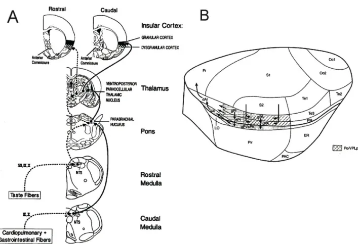
(16) 16. distension, etc.) is carried to the brain by primary sensory neurons. These visceral sensory axons enter the brain via cranial nerves and convey sensory information to the nucleus tractus solitarius (NTS), in a topographically organized manner (Saper 2002). The visceral inputs are relayed to the parabrachial nucleus, where the information is integrated and distributed to the hypothalamus, the visceral thalamus, and finally reaches the insular cortex, mostly to the granular region of the primary interoceptive posterior insular cortex (pIC, Fig. 1A). This pIC region is somehow coextensive with the anterior parietal granular IC of Shi and Cassell (1998), but included the adjacent dysgranular and agranular IC cortices (Fig. 1B). The anterior parietal granular IC is the recipient of axonal projections from the parvicellular part of the ventroposterior lateral thalamic nucleus (VPLpc, Shi and Cassell 1998), the thalamic relay of interoceptive information, and it is in a position to distribute information to other IC regions that are connected to the prefrontal cortex, the amygdala and the hippocampal formation..
(17) 17. Fig. 1. The insular cortex (IC) is the cortical component of the interoceptive system. A. Coronal sections of the rat brain showing the interoceptive pathway. Briefly, interoceptive information reaches the Nucleus tractus solitaries (NTS) at the brainstem level. Afferents further ascend trough the parabrachial to the parvicellular part of the ventroposterior lateral thalamic nucleus (VPLpc), to finally reach the granular IC. Adapted from Cechetto and Saper, 1990. B. Lateral view of the rat brain showing the location of the IC: above the piriform cortex and under the primary and secondary somatosensory cortices. The region receiving afferents from the visceral thalamus is showed with a coded shade. AI, anterior insula; PA, parietal insula; PI, posterior insula; a, agranular; d, disgranular; g, granular. Adapted from Shi and Cassell, 1998.. Evidence for a role of the pIC in the representation of interoceptive signals comes mainly from electrophysiological recordings of single neurons in the anesthetized rat. Cechetto and Saper (Cechetto & Saper 1987) reported that neurons from the pIC respond to a variety of.
(18) 18. interoceptive stimulus, such as baroreceptor activation (i.v. phenylephrine) chemoreceptor activation (i.v. cyanide or 10% CO2), mechanoreceptor activation (balloon inflation in the stomach), etc (Fig. 2). Moreover, it has been observed a correlation between fluctuations in blood pressure and neural activity of the pIC (Hanamori 2005). The insula has also strong connections with subnuclei providing interoceptive information, suggesting a modulatory action over them. In this sense, the insula has been proposed as a visceral sensorimotor cortex (Cechetto & Saper 1990). In fact, electrical stimulation of the pIC evoked blood pressure changes and gastric motor responses, further supporting the visceral sensorimotor character of the insula (Bagaev & Aleksandrov 2006)..
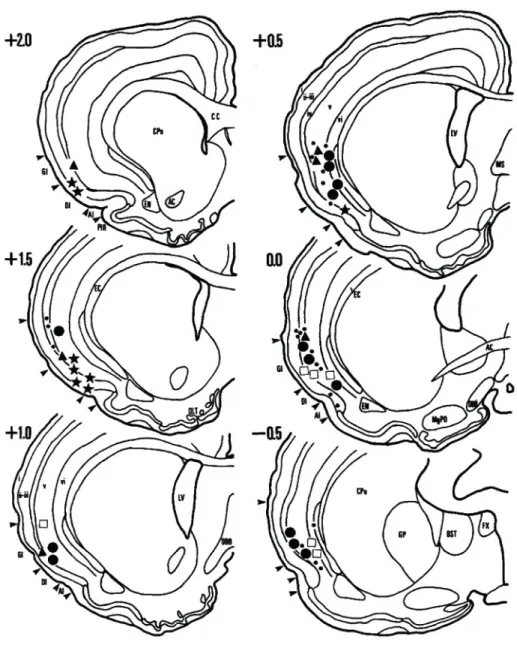
(19) 19. Fig. 2. The insular cortex (IC) is a sensory cortex. Schematic drawings of coronal sections from the rat brain, showing the sites where interoceptive responses were recorded. Recording sites were mostly within the granular IC. Neurons responded to several stimulus including taste (stars), gastric mechanoreceptors (triangles), respiratory chemoreceptors (circles) or cardiovascular baroreceptors (open squares). Small dots represent non responsive neurons. Adapted from Cechetto and Saper, 1987..
(20) 20. The IC has been implicated in a number of cognitive functions besides interoception, including emotion, memory, attention, etc (Kurth et al. 2010). Interestingly, a flow of information has been proposed from the posterior insula to its anterior subdivision (Cauda et al. 2012). This process would involve the integration of emotional and cognitive aspects of information in the anterior IC that starts with the collection of sensory information that reaches the posterior insula (Cauda et al. 2012). We think that the processing of interoceptive information within the insular circuits is necessary for the modulation of physiological responses to stimulus affecting homeostasis. In the present work we evaluated the role of the insula and particularly the pIC in the regulation of autonomic and behavioral responses to hypoxia. We also tested the idea that the activity of the pIC is associated to the behavioral expression of fear, to give an idea of the contribution of interoception to emotion. Finally, we tested a role for the pIC in the regulation of fear memories. 1.2 Hypoxia as an interoceptive stimulus. The processing of interoceptive information in the IC is crucial for homeostatic control (Cechetto & Saper 1987; Craig 2002). Hypoxia stimulates the carotid body (CB) chemoreceptor eliciting reflex cardiorespiratory responses, and arousal or even escapes behavior in conscious mammals (J. M. Marshall 1994). The episodes of behavioral arousal produced by the stimulation of the CB are consistent with the activation of hypothalamic defense areas (Marshall & Metcalfe 1990; J. M. Marshall 1994), and are abolished by the bilateral section of the carotid sinus nerves (Franchini & Krieger 1993; Koehler et al. 1980). Another structure involved in the behavioral responses elicited by CB stimulation is the periaqueductal grey matter (PAG). Indeed, stimulation of the rat CB with anoxia or KCN, or.
(21) 21. the electrical stimulation of the PAG produces a repertoire of behavioral responses that resemble panic attacks (Franchini & Krieger 1993; Schimitel et al. 2012). Dyspnea is defined as the uncomfortable awareness of respiratory distress, or the experience of air hunger (Meek et al. 1999). It has been proposed that the CB contributes to the dyspnea perception by increasing the respiratory motor output in response to hypoxia or hypercapnia, which can be perceived as unpleasant, or by inducing the sensation of dyspnea through the activation of ascending connections to the limbic system and sensory cortex, including the IC (Buchanan & Richerson 2009; Banzett et al. 2000; Liotti et al. 2001; von Leupoldt & Dahme 2005). In imaging studies conducted in humans, it has been found that the perception of an acutely CO2induced breathlessness activates the IC (von Leupoldt et al. 2008). Moreover, patients with one-hemispheric IC lesions, assessed with computer tomography or magnetic resonance imaging, showed a reduced perceptual sensitivity to dyspnea, suggesting that the dyspnea perception is processed in the IC (von Leupoldt et al. 2008; Schön et al. 2008). Dyspnea can be induced by an increase in CO2 or a decrease in O2, due to direct excitatory effects on the CB chemoreceptors, or by indirect effects through interactions with other respiratory afferents (i.e. lung mechanoreceptors). Since the IC has been involved in the perception of dyspnea, it is plausible that a hypoxic challenge would be able to induce a great activation of the IC. However, the role played by IC in the autonomic and behavioral responses to hypoxia remains largely unknown. Therefore, we hypothesized that the inactivation of the pIC disrupts the behavioral and autonomic responses to hypoxia. Accordingly, we tested the effects of the reversible inactivation of the pIC with the voltagedependent Na+ channel blockers bupivacaine on the escape behavior, and changes in arterial blood pressure, heart rate, and temperature in conscious rats exposed to hypoxia. Since the.
(22) 22. NTS plays a major role in the integration of the CB chemosensory inputs (Kline 2010), and the PAG produces defensive behavior (Schenberg et al. 2001; Schimitel et al. 2012), we also studied the effects of hypoxia on the number of Fos-immunorreactive (Fos-ir) neurons in the NTS and PAG in addition to the pIC. 1.3 Fear and fear memory. Fear or fright is elicited when animals or humans detect a threat that is concrete and sudden (Lazarus 1994). Several forebrain structures and neural circuits have been implicated in fear expression and fear memory in rats, including the amygdala complex, the medial prefrontal cortex, and the hippocampal formation (LeDoux 2000; Maren 2001). It is widely accepted that the amygdala complex is involved in the acquisition, storage and expression of learned fear (LeDoux 2000). It has been proposed that the induction of a fear-related body state by the amygdala is necessary for experiencing a feeling of fear (Damasio 2005). The anterior cingulate, the medial prefrontal cortex and the hippocampus have a key role in the regulation of fear responses (Etkin et al. 2011; Zelikowsky et al. 2014), while the sensory cortices are important for the long-term association between a sensory stimulus and fear responses (Sacco & Sacchetti 2010). However, none of these forebrain regions seems a good candidate for a perceptual or a cognitive representation of the diverse emotions including fear. The IC has been implicated in both bodily states representation (interoception) and emotional experience (Craig 2002). Imaging studies in humans reveal a marked engagement of insular and somatosensory cortices, as well as a number of subcortical structures, when subjects recall and re-experience different emotions (Damasio et al. 2000). There is a sustained metabolic activation of the IC during the expression of learned fear, in contrast to the transient activation.
(23) 23. of the amygdala, observed in both, humans (Phelps et al. 2001) and rats (Quirk et al. 1995). The sustained activation of the IC is compatible with a role in the cognitive representation of fear (Phelps et al. 2001). Furthermore, a correlation has been observed between insular volume and the magnitude of the autonomic response to conditioned fear (Hartley et al. 2011), suggesting that interoception may be an important factor in emotion perception, as proposed first by William James (Barbalet 1999). In the present work, we studied fear responses by means of classical conditioned fear. This behavioral paradigm is widely used to study the neural mechanisms mediating fear. It consists in the presentation of a normally harmless stimulus (light, sound, etc.) that co-terminates with an aversive stimulus (an electric foot shock in most cases). The harmless stimulus is known as conditioned stimulus (CS) and the aversive stimulus is called unconditioned stimulus (US). The repeated pairings between the CS and the US leads the animal to learn that the CS predicts the US. The process by which transient memory representations are transformed into a persisting longterm representation, is known as “consolidation” (Dudai 2012). On subsequent exposures to the CS alone, if the memory is consolidated, the animal remembers the CS-US association triggering the behavioral and physiological responses known as fear. On the other hand, when exposures to the CS in the absence of the US are repeated, a decrease in fear expression is observed as the animal learns that the CS does not predict the foot shock. This process is known as “extinction” and is considered as a new learning (Abel & Lattal 2001). The behavioral expression of fear measured here is freezing, the most widely used, which can be defined as the absence of all movements except for breathing (Blanchard & Blanchard 1969)..
(24) 24. Experimental studies in the rat have provided conflicting results regarding the relationship between the IC and fear. Christianson and colleagues (2008) showed that a pre-training lesion or the reversible inhibition of a region of the pIC during training, but not during testing (Christianson et al. 2011), prevents the effect of a safety signal on subsequent fear/anxiety-like behavior. Alves and coworkers (2013) found that inactivation of a more rostral IC region after training or before testing (but not before training) attenuated freezing and cardiovascular responses evoked by context. The lesion of the most caudal IC after training blocked fearpotentiated startle (Shi & Davis 1999). In contrast, no evidence was found for an involvement of this most caudal IC in fear conditioning (Brunzell & Kim 2001; Lanuza et al. 2004). When comparing these studies, it seemed that one key difference lies on the specific IC region that was intervened. On the other hand, none of these studies is focused in the interoceptive region of the pIC, from +0.95 mm to -1.50 mm from bregma, where the densest projection from the interoceptive thalamus are found (Shi & Cassell 1998; Contreras et al. 2007). Accordingly, a study conducted by Brydges and colleagues (2013) showed a marked increase in activity, measured by fMRI, of the granular pIC in response to a fear-conditioned stimulus. Furthermore, it has been shown that the long-term inactivation of the pIC, with the sodium channel blocker neosaxitoxin, reduced expression of conditioned fear to subsequent CS exposures (Madrid 2014). However, the electrical activity pattern during expression of conditioned fear remains unclear. We hypothesized a differential response of pIC neurons during high vs low fear states (freezing behavior). To address this issue we used fearconditioned rats and recorded the activity of pIC neurons during expression of conditioned fear to the CS in the absence of the US..
(25) 25. Sensory cortical areas has been proposed as the storage site of memories, since plasticity and sensory processing take place, thus encoding the emotional experience occurring during reactivation of memories (Sacco & Sacchetti 2010). In this sense, it has been reported that the activity of the pIC is important for the acquisition of spatial memory (Nerad et al. 1996), consolidation of taste-based memory (Berman & Dudai 2001) and for the processing of diverse stimulus-outcome associations (Contreras et al. 2007; Contreras et al. 2012; Gardner & Fontanini 2014). Moreover, the integration between perception and behavioral response in the pIC was recently demonstrated (Kusumoto-yoshida et al. 2014). Taking advantage of the conditioned fear paradigm, we were also able to study the role of the pIC in fear memory. We hypothesized that the activity of the pIC following conditioning training is important for the retention of conditioned fear. We studied the effect of interfering with this pIC region before and after classical auditory fear conditioning. We used reversible inactivation and inhibition of protein synthesis in order to determine the role of the pIC in the regulation of fear memories..
(26) 26. 2. Hypothesis. Our main objective is to determine the role of the pIC in the regulation of autonomic and behavioral responses to stimulus affecting the homeostasis. Here we examined the hypothesis that the activity of the rat interoceptive insula is involved in the control of behavioral and autonomic responses to interoceptive and emotional stimuli. Information about the condition of the physiological state of the body reaches the pIC, where the information is processed and distributed to other brain regions, in order to regulate autonomic variables and to display an appropriate behavior. The pattern of activity of the pIC during fear expression constitutes the neuronal correlate for the behavioral expression of this emotion. Accordingly, the pIC is responsible for the storage and regulation of fear memory, which is crucial for the behavioral expression of fear. This hypothesis is based on the following observations: First, the pIC is the region of the IC with the densest projections from the visceral sector of the thalamus. Second, neurons from the pIC respond to several interoceptive stimuli. Third, the pIC is strongly interconnected with brain structures that regulate homeostasis. Fourth, the inactivation of the pIC delays the behavioral expression of GI malaise induced by LiCl administration and disrupts drug craving. Fifth, the electrical activity of the pIC is associated with the behavioral expression of GI malaise. Sixth, the inactivation of the pIC with the long-acting sodium channel neosaxitoxin reduced the expression of conditioned fear. Seventh, the IC has been involved in the storage of several types of memories..
(27) 27. 3. Objectives. Determine the role of the pIC in the behavioral and autonomic response to hypoxia. - Assess the contribution of pIC activity for the behavioral and autonomic responses to hypoxia. - Determine the pattern of brain activity of rats subjected to hypoxia. Determine the role of the pIC in expression of conditioned fear. - Determine the pattern of IC activity underlying expression of conditioned fear. - Determine the activity of pIC neurons during expression of conditioned fear. - Compare changes in FR of pIC neurons in high vs low fear states. - Determine whether the activity of pIC neurons correlate with freezing time course. Determine the role of the pIC in fear memory. - Determine whether the activity of the pIC is necessary for retention of conditioned fear. - Determine whether protein synthesis in the pIC is necessary for retention of conditioned fear memory..
(28) 28. 4. Materials. Animals. Experiments were performed on adult male Sprague-Dawley rats weighing 250 g. Rats were housed in individual cages, fed with standard chow diet and water ad libitum, and kept on a 12:12 h light–dark cycle. Room temperature was kept between 23 and 25 ˚C. All the experimental procedures were approved by the Bio-Ethical Committee of the Facultad de Ciencias Biológicas, P. Universidad Católica de Chile, and were performed according to the National Institutes of Health Guide for the Care and Use of Laboratory Animals. Drugs. Ketamine, Xylacine, Ketophen and Enrofloxacin were acquired from Corso SA (Santiago, Chile). Bupivacaine and Saline were acquired from Sanderson (Santiago, Chile). Isoflurane was acquired from Baxter LTDA (Santiago, Chile). Muscimol and Anisomycin were acquired from Sigma-Aldrich (USA). Recording of physiological variables. We used radio-telemetric transmitters (PB52, Telemetry Research, New Zealand) endowed with Millar Mikro-Tip pressure transducers. These transmitters allow the measurement of blood pressure, temperature and heart rate. These variables are acquired with an analogue–digital system (PowerLAB8 SP; ADInstruments, Australia) and analyzed with the Chart 7.2 Pro software. Cortical infusion of drugs. Stainless steel guide cannulae with removable occluders (4.5 mm long, 26 G; Plastics One, USA) were used to directly infuse drugs into the pIC. Injection cannulae (33 G; Plastics One) coupled to a 10 µL Hamilton syringe by a polyethylene tubing (inner diameter 1.27 mm; Plastics One) were used for infusions..
(29) 29. Recording of neuronal activity. Tetrodes were made with NiCr wires (17 µm diameter) that were acquired from California Fine Wire Co. (USA). Commercial hyperdrives (Harlan 8, Neuralynx, USA) that allows individual manipulation of 6 tetrodes were used. A commercial headtage (HS-36, Neuralynx) was used to preamplify signals from the rat brain. A 32 channel computer-controlled setup with four Lynx-8 amplifiers (Neuralynx) was used to acquire neural signals. Custom software written in MATLAB was used for spike sorting and data analysis.. 5. Methods. 5.1 General. Surgical procedures for telemetric transmitter implantation. Rats anesthetized with 2–3% isoflurane (Baxter, Chile) in O2 were implanted with radio-telemetric transmitters (PB52, Telemetry Research, New Zealand) endowed with Millar Mikro-Tip pressure transducers in the abdomen to record body temperature, heart rate and arterial blood pressure from the descendent aorta. After surgery, rats received Ketoprofen 0.2mg/kg (Rhodia Merieux) and Enrofloxacin 20 mg/kg (Bayer) i.p. for 3 consecutive days. Rats were allowed to recover for 7 days prior to any manipulation. Surgical procedures for implantation of infusion cannulas into the pIC. Rats were anesthetized with Ketamine 100 mg/kg (Imalgene; Rhodia Merieux) and Xylazine 20 mg/kg (Rompun; Bayer) i.p., and placed in a stereotaxic apparatus. Stainless steel guide cannulae (4.5 mm long, 26 G; Plastics One, USA) were lowered bilaterally, angled 10˚ divergent from the.
(30) 30. vertical, aimed to the following coordinates to reach the pIC: 0.51 mm posterior to bregma; ±5.0 mm from the midline; and 6.5 mm from the cranial surface (Swanson, L.W. 1998). Three small screws were positioned in the surface of the skull, to facilitate adhesion of the dental acrylic. Cannulae were capped with occluders to keep the tissue unexposed. The tip of the injection cannulae (33 G; Plastics One) protrudes 2 mm below guide cannula. Correct placement was verified by analyzing the location of the tip of the microinjection cannula in Nissl-stained sections. After surgery and for the following 3 days, rats received Ketoprofen 0.2 mg/kg (Rhodia Merieux) and Enrofloxacin 20 mg/kg (Bayer) i.p. Rats were allowed 7 days to recover from surgery prior to any other manipulation. Cortical injections. Injection cannulas were coupled to a 10 µL Hamilton syringe by a polyethylene tubing (inner diameter 1.27 mm; Plastics One) filled with the GABAA agonist muscimol (MUS, 0.5 µg/µL, 0.5 µL/side; Sigma-Aldrich), the voltage-gated sodium channel blocker bupivacaine (26.5 µM, 1 µL/side, Sanderson), the protein synthesis inhibitor anisomycin (ANI, 100 µg/µL, 0.5 µL/side; Sigma-Aldrich) or with sterile saline (SAL, 0.5 µL/side). Microinjections took 1 min on each side. The injection needle was left in place for additional 2 min to allow diffusion, slowly removed the injection needle; and replaced the occluders back immediately. Surgical procedures for implantation of tetrodes into the pIC. Prior to surgery, animals were handled 10 min once daily for 3 consecutive days. Animals were anesthetized with 100 mg/kg of ketamine (Imalgene; Rhodia Merieux) plus 20 mg/kg of xylazine (Rompun; Bayer), placed in a stereotaxic apparatus, and implanted with a hyperdrive (Neuralynx) aimed to the pIC using the following coordinates: Bregma, -0.51 mm, midline, + 6.0 mm, and -6.5 mm.
(31) 31. from the cranial surface, according to the Swanson’s atlas (Swanson, L.W. 1998). The hyperdrive consists of 6 independently movable tetrodes. Tetrodes were constructed with four nickel-chrome (18 µm) wires twisted together with an impedance of 1-1.5 MΩ (adjusted by gold plating procedures). The hyperdrive was anchored to the skull with 5 screws stainlesssteel screws (Plastics One) and dental acrylic. One of those screws was used as animal ground. Right after surgery and for the following 3 days, rats were injected with Enrofloxacin 5% (19 mg/kg i.p., Bayer) and Ketoprofen (0.2 mg/kg i.p. Rhodia Merieux). Rats were allowed to recover for 7 days prior to any experimental procedure. Tetrodes were lowered 480 µm daily (3 turns per screw), in 40 µm steps, until they reach the pIC at ca. 4000 µm under dura. Histology. Upon completion of experiments, rats were perfused transcardially with 500 mL of saline followed by 500 mL of 10% formalin. Brains were removed, left in post fixation in 10% formalin for 2h, and then stored in 30% sucrose with 0.02% sodium azide in PBS until they sank. Brains were sectioned frozen under dry ice in the coronal plane, at 50µm thickness, using a sliding microtome. The sections were stained with Cresyl Violet and examined by light microscopy to determine cannula or tetrode placement. Rats with misplaced cannula/electrodes were discarded from the behavioral analysis. Rats implanted with tetrodes were previously anesthetized and electrolytic lesions were performed by applying anodic current of 25µA for 20s through two wires in each tetrode and the animal tail. They were killed 48 h later and were perfused as mentioned above. Immunohistochemistry for Zif268 and Fos expression. Coronal brain sections were incubated in 0.3% H2O2 in PBS for 30 min, rinsed in PBS and transferred to the blocking solution (0.4% Triton-X100, 0.02% sodium azide, 3% normal goat serum in PBS) for 1 h, and.
(32) 32. left incubated overnight at room temperature with the primary polyclonal antibody anti-Fos (Ab-5, rabbit polyclonal, from Oncogene, USA), diluted 1:20000, or anti-Zif268 (sc-110, rabbit polyclonal; Santa Cruz Biotechnology, USA) diluted 1:2000. The following day, sections were rinsed in PBS, and then were incubated in the secondary antibody solution for 1 h (goat anti-rabbit IgG BiotinSPconjugated, Jackson Immuno Research, diluted 1:1000). After rinsing for 40 min, sections were incubated for 1 h in Vectastain ABC Elite Kit (Vector Laboratories, USA), rinsed and incubated in a 0.05% DAB solution containing 0.003% H2O2, and 0.05% nickel chloride until obtain a dark blue reaction product. Cell counting. The number of Zif268-ir and Fos-ir neurons was determined live in coronal sections with a camera lucida coupled to a Nikon microscope fitted with a 10X objective. For the anterior, higher order subdivision of the IC, the rostral agranular IC (RAIC), from bregma + 4.85 to + 3.6 mm, we sampled 2 sections per rat, and used a 0.25 x 1 mm2 counting grid; for bregma + 2.80 mm, we sampled 1 section per rat and used a 0.5 x 1.25 mm2 counting grid; and from bregma + 1.70 to + 1.20 mm, we sampled 2 sections per rat and used a 0.5 x 1 mm2 counting grid. For the pIC, from bregma +0.95 to -0.26 mm, we sampled 4 sections per rat, and used a 0.25 x 1 mm2 counting grid; from bregma -0.51 to -2.45 mm, we sampled 4 sections per rat, and used a 0.5 x 1 mm2 counting grid. For the primary somatosensory cortex (SSp), from bregma -0.82 to -1.78 mm, we sampled 4 sections per rat, and used a 0.5 x 1 mm2 counting grid. For the prelimbic cortex (PL), we sampled 3 sections per rat, from bregma +4,85 to 2,80 mm2 and used a 0,96 x 1,04 mm2. The number of Fos-positive neurons in the NTS and PAG were measured bilaterally in coronal photomicrographs taken at 10X and 4X, respectively with a Nikon digital camera (DXm 1200) coupled to a Nikon E400 microscope, with the ImageJ software (National Institutes of Health). We counted in 3 different levels in.
(33) 33. the NTS, −13.3 to −14.3 mm from bregma (Swanson, L.W. 1998) and in 3 levels for the PAG, −7.00 to −7.80 mm from bregma (Paxinos & Watson 1998). Data were analyzed using Two Way ANOVA and Bonferroni post test, or unpaired t-test (one-tailed) when appropriate. 5.2 Objective 1: Determine the role of the pIC in the behavioral and autonomic response to severe hypoxia. Effects of IC inactivation on autonomic and behavioral responses to hypoxia. The hypoxic chamber consisted of an acrylic cylinder (18 cm diameter and 55 cm long), provided with a removable cap in one end coupled to a fan for air renewal protected with a metal mesh. The opposite end was connected to gas cylinders containing air or N2 trough two normally closed solenoid valves. In order to decrease O2 levels in the chamber, N2 was infused at a flow rate of 10 L/min, thus lowering O2 from 21% to 5–6% in about 150 s. The O2 level was continuously monitored with an O2 analyzer (Ohmeda 5120, BOC Healthcare, USA), placed into the top of the chamber. After recovery from surgeries (day 15), rats were habituated for 3 consecutive days to the hypoxic chamber for 20 min. During these habituation sessions, air was infused at a flow rate of 10 L/min during 150 s to habituate rats to the gas infusion and the sound of the solenoid valve. After habituation, rats were microinjected bilaterally with sterile saline (1 µL/side) or bupivacaine (26.5 µM, 1 µL/side, Sanderson) on days 18 and 19 in a counterbalanced fashion. Rats were placed in the chamber and after 5 min, 100% N2 was infused in order to lower O2 to 6% in about 150 s. To return to normoxia, N2 infusion was stopped, the fan was switched on, and rats were left inside the chamber for another 10 min. Physiological variables (arterial blood pressure, heart rate and body temperature) were continuously acquired with an analog-digital system PowerLAB 8SP (ADInstruments, Australia), stored in a computer and analyzed off-line with the Chart 7.2 Pro software. The.
(34) 34. behavioral response to hypoxia was recorded with a webcam coupled to a computer, where the videos were stored to posterior analysis off-line. All experiments were conducted at 23–25 ˚C in a sound-attenuated room. Data analysis. For the analysis of behavior and physiological responses, hypoxia was divided in two stages depending on the O2 level reached in the chamber: mild hypoxia (21%–13% O2) and severe hypoxia (13%–6% O2). Recording of temperature, heart rate and arterial blood pressure was initiated once the rat was inside the chamber. For the analysis, we compared autonomic data averaged for 40 s during normoxia, mild hypoxia, severe hypoxia and posthypoxic recovery. The data in normoxia corresponds to the average of the last 40 s prior to the N2 infusion; mild hypoxia to the 40 s measured 30 s after the onset of hypoxia, severe hypoxia to the last 40 s of hypoxia and the post-hypoxic recovery, to the 40 s measured 60 s after the end the hypoxic challenge. For the analysis of the behavioral responses during hypoxia, we measured the frequency (number of episodes), the duration (percentage of time spent), and latency (time after N2 infusion and the %O2 to trigger behavioral response) of the behaviors described in Table 1. Similar behaviors have been identified and described in detail as part of the escape behavioral repertoire in the rat elicited by the stimulation of the CB chemoreceptors with KCN (Schenberg et al. 2001; Schimitel et al. 2012), and by the electrical stimulation of the dorsal PAG (Schenberg et al. 2000; Vargas & Schenberg 2001). Data were expressed as mean + SEM of the episodes of running or rearing, percentage of time spent in immobility during hypoxia and time after the N2 infusion and %O2 at which escape behavior appeared..
(35) 35. Table 1. Behaviors exhibited by the rats exposed to hypoxia. Behavior. Description. Immobility. Periods of null locomotion, without any movement except for breathing.. Running. Brief episodes of fast and violent locomotion, with or without great displacement within the chamber.. Rearing. Brief episodes in which rats stand on its hind limbs and guide his head and front limbs to the top of the chamber, as if looking for a way out.. Effects of hypoxia on Fos-ir in the pIC, NTS and PAG. In another experimental series, 12 rats were habituated for 3 consecutive days to the hypoxia chamber for 20 min. During the habituation session, air was infused at a flow rate of 10 L/min during ca. 150 s to habituate rats to gas infusion and valve’s sound. On day 4, rats were randomly divided in two groups (normoxia and hypoxia) of 6 rats each. In the hypoxia group, each rat was individually introduced in the chamber and after 5 min, 100% N2 was infused to lower the O2 levels to 6% in about 150s. To return to normoxia, the fan was switched on and the rats were left inside the chamber for another 10 min. In the normoxia group, each rat was individually introduced in the hypoxia chamber, and after 5 min air was infused at a flow rate of 10L/min. Ninety minutes after hypoxia or normoxia exposure, rats were anesthetized, perfused and the brains extracted for Fos immunohistochemistry..
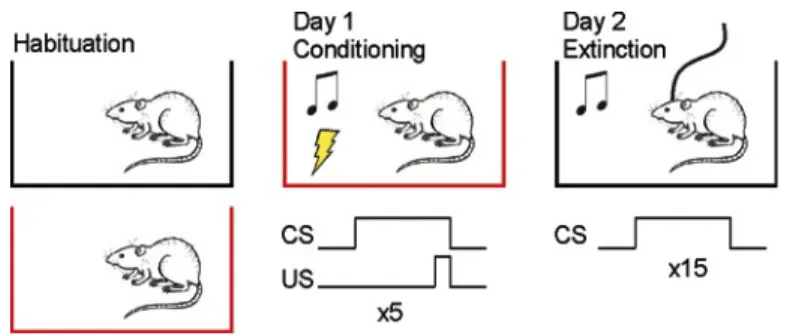
(36) 36. Data analysis. Data were expressed as mean + SEM. Significant differences (p<0.05) were assessed with unpaired t-test, paired t-test or two-way ANOVA followed by Bonferroni post test comparisons, when appropriate. 5.3 Objective 2: Determine the role of the pIC in expression of conditioned fear. Fear conditioning. Rats were conditioned in standard conditioning chamber (Harvard Apparatus Model LE1005; context A), equipped with a floor of steel rods to deliver electric foot shocks. Extinction and recordings took place in a different Plexiglas chamber of 64x38x30 cm (context B), situated in a different room, so that conditioning is restricted only to the auditory and not to the environmental cues. The training (day 1) consisted in presenting 5 habituation tones (CS, 80 dB, 5 kHz, 20 s), followed by a train of 5 tones that co-terminated with a mild foot shock (0.5 mA, 0.5 s). The pairings were separated by a variable interval averaging 2 minutes. Two min after the last CS-US pairing, the rats were taken back to their home cages. On day 2, rats were placed into the context B, where the CS was presented 15 times (extinction training) while neural activity was recorded. Behavior was recorded using a webcam plugged to a computer, where videos were stored for analysis off-line. Expression or early genes accompanying fear memory reactivation. This experiment was aimed to assess the expression of two early genes, c-fos and zif268, known to be involved in synaptic plasticity and used as markers of neural activity (Chaudhuri & Zangenehpour 2002). To assess the expression of Fos and Zif268 in the IC, some rats were conditioned in context A, and were killed 90 min after one CS presentation in the context B the following day (Fig. 3). A control group of rats was spared the foot shocks and exposed only to the CS in the conditioning chamber (control group) and killed 90 min after one CS presentation in context B.
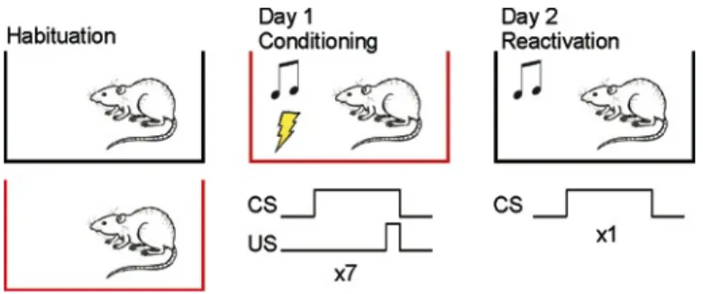
(37) 37. the following day. We selected this time point as allowed us to assess Fos and Zif268 at their peak expression (Maviel et al. 2004; Chaudhuri & Zangenehpour 2002).. Fig. 3. Protocol for fear conditioning to assess expression of c-fos and zif268. Rats were conditioned to tones of 20 s that co-terminated with a mild foot shock (0.5 mA) in the conditioning chamber (context A, in red). The rats were killed 90 after one CS presentation in the context B (in black). Tetrode construction and electrophysiological recordings. Extracellular recordings were performed with custom-made tetrodes mounted in a commercial hyperdive (Fig 4). This arrangement of electrodes provides a more reliable way to identify signals from individual neurons than single wire electrodes (Gray et al. 1995; Harris et al. 2000). Briefly, two segments (14 cm) of polyimide insulated NiCr wire (17 µm diameter) folded on themselves were twisted. Heat (250 ºC) was applied with a heat gun to facilitate adhesion between wires to keep this configuration. The tetrodes were inserted in polyimide tubes (Small Parts, USA) to protect them. A bundle of six stainless steel tubes (HTX-30T-30, outer diameter 300 µm; Small Parts, USA) of 2.5 cm each, was assembled within a Harlan 8 hyperdrive. Six tetrodes are inserted in the bundle (one for each tube) and were fixed to one of the 6 drives from the hyperdrive. Each wire from the tetrode was connected to an electrode interface board (EIB 36, Omnetics, USA), which provides the signal connection between tetrode wires and a 36-.
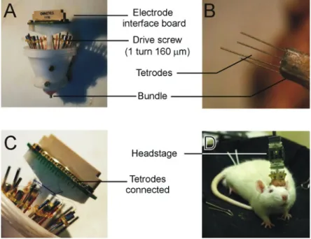
(38) 38. channel headstage pre-amplifier. A single silver wire (200 µm diameter; A-M systems) removed from its Teflon cover was attached to the EIB 36 board in one end, and tied to a small screw (TX1-3-C; Small Parts, USA) to be anchored in the skull as animal ground. Once it was checked there were no short circuit, the tip of each tetrode was gold-plated by passing a cathodal current of 10 µA to low the impedance to 1 MΩ. For recordings, a headstage preamplifier containing 36 channels of unity-gain amplification (HS-36, Neuralynx) was connected to the EIB. The headstage was connected to a 32-channel, computer-controlled system (Four units of the Lynx-8 amplifier, Neuralynx). Signals exceeding a voltage threshold of two standard deviation from the noise floor were amplified (15000x), bandpass filtered (0.6 kHz-6 kHz), sampled at 32 kHz and stored on a pc for analysis offline.. Fig. 4. Tetrodes array in the hyperdrive. A. Each tetrode is mounted inside one tube of the bundle and coupled to a drive. B. Tetrodes out of the bundle. C. Free ends of each tetrode are connected to the electronic interface board. D. Rat implanted and connected to the recording setup..
(39) 39. Recordings were performed on day 2 (Fig. 5) during the night (after 21:00 h) in a dark room, as rats are more active. Two hours before starting the experiment, the rat was connected to the recording system to search for unit activity, by gently lowering tetrodes in 40 µm steps until stable signals were acquired in most tetrodes.. Fig. 5. Fear conditioning protocol to record the activity of posterior insular cortex neurons. Rats were conditioned to tones of 20 s that co-terminated with a mild footshock (0.5 mA) in the conditioning chamber (context A, in red). Extinction and recordings were conducted on day 2 in the context B (in black).. Behavioral analysis. Freezing was defined as absence of movement except for breathing (Blanchard & Blanchard 1969). Freezing was calculated as the percentage of time rats spent in freezing during tone duration (20 s). Time spent in freezing during tone was scored offline. Differences in freezing were analyzed with repeated measures ANOVA. Freezing behavior was divided in two stages: early extinction, which averaged the time of freezing for the first 3 trials (maximal fear state), and late extinction, which considered the last 3 trials (minimal fear state). Electrophysiological analysis. Electrophysiological signals were analyzed using MClust (A.D. Redish, University of Minneapolis, USA) that classify them according to their shape.
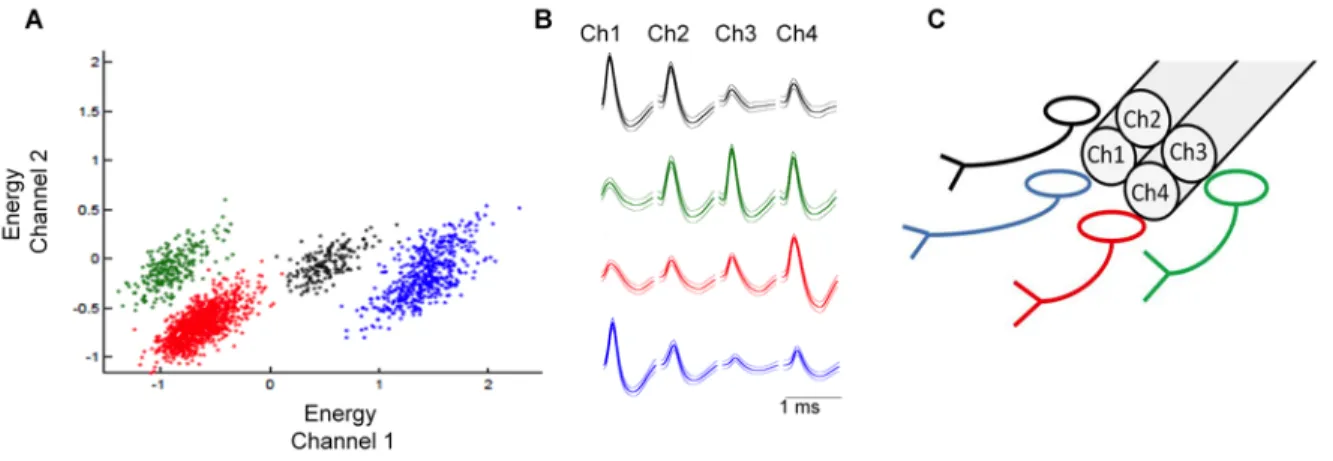
(40) 40. into individual clusters. Clusters formed by electrophysiological signals were further analyzed using the custom MATLAB script KlustaKwik (Kadir et al. 2014) that allows the manual selection of clusters with action potential shape signals. These signals are plotted in terms of relative amplitude (among other parameters) in different channels from the tetrode, with signals with similar characteristics forming an individual cluster. Signals were also plotted in the time domain to analyze their stability throughout the experiment. Next, waveforms in the 4 channels of the tetrode were analyzed again to confirm their action potential shape. Time interval between action potentials (inter spike interval, ISI) was also calculated. Neurons with more than 1% of action potentials with ISI less than 1 ms were excluded from the analysis, as they do not likely represent signals from one individual neuron. Figure 6 shows a representative individualization of recorded neurons. For the characterization of recorded units we calculated basal firing rate (FR) during the first 60 s of recording. Neurons with basal FR below 0.1 Hz were excluded from the analysis. We also calculated the AP (action potential) duration, defined as the time elapsed between peak and valley..
(41) 41. Fig. 6. Representative identification of four neurons recorded with one tetrode. A. Dispersion plot showing energy of waveforms in channel 1 vs channel 2 of one tetrode. B. Average waveforms recorded in each channel of the tetrode. C. Scheme representing the position of neurons.. A total of 100 units from 9 successfully implanted rats were included in the analysis. To detect changes in FR in response to the CS, we calculated the FR of each unit in 2 s bins, from 20 s before and 20 s after the CS onset and obtained average FR for the first 3 CS (early extinction) and for the last 3 CS (late extinction). To obtain a measure of dispersion, we then z-scored the data as follows: Z-score = FR during each 2 sec bin – mean pre-CS FR SD of pre-CS FR. Neurons with 3 or more bins above or below 1.96 standard deviation (p<0.05) were classified as excited or inhibited by the CS, respectively (Burgos-Robles et al. 2009). We also compared changes in FR of groups of neurons classified either as excited or inhibited in early vs late extinction using a two way ANOVA, using time (before and after tone onset) and protocol phase (early and late extinction) as main factors. The test of Pearson (Prism software) was.
(42) 42. used to establish the correlation between freezing behavior and the activity of pIC single units. For this analysis, FR and freezing were calculated in 5 s bins instead of 2 s to facilitate freezing measurement. All analyses were performed in custom-software written in MATLAB. 5.4 Objective 3. Fear memory consolidation in the pIC. Fear conditioning. For the acquisition of auditory fear conditioning, rats were placed in a 27x27x27 cm conditioning chamber (Harvard Apparatus Model LE1005; context A), equipped with a floor of steel rods to deliver electric foot shocks. A different chamber of 64x38x30 cm (context B), with Plexiglas® floor and situated in a different room, was used to assess conditioning. Prior to the experiments, rats were habituated to both contexts, A and B, by allowing them to explore during 15 min each one for 2 consecutive days. The training (day 1) consisted in presenting 5 habituation tones (CS, 80 dB, 5 KHz, 20 s) followed by a train of 7 tones that co-terminated with a mild footshock (0.7 mA, 0.5 s). The pairings were separated by a variable interval averaging 2 min. Two min after the last CS-US pairing, the rats were taken back to their home cages. On day 2, the rats were placed into the context B, where the CS was presented 15 times with a cadency of one event every 2 minutes. On day 3, the rats were placed back in the context B to test for reactivation of fear memory by presenting them two CS. Figure 7 summarizes this protocol. Effect of pIC inactivation on conditioned fear retention. The first experiment was aimed to determine whether the pIC has a role in the retention of conditioned fear. The rats were injected bilaterally with MUS or SAL into the pIC 30 min prior to training (arrow in Fig. 2A). The following days (Day 2 and Day 3), rats were tested for conditioned fear expression in the context B..
(43) 43. Effect of post-training inhibition of protein synthesis in the pIC on consolidation of conditioned fear. Immediately following fear conditioning, rats were infused bilaterally with ANI or SAL into the pIC (arrow in Fig. 3). In a separate group of rats, infusions of ANI or SAL took place 6 h after conditioning (Fig. 4). The following days (Day 2 and Day 3), rats were tested for conditioned fear expression in the context B.. Fig.7. Fear conditioning protocol to assess the effect of pharmacological manipulations of the posterior insular cortex on retention of conditioned fear. Rats were conditioned to tones of 20 s that co-terminated with a mild footshock (0.7 mA) in the conditioning chamber (context A, in red). Retention of fear was assessed on days 2 and 3 in the context B (in black).. Data analysis. Fear expression was assessed as the percentage of time each rat spent in a state of behavioral freezing during tone presentation. For this purpose, behavior was recorded in video by means of a webcam connected to a computer, where videos were stored for off-line analysis by an experimenter blind to treatment. Data were plotted as the average freezing of two trials + SEM. The odd number of trials used signifies that the last trial in every stage was excluded from the statistical analysis. Data were analyzed using Two Way ANOVA, with training phase and infusion as the main factors and unpaired t-test (two-tailed) when appropriate..
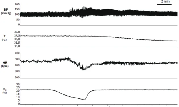
(44) 44. 6. Results.. 6.1 Objective 1: Determine the role of the pIC in the behavioral and autonomic response to severe hypoxia. Effect of pIC inactivation on behavioral and autonomic responses to hypoxia. The experimental protocol assessing the contribution of pIC activity on autonomic and behavioral responses to hypoxia is shown in Fig. 8. In awake behaving rats implanted with telemetric transmitters, saline or bupivacaine were infused through microinjection cannulae aimed to the pIC bilaterally (Fig. 8). Rats were subjected to an acute fast drop in %O2 (from 21 to 5–6% in 150 s) and the physiological variables and behavior were monitored. Hypoxia produced cardiovascular and thermal responses (Fig. 9). Hypoxia increased the arterial blood pressure and produced a biphasic response in heart rate depending on the level of O2 in the chamber. Initially, mild hypoxia produced tachycardia, but bradycardia was observed when O2 descended under 8%. In addition, hypoxia induced a significant decrease in body temperature, which persisted after the withdrawal of the hypoxic stimuli (Fig. 9)..
(45) 45. Fig. 8. Experimental protocol to assess the contribution of posterior insular cortex (pIC) activity on responses to hypoxia. a. Histological localization of the injection cannula in the pIC. Left panel depicts a representative example showing the tip of the cannula (arrow). Right panel shows the anatomical drawing adapted from Swanson’s stereotaxic atlas (1998). b. Time line to study the effect of pIC inactivation on autonomic and behavioral responses to hypoxia. c. Experimental setup showing the hypoxic chamber. CP, caudoputamen; pIC, posterior insular cortex; rf, rhinal fissure; SSs, secondary somatosensory area. Scale bar, 500 µm..
(46) 46. Fig. 9. Representative recording of autonomic response to acute hypoxia measured with telemetry in a freely moving rat. BP, arterial blood pressure; T, body temperature, HR, heart rate; O2, percentage of inspired oxygen..
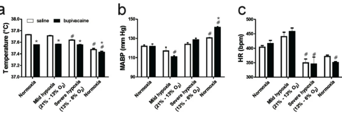
(47) 47. The inactivation of the pIC with bupivacaine induced a significant decrease in the baseline temperature of 0.2 ˚C compared to the saline, which persisted through the course of the hypoxia and the post-hypoxic recovery (p < 0.05, Two-way ANOVA, Bonferroni post test; Fig. 3a). In rats that were infused with saline into the pIC, a significant hypoxia-induced decrease in body temperature from baseline was observed in severe and post-hypoxia. After bupivacaine infusion into the pIC, the decrease on body temperature was only evident in the post-hypoxic recovery (p < 0.05, Two-way ANOVA, Bonferroni post test; Fig. 10a). Basal mean arterial blood pressure (MABP) in normoxia was unaffected by the pIC inactivation. Mild hypoxia induced a significant decrease in MABP compared to the normoxia only in the bupivacaine microinfused rats, whereas in the post-hypoxic recovery, both groups showed an increase in MABP, but the increase was more pronounced in rats infused with bupivacaine into the pIC (p < 0.05, Two-way ANOVA and Bonferroni post test; Fig. 10b). Baseline hear rate in normoxia was also unaffected by bupivacaine infusion into the pIC. A significant bradycardia was observed in both groups during severe hypoxia, however this effect was more prolonged in bupivacaine-infused rats as was also evident in the recovery to normoxia (p < 0.05, Two-way ANOVA and Bonferroni post test; Fig. 10c)..
(48) 48. Fig. 10. Effect of posterior insular cortex (pIC) inactivation on autonomic responses to acute hypoxia. Following saline or bupivacaine (26.5 µM 1 µL/side) infusions into the pIC, rats were introduced in the hypoxic chamber and were exposed to hypoxia. a. Severe hypoxia elicited a significant decrease in body temperature in rats infused with saline. Bupivacaine decreased basal temperature in normoxia by ca. 0.2 ˚C as compared with saline, and this difference remained during the hypoxia exposure and subsequent recovery to normoxia. b. Basal mean arterial blood pressure (MABP) was unaffected by pIC inactivation, and a more pronounced change was observed in rats infused with bupivacaine during mild and posthypoxia recovery. c. Basal heart rate (HR) was unaffected by pIC inactivation, whereas the bradycardic response to hypoxia was more prolonged in bupivacaine infused rats. Data represent mean + SEM in 4 rats. *p < 0.05 compared to saline, #p < 0.05 compared to baseline, Two way ANOVA Bonferroni post-test..
(49) 49. During the hypoxic challenge, we observed a repertoire of behaviors, namely: immobility, running and rearing, which can be cataloged as an escape response (See Table 1 for a detailed description). When O2 dropped between 21 and 13%, running and rearing episodes were practically absent and only immobility was observed (Fig. 11a–c). In contrast, during severe hypoxia, running and rearing episodes appeared in addition to the immobility. The pattern of this behavioral response to hypoxia was not disrupted by pIC inactivation with bupivacaine (p > 0.05, paired t-test). Additionally, in order to detect any impairment in the ability of rats to sense hypoxia due to pIC inactivation, we measured the latency and the %O2 threshold to running and rearing episodes under saline and bupivacaine infusion into the pIC. An increased latency to running in terms of both, time after N2 infusion and %O2 (p < 0.05, paired t-test, Fig. 11d) was observed, whereas the latency to rearing (p > 0.05, paired t-test, Fig. 11e) was unaffected by pIC inactivation..
(50) 50. Fig.11. Effect of posterior insular cortex (pIC) inactivation on the behavioral responses to acute hypoxia. Rats were infused bilaterally with bupivacaine (26.5 µM, 1 µL/side) or saline, introduced into the chamber and after 5 min were subjected to acute hypoxia for 150 s. Severe hypoxia triggered running (a) and rearing (b) behavior in both conditions (p > 0.05, paired ttest). Hypoxia-induced immobility (c) was unaffected by pIC inactivation (p > 0.05, paired ttest). An increase in the latency to running (d) but not rearing (e) was observed when the pIC was inactivated (*p < 0.05, paired t-test). Data represent mean + SEM in 4 rats..
(51) 51. Hypoxia-induced neuronal activity in the pIC, NTS and PAG. Since pIC inactivation had only minimal effect on hypoxia-induced escape behavior and autonomic responses, we studied the effects of hypoxia on the Fos-positive neurons in the pIC, the NTS and the PAG, nuclei involved in the processing of interoceptive information (Cechetto & Saper 1987) and defensive responses (Schimitel et al. 2012), respectively. Rats were subjected to hypoxia or normoxia (control) and were sacrificed 90 min after. In the pIC, we did not found any significant increase in the number of Fos-positive neurons (p > 0.05, unpaired t-test, Fig. 12). In contrast, we found a significant increase in the number of Fospositive neurons in the rostral and caudal NTS following hypoxia (−13.30 mm and −14.30 mm from bregma, respectively, p < 0.05, unpaired t-test, Fig. 13). In addition, in rats exposed to hypoxia we found a significant increase in the number of Fos-positive neurons in the dorsal PAG, particularly in the most rostral levels from the dorsolateral PAG, and in the lateral PAG (p < 0.05, unpaired t-test, Fig. 14),.
(52) 52. Fig. 12. Fos-immunoreactivity (Fos-ir) in the posterior insular cortex (pIC) is not modified following acute hypoxia. a. Representative photomicrograph showing Fos-ir in the pIC in normoxia (left) and hypoxia (right). Scale bar 100 µm. b. Summary of the effects of hypoxia on the number of Fos-positive neurons in the pIC of 6 rats. Note that hypoxia did not modify the number of Fos-positive neurons in the pIC (p > 0.05, unpaired t-test)..
(53) 53. Fig. 13. Effect of hypoxia on Fos-immunoreactivity (Fos-ir) in neurons from the rat Nucleus tractus solitarius (NTS). a. Representative photomicrograph showing positive Fos-ir in the NTS in normoxia (left) and hypoxia (right), scale bar 200 µm. b. Summary of the effects of hypoxia on the number of Fos-positive neurons in the NTS of 6 rats. Hypoxia induced a significant increase in the number of Fos-positive neurons in the rostral and caudal portions of the NTS (*p < 0.05, unpaired t-test)..
(54) 54. Fig. 14. Effect of hypoxia on Fos-immunoreactivity (Fos-ir) in neurons from the rat Periaqueductal gray matter (PAG). Quantification of the number of Fos-positive neurons in different regions of the PAG along the rostro-caudal axis. a. Dorsomedial PAG (PAGdm). b. Dorsolateral PAG (PAGdl). c. Lateral PAG (PAGl). d. Ventrolateral PAG (PAGvl). Hypoxia induced a significant increase in Fos-positive neurons in rostral PAGdl and rostral PAGl (*p < 0.05, **p < 0.01, unpaired t-test)..
(55) 55. 6.2 Objective 2: Determine the role of the pIC in the expression of conditioned fear. Expression of early genes accompanying fear memory reactivation. This experiment was aimed to assess the expression of two early genes, c-fos and zif268, known to be involved in synaptic plasticity and used as markers of neural activity (Chaudhuri & Zangenehpour 2002), in the IC underlying the expression of conditioned fear. To assess the expression of Fos and Zif268 in the IC, some rats were conditioned for one day and killed 90 min after one CS presentation in the context B the following day (see methods). A control group of rats was spared the foot shocks and exposed only to the CS in the conditioning chamber (control group) and killed 90 min after one CS presentation in context B the following day. We selected this time point as allowed us to assess Fos and Zif268 at their peak expression (Chaudhuri & Zangenehpour 2002; Maviel et al. 2004). Fear-conditioned rats (conditioned group) showed a robust conditioned response to CS on day 2 compared to control rats (Fig. 15A, t8=8.486, p<0.0001). The PL cortex, a cortical region known to be involved in the expression of conditioned fear (Burgos-Robles et al. 2009) showed an increased Fos-ir (t8=1.908, p=0.0464, Fig.15B). In contrast, no significant activation was observed in the SSp (t8=0.045, p=0.4826, Fig. 15B), a cortical region that served as a control for unspecific cortical activation. Expression of conditioned fear to tone was paralleled by an increase in Fos-ir and Zif268-ir, markers of neuronal activation and neural plasticity (Cole et al. 1989; Chaudhuri & Zangenehpour 2002). Fos-ir was increased in the pIC, specifically at levels +0.95 (t44=3.571, p<0.05) and +0.45 (t44=4.837, p<0.05) from bregma (Fig. 15C)..
(56) 56. Fig. 15. Reactivation of learned fear was paralleled by an increase in Fos expression in the insular cortex (IC). A. Freezing to the tone in context B. Conditioned rats showed higher levels of freezing to the tone relative to unconditioned rats. B. Quantification of Fos immunoreactive cells in prelimbic cortex (PL) and primary somatosensory cortex (SSp) in control and conditioned rats C. Quantification of Fos-ir in two different regions of the IC, the rostral agranular insular cortex (RAIC) and the granular field of the posterior insular cortex (pIC), at different levels from bregma. Below, representative photomicrograph of the pIC showing the near absence of Fos immunoreactivity (arrow) in control rats (left) and high Fosimmunoreactivity (arrow) in conditioned rats (right). Data are expressed as mean + SEM, *p<0.05, Two-Way ANOVA and Bonferroni post test. Scale bars indicate 100 µm..
(57) 57. In turn, conditioned rats showed also an increase in the expression of Zif268 (Fig. 16), in the RAIC, at level +1.2 (t44=3.657), and in the pIC, at the levels +0.45 mm (t44=4.986), -0.26 mm (t44=4.419) and -1.78mm (t44=4.649) from bregma. Taken together, these data show that reactivation of fear memory is accompanied by an increase in pIC activity and expression of early genes involved in neural plasticity, further supporting a role for the pIC in the long-term storage of fear memory.. Fig. 16. Reactivation of learned fear was paralleled by an increase in Zif268 expression in the insular cortex. Quantification of Zif268-ir in two different regions of the IC, the rostral agranular insular cortex (RAIC) and the granular field of the posterior insular cortex (pIC), at different levels from bregma. Below, representative photomicrograph of the pIC showing the near absence of Zif268 immunoreactivity (arrow) in control rats (left) and high Zif268immunoreactivity (arrow) in conditioned rats (right). Data are expressed as mean + SEM, *p<0.05, Two-Way ANOVA and Bonferroni post test. Scale bars indicate 100 µm..
Figure
+7


Documento similar