The role of gap junction channels during physiologic and pathologic conditions of the human central nervous system
20
0
0
Texto completo
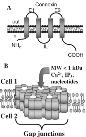
(2) 500. J Neuroimmune Pharmacol (2012) 7:499–518. A. E1. Connexin E2. second messengers through GJ and uHC results in the coordination of multiple physiologic functions (Sáez et al. 2003a). Here, we will review the pattern of Cx expression in each cell type of the central nervous system (CNS) and the function of GJ and uHC under normal and pathologic conditions.. out in NH2. IL COOH. B. MW < 1 kDa Ca2+, IP3, nucleotides. Cell 1. Cell 2 Gap junctions Fig. 1 Schematic diagram showing connexin (Cxs) membrane topology and a plaque of GJ channels. a Model showing membrane topology of Cx E1 and E2, represent the extracellular loops and IL, the intracellular domain. b Model of the GJ plaque between two cells (cell 1-cell 2) and its role in mediating communication by diffusion of second messengers smaller than 1 kDa, such as Ca 2+ , IP 3 and nucleotides. General introduction Gap junction (GJ) channels are formed by two hemichannels, each contributed by one cell, which are hexamers of homologous subunit proteins, termed connexins (Cxs), that connect the cytoplasm of adjacent cells (Bennett et al. 2003; Sáez et al. 2003a) (Fig. 1a). Connexin hemichannels (Cx HCs) can be formed by one (homomeric connexon) or several (heteromeric connexon) types of Cxs, while GJ channels can be formed by either two identical, homotypic, or different, heterotypic, hemichannel subunits. These different subunit combinations enable GJs to differ in their biophysical and permeability properties (Harris 2001, 2007). In addition, it was shown that unapposed hemichannels (uHC), before their cell-to-cell docking to form GJ, also open on the surface of the cell allowing exchange of small factors between the cytoplasm and the extracellular environment. Both GJ and uHC have an internal pore of approximately 12 Ao, allowing ions and intracellular messengers up to ~1 kDa in molecular mass to diffuse between connected cells or from the cytoplasm to the extracellular space (Bennett et al. 2003; Sáez et al. 2003a) (Fig. 1b). The diffusion of these. Expression of connexins in different cell types in the CNS Astrocytes. Astrocytes participate in many brain functions, including CNS differentiation, neuronal excitability, production of neurotrophic factors, control of extracellular synaptic metabolites, syncytial signaling, synaptic plasticity, formation of scar tissue after neuronal loss, immune activation, inflammation, and blood brain barrier (BBB) integrity. In all of these functions, GJ and uHC have a critical role (Rouach et al. 2002c; Kielian and Esen 2004; Sáez et al. 2005; Kielian 2008). Astrocytes form extended networks regions of the brain parenchyma by direct communication through GJs between coupled cells. Reverse transcription-PCR and protein analyses showed that Cx43 and Cx30 are the main Cxs expressed in astrocytes (Dermietzel et al. 1989, 1991; Giaume et al. 1991; Rash et al. 2001a, b; Nagy et al. 2003b; Nakase and Naus 2004). Cx43/Cx30 double-knockout mice only have minimal gap junctional communication (GJC) between astrocytes (Wallraff et al. 2006; Rouach et al. 2008), suggesting that both proteins are the main components of functional astroglial GJ channels. The original studies on Cx43 did not consider the expression of Cx30. However, recent data indicate that Cx30 plays an important role in hippocampus GJ coupling, cellular degeneration and cochlear function (Cohen-Salmon et al. 2007; Chang et al. 2008; Sun et al. 2009; Gosejacob et al. 2011). Thus, further studies are required to clarify the role of Cx43 and Cx30 in physiologic and pathologic conditions. Synaptic molecules released in response to neuronal activity, including K+, glutamate and other neurotransmitters, are normally taken up by astrocytes and extensively diluted in the astrocytic network through GJ channels. However, in pathologic conditions, GJC is compromised and these molecules can be toxic in the absence of functional GJ channels (Orkand et al. 1966; Rose and Ransom 1997; Wallraff et al. 2006). High neuronal activity enhances astrocyte GJ communication (Marrero and Orkand 1996), and also induces vasodilation of pial arterioles through connexin-based channels (Xu et al. 2008), suggesting a relationship between neuronal activity and blood supply through GJ, in eliminating these toxic molecules by providing better perfusion in areas subjected to high neuronal activity. In addition, GJC enables the coordination of several signaling events, including intercellular Ca2+ waves that control Ca2+-dependent glutamate release, and metabolic and electrical synchronization among.
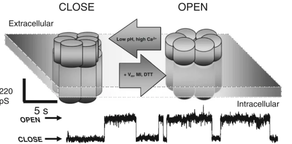
(3) J Neuroimmune Pharmacol (2012) 7:499–518. astrocytes and neurons and astrocyte and endothelial cells (ECs) to control synaptic plasticity, neuronal survival and/or vascular tone (Guthrie et al. 1999; Paemeleire and Leybaert 2000; Sáez et al. 2003b; Simard et al. 2003; Dale 2008). All these findings indicate that GJs play a key role in the coordination of astrocyte signaling and metabolic events usually altered in many brain-associated diseases. The opening of Cx uHC allows exchange of diverse molecules between the cytoplasm and extracellular environment, especially in conditions of cellular stress, to mediate autocrine/paracrine signaling. More recently, another gene family similar to the Cx family, encoding a set of three membrane proteins, named pannexins (Panxs 1–3), has been identified in different cell types, including astrocytes (Bruzzone et al. 2003). To date, the absence of ultrastructural evidence for GJ like structures by pannexins in mammalian cells suggests that the main function of pannexin is as pannexin (Panx) uHC. However, a recent study indicated that pannexin 3 expressed in osteblast can work as a uHC in the ER and the plasma membrane and also as a GJ, suggesting that this protein may have multiple functions depending on it cellular localization (Ishikawa et al. 2011). In astrocytes, along with Cx43 uHC, astrocytes also express functional Panx1 uHC (Iglesias et al. 2008, 2009; Iwabuchi and Kawahara 2011). However, the regulation of Panx1 uHC and their role in the function of astrocytes is still under investigation. Astroglial uHC are potential regulators of homeostatic imbalances present in diverse brain diseases or cellular stress conditions, but normally uHC are closed due to their high permeability that can result in cell death when the channels are opened (Abrams et al. 2002; Contreras et al. 2002; Iglesias et al. 2009; Orellana et al. 2011a). Astroglial dysregulation induced by ischemia-like conditions and metabolic inhibition resulted in opening of Cx43 uHC (Contreras et al. 2002). It was also demonstrated that fibroblast growth factor-1 (FGF-1), cytokines, hypoxia, oxidative stress and changes in intracellular/extracellular calcium result in opening of Cx43 uHC, allowing release of ATP, glutamate, nicotinamide adenine dinucleotide (NADH) and prostaglandins to the extracellular space mainly in pathologic conditions (Contreras et al. 2002; Stout et al. 2002; Retamal et al. 2006, 2007; Froger et al. 2009; Sánchez et al. 2009; Schalper et al. 2009; Garre et al. 2010; Orellana et al. 2010; Retamal et al. 2010; Sáez et al. 2010). Astrocytes provide metabolic and framework support to neurons. Therefore, damage associated with the opening of uHC has been proposed to increase neuronal susceptibility to insults (Sánchez et al. 2009a; Orellana et al. 2010; Sáez et al. 2010b; Orellana et al. 2011a). In agreement with this concept, it was demonstrated that opening of astroglial uHC potentiates glutamate-induced neurotoxicity by pro-inflammatory cytokines (Froger et al. 2010) and it has been shown that glutamate can be released through astroglial Cx43 uHC (Ye et al. 2003;. 501. Parpura et al. 2004; Malarkey and Parpura 2008; Orellana et al. 2011a), which can lead to neuronal death (Orellana et al. 2011a). Cx43 is the major Cx that forms uHC in astrocytes and Fig. 2 shows an example of activation of Cx43 uHC using whole cell electrophysiological recordings in HeLaCx43EGFP cells subjected to metabolic inhibition (MI). The cartoon in this figure summarizes some of the conditions that mediate opening/closing events of Cx43 uHCs, such as low pH, high calcium (high Ca2+), DTT, MI and changes to positive voltage (+Vm) (Fig. 2). Under control conditions, Cx43 uHC are open with relatively low probability (data not shown). MI increases the incidence of opening/closing events of Cx43 uHC with a conductance of ~220 pS (Fig. 2). Nontransfected HeLa cells do not show such activity of uHC in response to MI (Contreras et al. 2002). Opening of uHC, in addition to mediating the release of several intracellular factors, also participates in the coordination of signaling of other receptors, such as purinergic receptors. However, most of these data were obtained in immune cells. The complex between Panx-1/Cx uHC and ATP receptors mediates enhanced recognition of bacterial molecules, likely by autocrine release of ATP through uHC and subsequent activation of ATP receptors, including P2Y1 and P2Y2 (Kanneganti et al. 2007). This loop, involving uHC, ATP and purinergic receptors, participates in HIV infection and cell-to-cell fusion of immune cells, suggesting an essential role during the pathogenesis of HIV CNS disease (Lemaire et al. 2011; Seror et al. 2011). In agreement, a positive loop involving activation of Panx-1 uHC and ATP receptors has been described in many cell types (Dahl and Locovei 2006; Locovei et al. 2006). In addition, down regulation of Panx-1 uHC prevents the amplification of calcium waves (Locovei et al. 2007), usually spread through GJs and purinergic receptors, suggesting a co-participation of Panx-1 uHC, ATP receptors and GJ channels. Thus, in the CNS there may be interactions between GJs and uHC as well purinergic ATP receptors, which regulate cellular activation and inflammation. However, limited studies on these interactions have been performed in astrocytes. Under pathologic conditions, the role of astroglial GJs and UHCs is controversial. Contradictory data may be explained by differences in the models used, intensity of the injury and method of analysis. Some studies indicate that inhibition of GJ channels increases neuronal vulnerability to oxidative stress or ischemic insult (Blanc et al. 1998; Siushansian et al. 2001; Nakase et al. 2003; Nakase and Naus 2004; Nakase et al. 2006). In contrast, other studies indicate that functional GJ channels amplify ischemic damage (Lin et al. 1998), and apoptosis during HIV infection (Eugenín and Berman 2007; Eugenín et al. 2011). Our data obtained using HIV-infected astrocytes indicated that only ~5 % of cells are infected with minimal to undetectable viral production. However, bystander killing of neighboring.
(4) 502. J Neuroimmune Pharmacol (2012) 7:499–518. CLOSE. OPEN. Extracellular Low pH, high Ca 2+. + Vm, MI, DTT. 220 pS. 5s. Intracellular. OPEN CLOSE. Fig. 2 Cartoon representing some of the agents/conditions that open/ close hemichannels and an electrophysiological recording of their activity. The arrows in the diagram show the different conditions that open hemichannels, such as positive voltages (+Vm), metabolic inhibition (MI) and changes in redox potential (dithio-treitol, DTT), and. the conditions that close hemichannels, such as low pH and high extracellular calcium (high Ca2+). An example of an electrophysiological recording obtained in Hela cells transfected with Cx43-EGFP in whole cell patch, voltage clamp at +30 mV, is shown under metabolic inhibition conditions as described (Contreras et al. 2002). uninfected astrocytes, neurons and endothelial cells occurs by a GJ-dependent mechanism (Eugenín and Berman 2007; Eugenín et al. 2011) (see supplemental Fig. 1). We also demonstrated that HIV infection of a low percentage of astrocytes mediated endothelial apoptosis and BBB disruption by a mechanism that involved dysregulation of several signaling pathways present at the end-feet of the astrocytes that under physiologic conditions regulate blood flow (Eugenín et al. 2011). This BBB disruption was GJdependent, because blocking these channels with uncouplers, including 18-α-glycyrrhetinic acid (AGA, 32 μM), or carbenoxolone (CBX; 10 μM), reduced the spread of toxic signals from HIV infected astrocytes to uninfected neighboring astrocytes (Eugenín and Berman 2007), neurons (Supplemental Fig. 1A), and endothelial BBB cells (Eugenín et al. 2011). These data indicate that GJs channels in pathologic conditions, such as HIV infection of the CNS, promote the spread of apoptotic signals among connected cells. Our data indicated that extracellular glutamate does not play a role in bystander apoptosis, because although HIV infection increased glutamate release, blocking GJs increased further its release (Supplemental Fig. 1B). This is the same condition that was neuroprotective in Supplemental Fig. 1A. Thus extracellular glutamate did not contribute directly to HIV-astrocyte-neuronal apoptosis. In humans it is impossible to examine the dynamic role of GJ channels during the active process of different CNS diseases; therefore, most data addressing these processes were accrued using postmortem human tissue or rodent models. One of the best animal models to examine the role of Cx43 in astrocytes is the Cx43fl/fl;hGFAP-Cre mouse. These animals develop normally and upon activation of expression of Cre in astrocytes, Cx43 expression is. abolished in these cells specifically. This Cx43 deletion in astrocytes resulted in increased spreading depression and locomotor activity in these animals (Zhuo et al. 2001; Theis et al. 2003). One of the most striking findings using this mouse is the relationship between glial proliferation and the generation of gliotic areas, as astrogliosis was less pronounced in the Cx43 Cre(+) mice (lacking Cx43). Cx43 is important for control of astroglial proliferation in the penumbra area of the damaged brain (Nakase et al. 2004). In agreement with these reports the inhibition of GJs has been associated with increased levels of cyclins D1, D3, P21 and p27, all of which support proliferation (Sanchez-Alvarez et al. 2006; Tabernero et al. 2006), suggesting a close correlation between GJs, cellular proliferation and hypertrophy. Although there is a clear metabolic relationship between astrocytes and neurons, the role of GJs in this interaction is not fully understood. Cocultures of astrocyte-neurons increased Cx43 expression and GJC in astrocytes (Rouach et al. 2004a, b). Treatment of astrocyte-neuronal cultures with NMDA or acetylcholine resulted in a prominent reduction of astrocytic GJ channels (Rouach et al. 2002a), suggesting that neuronal activity can have different effects on GJ channels. Inflammatory and stress factors, such as IL-1β (John et al. 1999; Duffy et al. 2000), NO (Bolanos and Medina 1996), ATP (Meme et al. 2004), FGF-2 (Reuss et al. 1998), TGF-β (Reuss et al. 1998), arachidonic acid (Martinez and Saez 1999), endothelins (Giaume et al. 1992), glutamate/kainate (Muller et al. 1996), and acidification (H+ and lactic acid) (Morley et al. 1996, 1997; Dunina-Barkovskaya 1998; Trexler et al. 1999; Duffy et al. 2002; Yamaguchi and Ma 2003; Duffy et al. 2004; Gonzalez-Nieto et al. 2008) reduce Cx43 expression and opening of GJ channels. This indicates that the reduction in GJs and Cx expression perhaps reduces the.
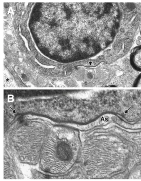
(5) J Neuroimmune Pharmacol (2012) 7:499–518. spread of damage. In contrast, some reports indicated that mild depolarization with K+ (Granda et al. 1998; De PinaBenabou et al. 2001), glutamate/kainate (Enkvist and McCarthy 1994; Robe et al. 2000) or TGF-β (Robe et al. 2000) enhances GJC. Therefore, the functional state of GJs is altered during inflammation or by neuronal activity depending on the intensity and nature of the damage. Neurons. Neurons express mainly Cx36, 30.2 and 45 and other Cxs with limited expression and localization (Rouach et al. 2002c; Sohl et al. 2005). Most neurons express Cx36, and Cx30.2 is mainly detected in interneurons in the retina and hippocampus (Kreuzberg et al. 2008; Muller et al. 2010). Our experiments using human fetal neurons obtained from the cortex and hippocampus indicated that these neurons also express Cx43. Additionally, Cx45 has been detected in neurons in the retina and the hippocampus (Schubert et al. 2005; Li et al. 2008; Zlomuzica et al. 2010; Blankenship et al. 2011). Interestingly, the function of these specific Cxs can be compensated in mouse KO models suggesting that their function can be replaced by other Cxs regardless of the differences in sequences and pore permeability (Frank et al. 2010). Despite the fact that extensive research on the Cxs in neurons has been performed, only a few examples of functional GJs were detected in vivo. Some of the best examples were demonstrated in GABAergic neurons in the striatum (Venance et al. 2004), neonatal/developmental cortex neurons (Peinado et al. 1993a, b; Bittman et al. 2002), motor neurons in the spinal cord (Chang et al. 1999; Chang and Balice-Gordon 2000), neurons of the inferior olivary nucleus (Benardo and Foster 1986; Devor and Yarom 2002a, b, c), interneurons in CA3 (Condorelli et al. 1998; Condorelli et al. 2003), visual cortex and dentate regions (Venance et al. 2000; Hormuzdi et al. 2001), in cortex fast-spiking interneurons (Galarreta and Hestrin 1999; Gibson et al. 1999), in the cerebellum (Mann-Metzer and Yarom 1999) and in almost all types of cells in the retina (Vaney 2002; Sohl et al. 2005). It is known that Cx36 GJs in the hippocampus are required for normal spatial coding and short term spatial memory between interneurons (Allen et al. 2011) and participate in neuronal remodeling by altering differentiation of neuronal stem cells (Hartfield et al. 2011). The proposed functions of these channels in all these systems are to coordinate neuronal firing, spike frequency modulation (Moortgat et al. 2000), fast oscillations (Friedman and Strowbridge 2003; Migliore et al. 2005), neuronal remodeling, and other synchronization properties required under physiologic conditions. Oligodendrocytes Oligodendrocytes express Cx29, Cx32, Cx31.3, Cx45 and Cx47 (Kunzelmann et al. 1997; Nagy and Rash 2000; Nagy et al. 2003a; Kleopa et al. 2004;. 503. Kamasawa et al. 2005; Rash et al. 2005; Ahn et al. 2008; Orthmann-Murphy et al. 2008; Sargiannidou et al. 2008; Sargiannidou et al. 2009; Maglione et al. 2010; Parenti et al. 2010; Magnotti et al. 2011). Cx29 is present along cell processes, especially in the juxtaparanodal region, but does not colocalize with Cx32 (Altevogt et al. 2002; Menichella et al. 2003; Nagy et al. 2003a; Meier et al. 2004). Cx32 is expressed in paranodal loops, Schmidt-Lanterman incisures and between the outer two layers of internodal myelin, between compact and uncompact myelin (Meier et al. 2004; Kamasawa et al. 2005). Cx47 colocalizes with Cx32 in GJ plaques (Menichella et al. 2003). Cx45 expression in oligodendrocytes is controversial (Dermietzel et al. 1997; Pastor et al. 1998; Maxeiner et al. 2003) and appears to be associated with the cerebral vasculature (Kleopa et al. 2004), most likely with smooth muscle cells close to vessels (Li and Simard 2001). It has also been proposed that oligodendrocytes form GJs with astrocytes (Butt and Ransom 1989; Menichella et al. 2003; Orthmann-Murphy et al. 2008), and perhaps with axons, although this has only been shown between axons and Schwann cells in sciatic nerve using electron microscopy (Dezawa and Nagano 1996). Our data clearly indicated that astrocytes oligodendrocyte interaction have a unique membrane specialization, such as desmosomes and GJ plaques that likely coordinate heterocellular signaling (see Fig. 3). These GJ structures are particularly dense in EM, due to a more intense accumulation of glial filaments (Soffer and Raine 1980). Desmosomes and GJs are common between glial processes or between astrocytes and oligodendrocytes (Fig. 3). In these cases, GJ have also been proposed to reduce high extracellular concentrations of K+ by providing a mechanism of lateral diffusion and dispersion. Thus, alterations in GJ channels as well as desmosomes may contribute to the pathology observed in several demyelinating diseases. In animal models, Cx32 deletion resulted in abnormalities in the sciatic nerve, associated with degeneration (Nelles et al. 1996; Anzini et al. 1997). However, Cx43/Cx47 compensatory mechanisms in the absence of Cx32 have been described (Nagy et al. 2003a). It also remains to be determined whether alterations in Cx32 or other oligodendrocytic Cxs impact different regions of the myelin sheath, such as uncompacted, compacted and/or internodal areas. Cx32 and Cx47 KO mice showed major abnormalities in myelin formation, such as hypomyelination and axonal loss, and oligodendrocyte survival (Odermatt et al. 2003). Similar myelin problems were found in human diseases, such as multiple sclerosis and Pelizaeus-Merzbacher disease (Martini 2000; Kleopa and Scherer 2002; Kleopa et al. 2004) (see section about MS). Experiments in the Cx47 mouse indicated that a missense mutation in the Cx47 gene causes PelizaeusMerzbacher disease and results in a pathologic phenotype in this animal model, suggesting that Cx47 significantly.
(6) 504. J Neuroimmune Pharmacol (2012) 7:499–518. A. B. Fig. 3 Electron microscopy in normal mice CNS sections between astrocytes and oligodendrocytes. a An oligodendrocyte in the optical nerve lies near the apical surface (*). Note the gap junctional complex along its lower surface (arrow). x12,500. b Details of the gap junctional plaque in A, between the oligodendrocyte (above), and an astrocyte process (As). The gap junction plaque is unusually long and is flanked by desmosome-like contacts (arrows). Adjacent astrocyte processes are rich in glial filaments and a small desmosome can be seen (below). x62,000. This is an extraordinary demonstration of large gap junctions between astrocytes and oligodendrocytes that support the concept that oligodendricyte and astrocyte communication is active and under pathological conditions, alterations in communication could result in oligodendrocyte damage. participates in the pathogenesis of this disease (Tress et al. 2011). The proposed role for GJs in oligodendrocyte physiology is to provide a shortcut for nutrients and second messengers across the different myelin layers to the axon, and therefore alterations in GJs might contribute to the pathogenesis of myelin/axonal diseases. Microglia Microglia express low to undetectable levels of Cx43 and Cx36 under resting conditions (Eugenín et al. 2001; Parenti et al. 2002; Dobrenis et al. 2005; Garg et al. 2005; Lee et al. 2005). Expression of Cx43 and formation of GJ channels can be induced by treatment of rat/mouse microglia with LPS or TNF-α plus IFN-γ (Eugenín et al. 2001), calcium ionophore plus PMA (Martínez et al. 2002), or Staphylococcus aureus-derived peptidoglycan (Garg et al. 2005). Cx36 expression in microglia has been shown using immunohistochemistry and RT-PCR under resting conditions (Parenti et al. 2002). Cxs in microglia may enable these cells to establish direct communication with other cells in the CNS to increase inflammation or to promote the repair of damaged tissue. Interestingly, activation of. microglia can down-regulate Cx43 expression and GJ channels among astrocytes when both cell types are in co-culture suggesting a cell-specific control of Cx43 expression (Rouach et al. 2002b; Faustmann et al. 2003; Meme et al. 2006). Findings in dendritic cells indicated that GJC could be used for the sharing of antigenic peptides (Neijssen et al. 2005; Matsue et al. 2006; Corvalan et al. 2007; Handel et al. 2007; Mendoza-Naranjo et al. 2007; Pang et al. 2009), suggesting the possibility that GJs between microglia also coordinate the CNS immune response. Our data using cultures of rat microglia showed that under control conditions, the release of TNF-α, IL-1β and IL-6 was minimal (Supplemental Fig. 2a and b, white bars). Treatment of microglia cultures with LPS plus IFN-γ for 1 to 9 h, a condition that increased GJC, increased secretion of TNFα and IL-1β (Supplemental Fig. 2A), but not IL-6 (Supplemental Fig. 2B, cross line bands). The secretion of these cytokines was partially blocked by a GJ blocker, AGA (Supplemental Fig. 2A, cross line bars), suggesting that functional GJs are important in microglia cytokine secretion. Thus, GJs in microglia are induced by specific inflammatory factors and the proposed function of these channels is to help to coordinate the microglial mediated inflammation. Blood–brain barrier (BBB) The BBB is composed of dynamic vessels that are capable of responding to rapid changes in the brain or in the blood stream (Gloor et al. 2001; Ballabh et al. 2004). The BBB is composed of endothelial cells (EC) in close contact with astrocytic end-feet across a basal lamina, perivascular macrophages and pericytes. These cellular interactions function in combination with other systems specialized for the transport of metabolites required for brain function, as well as tight junction proteins (TJP), which seal the intercellular gaps between EC-EC and EC-astrocytes, to establish impermeability to most macromolecules and blood cells (Ballabh et al. 2004). EC isolated from blood vessels of different organs express Cxs 37, 40 and 43 (Haefliger et al. 2004; Burnier et al. 2009). Cx45 has been detected in cerebral blood vessels but its expression is associated with smooth muscle cells (Li and Simard 2001). However, deletion of Cx45 results in defective vascular development, suggesting a critical role of Cx45 in the development of brain vasculature (Kruger et al. 2000). In the systemic vasculature, deletion of Cx40 results in hypertension (Firouzi et al. 2006a; Firouzi et al. 2006b; Hauer et al. 2006). Endothelial-specific deletion of Cx43 results in hypotension and bradycardia (Liao et al. 2001), suggesting that expression of these Cxs participates in the regulation of blood pressure. In agreement, the control of blood flow by a GJ-dependent mechanism has been described in peripheral arterioles (de Wit et al. 2000; de Wit et al. 2003). Our data demonstrated that HIV infection of just a few astrocytes in a BBB tissue.
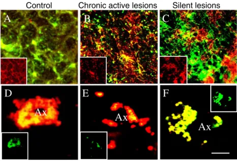
(7) J Neuroimmune Pharmacol (2012) 7:499–518. culture model amplifies endothelial apoptosis by dysregulation of astrocyte end-feet signaling in a GJ-dependent manner (Eugenín et al. 2011). However, little is known about the expression of Cxs and their role in the physiology and pathogenesis of the BBB under normal and pathologic conditions. Neuronal stem cells Neuronal precursors express Cx26, Cx30, Cx33, Cx36, Cx37, Cx40, Cx43 and Cx47 depending on the area of the brain from which these cells were isolated, the differentiation stage and the cell culture conditions (Rozental et al. 1998; Duval et al. 2002; Maxeiner et al. 2003; Trosko and Chang 2003; Elias et al. 2007; Elias and Kriegstein 2008; Wen et al. 2008; Cina et al. 2009). Expression of Cxs 30, 36, 37 and 43, but not Cxs 26, 32 or 47, has been reported in NT2/D1 progenitor cells. These cells were obtained from a teratocarcinoma progenitor line that can be induced to differentiate into hNT neurons and NT-G non-neuronal cells (Bani-Yaghoub et al. 1999). hNT/ NT-G cells differentiated with retinoic acid, express Cxs 36, 37 and 47. However, only undifferentiated cells are capable of dye transfer to other cells (Boucher and Bennett 2003). Functional GJ are required to maintain cortical neural progenitor cells in a proliferative state (Cheng et al. 2004) as well as for their radial migration in the neocortex (Elias et al. 2007). It has also been described that the carboxyl-terminal domain of Cx43 regulates neuronal differentiation (Cina et al. 2009; Santiago et al. 2010). In addition, Panx-2 uHC are expressed in postnatal hippocampus neuronal progenitors and also modulate the differentiation of neurons (Swayne et al. 2010). Thus, it is clear that GJs and uHC are critical for multiple functions involving neuronal differentiation and migration. However, the question whether GJ and uHC are involved in diseases involved in migration and differentiation is still under investigation. Gap junctions and connexin expression in human diseases Cx32 mutations linked to the X-linked hereditary motor and sensory neuropathy Charcot-Marie-Tooth (CMTX) More than 305 different Cx32 mutations have been associated with CMTX (see for details; http://www.molgen.ua.ac.be/ CMTMutations/), but only a few results in evident pathologic conditions (Kleopa et al. 2002). One explanation is that not all Cx32 mutations destabilize the myelin sheath and/or compromise communication in a significant manner. An alternative or additional explanation is that other Cxs expressed in oligodendrocytes or Schwann cells can be compensatory. Little is known about the functions of Cx32 in the PNS and CNS, due to difficulties in evaluating the function(s) of these channels in vivo, or during the pathogenesis of CMTX disease (Bergoffen et al. 1993; Ionasescu et al. 1996; Ionasescu 1998). Cx32-deficient mice did not show any evident oligodendrocyte-dependent myelination. 505. (Scherer et al. 1998). Neurons isolated from Cx32deficient mice did not display impaired excitability and synaptic formation/stability (Sutor et al. 2000). However, Cx32/Cx47 KO mice exhibited motor problems and a myelin defect (vacuolization) when compared to single Cx47 KO or Cx32 KO mice (Odermatt et al. 2003). This suggests that deficiencies in both Cxs are required to result in a pathologic phenotype. Cx32 is also important in mediating GJ communication between oligodendrocytes and astrocytes or neurons, and these interactions may be altered in CMTX (Angaut and Sotelo 1973; Sotelo and Angaut 1973; Kettenmann et al. 1983; Butt and Ransom 1989; Menichella et al. 2003; Orthmann-Murphy et al. 2008). Cxs and multiple sclerosis Multiple sclerosis (MS) is a primary demyelinating disease in humans. As described above, Cx32 plays a key role in maintaining the structure and function of the myelin sheath. Deletions or mutations in this gene in rodents and humans can result in myelin abnormalities, similar to those characterized in MS. In the EAE, an animal model of MS, Cx43 expression was downregulated and Cx30 upregulated in sites in close association with inflammation (Brand-Schieber et al. 2005). Our data using human tissue sections indicated that in control non inflammatory conditions, colocalization between myelin basic protein (MBP) and Cx32 was observed (Fig. 4). Tissue sections obtained from individuals with chronic active and silent MS lesions demonstrated that Cx32 is down regulated in oligodendrocytes in chronic active lesions at sites of inflammation (Fig. 4). In contrast, upregulation of Cx32 was observed in silent lesions from individuals with chronic silent MS, characterized by the absence of inflammation and re-myelination (Fig. 4). In active MS lesions, Cx32 staining was reduced (Fig. 4). Cx43, which is mainly expressed in astrocytes, was upregulated in areas around the lesion, suggesting that scar tissue has high expression of Cx43, most likely due to astrocyte proliferation (Fig. 4). However, astrocytes in the center of the lesions were negative for Cx43 in active chronic lesions (Fig. 4). Thus, our data indicated that Cx32 expression is associated with the degree of damage and remyelination, while Cx43 is associated with astrocyte activation/proliferation as well as inflammation. Future studies will be essential to understand the role of other Cxs expressed in oligodendrocytes, Schwann cells, astrocytes and neurons during the pathogenesis of demyelinating diseases. HIV and NeuroAIDS HIV infection of the CNS induces different degrees of cognitive and motor impairment, collectively termed HIV associated neurologic disorders (HAND). Approximately 60 % of individuals infected with HIV have HAND even in the current anti-retroviral era (González-Scarano and Martín-García 2005; Boisse et al..
(8) 506. J Neuroimmune Pharmacol (2012) 7:499–518. Control. Chronic active lesions. Silent lesions. A. B. C. D. E. F. Ax. Ax. Ax. Fig. 4 Distribution of Cx43 and Cx32 in human spinal cord sections obtained from normal individuals and individuals with MS. Confocal microscopy of Cx43 (FITC, green) and MBP (Cy3, red) staining, colocalization of both proteins is represented as orange staining in the last panel. A, B and C, represents staining of human tissue sections for GFAP (FITC, green) and Cx43 (Cy3, red), a small insert shows the Cx43 staining alone, from normal (a), MS with chronic active lesions (b) and chronic silent lesions (c). d, e and f, represents staining for. Cx32 (FITC, Green) and MBP (Cy3, red) in human sections obtained from spinal cords from individuals with normal tissue (d), MS with chronic active lesions (e) and silent lesions (f). The small inserts in each picture show the Cx32 staining alone. These tissue sections were already characterized for the kind of MS lesions and the damage in the lesion area (Calderon et al. 2006). Note that in MS tissue it is possible observe oligodendrocyte atrophy and disorganization of the brain parenchyma. Bar: 70 μm. 2008; Kraft-Terry et al. 2009). However, the mechanisms that mediate HAND in most HIV infected individuals are not completely understood. The neuropathology of HIVinfection includes microglial nodules, multinucleated giant cells and astrogliosis, as well as neuronal injury and loss (Albright et al. 1999; Kaul et al. 2001; González-Scarano and Martín-García 2005). Macrophages and microglia support high viral replication within the CNS (GonzálezScarano and Martín-García 2005). HIV-infected astrocytes have also been detected in vivo and in vitro (Tontsch and Bauer 1991; Tornatore et al. 1991; Conant et al. 1994; Tornatore et al. 1994a; Tornatore et al. 1994b; Bagasra et al. 1996; Ohagen et al. 1999; Gorry et al. 2003; Churchill et al. 2006; Eugenín and Berman 2007; Churchill et al. 2009; Eugenín et al. 2011) and was characterized by low to undetectable viral replication and a low numbers of cells that are infected (Tornatore et al. 1994a; Ohagen et al. 1999; Schweighardt and Atwood 2001; Eugenín and Berman 2007; Eugenín et al. 2011). In general, inflammation and infectious agents reduce Cx43 expression and GJC (see details below). However, HIV is different because despite its inflammatory nature, Cx43 expression and GJ channels are maintained in astrocytes (Eugenín and Berman 2007). Functional GJ channels promote the spread of toxic signals from a few HIV-infected astrocytes to uninfected astrocytes, neurons and endothelial cells resulting in the spread of toxic mediators and dysregulation of glutamate and CCL2 secretion (Eugenín and Berman 2007; Eugenín et al. 2011). Interestingly, the few HIV. infected astrocytes are protected from apoptosis by a viraldependent mechanism, resulting in a viral reservoir within the CNS to perpetuate the presence of the virus. Despite extensive evidence of pathological changes in the CNS of HIV-infected people, the role of GJs has been minimally examined. An accepted mechanism by which cognitive impairment and dementia occurs involves the transmigration of HIV-infected monocytes across the BBB into the CNS parenchyma and the accumulation of macrophages and microglia within the CNS in correlation with several inflammatory factors (Persidsky et al. 1997; Weiss et al. 1998; Eugenín et al. 2006; Roberts et al. 2010). Normally, macrophages/microglia express low to undetectable levels of Cxs, however, we and others demonstrated that macrophages/microglia express higher levels of Cxs under inflammatory conditions (Eugenín et al. 2001; Martínez et al. 2002; Parenti et al. 2002; Dobrenis et al. 2005; Garg et al. 2005). In Supplemental Fig. 3, our data demonstrate that during HIV associated dementia (HAD) or HIV encephalitis (HIVE), alterations in GJs occur in HIVinfected leukocytes, microglia and astrocytes. HIV-infected leukocytes after HIV infection begin to express high levels of Cx43 (Supplemental Fig. 3A) that may be necessary for transmigration across the BBB, as we previously described in uninfected peripheral blood mononuclear cells (Eugenín et al. 2003). In addition, confocal analyses of HIVE human tissue demonstrated that microglia/macrophages express Cx43 (Supplemental Fig. 3B) and Cx36 (data not shown).
(9) J Neuroimmune Pharmacol (2012) 7:499–518. in close contact with neuronal cell bodies (Supplemental Fig. 3B). An example is shown in Supplemental Fig. 3, showing Cx43 expression between a neuron and microglia/macrophage (CD68 positive cells) in HIVE tissue. We did not detect either Cx in microglia/macrophages in brain tissue sections obtained from normal or non-encephalitic HIV positive individuals (data not shown). In addition, Cx43 expressed in astrocytes and Cx36 in neurons were down-regulated in HIVE tissue sections as compared to cells in tissue sections obtained from uninfected brains. In addition, our findings indicated that uHC are opened in response to HIV infection in astrocytes, suggesting their participation in the pathogenesis of HIV CNS disease. These findings that Cxs participate in the pathogenesis of NeuroAIDS open a new avenue of investigation to study the mechanisms by which HIV “hijacks” this communication system, GJs and perhaps uHCs, to spread toxicity, inflammation, and increase leukocyte transmigration into the CNS. Viral and bacterial infections In general, both viral and bacterial infections reduce GJ channels and Cx expression. For example, swine Flu virus down-regulates endothelial Cx43 expression by an ERK and increased degradation dependent mechanism (Hsiao et al. 2010). Borna virus also down regulates Cx36 in the CNS in specific brain cell types (Koster-Patzlaff et al. 2007, 2008, 2009). Influenza viral infection during pregnancy alters development of the brain of the fetus, suggesting that viruses can impact neuronal development by affecting GJs (Fatemi et al. 2008) required for development and function of the CNS. Studies in Vero cells demonstrated that infection with Herpes Simplex Virus-2 (HSV-2), down-regulated GJ channels and Cxs expression (Fischer et al. 2001; Musee et al. 2002; Knabb et al. 2007). It was reported that an increase in tyrosine phosphorylation by HSV-2 and Rous sarcoma virus leads to an inhibition of GJ channels and Cx43 expression (Crow et al. 1990; Filson et al. 1990; Crow et al. 1992). Bovine papillomavirus type 4 E8, when bound to ductin, causes loss of GJ channels between primary fibroblasts (Faccini et al. 1996; Ashrafi et al. 2000). In contrast, our preliminary data show that HIV infection of astrocytes is different because it maintains/increases Cx43, Cx30 and GJ channels allowing toxic signals generated in a few infected cells to spread through GJs to uninfected cells (Eugenín and Berman 2007; Eugenín et al. 2011). In addition, GJ channels increase invasion and dissemination of Shigella in epithelial cells (Tran Van Nhieu et al. 2003), suggesting the possibility that specific infectious agents, such as Shigella and HIV, can use GJ channels to sensitize uninfected cells and spread infection/toxicity to healthy cells. Future experiments are necessary to identify the signals generated by infected cells that cross through GJs to sensitize uninfected cells and enable them to become targets of these infectious agents.. 507. Alternatively, GJs benefit the host immune system by mediating a phenomenon termed cross-antigen presentation. This enables coupled cells to share viral peptides (antigens) and trigger a response in CTL cells, even when some cells were never directly exposed to the pathogen (Neijssen et al. 2005). GJ-mediated immune coupling suggests the possibility that GJs expressed by monocytes/macrophages in inflammatory conditions cross-present antigens to lymphocytes and other inflammatory cells to maintain an immune memory in cells never exposed directly to specific antigen (Neijssen et al. 2005). In agreement, Cx43 is recruited to the immunologic synapse during T cell priming, suggesting that GJ and uHCs also participate in antigen presentation (Mendoza-Naranjo et al. 2011). Alzheimer’s disease (AD) Aβ peptide is required for CNS function. However, under certain conditions, this peptide aggregates and produces toxic effects (Palop and Mucke 2010; Parihar and Brewer 2010). Up-regulation of Cx43 has been detected in cortical astrocytes in the brain containing Aβ peptide plaques as compared to normal brains (Nagy et al. 1996; Mei et al. 2010). Experiments using rat astrocytes demonstrated that β/A4 amyloid resulted in an increase in the amplitude, velocity and travel distance of evoked calcium waves, by an ATP- and GJC-dependent mechanism (Haughey and Mattson 2003). Recently, it was shown that Aβ peptide induces the release of ATP and glutamate through glial uHC, which leads to further neuronal death by activation of Panx-1 uHC in neurons (Orellana et al. 2011b). Additionally, expression of β/A4 amyloid in PC12 cells increased Cx43 expression and dye coupling (Lynn et al. 1995), suggesting that Aβ peptide increases GJC in CNS cells by an unknown mechanism. However, in all of these studies it is unclear whether normal regulation of Cx is a product of a physiologic or pathogenic effect of Aβ or due to the damage observed in the end stages of the disease. Thus, further examination of the role of Cxs and GJ during the pathogenesis of AD is required. Parkinson’s disease (PD) PD is a neurodegenerative disease characterized by loss of dopaminergic neurons, especially in the substantia nigra-striatum, that results in progressive tremor, and muscle and gait abnormalities (Lim et al. 2002; Lotharius and Brundin 2002). The mouse MTPT model of PD showed increased levels of Cx43 in the stratium (Rufer et al. 1996), but this may be related to gliosis and not be specific to PD. The dysfunction of astrocytic Cx43 induced by rotenone to trigger a PD phenotype, as a model of PD, can be reversed by opening mitochondrial ATPsensitive potassium channels, suggesting the involvement of these channels in the mitochondria regulated Cx43 expression (Kawasaki et al. 2009; Zhang et al. 2010)..
(10) 508. Interestingly, treatment of the Cx36 KO mouse with harmaline, a beta-carboline derivative, induced tremors by altering rhythmogenesis in a similar way to PD, but did not show any differences when compared to wild type mice, suggesting that Cx36 may not play a role in the initial pathogenesis of the disease (Long et al. 2002a). However, a compensatory mechanism should not be ruled out, as it has already been described in the Cx36 KO mice in coordinating rhythmic activity between the neurons of the inferior olive and suprachiasmatic nucleus (Long et al. 2002b, 2005), which is believed to be the region that generates the tremors (Elble 1996). In conclusion, there are only a few studies that characterize the expression, function and localization of Cxs in PD brains or animal models and the participation of these channels in the pathogenesis of PD is unclear. Huntington disease Huntington disease is an autosomal dominant neurodegenerative disorder characterized by motor dysfunctions, cognitive impairment and personality changes that is due to mutations in the protein huntingtin (htt). Studies using human tissue sections obtained from individuals with Huntington disease showed a similar distribution of Cx32 and Cx26 as compared to normal brain tissue sections, but Cx43 immunoreactivity was increased in the caudate nucleus, especially in regions richer in GFAP staining (Vis et al. 1998), perhaps due to gliosis. Studies in retina using a transgenic R6/2 mice expressing the mutant form of htt demonstrated decreased expression of Cx45 while Cx36 and Cx43 were not significantly altered (Petrasch-Parwez et al. 2004). Further studies are required to dissect whether these changes in Cx expression participate in the pathogenesis of the disease or are a consequence of the cellular damage characteristic of Huntington disease. Epilepsy Epileptic seizures may be associated with abnormal stimulation of certain brain areas causing abnormal depolarization, which expands by spreading to neighboring areas (Ure and Perassolo 2000; Ure et al. 2006). This process can be mediated by glutamate (Meldrum 1994; Moldrich et al. 2003), auto antibodies that activate AMPA receptors (GluR3) (Levite et al. 1999; Levite and Hermelin 1999; Palmer et al. 1999), and aberrant activation or deactivation of NMDA receptors, GABA receptors, potassium channels, sodium channels and GJs (Naus et al. 1991; Carlen et al. 2000; Samoilova et al. 2003; Brooks-Kayal et al. 2009; Kang and Macdonald 2009; Planells-Cases and Jentsch 2009; Galanopoulou 2010). Astrocytic GJ channels increase in epileptic tissue of cats as demonstrated by morphological studies (Vaquero et al. 1978) and an increase in Cx43 mRNA was found in human epileptic tissue (Naus et al. 1991), suggesting a potential role of Cx43 in the pathogenesis of this disease. In addition, increased GJ channels have been shown between astrocytes isolated from human. J Neuroimmune Pharmacol (2012) 7:499–518. epileptic tissue as assayed by the enhanced propagation of a calcium wave in response to glutamate (Lee et al. 1995). Based on these data, it has been proposed that Cxs are involved in expanding the spread of an epileptic wave during seizures (Perez Velazquez and Carlen 2000; Perez Velazquez et al. 2001). This hypothesis is supported by studies showing that GJ blockers reduced seizures in different models of epilepsy in vitro and in vivo (Sohl et al. 2000; Jahromi et al. 2002; Samoilova et al. 2003; Gareri et al. 2004; Gajda et al. 2005; Gareri et al. 2005; Bostanci and Bagirici 2006; Nilsen et al. 2006; Meldrum and Rogawski 2007; Jacobson et al. 2010). Studies using the Cx36 KO mouse showed that loss of GJ channels reduced kainate- or 4-aminopyridine-induced seizures (Hormuzdi et al. 2001; Maier et al. 2002; Christie et al. 2005), suggesting a key role of GJ channels in the development and sustaining of epileptic seizures. In human tissue, upregulation of Cx43 and Cx32 has been detected in different types of epilepsy (Elisevich et al. 1997; Aronica et al. 2001; Li et al. 2001; Fonseca et al. 2002; Samoilova et al. 2008; Yao et al. 2009). Additionally, Cx30, which is normally expressed in astrocytes, was detected in neurons after kainate-induced seizures (Condorelli et al. 2002). All these data suggest that de novo synthesis of Cxs causes changes in permeability and distribution of Cxs during epileptogenesis. While these data in humans are consistent, results in animal models are more variable and difficult to interpret. This may be due to the use of different experimental approaches to generate the epileptic phenotype and the methods to examine the disease that may or may not be good models for epilepsy in humans resulting in different patterns of Cxs expression and functions of GJ channels. Thus, design of better animal models and studies in humans are required to compare both systems. CNS tumors Gliomas are the most common tumor in the brain (Behin et al. 2003), characterized by high intracranial pressure, brain edema and vessel occlusion (Behin et al. 2003). Cx expression and GJ channels are low in tumors, primary glioma cells or glioma cell lines (Huang et al. 1999; Zhang et al. 1999; Soroceanu et al. 2001; Lin et al. 2002; Pu et al. 2004; Oliveira et al. 2005; Kubota et al. 2006; Bates et al. 2007; Lai et al. 2007). Tumor cells can directly couple with normal cells through GJs (Zhang et al. 1999). This communication results in a phenotypic transformation of astrocytes that may contribute to the susceptibility of surrounding tissue to glioma invasion. There are reports indicating a beneficial role of Cxs in glioma treatment. Cx43 expression by itself is considered to be a tumor suppressor gene independent of formation of functional GJs (Huang et al. 1998; Yamasaki et al. 1999; Moorby and Patel 2001; Omori et al. 2001; Zhang et al. 2003a, b, c; Del Monte and Statuto 2004). In addition, transfection of Cx43 and herpes.
(11) J Neuroimmune Pharmacol (2012) 7:499–518. simplex virus thymidine kinase (HSVtk) resulted in bystander cell death in a Cx43 dependent manner upon treatment with ganciclovir (GCV), a nucleoside analogue (Shinoura et al. 1996; Cirenei et al. 1998; Grignet-Debrus et al. 2000; Marconi et al. 2000; Namba et al. 2001; Asklund et al. 2003; Huang et al. 2010). GVC is phosphorylated by HSVtk into a monophosphate form and subsequently to GCV-triphosphate by endogenous kinases, and is incorporated into the DNA of the target cell, leading to strand breaks and cell death. Interestingly, neighboring cells coupled by GJs also die, although these cells do not express the enzyme. This phenomenon is believed to be a bystander effect mediated by the transfer of toxic GVC-metabolites through GJs from the cell infected with HSVtk to uninfected neighboring cells (Dilber et al. 1997). Cx43 transfection into tumor cells results in functional coupling and in enhancement of the bystander effect in vivo (Mesnil et al. 1996; Cirenei et al. 1998; AndradeRozental et al. 2000) and in vitro (Mesnil et al. 1996; Cirenei et al. 1998; Andrade-Rozental et al. 2000; Marconi et al. 2000; Musee et al. 2002; Asklund et al. 2003; Huang et al. 2010). Currently, several groups continue to examine the potential role of this bystander effect in the treatment of different types of tumors. The discovery that glioma cells can express functional uHC also increases the possibility that during pathologic conditions, these channels contribute to the regulation of proliferation or in protecting tumor cells from damage induced by GVC or chemotherapy by pumping out toxic metabolites in similar ways to ABC transporters (Andrade-Rozental et al. 2000). In addition, future studies investigating the generation of other intracellular toxic metabolites, such as cytochrome C, which has been shown to cause bystander killing in the retina, could be beneficial.. Conclusions This review summarizes much of the current data on the role of Cxs in different cell types and their involvement in human central nervous system diseases. A more complete understanding of the role of Cxs in different human diseases require a more detailed study of the function of connexinand pannexin-based cell-cell channels, GJs and uHC, under normal and pathologic conditions. A critical point is to examine, in addition to the role of GJs and uHC in physiologic conditions, the role of these channels during the pathogenesis of human disease. Recent work indicated that GJs and uHC are key channels involved in resolving, but also in spreading, disease. Our study with HIV indicates that some pathogens use these communication systems to their advantage. Thus, a more complete understanding of these communication systems should provide essential information for the development of novel therapeutic approaches. 509 Acknowledgments We apologize to the authors and groups whose work we did not cite due to limitations on the numbers of references. This work was supported by the National Institute of Mental Health grants (MH076679 and MH096625 to E.A.E. and MH075679 and MH083497 to J.W.B.) and by the National Institute of Neurological Disorders and Stroke (NS072238 to FFB, and NS55363 to M.V.L.B, who is the Sylvia and Robert S. Olnick Professor of Neuroscience). We thank the National Multiple Sclerosis Society Grant RG-1001-K-11 and CSR was the Wollowick Family Foundation Professor for Multiple Sclerosis Research (to Dr. Cedric Raine). We thank the NIH Centers for AIDS Research Grant (CFAR) AI-051519, Anillo ATC-71 (JCS) and Centro interdisciplinario de Neurociencias P09-022-F (to JCS) and a CFAR pilot project at the Albert Einstein College of Medicine. Conflict of interest None. References Abrams CK, Bennett MV, Verselis VK, Bargiello TA (2002) Voltage opens unopposed gap junction hemichannels formed by a connexin 32 mutant associated with X-linked Charcot-Marie-Tooth disease. Proc Natl Acad Sci U S A 99:3980–3984 Ahn M, Lee J, Gustafsson A, Enriquez A, Lancaster E, Sul JY, Haydon PG, Paul DL, Huang Y, Abrams CK, Scherer SS (2008) Cx29 and Cx32, two connexins expressed by myelinating glia, do not interact and are functionally distinct. J Neurosci Res 86:992–1006 Albright AV, Shieh JT, Itoh T, Lee B, Pleasure D, O’Connor MJ, Doms RW, Gonzalez-Scarano F (1999) Microglia express CCR5, CXCR4, and CCR3, but of these, CCR5 is the principal coreceptor for human immunodeficiency virus type 1 dementia isolates. J Virol 73:205–213 Allen K, Fuchs EC, Jaschonek H, Bannerman DM, Monyer H (2011) Gap junctions between interneurons are required for normal spatial coding in the hippocampus and short-term spatial memory. J Neurosci Off J Soc Neurosci 31:6542–6552 Altevogt BM, Kleopa KA, Postma FR, Scherer SS, Paul DL (2002) Connexin29 is uniquely distributed within myelinating glial cells of the central and peripheral nervous systems. J Neurosci 22:6458–6470 Andrade-Rozental AF, Rozental R, Hopperstad MG, Wu JK, Vrionis FD, Spray DC (2000) Gap junctions: the “kiss of death” and the “kiss of life”. Brain Res Brain Res Rev 32:308–315 Angaut P, Sotelo C (1973) The fine structure of the cerebellar central nuclei in the cat. II. Synaptic organization. Exp Brain Res 16:431– 454 Anzini P, Neuberg DH, Schachner M, Nelles E, Willecke K, Zielasek J, Toyka KV, Suter U, Martini R (1997) Structural abnormalities and deficient maintenance of peripheral nerve myelin in mice lacking the gap junction protein connexin 32. J Neurosci 17:4545–4551 Aronica E, Gorter JA, Jansen GH, Leenstra S, Yankaya B, Troost D (2001) Expression of connexin 43 and connexin 32 gap-junction proteins in epilepsy-associated brain tumors and in the perilesional epileptic cortex. Acta Neuropathol 101:449–459 Ashrafi GH, Pitts JD, Faccini A, McLean P, O’Brien V, Finbow ME, Campo S (2000) Binding of bovine papillomavirus type 4 E8 to ductin (16 K proteolipid), down-regulation of gap junction intercellular communication and full cell transformation are independent events. J Gen Virol 81:689–694 Asklund T, Appelskog IB, Ammerpohl O, Langmoen IA, Dilber MS, Aints A, Ekstrom TJ, Almqvist PM (2003) Gap junction-mediated bystander effect in primary cultures of human malignant gliomas with recombinant expression of the HSVtk gene. Exp Cell Res 284:185–195 Bagasra O, Lavi E, Bobroski L, Khalili K, Pestaner JP, Tawadros R, Pomerantz RJ (1996) Cellular reservoirs of HIV-1 in the central.
(12) 510 nervous system of infected individuals: identification by the combination of in situ polymerase chain reaction and immunohistochemistry. AIDS 10:573–585 Ballabh P, Braun A, Nedergaard M (2004) The blood–brain barrier: an overview: structure, regulation, and clinical implications. Neurobiol Dis 16:1–13 Bani-Yaghoub M, Felker JM, Naus CC (1999) Human NT2/D1 cells differentiate into functional astrocytes. Neuroreport 10:3843–3846 Bates DC, Sin WC, Aftab Q, Naus CC (2007) Connexin43 enhances glioma invasion by a mechanism involving the carboxy terminus. Glia 55:1554–1564 Behin A, Hoang-Xuan K, Carpentier AF, Delattre JY (2003) Primary brain tumours in adults. Lancet 361:323–331 Benardo LS, Foster RE (1986) Oscillatory behavior in inferior olive neurons: mechanism, modulation, cell aggregates. Brain Res Bull 17:773–784 Bennett MV, Contreras JE, Bukauskas FF, Saez JC (2003) New roles for astrocytes: gap junction hemichannels have something to communicate. Trends Neurosci 26:610–617 Bergoffen J, Scherer SS, Wang S, Scott MO, Bone LJ, Paul DL, Chen K, Lensch MW, Chance PF, Fischbeck KH (1993) Connexin mutations in X-linked Charcot-Marie-Tooth disease. Science 262:2039–2042 Bittman K, Becker DL, Cicirata F, Parnavelas JG (2002) Connexin expression in homotypic and heterotypic cell coupling in the developing cerebral cortex. J Comp Neurol 443:201–212 Blanc EM, Bruce-Keller AJ, Mattson MP (1998) Astrocytic gap junctional communication decreases neuronal vulnerability to oxidative stress-induced disruption of Ca2+ homeostasis and cell death. J Neurochem 70:958–970 Blankenship AG, Hamby AM, Firl A, Vyas S, Maxeiner S, Willecke K, Feller MB (2011) The role of neuronal connexins 36 and 45 in shaping spontaneous firing patterns in the developing retina. J Neurosci Off J Soc Neurosci 31:9998–10008 Boisse L, Gill MJ, Power C (2008) HIV infection of the central nervous system: clinical features and neuropathogenesis. Neurol Clin 26:799–819, x Bolanos JP, Medina JM (1996) Induction of nitric oxide synthase inhibits gap junction permeability in cultured rat astrocytes. J Neurochem 66:2091–2099 Bostanci MO, Bagirici F (2006) The effects of octanol on penicillin induced epileptiform activity in rats: an in vivo study. Epilepsy Res 71:188–194 Boucher S, Bennett SA (2003) Differential connexin expression, gap junction intercellular coupling, and hemichannel formation in NT2/D1 human neural progenitors and terminally differentiated hNT neurons. J Neurosci Res 72:393–404 Brand-Schieber E, Werner P, Iacobas DA, Iacobas S, Beelitz M, Lowery SL, Spray DC, Scemes E (2005) Connexin43, the major gap junction protein of astrocytes, is down-regulated in inflamed white matter in an animal model of multiple sclerosis. J Neurosci Res 80:798–808 Brooks-Kayal AR, Raol YH, Russek SJ (2009) Alteration of epileptogenesis genes. Neurotherapeutics 6:312–318 Bruzzone R, Hormuzdi SG, Barbe MT, Herb A, Monyer H (2003) Pannexins, a family of gap junction proteins expressed in brain. Proc Natl Acad Sci U S A 100:13644–13649 Burnier L, Fontana P, Angelillo-Scherrer A, Kwak BR (2009) Intercellular communication in atherosclerosis. Physiology (Bethesda) 24:36–44 Butt AM, Ransom BR (1989) Visualization of oligodendrocytes and astrocytes in the intact rat optic nerve by intracellular injection of lucifer yellow and horseradish peroxidase. Glia 2:470–475 Calderon TM, Eugenin EA, Lopez L, Kumar SS, Hesselgesser J, Raine CS, Berman JW (2006) A role for CXCL12 (SDF-1alpha) in the. J Neuroimmune Pharmacol (2012) 7:499–518 pathogenesis of multiple sclerosis: regulation of CXCL12 expression in astrocytes by soluble myelin basic protein. J Neuroimmunol 177:27–39 Carlen PL, Skinner F, Zhang L, Naus C, Kushnir M, Perez Velazquez JL (2000) The role of gap junctions in seizures. Brain Res Brain Res Rev 32:235–241 Chang Q, Balice-Gordon RJ (2000) Gap junctional communication among developing and injured motor neurons. Brain Res Brain Res Rev 32:242–249 Chang Q, Gonzalez M, Pinter MJ, Balice-Gordon RJ (1999) Gap junctional coupling and patterns of connexin expression among neonatal rat lumbar spinal motor neurons. J Neurosci 19:10813–10828 Chang Q, Tang W, Ahmad S, Zhou B, Lin X (2008) Gap junction mediated intercellular metabolite transfer in the cochlea is compromised in connexin30 null mice. PLoS One 3:e4088 Cheng A, Tang H, Cai J, Zhu M, Zhang X, Rao M, Mattson MP (2004) Gap junctional communication is required to maintain mouse cortical neural progenitor cells in a proliferative state. Dev Biol 272:203–216 Christie JM, Bark C, Hormuzdi SG, Helbig I, Monyer H, Westbrook GL (2005) Connexin36 mediates spike synchrony in olfactory bulb glomeruli. Neuron 46:761–772 Churchill MJ, Gorry PR, Cowley D, Lal L, Sonza S, Purcell DF, Thompson KA, Gabuzda D, McArthur JC, Pardo CA, Wesselingh SL (2006) Use of laser capture microdissection to detect integrated HIV-1 DNA in macrophages and astrocytes from autopsy brain tissues. J Neurovirol 12:146–152 Churchill MJ, Wesselingh SL, Cowley D, Pardo CA, McArthur JC, Brew BJ, Gorry PR (2009) Extensive astrocyte infection is prominent in human immunodeficiency virus-associated dementia. Ann Neurol 66:253–258 Cina C, Maass K, Theis M, Willecke K, Bechberger JF, Naus CC (2009) Involvement of the cytoplasmic C-terminal domain of connexin43 in neuronal migration. J Neurosci 29:2009–2021 Cirenei N, Colombo BM, Mesnil M, Benedetti S, Yamasaki H, Finocchiaro G (1998) In vitro and in vivo effects of retrovirusmediated transfer of the connexin 43 gene in malignant gliomas: consequences for HSVtk/GCV anticancer gene therapy. Gene Ther 5:1221–1226 Cohen-Salmon M, Regnault B, Cayet N, Caille D, Demuth K, Hardelin JP, Janel N, Meda P, Petit C (2007) Connexin30 deficiency causes instrastrial fluid-blood barrier disruption within the cochlear stria vascularis. Proc Natl Acad Sci U S A 104:6229–6234 Conant K, Tornatore C, Atwood W, Meyers K, Traub R, Major EO (1994) In vivo and in vitro infection of the astrocyte by HIV-1. Adv Neuroimmunol 4:287–289 Condorelli DF, Parenti R, Spinella F, Trovato Salinaro A, Belluardo N, Cardile V, Cicirata F (1998) Cloning of a new gap junction gene (Cx36) highly expressed in mammalian brain neurons. Eur J Neurosci 10:1202–1208 Condorelli DF, Mudo G, Trovato-Salinaro A, Mirone MB, Amato G, Belluardo N (2002) Connexin-30 mRNA is up-regulated in astrocytes and expressed in apoptotic neuronal cells of rat brain following kainate-induced seizures. Mol Cell Neurosci 21:94–113 Condorelli DF, Trovato-Salinaro A, Mudo G, Mirone MB, Belluardo N (2003) Cellular expression of connexins in the rat brain: neuronal localization, effects of kainate-induced seizures and expression in apoptotic neuronal cells. Eur J Neurosci 18:1807–1827 Contreras JE, Sanchez HA, Eugenin EA, Speidel D, Theis M, Willecke K, Bukauskas FF, Bennett MV, Saez JC (2002) Metabolic inhibition induces opening of unapposed connexin 43 gap junction hemichannels and reduces gap junctional communication in cortical astrocytes in culture. Proc Natl Acad Sci U S A 99:495–500 Corvalan LA, Araya R, Branes MC, Saez PJ, Kalergis AM, Tobar JA, Theis M, Willecke K, Saez JC (2007) Injury of skeletal muscle.
(13) J Neuroimmune Pharmacol (2012) 7:499–518 and specific cytokines induce the expression of gap junction channels in mouse dendritic cells. J Cell Physiol 211:649–660 Crow DS, Beyer EC, Paul DL, Kobe SS, Lau AF (1990) Phosphorylation of connexin43 gap junction protein in uninfected and Rous sarcoma virus-transformed mammalian fibroblasts. Mol Cell Biol 10:1754– 1763 Crow DS, Kurata WE, Lau AF (1992) Phosphorylation of connexin43 in cells containing mutant src oncogenes. Oncogene 7:999–1003 Dahl G, Locovei S (2006) Pannexin: to gap or not to gap, is that a question? IUBMB Life 58:409–419 Dale N (2008) Dynamic ATP signalling and neural development. J Physiol 586:2429–2436 De Pina-Benabou MH, Srinivas M, Spray DC, Scemes E (2001) Calmodulin kinase pathway mediates the K+-induced increase in Gap junctional communication between mouse spinal cord astrocytes. J Neurosci 21:6635–6643 de Wit C, Roos F, Bolz SS, Kirchhoff S, Kruger O, Willecke K, Pohl U (2000) Impaired conduction of vasodilation along arterioles in connexin40-deficient mice. Circ Res 86:649–655 de Wit C, Roos F, Bolz SS, Pohl U (2003) Lack of vascular connexin 40 is associated with hypertension and irregular arteriolar vasomotion. Physiol Genomics 13:169–177 Del Monte U, Statuto M (2004) Drop of connexins: a possible link between aging and cancer? Exp Gerontol 39:273–275 Dermietzel R, Traub O, Hwang TK, Beyer E, Bennett MV, Spray DC, Willecke K (1989) Differential expression of three gap junction proteins in developing and mature brain tissues. Proc Natl Acad Sci U S A 86:10148–10152 Dermietzel R, Hertberg EL, Kessler JA, Spray DC (1991) Gap junctions between cultured astrocytes: immunocytochemical, molecular, and electrophysiological analysis. J Neurosci 11:1421–1432 Dermietzel R, Farooq M, Kessler JA, Althaus H, Hertzberg EL, Spray DC (1997) Oligodendrocytes express gap junction proteins connexin32 and connexin45. Glia 20:101–114 Devor A, Yarom Y (2002a) Coherence of subthreshold activity in coupled inferior olivary neurons. Ann N Y Acad Sci 978:508 Devor A, Yarom Y (2002b) Electrotonic coupling in the inferior olivary nucleus revealed by simultaneous double patch recordings. J Neurophysiol 87:3048–3058 Devor A, Yarom Y (2002c) Generation and propagation of subthreshold waves in a network of inferior olivary neurons. J Neurophysiol 87:3059–3069 Dezawa M, Nagano T (1996) Immunohistochemical localization of cell adhesion molecules and cell-cell contact proteins during regeneration of the rat optic nerve induced by sciatic nerve autotransplantation. Anat Rec 246:114–126 Dilber MS, Abedi MR, Christensson B, Bjorkstrand B, Kidder GM, Naus CC, Gahrton G, Smith CI (1997) Gap junctions promote the bystander effect of herpes simplex virus thymidine kinase in vivo. Cancer Res 57:1523–1528 Dobrenis K, Chang HY, Pina-Benabou MH, Woodroffe A, Lee SC, Rozental R, Spray DC, Scemes E (2005) Human and mouse microglia express connexin36, and functional gap junctions are formed between rodent microglia and neurons. J Neurosci Res 82:306–315 Duffy HS, John GR, Lee SC, Brosnan CF, Spray DC (2000) Reciprocal regulation of the junctional proteins claudin-1 and connexin43 by interleukin-1beta in primary human fetal astrocytes. J Neurosci 20:RC114 Duffy HS, Sorgen PL, Girvin ME, O’Donnell P, Coombs W, Taffet SM, Delmar M, Spray DC (2002) pH-dependent intramolecular binding and structure involving Cx43 cytoplasmic domains. J Biol Chem 277:36706–36714 Duffy HS, Ashton AW, O’Donnell P, Coombs W, Taffet SM, Delmar M, Spray DC (2004) Regulation of connexin43 protein complexes by intracellular acidification. Circ Res 94:215–222. 511 Dunina-Barkovskaya A (1998) pH dependence of junctional conductance. Membr Cell Biol 11:793–801 Duval N, Gomes D, Calaora V, Calabrese A, Meda P, Bruzzone R (2002) Cell coupling and Cx43 expression in embryonic mouse neural progenitor cells. J Cell Sci 115:3241–3251 Elble RJ (1996) Central mechanisms of tremor. J Clin Neurophysiol 13:133–144 Elias LA, Kriegstein AR (2008) Gap junctions: multifaceted regulators of embryonic cortical development. Trends Neurosci 31:243–250 Elias LA, Wang DD, Kriegstein AR (2007) Gap junction adhesion is necessary for radial migration in the neocortex. Nature 448:901– 907 Elisevich K, Rempel SA, Smith BJ, Edvardsen K (1997) Hippocampal connexin 43 expression in human complex partial seizure disorder. Exp Neurol 145:154–164 Enkvist MO, McCarthy KD (1994) Astroglial gap junction communication is increased by treatment with either glutamate or high K+ concentration. J Neurochem 62:489–495 Eugenín EA, Berman JW (2007) Gap junctions mediate human immunodeficiency virus-bystander killing in astrocytes. J Neurosci 27:12844–12850 Eugenín EA, Eckardt D, Theis M, Willecke K, Bennett MV, Saez JC (2001) Microglia at brain stab wounds express connexin 43 and in vitro form functional gap junctions after treatment with interferongamma and tumor necrosis factor-alpha. Proc Natl Acad Sci U S A 98:4190–4195 Eugenín EA, Branes MC, Berman JW, Saez JC (2003) TNF-alpha plus IFN-gamma induce connexin43 expression and formation of gap junctions between human monocytes/macrophages that enhance physiological responses. J Immunol 170:1320–1328 Eugenín EA, Osiecki K, Lopez L, Goldstein H, Calderon TM, Berman JW (2006) CCL2/monocyte chemoattractant protein-1 mediates enhanced transmigration of human immunodeficiency virus (HIV)-infected leukocytes across the blood–brain barrier: a potential mechanism of HIV-CNS invasion and NeuroAIDS. J Neurosci 26:1098–1106 Eugenín EA, Gonzalez HE, Sanchez HA, Branes MC, Saez JC (2007) Inflammatory conditions induce gap junctional communication between rat Kupffer cells both in vivo and in vitro. Cell Immunol 247:103–110 Eugenín EA, Clements JE, Zink MC, Berman JW (2011) Human immunodeficiency virus infection of human astrocytes disrupts blood–brain barrier integrity by a gap junction-dependent mechanism. J Neurosci Off J Soc Neurosci 31:9456–9465 Faccini AM, Cairney M, Ashrafi GH, Finbow ME, Campo MS, Pitts JD (1996) The bovine papillomavirus type 4 E8 protein binds to ductin and causes loss of gap junctional intercellular communication in primary fibroblasts. J Virol 70:9041–9045 Fatemi SH, Folsom TD, Reutiman TJ, Sidwell RW (2008) Viral regulation of aquaporin 4, connexin 43, microcephalin and nucleolin. Schizophr Res 98:163–177 Faustmann PM, Haase CG, Romberg S, Hinkerohe D, Szlachta D, Smikalla D, Krause D, Dermietzel R (2003) Microglia activation influences dye coupling and Cx43 expression of the astrocytic network. Glia 42:101–108 Filson AJ, Azarnia R, Beyer EC, Loewenstein WR, Brugge JS (1990) Tyrosine phosphorylation of a gap junction protein correlates with inhibition of cell-to-cell communication. Cell Growth Differ 1:661–668 Firouzi M, Bierhuizen MF, Kok B, Teunissen BE, Jansen AT, Jongsma HJ, Groenewegen WA (2006a) The human Cx40 promoter polymorphism -44 G––>A differentially affects transcriptional regulation by Sp1 and GATA4. Biochim Biophys Acta 1759:491–496 Firouzi M, Kok B, Spiering W, Busjahn A, Bezzina CR, Ruijter JM, Koeleman BP, Schipper M, Groenewegen WA, Jongsma HJ, de Leeuw PW (2006b) Polymorphisms in human connexin40 gene.
Figure
Documento similar