Formulation and in vitro characterization of metoprolol tartrate loaded chitosan microspheres
7
0
0
Texto completo
(2) Ars Pharmaceutica Formulation and in vitro characterization of metoprolol tartrate loaded chitosan microspheres. Bharti D. Adi1, Keservani K. Raj1, Sharma K. Anil1, Kesharwani K. Rajesh2, Husain M. Gulam3 1. School of Pharmaceutical Sciences, Rajiv Gandhi Proudyogiki Vishawavidyalaya, Bhopal, India-462036; 2. Division of Applied Science and Indo-Russian Center for Biotechnology [IRCB], Indian Institute of Information Technology, Allahabad, India-211012; 3. Department of Pharmaceutics, Banaras Hindu University, Varanasi, India- 221005.. Original Paper Artículo Original. RESUMEN. Correspondence/Correspondencia: Keservani K. Raj. School of Pharmaceutical Sciences, Rajiv Gandhi Proudyogiki Vishawavidyalaya, Bhopal, India-462036; Telf: +917897803904; E-mail: [email protected]. de tartrato de metoprolol (cardioselectivo β1-bloqueante), mediante la formulación de microesferas. Objetivos: El objetivo principal del presente estudio es el de facilitar la administración intranasal mucoadhesivas. Métodos: Las microesferas se preparan por precipitación iónica y el método químico de reticulación. Las microesferas se caracterizaron mediante determinación del tamaño de partícula, morfología de la superficie, la eficiencia de atrapamiento, estudio in vitro de la liberación del fármaco y por espectroscopia infrarroja.. Competing interest / Conflicto de interes: Author declares no conflict of interest. Fundings / Financiación: The present research was provided financial support from all India council for technical education, Delhi.. Resultados: El tamaño de partícula y retención máxima de formulación optimizada (MT 4) fue de 1897 nm y 94,19± 0,015%, respectivamente. La liberación del fármaco in vitro en una solución salina de tampón fosfato (pH=7,4) fue 97,88 ± 0,02% en la formulación de MF1. La tasa de liberación del fármaco puede ser adaptada mediante la manipulación del grado de cristalinidad, la menor cristalinidad favorece una liberación mas lenta. El fármaco empleado puede inducir la escisión de la cadena de polímeros a través de la degradación del nucleófilo. Los resultados de los análisis de infrarrojos no. Received: 14/02/2011 Accepted: 20/06/2012. moistraron ningún tipo de alteración producida por el proceso de microencapsulación Conclusiones: el tartrato de metoprolol liberado en la mucosa nasal es aceptable para conseguir reducir los niveles elevados de presión arterial. PALABRAS CLAVE: Tartrato de metoprolol, Microesferas de quitosano, Microfotografía.. ABSTRACT Aim: The main objective of present study was to facilitate intranasal delivery of Metoprolol tartrate (cardioselective β1-blocker) by formulating mucoadhesive microspheres. Methods: The microspheres were prepared by ionic precipitation and chemical cross-linking method. Microsphere formulations were characterized for particle size, surface morphology, entrapment efficiency, in vitro drug release and Infrared Spectroscopy. Results: The particle size and maximum entrapment of optimized formulation (MT 4) was found to be 1897 nm, 94.19±0.015 %, respectively. In vitro drug release in phosphate buffer saline (pH 7.4) was maximum (97.88±0.02 %) in formulation MF1. The drug release rate can be tailored by manipulating the degree of crystallinity, reduced crystallinity is favorable when slow release is desired. The drug employed can induce polymer chain scission through nucleophilic degradation. The results of the infrared analysis were in agreement with reference as no new chemical species after the microencapsulation process was observed. Conclusions: To conclude, the drug (metoprolol tartrate) thus released in nasal mucosa will attain therapeutic plasma concentration and reduce elevated blood pressure levels. KEY WORDS: Metoprolol tartrate, Chitosan Microspheres, Microphotograph.. Ars Pharm. 2012; 53(3): 13-18.. 13.
(3) Bharti AD, Keservani RK, Sharma AK, Kesharwani RK, Husain GM. INTRODUCTION The WHO has estimated that high blood pressure causes one in every eight deaths, making hypertension the third leading killer in the world. Globally, there are one billion hypertensives and four million people die annually as a direct result of hypertension. By 2010, 1.2 billion people will be suffering hypertension worldwide.1 Metoprolol tartrate is a Beta-adrenoreceptor blocking (antihypertensive) agent. It is the prototype of cardioselective (β1) blockers, its potency to block cardiac stimulation.2 Nasal drug delivery system has been practiced since very old times for systemic effects. In modern pharmaceutics, the nose had been considered primarily as a route for local drug delivery. The last 2 decades have witnessed a number of advances in pharmaceutical biotechnology resulting in possibilities for large scale production of biopharmaceuticals especially proteins and peptides. The inability to administer these drugs by routes other than parenteral route motivated scientists to explore other possibilities such as pulmonary and nasal administrations. On the other hand very good results were obtained with small organic molecules which led to the successful development of a number of products currently in the market.3 Examination of the causes of failure led to the conclusion that the short residence time of the formulation within the nasal cavity coupled to the low permeability of the later did play significant role. Consequently, the attention shifted to the evaluation of mucoadhesive polymers, some of which would even demonstrate additional permeation enhancing capabilities. The encouraging results and the desire to overcome some new challenges stimulated the development of new generations of polymers based on pH or thermal responsiveness,4,5 or modified existing polymers having improved bioadhesive or permeation enhancing properties.6-8 Even though a number of challenges are still to be overcome, especially with respect to toxicity, the potential of nasal drug delivery, including the ability to target drugs across the blood brain barrier (BBB), are very high and continues to provoke academics and industries. Due to their short residence of the time at the absorption site, the success of microspheres as drug delivery systems is narrow. The main advantage of this system is it provides an close contact with the absorbing membranes. It can be achieved by imparting bioadhesion characteristics to microspheres and developing novel delivery systems referred to as “bioadhesive microspheres”. Bioadhesive microspheres system includes microcapsules and microparticles of 1-1000 nm in diameter and consist of bioadhesive polymer or as an outer coating materials of it, respectively.9 The polymers such as hydroxypropylcellulose (HPC),10,11 chitosan,12-14 carbopol,15 carboxy-methylcellulose, 14. hyaluronic acid and polyacrylic acid have all shown promise as muco/bio-adhesive agents for potential use in pulmonary delivery,16 either alone, in combination with another carrier or incorporated into the structure of the carrier itself.17 The present work was carried out with the goal to deliver metoprolol tartrate via nasal route by formulating mucoadhesive particulates (microspheres) of chitosan. The influence of various process variables was observed on the particle size, entrapment efficiency (EE) and in vitro drug release of microsphere formulations.. MATERIALS AND METHODS Materials The drug metoprolol tartrate was procured as a gift sample from Torrent Pharmaceuticals, Aurangabad, India. Chitosan (Mw. 5 x10⁵-1x10⁶, 80% deacetylated) was provided at gratis from Central Institute of Fisheries Technology, Kochi, India. The Acetic acid, Sodium Sulphate and Glutaraldehyde were purchased from CDH Limited, New Delhi, India. The Tween 80 was purchased from Himedia Labs, Mumbai, India. All chemicals were of analytical grade. Methods Preparation of Microspheres The microsphere system was prepared by ionic precipitation and chemical cross linking method. A specific amount of chitosan was dissolved in 100 ml of acetic acid solution (2% v/v). To the above solution Tween-80 (1% v/v) was added with constant stirring. Then sodium sulphate (20% w/v) solution was added during the stirring process, drop wise, until uniform turbidity was achieved. To this, glutaraldehyde (1% w/v) was added as cross linking agent and solution was homogenized for additional 1hr to stabilize the microspheres. Then microsphere suspension was centrifuged at 3000 x g (GPR Centrifuge, Beckman Instruments, Palo Alto, CA, USA) for 15 min and microspheres were collected. The microspheres were washed twice with distilled water and freeze-dried. The drug was then adsorbed on the surface of so formed microspheres. The microspheres were suspended in distilled water and drug was added to this aqueous dispersion in micro test tubes at 20 °C with vigorous shaking at every 15 min. The pH of the loading medium was maintained at 4.0±0.2 with acetic acid. After 3 h, the samples were centrifuged for 30 min (Capsule HF-120, Tomy Seiko Co. Ltd, Tokyo, Japan) and the supernatant was measured spectrophotometrically after dilution with water.18. Ars Pharm. 2012; 53(3): 13-18..
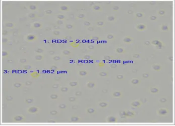
(4) Formulation and in vitro characterization of metoprolol tartrate loaded chitosan microspheres. Characterization of chitosan microspheres Particle Size Distribution and Surface Charge (Zeta Potential) Metoprolol tartrate loaded microspheres were suspended in distilled water for size distribution analysis with the help of Malvern Zeta sizer (Malvern ZS-90 instrument Ltd U.K.).18 Surface Morphology The morphology of the microspheres was determined by observation on a fluorescent microscope. The samples were prepared in distilled water and mounted on glass slide to determine the morphology of microspheres with the help of Fluorescent microscope (Radical Rx Lr 3T).18 Entrapment Efficiency (EE) The entrapment efficiency of microspheres prepared by ionic precipitation technique was determined by using cooling centrifuge. The microsphere dispersions were centrifuged at 3000 × g for 30 min in acetic acid solution. The clear supernatant was analyzed for metoprolol tartrate by UV-spectrophotometeric method at 274 nm. The drugloading efficiency was determined as the ratio between the analytical and theoretical drug content.19 All the experiments were performed in triplicate. It was calculated using following formula:. %EE =. Amount of metroprolol tartrate entrapped x 100 Total of metroprolol tartrate. Infrared Spectroscopy Infrared spectroscopy of the drug-loaded microspheres was performed at ambient temperature in the range of 400-4000cm-1, using potassium bromide pellets in FT-IR spectrometer (JASCO, Japan).20 In Vitro Drug Release. RESULTS AND DISCUSSION Preparation of Metoprolol Tartrate Loaded Chitosan Microspheres Precipitation and precipitation chemical cross-linking methods are rapid and simple techniques for producing metoprolol tartrate loaded microspheres with small size and good reproducibility. This production process is based on the solubility behavior of Chitosan, which is sparingly soluble in water. Addition of an acid improves its solubility as a result of protonation of amino groups. Chitosan solubility is also affected by other anions present in the solution. In the presence of phosphate, polyphosphate and sulphate ions, Chitosan shows a decreased solubility18. For this reason, sodium sulphate was chosen for microsphere formulations, since sulphate leads to a poorly soluble Chitosan derivative, facilitating microsphere formulation possible. Particles Size and Surface Morphology The chitosan microspheres were spherical and particle size of optimized formulation (MT 4) was found to be 1897 nm. The change in particle size was observed upon varying processing variables as concentration of chitosan and stirring speed. It was concluded that on decreasing the concentration of polymer and on increasing the stirring rate the size of microspheres was decreased. The crosslinked microspheres were smaller than the corresponding uncross-linked particles in compliance with expectation. These differences in size indicate that cross linked microspheres were more compact in structure because of the cross-linkage. Optical micrographs showed spherical particles (Figure 1).. Figure 1. Microphotograph of metoprolol tartrate loaded microspheres. The chitosan microspheres were suspended in small volume of phosphate buffer saline (pH 7.4) in order to determine drug release behavior. This suspension was placed in an egg membrane and suspended in beaker filled with 50 ml of dissolution medium. This solution was stirred at 100 rpm with magnetic stirrer at 37±1°C. Sink conditions were maintained during the drug dissolution study.19 Sampling was done at specific intervals. At each sampling, 3 ml of the solution was withdrawn and replenished with fresh medium. The drug concentration was measured at respective λmax in respective medium using UV/visible spectrophotometer (Shimadzu 1700, Japan).. Ars Pharm. 2012; 53(3): 13-18.. 15.
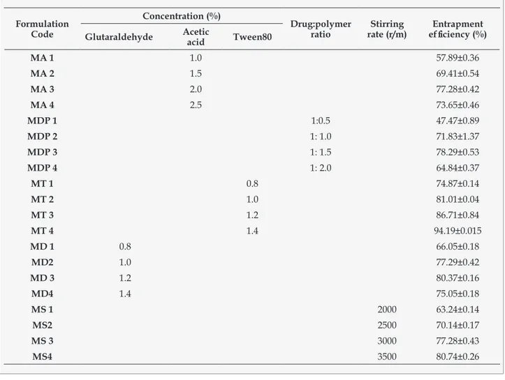
(5) Bharti AD, Keservani RK, Sharma AK, Kesharwani RK, Husain GM. Investigation of the Zeta potential is an important part of microsphere characterization. The phosphate buffer influenced measurement of Zeta potential, due to the effect of the counter ions on the positively charged chitosan microspheres. Chitosan microspheres were positively charged (Zeta Potential 5.59 mV-Formulation MT4), although sulphate ions were used as precipitant. This was concluded that only a part of the amino groups are neutralized during microsphere formation.. of microspheres decreases and the % drug entrapment increases. It was observed that entrapment efficiency increases with increase in surfactant concentration. The metoprolol tartrate loaded microspheres were produced by the ionic precipitation technique using different concentrations of cross linking agent (glutaraldehyde). Glutaraldehyde is probably able to interact with the amine groups of metoprolol tartrate, forming a complex that forms the microspheres. On increasing concentration of glutaraldehyde the amount of free drug was decreased (Table1).. Entrapment Efficiency The entrapment efficiency of metoprolol loaded microspheres was performed as per reported method in order to estimate the actual amount of drug being entrapped in microspheres. Encapsulation efficiency was estimated after freeze-drying. On the basis of entrapment efficiency the acetic acid concentration, drug polymer ratio, stirring speed, glutaraldehyde concentration, and tween 80 concentrations were optimized. In chitosan microspheres formulation, the increase in. In Vitro Drug Release In vitro drug release from microspheres was performed in phosphate buffer saline (pH 7.4) through egg membrane. The natural membranes (such as egg membrane, peach and tomato skin, inter-lamellar layer of the onion) used, have pores and channels with hydrophilic properties which permeates small to middles size hydrophilic drugs to diffuse in a manner similar to human skin, and because of its. stirring rate will disperse the precipitating agent in solution rapidly and also due to increase in shearing force, the size. availability can be used for in vitro diffusion studies. It was reported that on increasing the concentration of polymer. Table 1. Effect of various processing parameters on entrapment efficiency (EE) of microspheres. Formulation Code. 16. Concentration (%) Glutaraldehyde. Acetic acid. Tween80. Drug:polymer ratio. Stirring rate (r/m). Entrapment efficiency (%). MA 1. 1.0. 57.89±0.36. MA 2. 1.5. 69.41±0.54. MA 3. 2.0. 77.28±0.42. MA 4. 2.5. 73.65±0.46. MDP 1. 1:0.5. 47.47±0.89. MDP 2. 1: 1.0. 71.83±1.37. MDP 3. 1: 1.5. 78.29±0.53. MDP 4. 1: 2.0. 64.84±0.37. MT 1. 0.8. 74.87±0.14. MT 2. 1.0. 81.01±0.04. MT 3. 1.2. 86.71±0.84. MT 4. 1.4. 94.19±0.015. MD 1. 0.8. 66.05±0.18. MD2. 1.0. 77.29±0.42. MD 3. 1.2. 80.37±0.16. MD4. 1.4. 75.05±0.18. MS 1. 2000. 63.24±0.14. MS2. 2500. 70.14±0.17. MS 3. 3000. 77.28±0.43. MS4. 3500. 80.74±0.26. Ars Pharm. 2012; 53(3): 13-18..
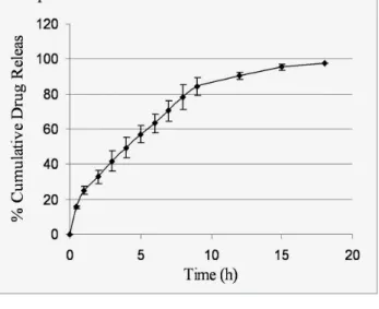
(6) Formulation and in vitro characterization of metoprolol tartrate loaded chitosan microspheres. the release rate of drug from chitosan microspheres were decreased, as the thickness of polymer was increased and diffusion distance for drug to diffuse out from microspheres was increased. The observations were made continuously for 18 hr. The release profiles were also dependent on the size of the microspheres, the rate of drug release was found to decrease with increasing particle size of microspheres. This may be useful for the controlled release of a drug inside the core matrix since the release rate is controlled by diffusion through a shell of uniform thickness. The results of in vitro drug release studies are showed in Figure 2, Figure 3 and Figure 4. Infrared Spectroscopy It was performed on metoprolol tartrate loaded chitosan microspheres to identify the drug polymer interactions. The characteristic IR spectra were very similar, showing all the bands of the functional groups of drug in metoprolol tartrate loaded microspheres. The results of the IR analysis were in an agreement for which no new chemical species after the microencapsulating process was observed but there are slight shift in peaks that is due to physical interaction as 841 nm instead of 822 nm, 1126 nm instead of 1111 nm, 1024 nm instead of 1023 nm.. CONCLUSION The present research work was an attempt to overcome pitfalls associated with conventional drug delivery systems such as fluctuation in plasma drug concentration, lack of target specificity. The chitosan (biocompatible polymer) microspheres of metoprolol tartrate delivered via nasal route ensure high degree of absorption and rapid transport of absorbed substances into the systemic circulation for initiation of therapeutic action.. Figure 2. In vitro % cumulative drug release profile in PBS pH-7.4 of formulation F1. Figure 3. In vitro % cumulative drug release profile in PBS pH-7.4 of formulation F2. Figure 4. In vitro % cumulative drug release profile in PBS pH-7.4 of formulation F3. ACKNOWLEDGEMENTS Authors are thankful to the Mr. Amit Kumar Singh, for kind cooperation during entire research work. The authors would like to express sincere gratitude towards Prof. Piyush Trivedi, Vice Chancellor, Rajiv Gandhi Proudyogiki Vishawavidyalaya, Bhopal for his kind permission and valuable suggestions to accomplish current research work. REFERENCES 1.. Alnasir FA. Hypertension the Silent Killer, 1st International Conference, in Partnership with the WHO. Qatar Primary Health Care. 2008. [cited 2010 Dec 19]. Available from:http://gis.emro.who.int/ HealthSystemObservatory/Workshops/QatarConference. Ars Pharm. 2012; 53(3): 13-18.. 2.. Tripathi KD. Essentials of medical pharmacology. 6th ed. New Delhi; 2004.. 3.. Behl CR. Effects of physicochemical properties and other factors on systemic nasal drug delivery. Adv Drug Deliv Rev. 1998; 29: 89-116. 17.
(7) Bharti AD, Keservani RK, Sharma AK, Kesharwani RK, Husain GM. 4.. Nakamura K. Uptake and release of budesonide from mucoadhesive, pH-sensitive copolymers and the application to nasal delivery. J Control Release. 1999; 61: 329-35.. 5.. Park JS. In situ gelling and mucoadhesive polymer vehicles for controlled intranasal delivery of plasmid DNA. J Biomed Mater Res. 2002; 59(1): 144-51.. 6.. 7.. Bernkop-Schnürch A, Kast CE, Richter MF. Improvement in the mucoadhesive properties of alginate by the covalent attachment of cysteine. J Control Rel 2001; 71(3): 277-85.. 8.. Wang JS. Aminated gelatin as a nasal absorption enhancer for peptide drugs: evaluation of absorption enhancing effect and nasal mucosa perturbation in rats. J Pharm Pharmacol. 2002; 54: 181-88.. 9.. Mathiowitz E, Chickering DE, Jacob JS inventors; Brown University Research Foundation, Providence, RI, assignee. Bioadhesive microspheres and their use as drug delivery and imaging systems. United States Patent. US6197346. 2001 Mar 6.. 10.. Sakagami M. Mucoadhesive beclomethasone microspheres for powder inhalation: their pharmacokinetics and pharmacodynamics evaluation, Journal of Controlled Release. 2002; 80 (1-3): 207-18.. 11.. Sakagami M, Sakon K, Kinoshita W, Makino Y. Enhanced pulmonary absorption following aerosol administration of mucoadhesive powder microspheres. Journal of Controlled Release 2001; 77 (1-2): 117-29.. 12.. 18. Kotze AF. Chitosan and chitosan derivatives as absorption enhancers for peptide drugs across mucosal epithelia. In: E Mathiowitz, D.E Chickering, C.-M Lehr editors. Bioadhesive Drug Delivery Systems, Fundamentals, Novel Approaches and Development. New York: Marcel Dekker; 1999. p 341-87.. Borchard G,. Luegen HL,. de Boer AG,. Verhoef JC,. Lehr CM, Junginger HE. The potential of mucoadhesive polymers in enhancing intestinal peptide drug absorption: III. Effects of chitosan-glutamate and carbomer on epithelial tight junctions in vitro. Journal of Controlled Release. 1996; 39(2-3): 131-38. 13.. Yuan Q, Shah J, Hein S, Misra RD. Controlled and extended drug release behavior of chitosan-based nanoparticle carrier. Acta biomaterialia. 2010; 6(3): 1140-48.. 14.. Saito K, Fujieda T, Yoshioka H, Feasibility of simple chitosan sheet as drug delivery carrier. Euro J Pharm Biopharm, 2006; 64 (2): 161-66.. 15.. Takeuchi H. Mucoadhesive properties of carbopol or chitosan-coated liposomes and their effectiveness in the oral administration of calcitonin to rats. Journal of Controlled Release. 2003; 86(2-3): 235-42.. 16.. Vanrell RH, Carballido AF, Frutos G, Cadorniga R. Enhancement of the mydriatic response to tropicamide by bioadhesive polymers. J Ocular Pharmacol Ther. 2000; 16: 419-28.. 17.. Lim ST, Forbes B, Martin GP, Brown MB. In vivo and in vitro characterization of novel microparticulates based on hyaluronan and chitosan hydroglutamate. AAPS Pharm Sci Tech. 2001; 2: 20.. 18.. Berthold A, Cremer K, Kreuter J. Preparation and characterization of chitosan microspheres as drug carrier for prednisolone sodium phosphate as model for antiinflammatory drugs. Journal of Controlled Release. 1996; 39(1): 17-25.. 19.. Krisnamoorthy R, Mitra AK. Mucoadhesive polymers in ocular drug delivery, New York: Marcel Dekker; 1993.. 20.. Junior AA, Matos J R. Thermal behavior and stability of biodegradable spray-dried microparticles containing triamcinolone. International Journal of Pharmaceutics. 2009; 368(1-2): 45-55.. Ars Pharm. 2012; 53(3): 13-18..
(8)
Figure
Documento similar