TítuloHydrogen sulfide and inflammatory joint diseases
17
0
0
Texto completo
(2) 1. INTRODUCTION Rheumatoid arthritis (RA) and osteoarthritis (OA) represent two of the most common arthritides in the developed world. RA, a chronic inflammatory autoimmune disease affecting diarthrodial joints, is characterized by joint swelling, tenderness and eventual destruction of synovial joints, leading to functional impairment and shortened life expectancy. While the etiology of RA is unknown, one hypothesis is that the disease ensues from an environmental trigger in a genetically predisposed individual [1, 2]. From disease onset, an inflammatory infiltrate' of mononuclear cells exists in the synovial membrane, followed shortly by synovitis. In RA, the two types of cells, macrophage-like synoviocytes (MLS) and fibroblast-like synoviocytes (FLS), contained in the synovial intimal lining become activated. MLS produce large amounts of tumor necrosis factor-a (TNF), interleukin (IL)-lβ, granuloeytemacrophage colony stimulating factor (GM-CSF) and chemokines, such as IL-8. These factors further stimulate the FLS to become the primary source of mediators contributing to joint destruction. Important among these mediators are matrix metalloproteinases (MMPs) , particularly collagenases, stromelysins and gelatinases, all of which are responsible for joint structural damage, other molecules that enhance the inflammatory response (IL-6, cyclooxygenase (COX)-2, prostaglandin (PG) E2 or IL-8) and mediators that increase vascular permeability and facilitate angiogenesis [vascular cell adhesion molecule (VCAM)l, vascular endothelial growth factor (VEGF) and fibroblast growth factor (FGF)]. The interactions between MLS and FLS involve complex cytokine networks that perpetuate synovitis, TNF-α, IL-1β and IL-6 are key cytokines causing inflammation in RA, with TNF-α having the dominant role because it is a potent paracrine inducer of other inflammatory cytokines [3,4]. In contrast, OA is characterized by the progressive loss of articular cartilage resulting from an imbalance of the catabolic and anabolic processes in chondrocytes. OA characteristically has a significant chronic inflammatory state [5]. Local inflammatory molecules contribute to progression of the disease in the early stages, finally resulting in chronic inflammation. These local inflammatory molecules include IL-lβ, TNF-α, IL-6, nitric oxide (NO) and PGE2. Particularly in OA, IL-1β is a key pro-inflammatory factor (Reviewed in [6-8]). While there are two receptors for IL-1β, IL-1RI and IL-1RII, only IL-1RI can transduce the IL-lβ signal [9]. The articular chondrocytes (AC) and FLS of OA patients express high levels of IL-1RI (10, 11] and increased numbers of chondrocytes synthesizing IL-lβ and TNF-α occur in OA cartilage [12]. When IL-lβ binds to IL-1RI, several signalling and transcriptional pathways, such as nuclear translocation of nuclear factor kappa-light-chain-enhancer of activated B cells (NFκB), activation of protein kinase (PK) C, p38, extracellular signal regulated kinases (ERK) 1/2 or c- Jun N-terminal kinases (JNK) are initiated [13, 14]. Therefore, in OA, IL-lβ is responsible for the inhibition of anabolism by the reduction in the production of collagen type II and aggrecan and the induction of catabolism by the upregulation of MMPs and aggrecanases in ACs [15]. IL-lβ also participates in the upregulation of PGE2 synthesis enzymes, COX-2 and microsomal prostaglandin E synthase-1 (PTGES), which leads to increased PGE2 production [16, 17]. In both RA and OA, reactive nitrogen species (RNS) and oxygen species (ROS) are also important players involved in inflammation. NO, peroxynitrite, H2O2 or O2 cause deleterious effects in cells, including oxidative damage to proteins, membranes and DNA and activation of the cells apoptotic pathways. NO contributes to the enhancement of the activation and production of MMPs, the inhibition of synthesis of anabolic molecules [18] and the promotion of chondrocyte apoptosis in both RA and OA [19]. During energy metabolism, the mitochondrial respiratory chain is the main source of ROS [20]. Evidence that mitochondrial dysfunction contributes to the pathophysiology of inflammatory joint diseases [21, 22] and to a wide range of other pathologies [23] is accumulating. Hydrogen sulfide (H2S), a gas detectable by its characteristic odor of rotten eggs, is very toxic in high concentrations. H2S exerts its toxicity by the inhibition of cytochrome C oxidase and reduction of ATP production [24]. Early studies were concerned with its environmental toxic effects and with measurement techniques for the detection and prevention of the risks associated with human exposure [25]. We have.
(3) since learned that H2S is an endogenous gasotransmitter capable of penetrating cell membranes without a specific transporter. H2S is produced in most tissues in considerable levels and participates in many physiologic and pathologic events (reviewed elsewhere [26-29]). Evidence is mounting that H2S plays an important role in inflammation [30, 31] and that exogenous supplementation of H 2S could be of therapeutic value in several diseases [29]. This review ad- dresses the relevance of H2S supplementation in the context of acute and chronic inflammation as it relates to RA and OA, including current methods for H2S exogenous administration, as available in current literature.. 2. HYDROGEN SULFIDE SOURCES 2.1. Endogenous Synthesis of Hydrogen Sulfide Endogenous H2S is chiefly produced by three enzymes, cystathionine β-synthase (CBS, EC 4.2.1.22), cystathonine γ-lyase (CTH, EC 4.4.1.1) and 3-mercaptopyruvate sulfurtransferase (MPST, EC 2.8.1.2). These enzymes, which utilize cystathionine, homocysteine or L-cysteine to produce hydrogen sulfide, are found in various tissues and their expressíon and distribution are tissue specific [28, 32, 33]. CBS is the predominant enzyme in the brain and heart and CTH is mainly expressed in the liver, kidney and intestine, and by vascular smooth muscle cells [32]. MPST has been reported in the brain, liver, kidney and heart [33]. CBS and CTH are exclusively cytosolic enzymes, while MPST is found both in the cytosol and mitochondria of cells [33]. 2.2. Endogenous H2S and Rheumatic Diseases Articular tissues also express enzymes associated with H2S synthesis. A recent publication reports that chondrocyte- like cells (chondrogenically differentiated mesenchymal pro- genitor cells, CH-MPCs) and primary human (h) Aes express both CBS and CTH, and that CTH is induced in both cell types by IL-lβ, TNF-α, IL-6 or lipopolysaccharide (LPS) treatment [34]. Our group bas found these three enzymes at mRNA and protein levels in cartilage, synovial membrane and subchondral bone from OA and normal patients [35]. Other studies have shown that synovial fluid (SF) H 2S concentrations in patients with RA [36, 37] or gout [37] were significantly higher than those in paired H2S plasma values and those in the SF of age-matched OA patients. In addition, one investigator reported that SF H 2S levels of RA patients correlated with the disease activity score in 28 joints and tender joint count [37]. This same study also reported that plasma H2S levels in RA and gout did not differ significantly from those of healthy controls, while those of OA patients were significantly higher [37]. In contrast, Whiteman et al. [36] and similar studies by our group did not reveal any differences in H2S concentration in serum between OA patients and healthy controls. On the contrary, we have preliminary evidence that indicates that H2S biosynthesis in OA joints might be reduced [35]. One hypothesis recently promulgated states that when H2S is increased in inflammatory diseases, it represents an endogenous compensatory mechanism by cells attempting to overcome inflammation, indicating that H2S may play an anti-inflammatory role [30, 38-40], although further studies are needed to test this hypothesis. 2.3. Exogenous Administration of H2S Many of the early studies investigating the inflammatory role of H 2S in in vivo models or in cells in vitro used fast-dissolving salts, such as Na2S, NaSH or Lawesson's reagent (LR), to generate and deliver H2S. These reagents produce an immediate burst of H2S that lasts a few seconds. However, this brief concentration is likely not a good model for in vivo physiologic H2S synthesis, which may occur in smaller quantities and much more slowly. In 2008, GYY4137 (morpholin-4-ium-4-methoxyphenyl.
(4) (morpholino) phosphino-dithioate), a new type of H2S-delivering compound, was synthesized [41]. This compound slowly produces H2S both in vitro and in vivo. Using an endotoxic shock rat model and also in RAW264. 7 macrophages, GYY 413 7 exhibited anti-inflammatory effects [42]. When GYY4137 was administered to conscious rats 1 or 2 h prior to LPS treatment, the ensuing rise in plasma proinflammatory cytokines (TNF-α, IL-1β and IL-6) and lung myeloperoxidase (MPO) activity was decreased, while plasma concentration of the anti-inflammatory cytokine IL-1O was increased. In macrophages, GYY 413 7 reduced NFKB activation, the expression of the inducible form of NO synthase (iNOS) and COX-2, as well as the generation of PGE2 and NO-derived nitrites/nitrates [42]. A more recent series of H2S-delivering compounds that target mitochondria has been synthesized. These compounds contain a mitochondria-targeting moiety (triphenylphosphonium) that accumulates in the mitochondria [43] coupled to an H2S-releasing moiety (dithiolethione or thiohydroxybenzamide). The effect of AP39 [(10-oxo-10-(4-(3-thioxo-3H- 1,2-dithiol-5yl)phenoxy)decyl)triphenyl-phosphonium bromide], one of these compounds, was recently tested on bioenergetics, viability and mitochondrial DNA integrity of resting cells and during oxidative stress in bEnd.3 murine microvascular endothelial cells [44]; the results demonstrated anti-oxidant and cytoprotective effects during oxidative stress. These compounds also seem to be more potent than other H 2S-forming compounds; the range of concentrations needed to exert beneficial effects being 30-100 nM, appreciably lower than those reported for NaSH, LR, Na2S or GYY4137 [44]. These new compounds are novel and useful research tools to investigate the impact of H2S on cell bioenergetics and are likely to produce important information about the etiology of inflammatory joint diseases, possibly offering new therapeutic approaches. Finally, we should mention that the pharmacological addition of H2S-fonning reagents or drugs is not the only way to supply H2S exogenously. Beneficial effects, in particular, anti-inflammatory and antioxidant effects, on patients with chronic lung disease [45, 46], chronic rhinosinusitis [47], allergic disorders [48] and rheumatologic diseases [49] when H 2S is administered in the form of sulphurous mineral water have been reported.. 3. H2S AND ACUTE AND CHRONIC ARTHRITIDES To date, most studies on the inflammatory effects of H2S used models of acute inflammation. Some of these studies have found exogenous H2S treatment to exert anti- inflammatory effects in such classic inflammation models as gastrointestinal inflammation [50] or endotoxic shock [42], although contradictory reports can be found in recent literature. Studies supporting the hypothesis that H 2S may be protective or beneficial that investigate the molecular mechanisms involved in rheumatic inflammatory joint pathologies are relatively recent (for a summary see Table 1)..
(5) Table 1. Summary of the literature concerning the effects of H2S forming compounds on intlammatory joint disease in vivo or in vitro models. H2S Compound and Concentration. Reference. Pathology. Animal Model/ Cellular Type. Stimuli. Sieghart el al. [51]. OA. FLS. IL-1β. NaSH (up to 1 mM, 20 min). Burguera el al. [52]. OA. hACs. IL-1β or LPS. NaSH and OYY4137 (501000 μM, 48 h). Burguera el al. [53]. OA. hACs. IL-1β (5 ng/ml, 48 h). NaSH and OYY4137 (501000 μ1M, 48h). Ha et al. [54]. OA. hACs. IL-1β (10 ng/mi, 2 h). NaSH (0.06-1.5 mM, 30 min pretreatment). Kloesch el al. [55]. RA and OA. FLS. -. NaSH (up to 1 mM, 20 min). Kloesch el al. [56]. RA. FLS. IL-1β (5 ng/ml, 1 h). NaSH (0.03-1.0 mM). Kloesch el al. [57]. -. Chondrocyte cell line C28/I2. IL-1β (5 ng/ml, 1 h). NaSH (up to 1 mM). Stulhmeier et al. [58]. RA. FLS. -. NaSH 0.05-5 mM. Fox et al. [34]. Normal. Chondrogenically differentiated MPCs and hACs. SIN-1 H2O2 4-HNE. GVY4137 200 and 500 μM. Li et al. [40]. Normal. hFLS and hCHs. LPS. GYY4137. Andruski et al. [59]. Acute knee inflammation. C57b1/6 mice. Kaolin/carrageenan intraarticular injection. Na2S 10, 30 or 50 μM. Main Results. Reduced spontaneous and IL-1β induced secretion of IL-6, IL-8 and RANTES. Inhibited the formation of hyperplastic lining layer in FLS micromasses. Reduced MMP-3 mRNA and protein. 200 μM OYY4137 reduced LPS-induced mitochondrial ROS. No effect on SOD2 or CAT mRNA. Reduced NO and iNOS (mRNA and protein), POE2 levels, COX·2 and PTOES mRNA; no effect on COX-1. Reduced IL-6 and MMP13 levels and mRNA. Reduced NO and iNOS (mRNA and protein), POE2 levels, COX-2 mRNA; MMP13 levels and mRNA, up to 0.6 mM. Best result with 0.3mM NaSH. NaSH >0.5 mM, 20 min increased IL-6, IL-8, COX-2 in OA and RAFLS, and increased MMP3 mRNA in RA-FLS. But it also reduced MMP-14 and MMP-2 in RA-FLS. NaSH short-term exposure (<0.125 mM, 60 min) reduced constitutive and IL-1β-induced IL-6. NaSH long-term exposure (0.125 mM, 6 h) increased IL-6. NaSH 0.125 mM, 30 min or 1 mM, 15 min reduced IL-1βinduced IL-6 and IL-8. NaSH 2mM, 45 min increased HO-1 and HSP70 mRNA and protein levels. But TNF, IL-8, IL1β and COX-2 gene expression and protein levels were also elevated. Reduced SIN·1, H2O2 or 4-HNE induced cell death, mitochondrial toxicity (collapse of mitochondrial membrane potential), oxidant stress induced cellular ATP depletion and cytoplasm accumulation of cytochrome C. Reduced NO, POE2, TNF-α and IL-6, also the catalytic activities of iNOS, COX-2. H2S reduced leukocyte adherence and increased leukocyte velocity, suggesting of an anti-inflammatory effect, but had no influence on pain..
(6) Fig. (1). Summary of the effects of the exogenous administration of hydrogen sulfide (H2S) in osteoarthritis (OA) and rheumatoid arthritis (RA) using in vitro and in vivo models. The key pro-inflammatory cytokines are tumor necrosis factor-α (TNF-α) in RA and interleukin-1β (IL-1β) in OA. In RA, macrophage-like synoviocytes produce large amounts of TNF-α and IL-1β, and chemokines, such as IL-8. These activate fibroblast-like synoviocytes, which become the primary source of mediators that contribute to joint destruction. These mediators include matrix metalloproteinases (MMPs), IL-6, cyclooxygenase (COX-2), prostaglandin (PG) E2 and other mediators that increase vascular permeability and facilitate angiogenesis. In OA, IL-1β is responsible for an increase in the production of lL-6, nitric oxide (NO), PGE2 and also MMPs. Reactive nitrogen (RNS) and oxygen species (ROS), are also among the critical players involved in inflammation in both RA and OA. Virtually all reports on the administration of exogenous H2S, in various forms, using in vitro and in vivo models and human trials related to OA, provide evidence of beneficial effects, including the reduction of some oxidation, inflammation and markers of catabolism mentioned above. On the other hand, most published studies using RA models show exacerbation of inflammation symptoms which, collectively, do not support the idea that H2S supplementation might be beneficial to RA patients.. 3.1. H2S and In Vitro Models of Chronic inflammation Several groups, including ours, have used in vitro models with FLS or ACs from RA or OA patients to shed light on the role of H2S in these two rheumatic diseases. To evaluate its effects, these studies have typically used IL-1β or other pro-inflammatory stimuli, including LPS, to produce an inflammatory environment in vitro and NaSH or GYY4137 as H2S-fonning reagents. For a graphic representation of the effects of H2S on OA and RA cells using in vitro models (Fig.1). 3.1.1. Hydrogen Sulfide and OA Studies by our group [52, 53] and others [54] demonstrated the in vitro anti-inflammatory and anticatabolic effects of NaSH and GYY4137 on OA hACs. Our results showed that co-stimulation of these cells for 48 h with IL-1β (5 ng/ml) and either H2S generator led to significant reductions in NO, PGE2, IL6, MMP3 and MMP13 levels. In these stimulated cells, the mRNA expression of relevant genes involved in the synthesis of these molecules, including iNOS, COX-2 and PTGES, IL-6, MMP3 and MMP13, was also downregulated by the action of H2S. Protein levels of MMP3, MMP13 and iNOS were also reduced as demonstrated by immunocytochemistry. After pre-treating OA hACs with NaSH for 30 min prior to stimulation with 10 ng/ml of IL-1β for two additional hours, Ha and co-workers [54] found reductions in.
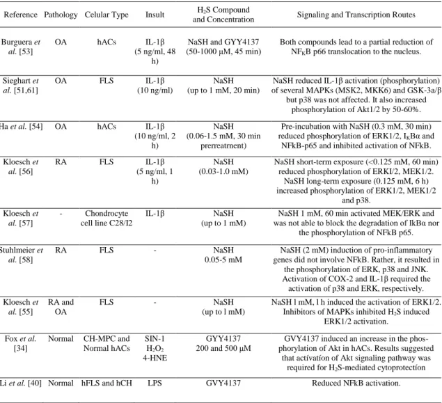
(7) the production of NO and PGE2 and MMP13 proteins by ELISA, as well as reduced mRNA expression of iNOS, COX-2 and MMP13. In our studies, there was also a partial reduction of the nuclear translocation of NFKB p65 induced by IL-1β treatment [53]. In agreement with this observation, Ha et al. [54] pretreated cells with 0.3 mM NaSH prior to a 2 h exposure to 10 ng/ml IL-1β and found that the activation of NFKB was inhibited. Table 2 and Fig. (2) provide a summary and a graphical representation, respectively, of the signalling and transcription routes that have been implicated in the effects of H 2S on inflammatory joint cells.. Table 2. Signaling and transcription routes known to be implicated in the effects of H2S on inflammatory joint cells Reference Pathology Celular Type. Insult. H2S Compound and Concentration. Signaling and Transcription Routes. Burguera et al. [53]. OA. hACs. IL-1β (5 ng/ml, 48 h). NaSH and GYY4137 (50-1000 μM, 45 min). Both compounds lead to a partial reduction of NFKB p66 translocation to the nucleus.. Sieghart et al. [51,61]. OA. FLS. IL-1β (10 ng/ml). NaSH (up to 1 mM, 20 min). NaSH reduced IL-1β activation (phosphorylation) of several MAPKs (MSK2, MKK6) and GSK-3a/β but p38 was not affected. It also increased phosphorylation of Akt1/2 by 50-60%.. Ha et al. [54]. OA. hACs. IL-1β (10 ng/ml, 2 h). NaSH (0.06-1.5 mM, 30 min prerreatrnent). Pre-incubation with NaSH (0.3 mM, 30 min) reduced phosphorylation of ERK1/2, IKBα and NFkB-p65 and inhibited activation of NFkB.. Kloesch et al. [56]. RA. FLS. IL-1β (5 ng/ml, 1 h). NaSH (0.03-1.0 mM). NaSH short-term exposure (<0.125 mM, 60 min) reduced phosphorylation of ERKI/2, MEK1/2. NaSH long-term exposure (0.125 mM, 6 h) increased phosphorylation of ERK1/2, MEK1/2 and p38.. Kloesch et al. [57]. -. Chondrocyte cell line C28/I2. IL-1β. NaSH (up to 1 mM). NaSH 1 mM, 60 min activated MEK/ERK and was not able to block the degradation of IkBα nor the phosphorylation of NFkB p65.. Stuhlmeier et al. [58]. RA. FLS. -. NaSH 0.05-5 mM. NaSH (2 mM) induction of pro-inflammatory genes did not involve NFkB. Rather, it resulted in the phosphorylation of ERK, p38 and JNK. Activation of COX-2 and IL-1β required the activation of p38 and ERK, respectively.. Kloesch et al. [55]. RA and OA. FLS. -. NaSH (up to l mM). NaSH l mM, l h induced the activation of ERK1/2. Inhibitors of MAPKs inhibited H2S induced ERK1/2 activation.. Fox et al. [34]. Normal. CH-MPC and Normal hACs. SIN-1 H2O2 4-HNE. GYY4137 200 and 500 μM. GVY4137 induced an increase in the phosphorylation of Akt in hACs. Results suggested that actívatíon of Akt signaling pathway was required for H2S-mediated cytoprotectíon. LPS. GVY4137. Reduced NFkB activation.. Li et al. [40] Normal hFLS and hCH.
(8) Fig. (2). Schematic representation of the signalling and transcription routes implicated in the effects of the exogenous administration of hydrogen sulfide compounds to articular cells. Hydrogen sulfide has been shown to influence several members of the mitogen activated protein kinase family (incIuding ERK1/2, MEK1/2, JNK or p38), other serine/threonine protein kinases (Akt1/2, GSK3α/β), as well as to interact with the NFkB signalling route. The reported effects varied depending on the cell type studied, the H2S forming compound used and its concentration, and the incubation time. Abbreviations: H2S, hydrogen sulfide; FLS, fibroblast-like synoviocytes; AC, articular chondrocytes; CH-MPC, chondrogenically differentiated mesenchymal progenitor cells; OA, osteoarthritis; RA, rheumatoid arthritis; ERK, extracellular signal regulated kinase; MEK, MAPK/ERK kinase; MSK, mito gen- and stress-activated protein kinase; MKK, mitogen-activated protein kinase kinase; Akt, protein kinase B; GSK-3, glycogen synthase kinase 3; IkB, nuclear factor of kappa light polypeptide gene enhancer in B-cells inhibitor; JNK, c-Jun N-terminal kinases; NFkB, nuclear factor kappa-light-chain-enhancer of activated B cells.. However, a different study found that NaSH treatment was detrimental to OA hFLS [55]. NaSH concentrations above 0.5 mM applied for 20 min treatments stimulated the express ion of IL-6, IL-8 and COX-2 mRNA for as long as 6 h afterward. Nevertheless, further investigations by the same group concluded that pre-treatment with NaSH dose- dependently reduced spontaneous, as well as IL-1βinduced, IL-6, IL-8 and RANTES (regulated on activation, normal T cell expressed and secreted) production in OA hFLS, with 1 mM NaSH proving to be the most effective concentration [51]. These authors also examined signalling routes by spot blot assay and determined the activation of 26 mitogenactivated protein (MAP) kinases and other serine/threonine kinases by IL-1β. Significantly activated were extracellular signal regulated kinases (ERK) 1/2, mitogen- and stress- activated protein kinase (MSK) 2, mitogen-activated protein kinase (MKK) 3/6, heat shock protein (HSP) 27, p38 units α, β, γ, and JNK2, among others, while RAC-beta serine/threonine-protein kinase B (Akt2) phosphorylation was slightly reduced. NaSH treatment significantly reduced the IL-1β-induced phosphorylation of MSK2, MKK.6 and glycogen synthase kinase (GSK) 3α/β. However, p38 MAP kinases, which were highly activated by IL1β, were not affected. Interestingly, NaSH treatment increased the phosphorylation of Akt1/2 by 50-60% and completely abolished the lining layer hyperplasia of hFLS micromasses stimulated with IL-1β..
(9) 3.1.2. Hydrogen Sulfide and RA There have been a few recent studies reporting of the effects of H2S on in vitro models of RA. Kloesch et al. [56] analyzed the effects of treatment of RA hFLS with different concentrations of NaSH for 1 h on IL-6. They found that NaSH concentrations lower than 0.125 mM reduced constitutive IL-6 levels and deactivated p44/42 MAPK (ERK1/2). Pre-incubation with 0.125 mM NaSH also blocked IL1β- induced.(5 ng/ml for 1 h) IL-6 expression. Conversely, long term exposure of hFLS to H2S (0.125 mM NaSH for 6h) produced elevated IL-6 expression and activation of p38 MAPK, MEK1/2 and ERK1/2. In another study, Kloesh et al. corroborated these results using chondrocyte cell line (C-28/I2) [57]. As found for RA hFLS, these C-28/I2 cells also expressed high constitutive levels of IL-6 and IL-8, .which were decreased following stimulation with NaSH (0.125 mM for 30 min or 1 mM for 15 min). In agreement with their previous findings [56], pretreatment with NaSH (1 mM, for up to 60 min) of C-28/I2 cells transiently activated the MEK/ERK pathway, the effect of which disappeared at 60 min. Additionally, in C-28/I2 cells stimulated with IL-1β, H2S was unable to block the degradation of IkB (nuclear factor of kappa light polypeptide gene enhancer in B-cells inhibitor) a or the phosphorylation of NFkB p65. A later study confirmed the adverse effects of NaSH exposure to RA hFLS [55]. The expression of IL6, IL-8 and COX-2 mRNA was induced in RA hFLS for up to 6 h after 20 min of stimulation with NaSH concentrations above 0.5 mM. MMP3 mRNA was also upregulated in these RA-FLS. In a positive observation, these authors report that MMP14 and MMP2 were downregulated [55]. It is worthy of note that both MMP2 and its molecular regulator MMP14 are enzymes relevant to RA; both are expressed at elevated levels in the synovial membrane and at sites of joint destruction in RA [62-64]. As previously reported [57] in RA and OA hFLS, NaSH treatment at 1 mM for 1 h resulted in phosphorylation of ERK1/2 at 15 min after treatment. Inhibitors of MAPKs inhibited the H 2S-induced expression of IL-6, IL8 and COX-2 mRNA, as well as ERK1/2 activation in both cell types. Another study confirming detrimental effects of H2S on RA hFLS [58] used commercially available RA hFLS treated with 0.05-5 mM NaSH. Short term exposure of the cells to 2 mM NaSH for 45 min induced a stress response characterized by increased mRNA and protein levels of heme-oxygenase (HO)-l and HSP70; although the gene expression and concomitant protein levels of TNF, IL-8, IL-1β and COX-2 were also significantly elevated. However, this NaSH induction of pro-inflammatory genes occurred without the involvement of NFkB transcription factor. Conversely, exposing hFLS to 2 mM NaSH resulted in the phosphorylation of ERK, p38 and JNK. Further experiments in hFLS with MAPK inhibitors demonstrated that the activation by NaSH of the COX-2 gene depends on activation of p38 and that of the IL-1β gene depends on ERK. Although there are some indications of beneficial results, taken collectively, available evidence from in vitro studies does not support any beneficial effects of H2S for RA (Fig. 1). However, because only NaSH was used to form H2S in these studies, it is possible that other H2S sources or concentrations might produce different effects. 3.1.3. Hydrogen Sulfide and Mitochondrial Dysfunction Mitochondrial dysfunction has emerged as a contributing factor to the pathophysiology of both RA and OA. Compared to normal cells, diverse in vitro analyses of mitochondrial respiratory chain activity have shown decreased complexes II and III activities in OA hACs [65]. This mitochondrial dysfunction perhaps potentiates cytokine-induced inflammation in hFLS and hACs [22, 66] and may modulate MMPs expression in hACs [67], Mitochondrial dysfunction may increase ROS production in OA hACs [68]. GYY4137 (200 μM or 500 μM) was employed to investigate the effects of pharmacological H2S on cellular oxidative stress in hCH- MPC and hACs. In these cells, the mitochondrial toxicity (mitochondrial membrane potential collapse), the decrease in mitochondrial ATP formation and the reduction of the cytoplasmic accumulation of cytochrome C induced by three oxidative stimuli [H 2O2, 3morpholinosydnonimine (SIN-1, a peroxynitrite donor) and hydroxynonenal (4-HNE)], as well as in.
(10) CTH- or CBS-siRNA treated cells, was significantly inhibited by GYY413. Collectively, these results suggest a cytoprotective role against oxidative injury for H 2S)in human joint cels [34]. Our group has investigated the anti-oxidant properties of GYY4137 and NaSH using OA hACs and found that only 200 μM GYY4137 reduced mitochondrial ROS (measured by 1,2,3-dihydrorhodamine and flow cytrometry), in LPS-, but not IL-1β-, stimulated cells; this level had no effect on the mRNA expression levels of the anti-oxidant enzymes mitochondrial superoxide dismutase (SOD2) and catalase (CAT) [52]. However, in an ischemic/reperfusion model, increased levels of SOD2 and reduced ROS levels have been reported [69]. As previously mentioned, compounds that deliver H 2S directly to the mitochondria, such as AP39, have recently been developed. To the best of our knowledge, these new compounds have not yet been tested on models of joint inflammatory diseases. 3.1.4. Hydrogen Sulfide and Normal Joint Cells GYY4137 was also employed to investigate the effects of H2S on cellular death induced by H2O2, SIN-l and 4-HNE in hCH-MPC and hACs [34]. When cells were treated with D,L-propargylglycine (PAG, CTH inhibitor), aminooxyacetate (AOAA, CBS inhibitor) or CTH-, CBS-siRNA silencing, GYY4137 preserved cellular viability against all three stimuli, dependent on concentration. A time and concentration-dependent increase in the phosphorylation of Akt, the pro-cell survival protein, was also induced in hACs by GYY4137. Experiments with Akt inhibitors showed that activation of Akt was required for GYY4137 cytoprotection against cell death. Another in vitro study revealed that GYY4137 exerted anti-inflammatory properties on LPS-treated normal hFLS and hACs [40]. When cells were treated with GYY4137 previously (for 1 h prior to LPS addition), and subsequently (at 6 or 18 h after LPS addition) the levels of pro-inflammatory markers PGE2, TNF-α, IL-6 and NO were reduced. Intracellular levels of COX-2 and iNOS and NFKB activation in both cell types were also reduced when GYY4137 was administered 1 h prior to LPS. 3.2. H2S and In Vivo Models of Acute Arthritis In addition to the in vitro studies described above, there are a few references using in vivo animal models. In 2008, the effects of an intra-articular injection of Na2S on acutely inflamed joints, induced by kaolin/carrageenan intra-articular injections, in C57B1/6 mice were examined [59]. These effects were evaluated by the following parameters: leukocyte recruitment and trafficking (by intravital microscopy), synovial blood flow (by laser Doppler perfusion imaging), and joint pain (by hindlimb incapacitance and von Frey hair algesiometry). While the local administration of Na 2S caused a dose-dependent reduction in leukocyte adherence and increased leukocyte velocity, suggesting an anti-inflammatory effect, no effect on joint pain was observed. However, the administration of LR to alleviate carrageenan-induced synovitis in rat knees produced a positive effect on pain signs [60]. In this study, Wistar rats were pretreated 60 min prior to induction of synovitis with either a non-selective COX inhibitor (indomethacin), LR, or an inhibitor of endogenous H2S production (PAG). In these rodents, pre-treatments with indomethacin and LR significantly reduced two common signs of pain, impaired gait and secondary tactile allodynia of the ipsilateral bind paw. In addition, the accompanying inflammatory response, characterized by joint swelling, inflammatory cell infiltration and increased synovial MPO activity, was also reduced. Interestingly, they found that pre- treatment with LR reduced the increased concentration of IL-1β that bad been induced by carrageenan in the rat knee cavity, but not those of TNF-α or IL-6. H2S pretreatment, however, bad no effect on carragenan-induced oxidative stress markers, including NO production, iNOS activity and nitration of protein tyrosine residues. Also noted was that the inhibition of endogenous H2S with PAG enhanced synovial iNOS activity and NO production, but had no effect on other tested markers of inflammation..
(11) In an in vivo model of acute joint inflammation produced by complete Freund's adjuvant (CFA) in mice [40], GYY4137 reduced inflammation when injected 6 h after CF A treatment, but did not have a prophylactic effect; it actually increased joint swelling when injected 1 h prior to CFA. Those SF parameters evaluated that were improved with GYY4137 treatment inc1uded SF concentrations of TNFα, IL-1β, IL-6 and IL-8, as well as reduced MPO activity and N-acetyl-D-glucosaminidase (NAG) concentration. GYY4137 was also anti-inflammatory when injected 18 h after CFA treatment, though to a much lesser extent. Notably, the H2S concentration in CFA-injected knees was higher than that of the saline-injected control knees. 3.3. H2S and Human Clinical Studies Finally, there are several clinical studies reporting positive outcomes when H 2S is administered to patients with rheumatologic diseases by soaking in sulphurous thermal water. A double-blind randomized controlled follow-up trial (RCT) in which hand OA patients received balneotherapy with sulphurous water 5 days a week for 3 weeks, while the control group bathed in tap water, was performed by Kovacs et al. [70]. Clinical parameters evaluated included hand pain, morning stiffness in band joints, grip strength of both hands and the Health Assessment Questionnaire Disability Index (HAQ), the Australian/Canadian Hand Osteoarthritis Index (AUSCAN) Hand Osteoarthritis Index, and EuroQol quality of life questionnaire. At treatment termination, the experimental group showed significant improvement in every parameter under the scope of the study. At the end of follow-up at 6 months, the values for pain parameters, and the HAQ and AUSCAN assessments continued to be significantly better in comparison with baseline values. In another prospective, single blinded RCT [71], treatment with intermittent sulphur baths (a 20 min bath 2 times a week for 6 weeks) was applied to patients with bilateral knee OA, while the control group bathed in tap water with the same regime and temperature (3536°C). Significant improvement in the Lequesne index of OA severity and the Western Ontario and McMaster Universities Arthritis Index (WOMAC) pain and stiffness scores for up to 6 months after treatment, and in self-reported pain (VAS) up to one month after treatment, was seen in the treatment group. The control group showed improvement in the Lequesne index only up to one month after treatment and in pain (VAS) only at the 3-month visit. The SF-36 bodily pain score significantly improved in both groups. In addition, at least three recent systematic reviews [72- 74], and a Cochrane Collaboration Review [75], have concluded that balneotherapy with sulphurous waters might indeed offer therapeutic value in rheumatic diseases. In fact, the Cochrane Collaboration review found "silver evidence concerning the beneficial effects of mineral baths compared to no treatment." Furthermore, the most recent OARSI guidelines for the nonsurgical management of knee OA in- corporate balneotherapy, including sulphurous waters, for individuals with multiple-joint OA and relevant comorbidities [76]. However, all these reviews of balneotherapy with sulphurous waters also point out that available information remains insufficient and that additional large and well-designed controlled randomized clinical trials are needed. The mechanisms responsible for the effects of sulphurous waters on rheumatic patients are not fully understood. It has been suggested that the benefits are the result of a combination of several elements. Chemical effects are thought to play an important role, but mechanical and thermal factors also contribute prominently. It is believed that the chemical components present in the water, including H2S, are absorbed through the skin and then act at a systemic level [77, 78]. For instance, decreased lipid (malondialdehyde) and protein oxidation products (carbonyls and advanced oxidation protein products) along with increased total antioxidant capacity (TAC) were found in the plasma of healthy volunteers that underwent a cycle of hydropinic therapy with sulphurous drinking water, compared with controls that did not [79]. Similar results were obtained in OA patients subjected to sulphur baths [80] or the combination of sulphur based mud baths and hydropinotherapy [49]. In addition, reduced serum TNF-α and cartilage oligomeric matrix protein concentrations were also found and the patients reported significantly lower pain scores [49]..
(12) CONCLUSION The investigation of the involvement of H2S in inflammatory joint pathologies is a rapidly expanding field. To date, in vitro studies using cells from OA patients have found anti- inflammatory, anti-catabolic and anti-oxidant properties for H2S-producing compounds. The available in vitro evidence is still inconclusive in the case of RA. Several in vivo animal models of acute arthritis have shown evidence of anti-inflammatory and pain relieving effects using NaSH, Lawesson's reagent or GYY4137 as H2S- producing reagents. New mitochondria-targeting H2S delivering compounds are promising new tools for investigation of the effects of H2S in the cellular bioenergetics known to be altered in RA and OA. The exogenous supplementation of H2S using sulphurous mineral water baths might also be an option to deliver H 2S treatment to patients. Overall, although further research is needed to confirm current working hypotheses, studies suggest that exogenous supplementation with H2S could have therapeutic value in inflammatory joint diseases, particularly OA. Current knowledge suggests that investigations in this field may produce useful information concerning the etiology of inflammatory joint diseases, as well as possibly offering new therapeutic options for treatment of arthritic patients.. LIST OF ABBREVIATIONS 4-HNE ACs Akt AP39. = = = =. AUSCAN CAT CBS CFA CH-MPC COX-2 CTH ERK FGF FLS GM-CSF GSK GYY4137 h H 2S HAQ HO HSP IL-1β IL-1R iNOS IkB JNK LPS LR MAPK MEK MKK MLS. = = = = = = = = = = = = = = = = = = = = = = = = = = = = =. Hydroxynonenal Articular chondrocytes Protein kinase B (10-oxo-10-( 4-(3-thioxo-3H-1,2-dithiol-5yI)phenoxy)decyl) triphenylphosphonium bromide Australian/Canadian Hand Osteoarthritis Index Catalase Cystathionine β-synthase Complete Freund's adjuvant Chondrogenically differentiated mesenchymal progenitor cells Cyclooxygenase 2 Cystathionine γ-lyase ExtracelIular signal regulated kinases Fibroblast growth factor Fibroblast-like synoviocytes Granulocyte-macropbage colony stimulating factor Glycogen synthase kinase Motpholin-4-ium-4-methoxyphenyl (morpholino) phosphinodithioate Human origin Hydrogen sulfide Health Assessment Questionnaire Disability Index Heme-oxygenase Heat shock protein Interleukin-1β Interleukin-1β receptor Inducible NO synthase Nuclear factor of kappa light polypeptide gene enhancer in B-cells inhibitor c-Jun N-terminal kinases Lipopolysaccharide Lawesson's reagent Mitogen-activated protein kinases MAPK/ERK kinase Mitogen-activated protein kinase kinase Macrophage-like synoviocytes.
(13) MMPs MPO MPST MSK NAG NFkB NO OA PAG PGE2 PKC PTGES RA RANTES RCT RNS ROS SF SIN-I SOD2 TAC TNF-α VAS VCAM VEGF WOMAC. = = = = = = = = = = = = = = = = = = = = = = = = = =. Matrix metalloproteninases Myeloperoxidase 3-Mercaptopyruvate sulfurtransferase Mitogen- and stress-activated protein kinase N-acetyl-D-glucosaminidase Nuclear factor kappa-light-chain-enhancer of activated B cells Nitric oxide Osteoarthritis D,L-propargylglycine Prostaglandin E-2 Protein kinase C Microsomal prostaglandin E synthase-l Rheumatoid arthritis Regulated on activation, normal T cell expressed and secreted Randomized controlled trial Reactive nitrogen species Reactive oxygen species Synovial fluid 3-morpholinosydnonimine Mitochondríal superoxide dismutase Total antioxidant capacity Tumor necrosis factor-α Visual analogue scale Vascular ceIl adhesion molecule Vascular endothelial growth factor Western Ontario and McMaster Universities Arthritis Index. CONSENT FOR PUBLICATION Not applicable.. CONFLICT OF INTEREST The authors declare no conflict of interest, financial or otherwise.. ACKNOWLEDGEMENTS Authors want to express their gratitude to the patients and staff of the Rheumatology and Orthopedic Services of the University Hospital A Coruña (CHUAC). EFB was supported by Axencia Galega de Innovación (IPP program) and Ciber-BBNIISCIII. The Biomedical Research Networking Center in Bioengineering, Biomaterials, and Nanomedicine (CIBER-BBN) is a national initiative of ISCIII.. AUTHOR CONTRIBUTIONS EF Burguera and R Meijide-Faílde searched the literature and reviewed data for the article. All authors made contributions to the discussion of the content. EF Burguera and FJ Blanco wrote the article. All authors performed re- view/editing of the manuscript before submission..
(14) REFERENCES [1] [2]. [3] [4] [5]. [6] [7] [8] [9] [10]. [11]. [12] [13] [14] [15] [16] [17]. [18] [19] [20] [21] [22]. [23]. [24] [25] [26]. Gibofsky A. Overview of epidemiology, pathophysiology, and diagnosis of rheumatoid arthritis. Aro J Manag Care 2012; 18: S295-S302. Aletaha D, Neogi T, Silman AJ, el al. 2010 Rheumatoid Arthritis Classification Criteria An American College of Rheumatology/European League Against Rheumatism Collaborative Initiative. Arthritis Rheum 2010; 62: 2569-81. Firestein GS. Invasive fíbroblast-like synoviocytcs in rheumatoid arthritis - Passive responders or transformed aggressors? Arthritis Rheum 1996; 39: 1781-90. Choy EHS, Panayi GS. Mechanisms of disease: Cytokine pathways and joint inflammation in rheumatoid arthritis. New Engl J Med 2001;344:907-16. Kraus VB, Blanco FJ, Englund M, Karsdal MA, Lohmander LS. Call for standardized definitions of osteoarthritis and risk stratification for clinical trials and clinical use. Osteoarthritis Cartilage 2015;23: 1233-41. Goldring MB, Otero M. Inflammation in osteoarthritis. Curr Opin Rheumatol 2011; 23: 4718. Daheshia M, Yao JQ. The interleukin 1 beta pathway in the pathogenesis of osteoarthritis. J Rheumatol 2008; 35: 2306-12. Kapoor M, Martel-Pelletier J, Lajeunesse D, Pelletier JP, Fahmi H. Role of proinflammatory cytokines in the pathophysiology of osteoarthritis. Nat Rev Rheumatol 2011; 7: 33-42. Braddock M, Quinn A. Targeting IL-1 in inflammatory disease: New opportunities for therapeutic intervention. Nat Rev Drug Discov 2004; 3: 1-10 Martel-Pelletier J, McCollum R, Dibattista J, et al. The Interleukin- 1 receptor in normal and osteoarthritic human articular chondrocytes - Identification as the type-I receptor and analysis of binding- kinetics and biologic function. Arthritis Rheum 1992; 35: 530-40. Sadouk M, Pelletier JP, Tardif G, Kiansa K, Cloutier JM, Martel- Pelletier J. Human synovial fibroblasts coexpress IL-1 Receptor- Type-I and Type-II Messenger-RNA - the increased level of the IL- 1 Receptor in osteoarthritic cells is related to an increased level of the Type-I Receptor. Lab Invest 1995; 73: 347-55. Shinmei M. Masuda K, Kikuchi T, Shimomura Y, Okada Y. Production of cytokines by chondrocytes and its role in proteoglycan degradation. J Rheumatol Supp11991; 27: 89-91. Kasza A. IL-1 and EGF regulate expression of genes important in inflammation and cancer. Cytokine 2013; 62: 22-33. Lee AS, Ellman MB, Yan D, et al. A current review of molecular mechanisms regarding osteoarthritis and pain. Gene 2013; 527: 440-7. Shlopov BV, Gumanovskaya ML, Hasty KA. Autocrine regulation of collagenase 3 (matrix metalloproteinase 13) during osteoarthritis. Arthritis Rheum 2000; 43: 195-205. Kobayashi M. Squires GR, Mousa A, et al. Role of interleukin-l and tumor necrosis factor a in matrix degradation of human osteoarthritic cartilage. Arthritis Rheum 2005; 52: 128-35. Kojima F. Naraba H, Miyamoto S, Beppu M, Aoki H, Kawai S. Membrane-associated prostaglandin E synthase-1 is upregulated by proinflammatory cytokines in chondrocytes from patients with osteoarthritis. Arthrit Res Ther 2004; 6: R355-R65. Abramson SB. Nitric oxide in inflammation and pain associated with osteoarthritis. Arthrit Res Ther 2008; 10 Suppl 2 doi: 10.1186/ar2463. Blanco FJ, Ochs RL, Schwarz H, Lotz M. Chondrocyte Apoptosis Induced by Nitric-Oxide. Am J Pathol 1995; 146: 75-85. Murphy MP. How mitochondria produce reactive oxygen species. Biochem J 2009; 417: 1-13. Blanco FJ, Rego I, Ruiz-Romero C. The role of mitochondria in osteoarthritis. Nat Rey Rheumatol 2011; 7: 161-9. Valcarcel-Ares MN, Riveiro-Naveira RR, Vaamonde-Garcia e, et al. Mitochondrial dysfunction promotes and aggravates the inflammatory response in normal human synoviocytes. Rheumatology 2014; 53: 1332-43. Pagano G, Talamanca AA, CasteIlo G, et al. Oxidative stress and mitochondrial dysfunction across broad-ranging pathologies: toward mitochondria-targeted clinical strategies. Oxid Med Cell Longev 2014; 2014: 541230. Khan AA, Schuler MM, Prior MG, et al. Effects of hydrogen- sulfide exposure on lung mitochondrial respiratory-chain enzymes in rats. Toxicol Appl Pharmacol 1990; 103: 482-90. Lawrence NS, Davis J, Compton RG. Analytical strategies for the detection of sulfide: a review. Talanta 2000; 52: 771 -84. Lee Predmore B, Joseph Lefer D, Gojon G. Hydrogen sulfide in biochemistry and medicine. Antioxid Redox Signal 2012; 17: 119- 40..
(15) [27] [28] [29] [30] [31] [32]. [33] [34]. [35]. [36]. [37]. [38] [39]. [40]. [41]. [42]. [43] [44]. [45]. [46]. [47] [48] [49]. Szabo C. Hydrogen sulfide and its therapeutic potential. Nat Rev Drug Discov 2007; 6: 91735. Kimura H. Hydrogen sulfide: its production, release and functions. Amino Acids 2011; 41: 113-21. Rivers JR, Badiei A, Bhatia M. Hydrogen sulfide as a therapeutic target for inflammation. Expert Opin Ther Targets 2012; 16: 439- 49. Whiteman M, Winyard PG. Hydrogen sulfide and inflammation: the good, the bad, the ugly and the promising. Expert Rey Clin Pharmacol 2011; 4: 13-32. Bhatia M. Role of hydrogen sulfide in the pathology of inflammation. Scientifica 2012; 2012: 159680. Renga B. Hydrogen sulfide generation in mammals: the molecular biology of cystathioninebeta- synthase (CBS) and cystathionine- gamma-lyase (CSE). Inflamm Allergy Drug Targets 2011; 10: 85- 91. Kamoun P. Endogenous production of hydrogen sulfide in mammals. Amino Acids 2004; 26: 243-54. Fox B, Schantz JT, Haigh R, et al. Inducible hydrogen sulfide synthesis in chondrocytes and mesenchymal progenitor cells: is H2S a novel cytoprotective mediator in the inflamed joint? J Cell Mol Med 2012; 16: 896-910. Vela-Aneto AA, Gato-Calvo L, Ruiz-Romero C, Meijide-Failde R, Blanco FJ, Burguera EF. Endogenous hydrogen sulfide production is reduced in OA cartilage. Possible contribution to the pathogenesis of OA. Osteoarthritis Cartilage 2015; 23: A311. Whiteman M, Haigh R, Tarr JM, Gooding KM, Shore AC, Win- yard PG. Detection of hydrogen sulfide in plasma and knee-joint synovial fluid from rheumatoid arthritis patients: relation to clinical and laboratory measures of inflammation. Ann N Y Acad Sci 2010; 1203: 146-50. Muniraj N, Stamp LK, Badiei A, Hegde A, Cameron V, Bhatia M. Hydrogen sulfide acts as a pro-inflammatory mediator in rheumatic disease. Int J Rheum Dis 2014; doi: 10.1111/1756185X.12472. Whiteman M, Moore PK. Hydrogen sulfide and the vasculature: a novel vasculoprotective entity and regulator of nitric oxide bioavailability? J Cell Mol Med 2009; 13: 488-507. Winyard PG, Ryan B, Eggleton P, et al. Measurement and meaning of markers of reactive species of oxygen, nitrogen and sulfur in healthy human subjects and patients with inflammatory joint dis- ease. Biochem Soc Trans 2011; 39: 1226-32. Li L, Fox B, Keeble J, et al. The complex effects of the slow- releasing hydrogen sulfide donor GYY4137 in a model of acute joint inflammation and in human cartilage cells. J Cell Mol Med 2013; 17: 365-76. Li L, Whiteman M, Guan YY, et al. Characterization of a novel, water-soluble hydrogen sulfide - Releasing molecule (GYY4137): New insights into the biology of hydrogen sulfide. Circulation 2008; 117: 2351-60. Li L, Salto-Tellez M, Tan CH, Whiteman M, Moore PK. GYY4137, a novel hydrogen sulfide-releasing molecule, protects against endotoxic shock in the rat, Free Radical Biol Med 2009; 47: 103-13. Smith RA, Hartley Re, Murphy MP. Mitochondria-targeted small molecule therapeutics and probes. Antioxid Redox Signa12011; 15: 3021-38. Szczesny B, Modis K, Yanagi K, et al. AP39, a novel mitochondria-targeted hydrogen sulfide donor, stimulates cellular bioenergetics, exerts cytoprotective effects and protects against the loss of mitochondrial ONA integrity in oxidatively stressed endothelial cells in vitro. Nitric Oxide 2014; 41: 120-30. Prandelli C, Parcia e, Buizza L, et al. Sulphurous thermal water increases the release of the anti-inflammatory cytokine IL-10 and modulates antioxidant enzyme activity. Int J Immunopathol Pharmacol20 13; 26: 633-46. Braga PC, Sambataro G, Dal Sasso M, Culici M, Alfieri M, Nappi G. Antioxidant effect of sulphurous thermal water on human neutrophil bursts: Chemiluminescence evaluation. Respiration 2008; 75: 193-201. Salami A, Dellepiane M, Strinati F, Guastini L, Mora R. Sulphurous thermal water inhalations in the treatment of chronic rhinosinusitis. Rhinology 2010; 48: 71-6. Valitutti S, Castellino F, Musiani P. Effect of sulfurous (thermal) water on lymphocyte-T proliferative response. Ann Allergy 1990; 65: 463-8. Benedetti S, Canino C, Tonti G, et al. Biomarkers of oxidation, inflammation and cartilage degradation in osteoarthritis patients undergoing sulfur-based spa therapies. Clin Biochem 2010; 43: 973-8..
(16) [50]. [51]. [52]. [53]. [54]. [55]. [56]. [57]. [58] [59]. [60]. [61]. [62]. [63] [64]. [65] [66]. [67]. [68] [69]. [70]. [71]. Chan MV, Wallace JL. Hydrogen sulfide-based therapeutics and gastrointestinal diseases: translating physiology to treatments. Am J Physiol Gastrointest Liver Physiol 2013; 305: G467-G73. Sieghart O, Liszt M, Wanivenhaus A. et al. Hydrogen sulfide decreases IL-1 beta-induced activation of fibroblast-like synoviocytes from patients with osteoarthritis. J Cell Mal Med 2015; 19: 187-97. Burguera E, Vela-Aneto A, Meijide-Failde R, Blanco F. Hydrogen sulfide donors alleviate IL-1 beta induced inflammation-like effects in human articular osteoarthritic chondrocytes. Osteoarthritis Cartilage 2013; 21: S241. Burguera EF, Vela-Anero A, Magalhaes J, Meijide-Failde R, Blanco F. Effect of hydrogen sulfide sources on inflammation and catabolic markers on interleukin 1 beta-stimulated human articular chondrocytes. Osteoarthritis Cartilage 2014; 22: 1026-35. Ha C, Tian S, Sun K, Wang O, Lv J, Wang Y. Hydrogen sulfide attenuates IL-1 beta-induced inflammatory signaling and dysfunction of osteoarthritic chondrocytes. Int J Mol Med 2015; 35: 1657- 66. Kloesch B, Liszt M, Krehan O, Broell J, Kiener H, Steiner G. High concentrations of hydrogen sulfide elevate the expression of a series of pro-inflammatory genes in fibroblastlike synoviocytes derived from rheumatoid and osteoarthritis patients. Immunol Lett 2012; 141: 197-203. Kloesch B, Liszt M, Broell J. H2S transiently blocks IL-6 expression in rheumatoid arthritic fibroblast-like synoviocytes and deactivates p44/42 mitogen-activated protein kinase. Cell Biol Int 2010; 34: 477-84. Kloesch B, Liszt M, Steiner G, Broell J. Inhibitors of p38 and ERKI/2 MAPkinase and hydrogen sulfide block constitutive and IL-1 beta-induced IL-6 and IL-8 expression in the human chondrocyte cell line C-28/12. Rheumatol Int 2012; 32: 729-36. Stuhlmeier KM, Broell J, Iliev B. NF-KappaB independent activation of a series of proinflammatory genes by hydrogen sulfide. Exp Biol Med 2009; 234: 1327-38. Andruski B, McCafferty OM, Ignacy T, Millen B, McDougall JJ. Leukocyte trafficking and pain behavioral responses to a hydrogen sulfide donor in acute monoarthritis. Am J Physiol Regul Integr Comp Physiol 2008; 295: R814-R20. Ekundi-Valentim E, Santos K, Camargo E, et al. Differing effects of exogenous and endogenous hydrogen sulfide in carrageenan- induced knee joint synovitis in the rat, Br J Pharmacol 2010; 159: 1463-74. Sieghart O, Kiener H, Kloesch B, Steiner G. Hydrogen sulfide reduces IL-1 beta-induced activation of fibroblast-like synoviocytes from patients with osteoarthritis. Nitric Oxide 2014; 39: S28. Goldbach-Mansky R, Lee JM, Hoxworth JM, et al. Active synovial matrix metalloproteinase2 is associated with radiographic erosions in patients with early synovitis. Arthritis Res 2000; 2: 145-53. Itoh Y. MTI-MMP: A key regulator of cell migration in tissue. IUBMB Life 2006; 58: 58996. Konttinen YT, Ainola M, Valleala H, et al. Analysis of 16 different matrix metalloproteinases (MMP-1 to MMP-20) in the synovial membrane: different profiles in trauma and rheumatoid arthritis. Ann Rheum Dis 1999; 58: 691-7. Maneiro E, Martin MA, de Andres MC, et al. Mitochondrial respiratory activity is altered in osteoarthritic human articular chondrocytes. Arthritis Rheum 2003; 48: 700-8. Vaamonde-Garcia C, Riveiro-Naveira RR, Valcarcel-Ares MN, Hermida-Carballo L, Blanco FJ, Lopez-Armada MJ. Mitochondrial Dysfunction Increases Inflammatory Responsiveness to Cytokines in Normal Human Chondrocytes. Arthritis Rheum 2012; 64: 2927-36. Cillero-Pastor B, Rego-Perez I, Oreiro N, Fernandez-Lopez C, Blanco FJ. Mitochondrial respiratory chain dysfunction modulates metalloproteases -1,-3 and -13 in human normal chondrocytes in culture. BMC Musculoskel Disord 2013; 14: 235. Blanco FJ, Lopez-Armada MJ, Maneiro E. Mitochondrial dysfunction in osteoarthritis. Mitochondrion 2004; 4: 715-28. Sun WH, Liu F, Chen Y, Zhu YC. Hydrogen sulfide decreases the levels of ROS by inhibiting mitochondrial complex IV and increasing son activities in cardiomyocytes under ischemialreperfusion. Biochem Biophys Res Commun 2012; 421: 164-9. Kovacs C, Pecze M, Tihanyi A, Kovacs L, Balogh S, Bender T. The effect of sulphurous water in patients with osteoarthritis of hand. Double-blind, randomized, controlled follow-up study. Clin Rheumatol 2012;31: 1437-42. Sherman G, Zeller L, Avriel A, Friger M, Harari M, Sukenik S. lntermittent Balneotherapy at the Dead Sea Area for Patients with Knee Osteoarthritis. Isr Med Assoc J 2009; 11: 88-93..
(17) [72] [73]. [74] [75] [76] [77] [78] [79] [80]. Forestier R, Francon A. Crenobalneotherapy for limb osteoarthritis: Systematic literature review and methodological analysis. Joint Bone Spine 2008; 75: 138-48. Fortunati NA, Fioravanti A, Seri G, Cinelli S, Tenti S. May spa therapy be a valid opportunity 10 treat hand osteoarthritis? A review of clinical trials and mechanisms of action. Int J Biometeorol 2016; 60: 1-8. Falagas ME, Zarkadoulia E, Rafailidis PI. The therapeutic effect of balneotherapy: evaluation of the evidence from randomised con- trolled trials. Int J Clin Pract 2009; 63: 1068-84. Verhagen A, Bierma-Zeinstra S, Larnbeck J, et al. Balneotherapy for osteoarthritis. A cochrane review. J Rheumatol 2008; 35: 1118- 23. McAlindon TE, Bannuru RR, Sullivan MC, et al. OARSI guidelines for the non-surgical management of knee osteoarthritis. Osteoarthritis Cartilage 2014; 22: 363-88. Fioravanti A, Cantarini L, Guidelli GM, Galeazzi M. Mechanisms of action of spa therapies in rheumatic diseases: what scientific evidence is there? Rheumatol Int 2011; 31: 1-8. Nasennoaddeli A, Kagamimori S. Balneotherapy in medicine: A review. Environ Health Prev Med 2005; 10: 171-9. Benedetti S, Benvenuti F, Nappi G, et al. Antioxidative effects of sulfurous mineral water: protection against lipid and protein oxidation. Eur J Clin Nutr 2009; 63: 106-12. Ekmekcioglu C, Strauss-Blasche G, Holzer F, Marktl W. Effect of sulfur baths on antioxidative defense systems, peroxide concentrations and lipid levels in patients with degenerative osteoarthritis. Forsch Komplementarmed Klass Naturheilkd 2002; 9: 216-20..
(18)
Figure

Documento similar