Are miRNAS an useful tool asessing treatment response inmultiple scleriosis?
41
0
0
Texto completo
(2) INDEX CONTENTS. TUTORESS AUTHORIZATION ............................................................................................. 2 ABSTRACT ......................................................................................................................... 3 1. INTRODUCTION............................................................................................................. 5 1.1 DEFINITION OF MULTIPLE SCLEROSIS .................................................................................. 5 1.2 CLASSIFICATION ................................................................................................................... 7 1.3 DIAGNOSIS ........................................................................................................................... 9 1.4 ETIOPATHOGENY ............................................................................................................... 11 1.4.1 GENETIC FACTORS ..................................................................................................................... 11 1.4.2 ENVIRONMENTAL FACTORS ...................................................................................................... 12. 1.5 THE PAPER OF MICRORNAS ............................................................................................... 14 1.5.1. DYSREGULATION OF MIRNAS IN MS ........................................................................................ 15. 3. STUDY JUSTIFICATION AND OBJECTIVES .................................................................... 15 4. METHODS ................................................................................................................... 16 4.1 SEARCH STRATEGY ............................................................................................................. 16 4.2 SELECTION CRITERIA .......................................................................................................... 17 4.3 DATA EXTRACTION............................................................................................................. 18. 5. RESULTS ...................................................................................................................... 20 5.1 SUMMARY BY ARTICLE ...................................................................................................... 20 5.2 METHODOLOGIC AND BIAS ASESSMENT ........................................................................... 26. 6. DISCUSSION ................................................................................................................ 28 7. CONCLUSION .............................................................................................................. 31 8. ACKNOWLEDGEMENTS............................................................................................... 32 9. REFERENCES ................................................................................................................ 32 10. SUPPLEMENTARY MATERIAL .................................................................................... 38. 1.
(3) TUTORESS AUTHORIZATION. TRABAJO DE FIN DE GRADO (TFG) - MEDICINA. EL/LA PROFESOR/A TUTOR/A hace constar su AUTORIZACIÓN para la Defensa Pública del Trabajo de Fin de Grado y CERTIFICA que el/la estudiante lo ha desarrollado a lo largo de 6 créditos ECTS (150 horas). TÍTULO del TFG: ¿ARE miRNAs AN USEFUL TOOL ASESSING TREATMENT RESPONSE IN MULTIPLE SCLEROSIS? ALUMNO/A: EMILIO ANTONIO PRIETO ZAMORA DNI: 48412116-Z PROFESOR/A TUTOR/A: ANA MARÍA SÁNCHEZ-PÉREZ. Fdo (Tutor/a): .......................................................... COTUTOR/A INTERNO/A (Sólo en casos en que el/la Tutor/a no sea profesor/a de la Titulación de Medicina):. Fdo (CoTutor/a interno): .......................................................... 2.
(4) RESUM S'està fent un esforç notable a tot el món per trobar nous biomarcadors adequats per a l'esclerosi múltiple (EM). Es prioritari trobar una font d'informació estandarditzada i fiable que permeta no només un diagnòstic precoç i fiable, sinó també avaluar la resposta del tractament, i anar més enllà, com a manera de dilucidar el complex mecanisme immunològic subjacent a la EM. Un grup de miRNA pot ser el futur contribuent a aquest objectiu. Els microARN són petites molècules d'ARN monocatenàries no codificants que actuen com a reguladors posttranscripcionals, silenciant l'ARN missatger (ARNm). Estan relacionats estrictament amb múltiples funcions indispensables, com la diferenciació cel·lular, la proliferació, l'apoptosi i el desenvolupament de teixits, i el que és més important en la EM, estan implicats en la regulació de la resposta immune, mitjançant la modulació i la diferenciació de les cèl·lules immunitàries, l'alliberament de mediadors inflamatoris i la producció d'anticossos.. En aquesta revisió, ens centrem en quatre assajos clínics dirigits a canvis en l'expressió de miRNA durant o abans i després de diferents tractaments, per tal de valorar si són vàlids com a biomarcadors de resposta al tractament de la EM. Una cerca estructurada es va dur a terme en tres bases de dades (PubMed, the Cochrane Library i WebOfScience) i un cribratge posterior va donar lloc a la selecció final. Per avaluar la qualitat metodològica vam utilitzar el manual Cochrane per a revisions sistemàtiques d'intervencions (versió 5.1.0). Després d'elaborar aquest treball, vam arribar a la conclusió que els miRNA tenen aplicacions molt prometedores en aquest camp, però són necessaris estudis millor dissenyats per a confirmar noves troballes i identificar els miRNAs més sensibles i específics per avaluar la resposta al tractament.. ABSTRACT A notable effort is being made worldwide in order to find the right biomarkers for multiple sclerosis (MS). It is a pressing subject to find a standardized and reliable source of information that allows not only an early and trusty diagnosis, but also to assess treatment response, and going further, as a way to elucidate the complex immunological mechanism underlying MS. A group of miRNAs might be the future contributors to this objective. MicroRNAs are small, noncoding, single-stranded RNA molecules that act as posttranscriptional regulators, silencing complementary messenger RNA (mRNA). They are strictly related to multiple indispensable functions, such as cellular differentiation, proliferation, apoptosis and tissue development, and more importantly in MS, they are involved in the regulation of immune response, via modulation. 3.
(5) and differentiation of immune cells, release of inflammatory mediators and antibody production. In this review, we center our focus on 4 clinical trials targeting changes in miRNA expression during or before and after different treatments, in order to assess whether they are valid as treatment response biomarkers in MS. A structured search was conducted in three databases (PubMed, The Cochrane library and WebOfScience) and a posterior revision yielded the final articles. To evaluate the methodologic quality we used the Cochrane Handbook for Systematic Reviews of Interventions (version 5.1.0). After elaborating this work, we conclude that miRNAs have very promising applications in this field, but better designed studies are much needed to confirm new findings and identify the most sensible and specific miRNAs to evaluate treatment response. Keywords: miRNAs, multiple sclerosis, biomarkers, natalizumab, glatiramer acetate, nanocurcumin.. 4.
(6) 1. INTRODUCTION 1.1 DEFINITION OF MULTIPLE SCLEROSIS. Multiple sclerosis (MS) is an inflammatory, chronic and degenerative central nervous system disease characterized by an acquired demyelination and posterior axonal degeneration, caused by a combination of autoimmune, genetic and environmental factors. Thus, susceptible individuals if exposed to specific external triggers and/or endogenous antigens, develop an autoimmune cascade in which the myelin basic protein (produced by oligodendroglia) and ultimately axons are recognized as foreign and attacked by the immune system (1). The result is a severely damaged neuronal transmission with fatal consequences (2)(3). This pathology represents the most common nontraumatic cause of neurological disability in young adults, specially between 20 and 40-45 years of age, but it can also appear in children or in older adults. It affects women two to three times more than men. It is present worldwide, affecting more than two million people (4), but seems to be more prevalent in countries far from equator, as latitude represents the second risk factor after controlling for ethnicity (5), being good examples of high prevalence southern Australia and New Zealand, the Scandinavian countries, Canada, Northern Ireland and Scotland, but with exceptions such as Spain, Greece, Italy or Saudi Arabia, which are closer to the equator but have a medium-high prevalence as well (6). Incidence varies greatly even between provinces. The estimated incidence in Spain is 15-25 cases for each 100.000 inhabitants/year (7). Up to date, there is no known cure for MS. Clinical manifestations of MS may range from modest to very serious neurological symptoms. Generally, MS is characterized by motor and sensory alterations. Motor symptoms include a limb or limbs that feel heavy, rigid, weak or clumsy, tremor, coordination impairment, bowel and urinary disorders, dysarthria, dysphonia and dysphagia. Frequent sensory symptoms reported are numbness, tingling, visual alterations, stiffness, fatigue and vertigo (8). Often MS is also accompanied by emotional and cognitive alterations and sexual disabilities.(9) These symptoms appear during or after a relapse or flare, which corresponds to the clinical manifestation of a new lesion in the CNS. The evolutional pattern of these flares is what drives the classification explained in the next section. In addition, the disease can follow different patterns depending on each case; some patients may only present a few relapses and stay relatively healthy for years or even decades, whereas in other cases, patients show a fast symptom worsening that can lead to a harsh reduction -7 to 14 years- in life expectancy (2). 5.
(7) The underlying reasons why myelin basic protein (MBP) and other myelin proteins are recognized by the immune system as foreign molecules remains unknown. It is generally accepted that that a group of myelin-specific autoreactive lymphocytes get activated by an unspecified environmental factor, which allows them to cross the blood-brain barrier (BBB) and cause havoc in the CNS. IFN-γ producing CD4+ Th1 cells and IL-17 producing CD4+ Th17 cells seem to be primarily involved in the pathogenesis of MS (10); on the contrary, IL-4 producing CD4+ Th2 and IL-10 and TGF-β secreting CD4+ Treg cells suppress harmful mechanisms present in the disease (11). Once activated, T cells cross the BBB and, in the brain tissue, they get re-stimulated by APCs, leading to disease induction and progression. As disease worsens, more myelin antigens are presented by APCs (epitope spreading), leading to further activation of newly arrived T cells (12) which trigger an attack first to the myelin sheath and then the axons, leading both of these injuries to an impairing or interruption of nerve function, responsible for the clinical manifestations of MS (13,14). T cells were pointed for years as the main responsible for the development and progression of MS, but B lymphocytes are now “in the spotlight”, as they are able to present antigens and thus activate T cells much more efficiently than dendritic cells, one of the reasons why B-cell depletion with treatments like rituximab and ocrelizumab are often used in MS trials, and have shed some light on the implication of B lymphocytes in MS (15). B cells can secrete both proinflammatory (like IL-6) and anti-inflammatory (such as IL-10) cytokines, but in MS patients proinflammatory features tend to dominate (16). Oligoclonal bands (typically IgG, but IgM can be present) can be found in both the CSF or the parenchyma. They are a product of these B cells once they cross the BBB, where they also differentiate to plasmatic cells(17); microglia and oligodendrocytes can promote B-cell survival and thriving by the secretion of B cell activation factor (BAFF), a proliferation inducing ligand (APRIL) and IL-15. These antibodies have been related to the perpetuation of immune response and neurodegeneration itself (18).. 6.
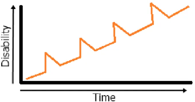
(8) 1.2 CLASSIFICATION. Patients with multiple sclerosis can be classified in four main groups depending on the unfolding of the disease: 1. CLINICALLY ISOLATED SYNDROME (CIS). CIS is the definition received by the first clinical manifestation of a demyelinating lesion. Patients are classified in this group as long as they don’t develop another episode or attack or a new radiologic lesion. 2. RADIOLOGICALLY ISOLA TED SYNDROME (RIS). A RIS consists of a lesion found incidentally that is compatible with MS in the brain and/or spinal cord but has not shown any clinical presentation or symptoms of the disease, and thus the patient doesn’t have any impairment related to MS. To be considered RIS, these findings should not be explained by another diagnosis, and the patient cannot have past or current clinical symptoms found on a neurological exam. 3. RELAPSING-REMMITING MULTIPLE SCLEROSIS (RRMS). Figure 1. Representation of the timeline of RRMS; following a relapse new symptoms may disappear without any increase of disability, or do so partially, resulting in a relative increase in disability. The duration of the episode and the duration of the intervals are variable. However, the recovery is less and less efficient with time (22).. RRMS is the most common modality of the disease, affecting up to 80-85% of MS patients. Initially, there may be no symptoms at all, which can go along years before it is diagnosed, even when CNS lesions are already developed; this can be deducted when old lesions are found on MRI scans that don’t correlate with any clinical signs or symptoms recorded by clinicians or recalled by the patients themselves. What better defines RRMS, is that there is no progression in disability during periods of remission. It can be additionally classified as either active (with relapses and/or new MRI activity) or not active, and as worsening (with an increase in disability following a relapse/flare) or not worsening (9).. 7.
(9) 4. SECONDARY PROGRESSIVE MULTIPLE SCLEROSIS (SPMS). Figure 2. Representation of the timeline of SPMS. Relapses are relatively frequent, while disability tends to progress between relapses, with or without evidence of disease activity (clinical or MRI changes). Between relapses or progression without them there can be periods of stability (22).. SPMS appears in patients that already suffer RRMS, most of which will develop this form sooner or later. It is characterized by the persistence of disability or even worsening between relapses, and it is considered an advanced form of MS. Even though most patients can slow down this process with disease-modifying treatments, the disease keeps worsening, but can also present phases of relative stability. This MS variant can be classified or characterized as active (relapses and/or new MRI activity) or not active, as well as with progression (disease worsening over time, with or without relapses) or without progression. 5. PRIMARY-PROGRESSIVE MS (PPMS). Figure 3. Representation of the timeline typically presented in this modality of the disease. PPMS can show brief periods of stability and have minor relapses, but what characterizes it the most is the increasing disability from onset (22).. 8.
(10) PPMS affects around 10-15%(19)(20) of MS patients, having a near distribution between women and men (F:M 1.1–1.3 to 1) (21). It is characterized by an almost complete absence of recurrences, with a slow beginning and a progressive worsening of symptoms, without any remission between them. There can be stages of stability, but any improvement is typically mild and transient. As it doesn’t present major relapses, and most disease-modifying therapies center their effect on reducing inflammation in the CNS, it is the MS variant with the smallest response to the available treatments, since the core of the disease is nerve degeneration rather than inflammation. 6. PROGRESSIVE -RELAPSING MS (PRMS). Figure 4. Representation of the timeline of PRMS. This modality of the disease shows a progressive disability from onset and also frequent relapses in between (22).. PRMS is an infrequent presentation, affecting around 5% of patients with the condition. It is a combination of the progressive form with relapses. This classification has been proposed to be eliminated in recent guidelines, since it can be defined as a primary-progressive MS (PPMS) with disease activity (22). 1.3 DIAGNOSIS. The diagnosis of MS is made based on the McDonald criteria. They were defined by an international panel directed by William Ian McDonald jointly with the National Multiple Sclerosis Society, which recommended these revised criteria in 2001 (23). Since their publication, the criteria have been revised in 2005, 2010 and 2017, and nowadays they are considered the best directives to diagnose MS (24–26).. 9.
(11) Table 1: The 2017 McDonald criteria for diagnosis of multiple sclerosis in patients with an attack at onset. Clinical attacks ≥2 ≥2. Number of lesions with Additional data needed for a diagnosis of MS objective clinical evidence ≥2 1 (as well as clear historical evidence of a previous attack involving a lesion in a distinct anatomical location†). None* None*. Dissemination in space implicating different CNS lesion sites visualized by MRI Dissemination in time demonstrated by MRI and /or additional clinical attacks OR demonstration of CSF1 ≥2 specific oligoclonal bands ¶. Dissemination in space AND time OR presence of 1 1 CSF-specific oligoclonal bands ¶. Table 1. If the 2017 McDonald Criteria are fulfilled and there is no other explanation for the clinical symptoms the diagnosis is MS. If MS is suspected but the 2017 McDonald Criteria are not completely met, the diagnosis is possible MS. If another diagnosis arises during the evaluation that better explains the clinical presentation, the diagnosis is not MS. *No additional tests are required to demonstrate dissemination in space and time. Brain MRI should be obtained in all patients where MS diagnosis is being considered. In addition, spinal cord MRI or CSF examination should be carried out in patients with insufficient clinical and MRI evidence supporting MS with a presentation other than a typical clinically isolated syndrome, or with atypical features. If imaging or other tests (eg, CSF) are negative, alternative diagnoses should be considered. † Clinical diagnosis based on objective clinical findings for two attacks is most secure. Reasonable historical evidence for previous inflammatory demyelinating attack, in the absence of documented neurological findings, can include historical events and patient and family recollection of symptoms. However at least one episode must be supported by objective findings, otherwise caution is needed. ¶The presence of CSF-specific oligoclonal bands can substitute for the requirement for demonstration of dissemination in space and time.. ≥2. 1. Initially, using the McDonald criteria, MS was diagnosed only by the clinical manifestations of the disease. Nowadays, both clinical symptoms and image techniques to visualize CNS lesions are used for diagnosis. Magnetic Resonance Imaging (MRI) is widely used to see lesions in white matter that are characteristically enhanced in T2 sequences. Generally, T2 sequences provide information about disease burden or lesion load (the total area affected by new and old lesions), while T1 sequences grant information about current disease activity, as it highlights areas with active inflammation. In the most recent revision of the McDonald criteria, a third diagnostic tool, cerebrospinal fluid (CSF) examination has been suggested to increase diagnostic confidence as the evidence of presence of intrathecal antibodies (oligoclonal bands) supports MS diagnosis. On the other hand, atypical findings in the CSF, such as pleocytosis (an increase in white blood cell count), elevated protein concentration or atypical cells may be associated to a different disease. CSF examination is especially useful in the following cases: •. When clinical and MRI evidence are insufficient to support a diagnosis. * 10.
(12) •. When there is clinical presentation other than a CIS, including a progressive course at onset (PPMS).. •. When clinical, imaging or laboratory results are atypical of MS.. •. Groups where MS is less common (children, non-white or older individuals).. *Particularly if starting any disease-modifying therapies is contemplated. Even though cerebrospinal fluid specific oligoclonal bands are decisive to diagnose MS in different situations (see table 1) (26) and their non-existence has a very high negative predictive value, still their absence does not dismiss MS. Thus alternative diagnoses must be investigated in such situations, but keeping in mind that early stages of the disease and children do not always show CSF-OCB (27). 1.4 ETIOPATHOGENY 1.4.1 GENETIC FACTORS. A genetical disorder underlying the onset of the disease is widely accepted because familiar recurrence is observed, and if the father has suffered MS it is generally more devastating in its descendance. When first degree relatives are affected, the risk of developing MS is multiplied 15 to 35 times (5). Relatedness. Recurrence. Monozygotic twins. 25.3%* (SE ± 4.4)(28). Heterozygotic twins. 5.4% (±2.8)(28). Non-twin brothers. 2.9% (±0.6) (28). Both parents affected by MS. 12.2%(29). One parent affected by MS. 2-5%(30). Table 2. Recurrence of MS depending on the familial relatedness. *Recurrence rate can vary greatly depending on the study, ranging from 5,9 to 50% (28).. Particular chromosomal regions have been related to the disease, specifically in the short arm of the chromosome 6 (6p21-23); located in this region, the human leukocyte antigen complex encodes for HLA-DR, an MHC class II cell surface receptor. In northern Europe, HLA-DR2 and more importantly HLA-DRB1*15 variants have been associated with MS (8)(31). Interestingly, Sardinia (Italy) has a very high prevalence of the disease, where HLA-DRB1*0301, HLA-DRB1*0405 and HLA-DRB1*1303 have been related to higher MS prevalence. Other genes related to a greater susceptibility to develop MS are interleukin 2 receptor alpha (IL2RA); tumor 11.
(13) necrosis factor receptor superfamily member 1A (TNFRSF1A), a main regulator of inflammation; IRF8 (Interferon regulatory factor 8), related to the regulation of lineage commitment and in myeloid cell maturation; cell adhesion antigen 58 (CD58) also called lymphocyte functionassociated antigen 3, present in APCs such as macrophages); Ecotropic viral integration site 5 (Evi5), that belongs to a small subfamily of the Tre-2/Bub2/Cdc16 (TBC) domaincontaining proteins and has been shown to increase susceptibility to MS, in particular in HLADRB1 positive patients; Iinterleukin-7 receptor α chain (IL7RA); CD6 (present in T-lymphocytes and other immune cells); and C-type lectin domain family 16 (CLEC16A), highly expressed on Blymphocytes, NK and dendritic cells, with a poorly understood function so far (32)(33). 1.4.2 ENVIRONMENTAL FACTORS Although it is undeniable that genetics are key determining the susceptibility to suffer MS, the importance of environmental factors is also recognized for disease development. As described earlier, monozygotic twins (that by definition share the same genome) can show a huge variation in concordance rates, suggesting that other not inheritable factors may be necessary for the initiation of the autoimmune response. There are several microbiologic and non-infectious precipitants identified as MS risk increasing factors. INFECTIOUS FACTORS The most studied infectious element related to MS is the Epstein-Barr virus (EBV), which belongs to the Herpesviridae family. This virus has been associated to infectious mononucleosis (IM) and to the onset of different types of cancer such as nasopharyngeal carcinoma, among many other entities (34). However EBV has been mostly associated to lymphoproliferative diseases such as Burkitt’s and Hodgkin’s lymphoma (35). EBV infects B lymphocytes, where it persists in resting B cells (memory B cells) for the rest of the host’s lifetime, allowing a chronic T cell cytotoxic response and the persistence of anti-EBNA titers (EBV nuclear antigen IgG) that usually remain stable throughout life (36). There is cross-sectional evidence where people with a history of EBV infection or acute IM acquire an increased risk to develop MS. For a person with IM history, a 2.3 fold increased relative risk of MS has been observed against those without EBV infection (95% CI) 1.7–3.0; p<10−8(37). Similarly, another meta-analysis found that the risk of developing MS following IM was higher (RR of 2.17 (95% CI) 1.97–2.39; p<10−54 (38)). In addition, EBV positive patients show a risk 15 times higher than EBV-negative individuals when acquired in childhood and 30 times higher among those infected in adolescence or later (39). The previous results are reinforced by longitudinal evidence; in a study where 200,000 individuals where followed for seroconversion to EBV and MS incidence, all the documented cases of MS in EBV positive individuals (10 cases. 12.
(14) in 200,000), were positive for the virus prior to the development of MS symptoms, adding temporal data to the available evidence (40). NON-INFECTIOUS FACTORS Three main non-infectious factors have been related to MS: scarce UV light exposure, low Vitamin D levels, and smoking. VITAMIN D. Vitamin D is a liposoluble secosteroid essential for calcium metabolism and bone mineralization, but it is also known to mediate multiple physiological functions as a hormone when in its active form (1,25-dihydroxyvitamin D, also known as calcitriol) binding to its intracellular receptor, which ultimately regulates the expression of several hundred genes (at least 500), some of them involved in immunomodulation, and therefore especially valuable for MS. Vitamin D was first brought into discussion because there is a clear latitudinal gradient of MS prevalence, as UV light penetrance required for vitamin D synthesis declines vastly as we get far from the equator, which might explain MS distribution worldwide (higher prevalence with less sunshine, but not always, as described at 1.1), and also the fact that MS risk decreases for individuals who migrate from places far from the equator to closer ones (41). Vitamin D levels are very dependent of sunlight exposure, as it is the main source for humans, with a low contribution from dietary sources that is limited to fortified foods, fatty fish or derivates and vitamin supplements (42). Calcidiol or 25-(OH)-D is a prohormone produced in the liver by hydroxylation of vitamin D3, given its long half-life (20 to 60 days), calcidiol concentration is used for analysis, giving more information about the reserves in the body and translating more precisely the measure of vitamin D coming from both diet and UV-B exposure (43). Normal physiological serum 25-(OH)D levels range from 30 to 60ng/mL; studies in Swedish pregnant women have found an inverse relationship between serum 25-(OH)-D levels and MS risk, where levels above 30ng/mL had a 61% lower MS risk than those with less than 30ng/mL (44). This effect was observed in newborns too, where a 25ng/mL elevation in neonatal 25-OH-D serum level was associated to a 30% reduction in risk to develop MS 15-30 years later (OR 0.70, 95% CI 0.57–0.84)(45). UVB LIGHT. UV light exposure was mentioned at the beginning of this section along with Vitamin D because it is a potential confounder, as it is very difficult to separate both as most of the vitamin D is obtained through exposure to this type of radiation. UV-B light has direct immunomodulatory 13.
(15) effects through induction of skin-derived tolerogenic (IL-10-producing) dendritic cells and regulatory T cells, confirmed by skin biopsies from MS patients exposed to UV-B phototherapy (46), so the immunomodulatory effects of vitamin D could be less potent than thought. UV-B light is the most important on a physiological level since it is the only fraction of the light spectrum that promotes vitamin D synthesis in the skin; however excess UV-B is deleterious, causing DNA damage through inducing a cascade of cytokines, vasoactive and neuroactive mediators that altogether create an inflammatory response that leads to the sign and symptoms known as “sunburn”(47). TOBACCO SMOKING. Smoking has been elevated to a subject of main interest as a risk factor for MS and also as very influencing on disease progression. Because it represents a completely modifiable exposure, it seems a priority to give patients every help and recommendation they need to abandon this addictive and pernicious product. The prospective studies Nurses Health Study I and II have shown that smoking is a risk factor for MS, obtaining a RR of 1.7 (95% CI: 1.2-2.4, p<0.01) for women who smoked 25 or more pack-years compared to individuals that never smoked, even controlling for ancestry and latitude. Furthermore, smoking is associated with a faster transition from CIS to confirmed MS, increases the risk of conversion from RRMS to SPMS and a quicker neurological loss with greater clinical disability in progressive phases (36). 1.5 THE PAPER OF MicroRNAs MicroRNAs (miRNA) are a type of small, non-coding RNA molecules with a length comprised between 19 and 22 nucleotides disposed in a simple strand. They act as posttranscriptional regulators, silencing complementary messenger RNA (48). miRNAs regulate a myriad of cellular events, differentiation, proliferation, apoptosis and tissue development. In addition, miRNAs are involved in the regulation of immune response, via modulating growth and differentiation of immune cells, antibody and inflammatory mediator production. Accordingly, dysregulation of miRNAs expression could lead to multiple biological alterations and pathologies, including MS. The use of miRNAs as biomarkers in MS has acquired special attention in the past 10 years. They can be isolated from peripheral blood mononuclear cells (lymphocytes and monocytes), immune cells, serum itself and cerebrospinal fluid. Differences in miRNA levels have been correlated to relapses, disease severity and response to some treatments (i.e. natalizumab) (11).. 14.
(16) 1.5.1. DYSREGULATION OF miRNAs IN MS miRNAs are involved in the development of any acute inflammatory reaction since they are direct regulators of antibody production in B cells and also intervene in the release of inflammatory cytokines. miRNA can be classified as anti-inflammatory or pro-inflammatory, depending if their function is to promote inflammatory response (ie upregulating antiinflammatory cytokines or degrading pro-inflammatory cytokines, respectively). Thus, deregulation of miRNA will influence inflammatory status. Some of the miRNAs implicated in the autoimmune war present in MS are the following: •. miR-16: (anti-inflammatory) is normally highly expressed in cells involved in the inflammatory response (monocytes, B lymphocytes, both CD4 and CD8 T cells and neutrocytes). When correctly regulated, it intervenes in the degradation of mediators like IL-6 and TNF-α.. •. miR520/373: (anti-inflammatory) reduces the expression of inflammatory cytokines (IL6 and IL-8), thus inhibiting signaling pathways triggered by NF-KB (a transcription factor that controls antibody transcription) (49).. •. miR-146 and miR-155: (proinflammatory) derived from the activation of TLR4, leading to the signaling pathway of NF-KB and cytokine production (50)(51).. •. miR-146a: (anti-inflammatory) has been identified as key in the regulation of innate immunity and critical for the suppressor function of T regulatory cells (52).. •. miR-126: (anti-inflammatory) is related to the regulation of VCAM-1 in endothelial cells, thus its downregulation increases the presence of this adhesion molecule, increasing leukocyte adhesion and neuroinflammation (53).. •. miR-31: (pro-inflammatory) it negatively regulates FOXP3, the master of Treg lymphocyte development and function (54).. There are dozens of other miRNAs involved in the inflammatory response and perpetuation in MS and other autoimmune diseases. Some of the miRNA in this list will be mentioned in the results and discussion sections. 3.STUDY JUSTIFICATION AND OBJECTIVES Multiple sclerosis is an unpredictable, uncurable and disabling disease that can alter every dimension of a patient’s life. Approved disease modifying treatments reduce the frequency and severity of relapses, and in RRMS, treatments can reduce the accumulation of lesions in the CNS,. 15.
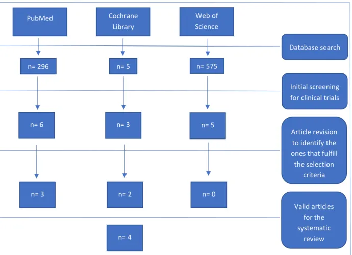
(17) but they have very little effect in the progressive forms of MS, in which a lot of patients end up being classified (55). As miRNAs seem to have great potential as diagnostic and prognostic biomarkers, the objective of this work is to perform a systematic review of the available evidence regarding miRNA expression in response to different treatments (disease modifying therapies and alternative if any), to assess if any of them responds commonly to the different pharmacological approaches and thus identify new trusty biomarkers for MS. Specific investigation questions for this review are: -¿Are there expression changes in miRNA after any pharmacological treatment of MS? (and therefore, considered as biomarkers of treatment response). -¿Is there any similitude between expression changes in conventional disease modifying therapies and alternative therapies in MS? 4.METHODS 4.1 SEARCH STRATEGY. The PRISMA-P statement was followed to conduct this search. As this systematic review is focused on a variety of treatment strategies and miRNA expression, the following search terms were used to search all trials in the PubMed database: a) The first one, with the MeSH terms "multiple sclerosis" AND "microRNA" yielded 296 results. Then, the search was modified to ((Multiple Sclerosis) AND Micro RNA) AND Trial, which narrowed down the results to 6 articles. The “published in the last 10 years” filter was applied, which didn’t change the results. The following search terms and filters were applied to search all trials in the Cochrane Library database: b) The same terms were used for the search, that is “Multiple Sclerosis” and “microRNA”, which yielded 5 total results and 3 results at the “trials” tab. Then, a temporal filter was applied to obtain results published only in the past 10 years, which didn’t change the results. In order to widen the sources for this review, a third search was conducted in the Web of Science database:. 16.
(18) c) The search terms “Multiple Sclerosis” and “microRNA” were used, which yielded 575 results. The term “clinical trial” was added to the search, which yielded 5 results. The temporal publication filter was set to the past 10 years, which didn’t change the results. These three searches were performed repeatedly from 24/01/2019 to 15/05/2019, with no variations in the final outcome. 4.2 SELECTION CRITERIA. •. As inclusion criteria: -. Patients diagnosed with Relapsing-remitting multiple-sclerosis (RRMS).. -. Samples withdrawn preferably before and after but at least after or during any treatment for RRMS.. -. Presence of a control group.. -. Analysis of microRNA expression.. •. As exclusion criteria: -. Papers that don’t focus on multiple sclerosis.. -. Duplicates.. -. Studies performed on animals.. -. Studies published more than 10 years ago.. After the preliminary revision of all articles obtained with all the filters applied, a total of 4 articles were selected: •. “Glatiramer Acetate Treatment Normalizes Deregulated microRNA Expression in Relapsing Remitting Multiple Sclerosis” by Waschbisch A et al.. •. “Altered microRNA expression in B lymphocytes in multiple sclerosis towards a better understanding of treatment effects” by Claudia Sievers et al.. •. “Nanocurcumin is a potential novel therapy for multiple sclerosis by influencing inflammatory mediators” by Sanam Dolati et al.. •. “Nanocurcumin restores aberrant miRNA expression profile in multiple sclerosis, randomized, double-blind, placebo-controlled trial” by Sanam Dolati et al.. 17.
(19) Cochrane Library. PubMed. Web of Science Database search. n= 5. n= 296. n= 575 Initial screening for clinical trials. n= 6. n= 3. n= 5. n= 3. n= 2. n= 0. n= 4. Article revision to identify the ones that fulfill the selection criteria. Valid articles for the systematic review. Figure 5. Flowchart illustrating the search steps for this review.. 4.3 DATA EXTRACTION. For a proper gathering and simplification of all the data of interest, a table was designed containing the following: • • • • • • • • • • •. Author, journal and date of publication. Country where the study has been conducted. Type of study. Objectives. Number of patients that participated (dividing each group). Number of losses if any. Treatment received Results Conclusion Levels of evidence: SIGN grading system for randomized controlled trials (56) and USPSTF for non-randomized trials (57) (supplementary figures 4 and 5 respectively). Methodologic quality: evaluated using the Cochrane Handbook for Systematic Reviews of Interventions (Version 5.1.0).. 18.
(20) Author, journal and date Anne Waschbisch et al. PLOS ONE 6(9): e24604. Sep 2011. Claudia Sievers et al. Clinical Immunology 144, 70–79. May 2012.. Sanam Dolati et al. Journal of Cellular Physiology. Vol 233, Issue7 Pages 5222-5230. July 2018. Sanam Dolati et al. Pharmacological Reports Vol 70, Issue 6, Pages 1158-1167. Dec 2018.. Country Germany. Switzerland. Iran. Iran. Type of study Controlled clinical trial. Controlled clinical trial. Objectives Analyze expression response treatment. miRNA in to. Analyze miRNA expression in treated patients vs untreated vs healthy volunteers. Randomized, double blind, placebocontrolled clinical trial. Identify nanocurcumin effects on microRNAs in the peripheral blood. Randomized, single blind, placebocontrolled clinical trial. Assess the effects of nanocurcumin on inflammatory mediators and miRNA expression. Sample size. Losses. 74 RRMS patients 32 HV. None. Profiling study: 20 RRMS patients 10 HV Validation cohort: 30 RRMS patients 7 HV 50 RRMS patients 35 HV. 50 RRMS patients 35 HV. None. 5 in test group 4 in placebo group 9. Treatment received -Glatiramer acetate (Copaxone®) -IFN-β (Avonex®, Betaferon®, Rebif 22 and 44®) Natalizumab. Nanocurcumin. -Nanocurcumin -IFN-β-1a (Actovex®) stopped at least 3 months before the intervention.. Results. Level of evidence. Bias risk. II-1. Moderate. USPSTF. Changes in miRNA expression after treatment (up and downregulation), they are further explained in the Results section.. II-1. Low. USPSTF. 1+. Low. SIGN. 1+. Moderate. SIGN. Table 3. Main characteristics of the analyzed studies. RRMS: relapsing remitting multiple sclerosis. HV: healthy volunteers.. 19.
(21) 5. RESULTS 5.1 SUMMARY BY ARTICLE. ARTICLE 1. Anne Waschbisch et al. PLOS ONE 6(9): e24604. 2011 . “Glatiramer acetate treatment normalizes deregulated microRNA expression in relapsing remitting multiple sclerosis”. After informed consent, a non-randomized controlled trial was performed with a total of 106 individuals (74 RRMS patients and 32 healthy volunteers), >97% of Caucasian origin.. Table 4. Clinical and demographic details of patients and healthy controls.. Regarding the RRMS patient group, 36 were treatment-naïve, 18 were treated with Interferonbeta (Avonex® n = 5, Rebif® n =8, Betaferon® n =5) and 20 were treated with Glatiramer-acetate (Copaxone®), both treated groups followed the therapies for at least 3 months. As a control group, 32 healthy volunteers were enrolled. Blood samples were drawn from all the participants and were rapidly processed for isolation of peripheral blood mononuclear cells (PBMC). The specifics about PBMC processing, miRNA isolation and posterior Real-Time PCR are summarized in the original article (52). RESULTS BY GROUPS: HEALTHY VOLUNTEERS AND TREATMENT NAÏVE RRMS PATIENTS. After processing and isolation of miRNA, expression was studied in five selected by the authors (miR-20b, miR-142-3p, miR-146a, miR-155 and miR-326) in treatment naïve RRMS patients and healthy controls (HC). No significant difference in the expression of miR-20b (a member of miR106a cluster, which characteristics are briefly described in the next summary) was found, but the expression of the rest miRNA was multiplied by 2 to 3 in treatment naïve patients in comparison to HC. Interestingly, miR-326 is overexpressed during relapses but falls down to normal levels in remission states, so it has been proposed as a biomarker for relapse and remission phases in patients with RRMS (58).. 20.
(22) RESULTS BY GROUPS: GLATIRAMER ACETATE AN D INTERFERON-BETA TREATED PATIENTS VS TREATMENT NAÏVE PATIENTS. The expression of the previously described miRNAs did not differ between IFN-β and treatment naïve (TN) patients, but it did in glatiramer acetate (GA) treated patients, specifically the expression of miR-142-3p (p=0,003) and miR-146a (p=0,028) was decreased to values similar to their normal levels, while miR-155 and miR-326 were comparable between GA and TN patients. In other words, this study would support the predictive value of miR142-3p and mir146a, but not miR-155 and miR-326. Interestingly, in vitro stimulation of PBMC derived from treatment-naïve patients with glatiramer acetate during 72 hours (with a of 40 µg/ml dose) did not result in the downregulation of the reviewed miRNAs, inferring that a more complex immunological process needs to occur for these mechanisms to function the way they do in vivo. ARTICLE 2. Claudia Sievers et al. CLINICAL IMMUNOLOGY 144, 70–79. 2012. Altered microRNA expression in B lymphocytes in multiple sclerosis. After informed consent, a non-randomized controlled trial was performed with a total of 89 individuals: Profiling group. Validation cohort. 10 untreated RRMS patients*. 30 untreated RRMS patients*. 10 treated with Natalizumab. 22 treated with Natalizumab. 10 healthy age and gender 7 healthy volunteers matched volunteers Table 5. Patient and control distribution. *Older than HVs (p<0.05) and natalizumab treated patients (p<0.05).. All untreated patients received no MS specific treatments in 6 months before or throughout the course of this study, and all the patients treated with natalizumab were responders (assessed by clinical changes). After blood was drawn, B lymphocytes where isolated by a physical method and then analyzed by flow cytometry for purity. RNA isolation and profiling, target prediction and miRNA/mRNA expression analysis are all described in detail in the original article (59). A total of 1059 miRNAs were tested in B cells from the profiling group.. 21.
(23) RESULTS BY GROUPS: E XPRESSION OF miRNAs IN UNTREATED PATIEN TS VS HEALTHY CONTROLS. 49 miRNAs were significantly downregulated in B-cells of untreated RRMS patients compared with healthy controls, and no significantly up-regulated miRNAs were found. Among the downregulated miRNAs, three members of the miR-106b-25 cluster were identified (miR-25, miR-106b and miR-93). Interestingly, this family of microns is found altered (down or up) in multiple affections such as different types of cancer, cardiac hypertrophy and MS (60)(61). Also, a member of the miR-17-92 cluster (miR-19b) was identified. This miRNA family is related to the negative regulation of NF-Kb, linked to chronic inflammation in rheumatoid arthritis (62). Two other miRNAs selected in the study where identified, miR-181a (an intrinsic modulator of T Cell sensitivity and positive and negative selection (63)); and miR-93, which contribution to MS in humans is still to be elucidated (64). Also 3 viral miRNAs (EBV-miR-BART7, BART19-5p and BART11-5p) were down-regulated.. RESULTS BY GROUPS: E XPRESSION OF miRNAs IN NATALIZUMAB TREA TED PATIENTS VS UNTREATED RRMS PATIE NTS. 6 months after initiating the treatment with natalizumab, 10 miRNAs were upregulated in treated compared with untreated RRMS patients. Half of them (miR-106b, miR-19b, miR-551a, miR-191 and EBV-BART11-5p) were upregulated comparing untreated RRMS patients with healthy volunteers, but significantly downregulated in natalizumab treated patients versus untreated RRMS patients.. Figure 6. Differentially expressed miRNAs in B lymphocytes between groups. Green circle corresponds to healthy volunteers. Red circle represents patients treated with natalizumab.. 22.
(24) In the follow up of natalizumab treated patients 12 months after starting the treatment, miR19b expression remained stable in 5 out of 8 patients, while miR-106b expression was downregulated in 7 out of 8 patients. From these results, we can assume natalizumab has a direct impact on miRNA regulation, at least in miR-106b, miR-19b, miR-551a, miR-191 and EBV-BART11-5p, as they almost normalize after treatment compared to untreated RRMS patients. ARTICLE 3. SANAM DOLATI ET AL. JOURNAL OF CELLULAR PHYSIOLOGY. VOL 233, ISS. 7. P5222-5230. JULY 2018. NANOCURCUMIN RESTORES ABERRANT MIRNA EXPRESSION PROFILE IN MULTIPLE SCLEROSIS, RANDOMIZED, DOUBLE-BLIND, PLACEBO-CONTROLLED TRIAL. A randomized, double-blind, placebo-controlled trial was performed with a total of 50 patients and 35 healthy volunteers. After losses, the final distribution was 20 RRMS patients treated with nanocurcumin, 21 patients assigned to placebo and 35 healthy controls. Informed consent was obtained prior to participating. The objective of this study was to analyze miRNA expression (obtained by qPCR) at the beginning of the trial and after 6 months of treatment with nanocurcumin, as a way to find the immunomodulatory impact of this polyphenol on miRNA expression. For this purpose, blood was drawn (10-15ml) to all patients prior to the administration of nanocurcumin or placebo capsules (1 daily) for the next 6 months. Healthy volunteers only had to provide one blood sample at the beginning of the study. RNA extraction methods and qPCR analysis of miRNA expression are described at the original paper (65). RESULTS BY GROUPS: RRMS PATIENTS AT BASELINE VS HEALTHY CONTROLS. 27 miRNAs from all RRMS patients and controls were analyzed at baseline. This analysis revealed 13 upregulated miRNAs in RRMS patients: miR-16, miR-17-5p, miR-17-92, miR-27, miR-29b, miR-126, miR-128, miR- 155, miR-326, miR-340, miR-550, and miR-340 in CD4+ T cells and mir-132 in B cells, compared with healthy controls. The analysis also unveiled 16 downregulated miRNAs: miR-15a, miR-16-1, miR-18a, miR-20b, miR-25, miR-106b, miR-363, miR-31, miR-181c, miR-374a and miR-150 in CD4 T cells and in miR16, miR-19b, miR-320a, miR-340 and miR- 599 in B cells of untreated RRMS patients compared with healthy controls. 23.
(25) RESULTS BY GROUPS: NANOCURCUMIN VS PLACEBO GROUP. Nanocurcumin treatment significantly decreased the expression of the upregulated miRNAs listed above compared with the placebo group (complementary figure 1) with two exceptions in miR-17-5p and miR-223 which showed no significant changes. Regarding the downregulated miRNAs, the expression of miR-15a, miR-16, miR-19b, miR-106b, miR-320a, miR-363, miR-31, miR-181c, miR-150, miR-340, and miR-599 was significantly raised in nanocurcumin treated RRMS patients compared to the placebo group (compl. figure 2). From these results it is assumable that nanocurcumin has direct positive effects on multiple miRNAs, turning their expression almost back to normal in some cases, which might open the path to bigger and strongly built clinical trials in the near future, since it is a seemingly innocuous substance (no systemic adverse effects were observed during the 6 month treatment with nanocurcumin). ARTICLE 4. SANAM DOLATI ET AL. PHARMACOLOGICAL REPORTS. VOL 70, ISSUE 6, PAGES 1158-1167. DEC 2018. NANOCURCUMIN IS A POTENTIAL NOVEL THERAPY FOR MULTIPLE SCLEROSIS BY INFLUENCING INFLAMMATORY MEDIATORS. Even though the patient sample seems to be the same for this study and the previously summarized, differences in design have been spotted looking deeper in their execution, there are different authors in both publications, and as this study targets one miRNA not analyzed in the previous study. In addition, this study evaluates miRNA dependent targets. A randomized, single-blind, placebo-controlled trial was performed with a total of 50 patients (at start) and 35 healthy volunteers. After losses, the final distribution was 20 RRMS patients treated with nanocurcumin, 21 patients assigned to placebo, and 35 healthy controls. Demographic details are summarized in the complementary figure 3. Informed consent was obtained prior to participation. The objective of this study was to identify the effects of nanocurcumin on inflammatory mediators in patients with RRMS. For this review, we will only focus on the miRNA analyzed and their targets, which were miR-132, miR-145 and miR-16. PBMC isolation, RNA extraction methods, cDNA synthesis and RT-PCR used for the assessment of miRNA expression are described at the original work (66). The expression levels of miR-145, miR-132 and miR-16 dependent targets were analyzed before and after 6 months of treatment with nanocurcumin or placebo. In the nanocurcumin group, the expression of miR-145 was significantly decreased compared with baseline. At endpoint, the expression of this miRNA was significantly higher in the placebo 24.
(26) group compared with the treatment group (0.94 ± 0.15 versus 0.32 ± 0.17, p<0.0001). The target of miR-145 is Sox2, a transcription factor crucial for oligodendroglial proliferation and differentiation, necessary for myelin formation and repair in multiple sclerosis and other myelinrelated neurological disorders (67). The expression level of Sox2 was increased in the nanocurcumin group compared with baseline and with the placebo group. Along the same path, miR-132 expression was decreased in the nanocurcumin group compared with baseline, and also with the placebo group (1.05 ± 0.25 in placebo vs 0.69 ± 0.29 in nanocurcumin, p=0.0039). The target of miR-132 is sirtuin1, which is known to regulate, among many other things, the NFkB-light-chain-enhancer of activated B cells and biological processes related to axonal integrity (68), reason why it has been proposed as a potential biomarker of response to treatment in MS (69). Accordingly, the expression level of sirtuin1 was significantly increased after treatment with nanocurcumin compared with baseline and also the placebo group. Finally, miR-16 expression was significantly reduced in the nanocurcumin treated group compared with baseline, but with no significant differences between the treatment and placebo group (p=0.69). miR-16 dependent targets are Foxp3 (like miR-31, described in 1.5.1.) and PDCD1 (programmed death-1, an inhibitory receptor on antigen activated T-cells that plays a critical role in induction and maintenance of immune self-tolerance)(70). Even after finding no significant differences between the treatment and placebo group in terms of miR-16 expression, its targets were significantly increased in the nanocurcumin group compared to baseline and the placebo group. These results suggest a direct impact of nanocurcumin on miRNA expression and/or their molecular targets, opening a new path of investigation for the treatment of MS. In regard of clinical changes, EDSS scores (Expanded Disability Status Scale (74), the most used disability quantification method In MS patients) were measured before and after treatment with nanocurcumin and placebo, yielding a significative improvement in the EDSS score of nanocurcumin treated patients, although they had low disability at baseline, but still had a significative difference with the placebo group (results are in complementary figure 6).. 25.
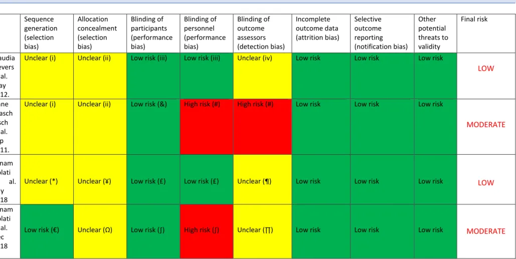
(27) 5.2 METHODOLOGIC AND BIAS ASESSMENT. Claudia Sievers et al. May 2012. Anne Wasch bisch et al. Sep 2011. Sanam Dolati et al. July 2018 Sanam Dolati et al. Dec 2018. Sequence generation (selection bias). Allocation concealment (selection bias). Blinding of participants (performance bias). Blinding of personnel (performance bias). Blinding of outcome assessors (detection bias). Incomplete outcome data (attrition bias). Selective outcome reporting (notification bias). Other potential threats to validity. Unclear (i). Unclear (ii). Low risk (iii). Low risk (iii). Unclear (iv). Low risk. Low risk. Low risk. Final risk. LOW. Unclear (i). Unclear (ii). Low risk (&). High risk (#). High risk (#). Low risk. Low risk. Low risk. MODERATE. Unclear (*). Unclear (¥). Low risk (£). Low risk (£). Unclear (¶). Low risk. Low risk. Low risk. LOW. Low risk (€). Unclear (Ω). Low risk (∫). High risk (∫). Unclear (∏). Low risk. Low risk. Low risk. MODERATE. Table 6. Assessment of the methodological quality according to the Cochrane Handbook for Systematic Reviews of Interventions (Version 5.1.0).. (i) The sample origin is not withdrawn, and no reference to any sequence generation is disclosed. The selection criteria can be partially deducted (treatment assigned or absence of it, being a healthy individual for the controls) but are not described in the papers. (ii) Patients were already in treatment by their neurologists at the beginning of the study. There isn’t a placebo group.. 26.
(28) (iii) There was not any blinding, but because of the study design and absence of conflict of interest of the main investigators, we don’t think it is a significant source of bias. (iv) Blinding of outcome assessors is not mentioned. & As patients were already on treatment by their neurologists, their blinding is unnecessary. # Because of the multiple conflicts of interest (the main investigator (AW) has received funding for travel or speaker honoraria from all the companies that manufacture the investigated drugs (Teva: Copaxone, Bayer: Betaferon, Biogen: Avonex, Merck: Rebif), while others have also received funding from these companies and others), we think the blinding in personnel and outcome assessors should have been used for this trial in order to reduce potential bias. * Randomization is mentioned in the title but not in the rest of the article. There is a table where patient characteristics are displayed, but there’s no mention to the randomization type. ¥ There is no mention about allocation concealment. Double-blinding is mentioned in the title, but again, not in the rest of the article. Nothing is mentioned about the placebo appearance besides it being in capsule form (the real treatment is also in capsule form). £ The only mention about blinding is in the title, as “double-blind”. No other reference in the rest of the paper, but it is specified as double blinded (participant and care provider) in the clinical trial registry. ¶ As the only mention is “double-blind”, we have to assume they mean blinding of both participants and personnel, but not outcome assessors. € Blocked randomization was used. ** Ω According to the investigators single blinding was used, so they knew who got treatment or placebo, and no description of the placebo is provided besides it being in capsule form, and at one point in the paper they describe the actual treatment as a “softgel”, then refer to it as in capsule form too, so it is highly unclear. ** ∫ The clinical trial register says single blinding was used, so the participants didn’t know if they were getting placebo or the real treatment, but the authors apparently did ** ∏ The study did not address this outcome. ** **This data was partially extracted from the Iranian Registry of Clinical Trials, as it was not fully present in the actual paper.. 27.
(29) 6. DISCUSSION The diagnosis of multiple sclerosis is fundamentally based in clinical procedures (medical history and neurological exam), MRI imaging and cerebrospinal fluid analysis. Other frequently used tools are evoked potentials (to assess demyelination) and optical coherence tomography (to analyze optic nerves, mapping retinal structure). All these instruments have proven their value, but there’s still need for new appliances that are able to measure relevant outcomes in treatment response, especially for monitoring response to disease modifying drugs. In this regard, most clinical trials register either clinical or MRI aspects of multiple sclerosis, but biomarkers lack a proper flagship when speaking about treatment response, since oligoclonal bands in cerebrospinal fluid (CSF) have a great prognostic value (their absence is related to a more benign course, and their presence with a higher probability of progression from CIS to clinical MS) but they are not affected by therapeutic agents; in fact, they tend to persist in the CSF after B-cell depletion using Rituximab (71) (an Anti-CD20 monoclonal antibody), or even after an autologous hematopoietic stem cell transplantation (72). Another problem related to OCB is that a lumbar puncture is needed to obtain CSF samples, which is invasive and if needed to perform repeatedly can become very unpleasant for the patient. In this path, numerous CSF parameters are being investigated as biomarkers, such as factors of inflammation, demyelination, remyelination, repair and neuronal damage. Interestingly, polyspecific antibodies against measles, rubella and varicella zoster -so called the MRZ antibody reaction- are becoming a more specific but less sensitive biomarker for MS, since they predict better (with a higher positive predictive value) the conversion from CIS to MS than MRI or oligoclonal bands. Other antibodies related to MS treatment are those generated against disease modifying treatments like natalizumab and IFN-β, which affects their bioactivity, or AntiJC virus antibodies, which high titers are related to an elevated overall incidence of progressive multifocal encephalopathy, especially if natalizumab therapy is extended for years. But as for treatment response, options are generally lacking applicability. We have performed a systematic review about multiple sclerosis since it is an incurable disease to this day, and centered around miRNAs because they are a flourishing line of investigation for a myriad of illnesses. miRNAs in MS seem to have a place, at least as dynamic markers of biochemical treatment response and disease status (58), but bigger and better designed studies are much needed for miRNAs to become a reliable and standardized tool in this field, be it as diagnostic or as prognostic markers. And perhaps therapeutic targets. We have found 4 registered clinical trials specifically focused on miRNA expression analysis in response to 28.
(30) different treatments: Ocrelizumab (anti-CD20 monoclonal antibody) with a cohort design and 130 participants, an open label study with recombinant IFN-β (Rebif and Avonex) in 50 participants, a randomized, double blind but not controlled trial with Vitamin D in a target sample size of 95 individuals, and another randomized, double blind, placebo controlled trial using nanocurcumin. Access to these trials is listed right after the reference list. For this work, 4 studies were selected (52,59,65,66) fulfilling inclusion and exclusion criteria, in which patients had been diagnosed with MS (specifically RRMS, the most common presentation), and treated with multiple pharmacological and alternative approaches (glatiramer acetate, IFN-β, natalizumab and nanocurcumin). All the studies selected are centered around the analysis of miRNA expression during or before and after treatment. For this purpose, in all of the studies reviewed (54,56,62,66) blood samples were withdrawn for PBMC isolation, cultivation and PCR procedures to amplify the target miRNAs. In all four studies expression patterns in miRNA changed after treatment; with glatiramer acetate (partially restoring the expression of miR-146a and miR-142-3p), natalizumab (enhancing the expression of miRNAs downregulated in untreated RRMS patients acting as controls, affecting miR-19b, miR-551a, miR-106b, miR-191 and EBV-miR-BART11-5p) and nanocurcumin (influencing several miRNAs and quite frequently downregulating the upregulated and upregulating the downregulated, with encouraging results but with a study design with room for improvement). After displaying study results, the most important matter is trying to answer the investigation questions raised earlier in this review. As for if there are expression changes in miRNA after any pharmacological treatment, we can extract from Waschbisch et al. that MS patients receiving treatment with glatiramer acetate have a different miRNA expression pattern than treatment naïve MS patients. And from Sievers et al, a similar conclusion can be reached about natalizumab, as treated patients also present a different miRNA expression compared to untreated RRMS patients and healthy controls. But in these two studies patients were already receiving their treatment when the first sample was withdrawn, so we don’t know how their miRNA expression was before treatment, and this represents the greatest flaw in both studies. We can’t be sure their miRNA expression before treatment was similar to the groups of untreated MS patients present in both studies. Another flaw in Sievers et al. is the small sample size, with only 10 individuals in the treatment group; this conditions low statistical power, low. 29.
(31) reproducibility and it can both reduce the chance of detecting a true effect and also reduces the likelihood that a statistically significant result reflects a true effect (73). During this review we could only find one randomized, double blind placebo-controlled trial with treated MS patients that also analyzed miRNA expression. And from this study, even with flaws in its development and in need of new studies reproducing their results, we can answer more robustly to the question that concerns us: are there expression changes in miRNA after any pharmacological treatment of MS? Or maybe more importantly, is there any good quality evidence supporting this claim? To our opinion, we finally have something to hold on. Sanam Dolati as the main investigator and her team from the Tabriz University of Medical Sciences (East Azerbaijan Province, Iran) have put an effort to design two studies with miRNA expression analysis exactly before starting the treatment they are testing and after a reasonable time from baseline (6 months). And the findings are encouraging. After treatment with nanocurcumin, several miRNAs related to immunological mechanisms that directly impact MS pathogenesis are upregulated or downregulated in the direction to their normal status, for example miR-106 and miR-19b (necessary for B cell development), which are typically downregulated in MS patients, after treatment with nanocurcumin are significantly upregulated (2.23±0.88 and 1.55±0.23 fold respectively) compared with baseline. Another question we asked ourselves at the beginning of this review is if is there any similitude between expression changes in conventional disease modifying therapies and alternative therapies in MS. The only miRNAs that show a significant change in expression in a widely used and an alternative treatment are miR-19b and miR-106b.. TREATMENT EFFECT ON miRNA EXPRESSION. miRNA with Natalizumab Claudia Sievers significant et al. May 2012. changes miR-19b Upregulation* miR-106b. Upregulation*. Nanocurcumin Sanam Dolati et al. July 2018. Upregulation** Upregulation**. Table 7. miRNA expression changes with different treatments. *Compared with untreated RRMS patients. **Compared with RRMS patients taking placebo.. These results suggest that some changes in miRNA expression can coincide between treatments, but this doesn’t mean much on its own. One question was if clinical trials support the value of miRNAs as biomarkers for treatment response in multiple sclerosis. We can intuit it is, at least in MS patients with mild disability; in Sanam Dolati et al. Dec 2018 EDSS changes were measured before and after treatment. In this study there was as significant reduction from 1.77±0.33 to 0.98±0.29 in the final EDSS score. This is encouraging, as the result was significantly different from the placebo group at endpoint. 30.
(32) On the other hand, In Sanam Dolati et al. July 2018 EDSS is depicted as <5/5 at baseline but there is no further analysis, in Sievers et al. EDSS status is not shown and in Waschbisch et al. they describe the scores by group, but made no longitudinal comparation. So, in light that we made this affirmation based on just one article, we’ll have to wait for other groups to reproduce similar study designs.. 7.CONCLUSION The most important question in this review is if miRNAs are useful biomarkers for the treatment response in MS. After revision of the selected papers and specific literature about the use of miRNA as biomarkers, we’d have to change the word “useful” and replace it with “promising”. miRNAs have been investigated as MS biomarkers for the past 10 years but the field is still in its first steps, mainly because of the lack of strong studies evaluating their true potential. It will be fundamental for this line of investigation to develop well designed prospective studies with the right objectives, mainly well-defined cohorts with sufficient sample size, blinding of personnel and outcome assessors, and invariably a meticulous management of all the obtained data to establish the right miRNAs as biomarkers. A very important part of all this effort needs to be put in standardization of the processes used to assess miRNA expression, from the sample withdrawal to each step of processing and analysis, for all this data to be as comparable as possible with other publications. Of course, all this evidence will not be useful if validation studies are not carried on to add strength to a generalization of their use. Another interesting line of investigation would be the use of miRNAs as therapeutic targets, as they play a fundamental role in multiple inflammatory and autoimmunity mechanisms.. 31.
(33) 8. ACKNOWLEDGEMENTS. A mi tutora por su dedicación, iopor encauzar el TFG mil y una veces y por estar disponible siempre que lo he necesitado. A Rebeca, por haber hecho todo lo posible para que hayamos terminado esta carrera a la vez. No me cansaré de decir que sin ti no habría tenido cimientos ni para esto ni para nada en mi vida actual, y te estaré eternamente agradecido por ello. Cada día de convivencia, estudio y trabajo contigo ha sido indispensable para convertirme en la mejor versión de mí que he conocido. A tu familia, por acogerme desde el primer día y hacerme partícipe de tantas cosas importantes. A mis padres, mi hermano y mi abuela por permitir, con su incalculable esfuerzo e interés, que fuese capaz de alcanzar la independencia necesaria para hoy estar donde estoy. A mis amigos y amigas, por acoger mis desahogos y apoyarme en esta senda aun cuando ni la había iniciado. A mis “amigos de la UJI”, denominación que fuera de mi control consciente habéis ido perdiendo cada uno y una en su momento, para convertiros en una parte imprescindible de mis pensamientos. Por haberme dado tanta vida durante estos años y haberme hecho sentir, al fin, en comunidad. No os imagináis, o quizá sí, lo importante que esto último es para mí. A mis compañeros y compañeras de trabajo, en las residencias de ancianos de Cabanes y Villareal y en el hospital Rey don Jaime, por haber hecho innumerables cambios de turno conmigo y haberse solidarizado con mi situación para permitir que nunca haya tenido una interferencia sin solución. Y, sobre todo, por haberme hecho sentir en unas prácticas remuneradas todos y cada uno de los cientos de días que hemos pasado juntos. Sois una parte fundamental de mi aprendizaje, y nunca voy a olvidar que la única forma de avanzar es hombro a hombro de forma transversal, sin clases ni barreras de ningún tipo. A todos los profesores y profesoras que, durante estos años, me han visto crecer como médico y como persona, y han conseguido que aprenda y sobre todo que entienda. Y muy en especial, a aquellos y aquellas que con su esfuerzo hicieron florecer a la medicina en esta universidad. Por ello tenéis mi mayor respeto y admiración. And finally to Sanam Dolati and her team, for being brave enough to start studies defying the status quo in pharmacology, and being truly passionate and fruitful towards her profession. Ha sido un camino precioso.. 32.
(34) 9.REFERENCES 1.. Mallucci G, Peruzzotti-Jametti L, Bernstock JD, Pluchino S. The role of immune cells, glia and neurons in white and gray matter pathology in multiple sclerosis. Progress in Neurobiology. 2015.. 2.. Scalfari A, Knappertz V, Cutter G, Goodin DS, Ashton R, Ebers GC. Mortality in patients with multiple sclerosis. Neurology. 2013.. 3.. Goverman J. Autoimmune T cell responses in the central nervous system. Nat Rev Immunol [Internet]. 2009 Jun;9(6):393–407. Available from: http://www.nature.com/articles/nri2550. 4.. Zwibel HL, Smrtka J. Improving quality of life in multiple sclerosis: an unmet need. Am J Manag Care [Internet]. 2011 May;17 Suppl 5:S139-45. Available from: http://www.ncbi.nlm.nih.gov/pubmed/21761952. 5.. Ramagopalan S V., Sadovnick AD. Epidemiology of Multiple Sclerosis. Neurol Clin [Internet]. 2011 May;29(2):207–17. Available from: https://linkinghub.elsevier.com/retrieve/pii/S0733861910001635. 6.. Browne P, Chandraratna D, Angood C, Tremlett H, Baker C, Taylor B V., et al. Atlas of multiple sclerosis 2013: A growing global problem with widespread inequity. Vol. 83, Neurology. 2014. p. 1022–4.. 7.. L. Gallud, J.V. Bagan, A. Cervelló, Y. Jiménez, R. Poveda CG. Multiple sclerosis as first manifestation in oral and facial area: Presentation of four cases. Med oral patol oral cir.bucal [Internet]. 2006;vol.11(2). Available from: http://scielo.isciii.es/scielo.php?script=sci_arttext&pid=S1698-69462006000200010. 8.. Muller M, Terry R, D. S, R. D. Current Theories for Multiple Sclerosis Pathogenesis and Treatment. In: Autoimmune Diseases - Contributing Factors, Specific Cases of Autoimmune Diseases, and Stem Cell and Other Therapies. 2012.. 9.. Goldenberg MM. Multiple sclerosis review. P T. 2012;. 10.. McFarland HF, Martin R. Multiple sclerosis: a complicated picture of autoimmunity. Nat Immunol [Internet]. 2007 Sep 1;8(9):913–9. Available from: http://www.nature.com/articles/ni1507. 11.. Huang Q, Xiao B, Ma X, Qu M, Li Y, Nagarkatti P, et al. MicroRNAs associated with the pathogenesis of multiple sclerosis. J Neuroimmunol [Internet]. 2016 Jun;295–296:148–61. Available from: https://linkinghub.elsevier.com/retrieve/pii/S0165572816300911. 12.. Chastain EML, Duncan DS, Rodgers JM, Miller SD. The role of antigen presenting cells in multiple sclerosis. Biochim Biophys Acta - Mol Basis Dis [Internet]. 2011 Feb;1812(2):265–74. Available from: https://linkinghub.elsevier.com/retrieve/pii/S0925443910001456. 13.. Ciccarelli O, Barkhof F, Bodini B, Stefano N De, Golay X, Nicolay K, et al. Pathogenesis of multiple sclerosis: Insights from molecular and metabolic imaging. Lancet Neurol [Internet]. 2014;13(8):807–22. Available from: http://dx.doi.org/10.1016/S1474-4422(14)70101-2. 14.. Engelhardt B, Ransohoff RM. The ins and outs of T-lymphocyte trafficking to the CNS: Anatomical sites and molecular mechanisms. Trends Immunol. 2005;26(9):485–95.. 15.. Milo R. Therapies for multiple sclerosis targeting B cells. Croat Med J [Internet]. 2019 Apr;60(2):87–98. Available from: https://www.ncbi.nlm.nih.gov/pmc/articles/PMC6509632/. 16.. Krumbholz M, Derfuss T, Hohlfeld R, Meinl E. B cells and antibodies in multiple sclerosis pathogenesis and therapy. Nat Rev Neurol [Internet]. 2012;8(11):613–23. Available from: 33.
(35) http://dx.doi.org/10.1038/nrneurol.2012.203 17.. von Büdingen H-C, Gulati M, Kuenzle S, Fischer K, Rupprecht TA, Goebels N. Clonally expanded plasma cells in the cerebrospinal fluid of patients with central nervous system autoimmune demyelination produce “oligoclonal bands.” J Neuroimmunol [Internet]. 2010 Jan;218(1–2):134– 9. Available from: https://linkinghub.elsevier.com/retrieve/pii/S016557280900397X. 18.. Pryce G, Baker D. Oligoclonal bands in multiple sclerosis; Functional significance and therapeutic implications. Does the specificity matter? Mult Scler Relat Disord [Internet]. 2018;25:131–7. Available from: https://doi.org/10.1016/j.msard.2018.07.030. 19.. Ontaneda D, Fox RJ. Progressive multiple sclerosis. Curr Opin Neurol . 2015;. 20.. Gajofatto A, Turatti M, Benedetti MD. Primary progressive multiple sclerosis: current therapeutic strategies and future perspectives. Expert Review of Neurotherapeutics. 2017.. 21.. Rice CM, Cottrell D, Wilkins A, Scolding NJ. Primary progressive multiple sclerosis: Progress and challenges. J Neurol Neurosurg Psychiatry. 2013;84(10):1100–6.. 22.. Lublin FD, Reingold SC, Cohen JA, Cutter GR, Sørensen PS, Thompson AJ, et al. Defining the clinical course of multiple sclerosis: The 2013 revisions. Neurology. 2014.. 23.. McDonald WI, Compston A, Edan G, Goodkin D, Hartung HP, Lublin FD, et al. Recommended diagnostic criteria for multiple sclerosis: guidelines from the International Panel on the diagnosis of multiple sclerosis. Ann Neurol [Internet]. 2001 Jul;50(1):121–7. Available from: http://www.ncbi.nlm.nih.gov/pubmed/11456302. 24.. Polman CH, Reingold SC, Edan G, Filippi M, Hartung H-P, Kappos L, et al. Diagnostic criteria for multiple sclerosis: 2005 revisions to the “McDonald Criteria.” Ann Neurol [Internet]. 2005 Dec;58(6):840–6. Available from: http://doi.wiley.com/10.1002/ana.20703. 25.. Polman CH, Reingold SC, Banwell B, Clanet M, Cohen JA, Filippi M, et al. Diagnostic criteria for multiple sclerosis: 2010 Revisions to the McDonald criteria. Ann Neurol. 2011;69(2):292–302.. 26.. Thompson AJ, Banwell BL, Barkhof F, Carroll WM, Coetzee T. Diagnosis of multiple sclerosis: 2017 revisions of the McDonald criteria. Lancet Neurol. 2018;. 27.. Deisenhammer F, Zetterberg H, Fitzner B, Zettl UK. The Cerebrospinal Fluid in Multiple Sclerosis. Front Immunol [Internet]. 2019 Apr 12;10. Available from: https://www.frontiersin.org/article/10.3389/fimmu.2019.00726/full. 28.. Willer CJ, Dyment DA, Risch NJ, Sadovnick AD, Ebers GC. Twin concordance and sibling recurrence rates in multiple sclerosis. Proc Natl Acad Sci [Internet]. 2003 Oct 28;100(22):12877– 82. Available from: http://www.pnas.org/cgi/doi/10.1073/pnas.1932604100. 29.. Ebers GC, Yee IML, Sadovnick AD, Duquette P. Conjugal multiple sclerosis: Population-based prevalence and recurrence risks in offspring. Ann Neurol [Internet]. 2000 Dec;48(6):927–31. Available from: http://doi.wiley.com/10.1002/15318249%28200012%2948%3A6%3C927%3A%3AAID-ANA14%3E3.0.CO%3B2-F. 30.. Herrera BM, Ramagopalan S V., Lincoln MR, Orton SM, Chao MJ, Sadovnick AD, et al. Parent-oforigin effects in MS: Observations from avuncular pairs. Neurology [Internet]. 2008 Sep 9;71(11):799–803. Available from: http://www.neurology.org/cgi/doi/10.1212/01.wnl.0000312377.50395.00. 31.. Cantó Puig E, Comabella M. Biomarcadores en la esclerosis múltiple : estado actual. Rev Española Escler Mult. 2012;. 32.. Gourraud P-A, Harbo HF, Hauser SL, Baranzini SE. The genetics of multiple sclerosis: an up-todate review. Immunol Rev [Internet]. 2012 Jul;248(1):87–103. Available from: 34.
Figure


+7


Documento similar
This study sought to analyze tumor CXCL5 gene expression in six patients with different efficacy of BVZ-containing CT in terms of the tumor response to treatment..
The increase in UCP3 expression in response to H 2 O 2 or HNE treatment that we found in this work, together with its proposed function in the control of mitochondrial
Additionally, deregulation of miR-1246, miR-5100 and miR-338-3p was observed in severe asthmatic patients after eight weeks of therapy, and a correlation was found between miR-1246
Taken together, miRNA and mRNA expression analysis identified a large number of miRNAs and genes in resistance arteries that are differentially expressed upon maturation, some of
We wondered whether this weak upregulation of SNAIL1 in MDCK-NBL cells was due to the high PRRX1 levels which in turn could activate the expression of miR-15
Our finding of lower 30-day mortality in patients with elevated levels of miR-16-5p and miR-146a at admission for CAP could reflect a better inflammatory response against the
To identify genes potentially regulated by miR-183-3p and miR-21-3p, we considered only samples from the discovery series with miRNA and mRNA expression data available (n=434),
In summary we observed a remarkable juxtaglomeru- lar apparatus hyperplasia and increased renin expression in a focal segmental glomerulosclerosis patient on long term treatment
