Alejandra Fernández, Javier Fernández, Maureen Marshall, René Martínez, Sven Niklander & Ziyad S. Haidar.
Facultad de Odontología, Univer-sidad Andrés Bello, Chile. BioMAT’X, Centro de Investigación Biomédica, Facultad de Medi-cina, Universidad de Los Andes, Chile. Facul-tad de Medicina, Universidad de Chile, Chile.
Facultad de Odontología, Universidad de Los Andes, Chile.
Ziyad S. Haidar. Universidad de Los Andes, BioMAT´X, Facultad de Odontología. Mons. Álvaro del Portillo 12.455 - Las Condes, Santiago, Chile. Phone: (56-2) 26181372 Ext. (56-2) 22149468 . E-mail: [email protected]
To evaluate the expression of the epidermal growth factor receptor (EGFR) and mean vascular density (MVD) in normal oral mucosa (NOM), oral epithelial dysplasia (OED) and oral squamous cell carcinoma (OSCC). Material and methods: Descriptive case study. Nineteen histological samples diagnosed with NOM, 18 diag-nosed with OED, and 19 with OSCC, were analyzed with immunohistochemistry against EGFR and CD31. EGFR expression was evaluated by extent and intensity of its expression in normal, dysplastic and neoplastic epithelium. MVD was determined through the detection of blood vessels by antibodies against CD31. Results: Extension of EGFR expression was highest in OSCC followed by OED and lowest in NOM, resulting in significant different between the degrees of extension (p<0.001). Intensity of EGFR was similar in NOM, OED and OSCC, without differences in its expres-sion (p=0.533). Differences in MVD were found between NOM and OSCC groups (p<0.01), and between OED and OSCC groups (p<0.01), with no differences between NOM and OED groups (p=0.91). MVD was 21.17±4.98 in NOM, 23.40±5.77 in OED and 33.92±8.39 in OSCC. Conclusion: EGFR is expressed in normal, dysplastic or neoplastic oral epithelium. However, the extent of its expression is greater as malig-nancy increases. MVD varies according to the diagnosis.
Oral cavity, mouth neoplasms, epidermal growth factor receptor, pathologic angiogenesis.
Oral squamous cell carcinoma (OSCC) is the most common malignant neoplasm of the oral cavity. An incidence of 500,000 cases per year has been reported worldwide. OSCC may originate from malignant transformation of normal oral mucosa (NOM), and from potentially malignant lesions with different degrees of oral epithelial dysplasia (OED).
The genesis of malignant neoplasms, such as OSCC, is a complex process involving a breakdown in the regulation pathways of cell division, differentiation, death and angiogenesis. In this regard, protooncogenes are the physiological regulators of proliferation and differentiation of normal cells. Overexpression of their mutated counterparts, the oncogenes, plays a key role in carcinogenesis. Oncogenes encode, among others, growth factor receptors, which may cause uncontrolled cell proliferation. In the case of carcinomas, one key receptor is the epidermal growth factor receptor (EGFR). Its mutation stimulates mitosis and inhibits the apoptosis of neoplastic keratinocytes. This receptor is expressed both in normal epithelia and in those subjects with pathologies such as epithelial dysplasia and carcinomas, where proliferation and differentiation of
None.
The study was approved by the Bioethics Committee of Universidad An-drés Bello (Folio No 033).
None.
This work was carried out in collaboration between all authors. All au-thors read and approved the final manuscript.
None.
Fernández A, Fernández J, Marshall M, Martínez R, Niklander S & Haidar ZS. Difference in EGFR expression and mean vascular density in normal oral mucosa, oral epithelial dysplasia and oral squamous cell carcinoma. J Oral Res 2017; 6(2): 39-45.
01/13/2017 01/30/2017
keratinocytes are altered.
The complex interaction between neoplastic cells and their environment plays a central role in carcinogenesis, as environmental changes may facilitate cell growth, invasion and metastasis. In the case of OSCC, the microenvironment consists of fibroblasts, deposits of the extracellular matrix, immune system cells, lymphatic vessels and blood vessels. The vascular system supplies oxygen and nutrients to the neoplastic cells. In addition, newly formed endothelial cells secrete growth factors that act on themselves and on adjacent neoplastic cells stimulating their proliferation. Malignant neoplasms induce angiogenesis in a volume up to 2-3 mm³ and this value represents the critical distance by which nutrients and oxygen can diffuse from the blood vessels. Therefore, the understanding of angiogenesis is critical to comprehend the malignant transformation of epithelial lesions of the oral cavity such as OED or OSCC.
CD31 is one of the most important molecular markers for evaluating angiogenesis, through the calculation of the mean vascular density (MVD). CD31 is a protein present in the intercellular junction of endothelial cells in developing or already developed blood vessels.
The aim of this study was to evaluate EGFR expression and angiogenesis, through the mean vascular density (MVD), in NOM, OED and OSCC.
A descriptive case study was designed. The study was appro-ved by the Bioethics Committee of Universidad Andrés Bello (Folio No 033). Participants were asked to sign an informed consent.
Nineteen samples diagnosed with NOM, 18 diagnosed with OED, and 19 with OSCC, were collected. Samples of NOM were obtained from alveolar ridge mucosa of mandibu-lar third momandibu-lars, from individuals who had undergone surgery at the School of Dentistry of Universidad Andrés Bello, Viña del Mar, between March and July 2014. Samples of OED and OSCC were obtained from paraffin-embedded samples co-llected between 2004 and 2012 by the Oral Histopathology Service at the School of Dentistry, Universidad Andrés Bello, Viña del Mar.
Inclusion criteria included paraffin-embedded samples
with enough tissue to obtain three histological section of 4 microns each, diagnosed histologically with NOM, OED or OSCC, with information regarding the patient’s age, gender and place of residence. All age ranges, both sexes and location of the lesion in the oral mucosa were included. Exclusion cri-teria consisted of OSCC samples from secondary to metastasis stage and histological lamellae with methodological artifacts.
To confirm the diagnosis of NOM, OED and OSCC of the selected samples, two independent, previously standardi-zed and calibrated pathologists examined hematoxylin-eosin stained sections under Olympus® CX-31 light microscopy (Olympus Corporation, Japan). In order to make the diagno-sis, they considered the criteria proposed by the World Health Organization in a double-blind examination.
Four micron sections were obtained and mounted on xyla-nized, dewaxed slides and hydrated with distilled water. Once hydrated, an antigenic recovery process was performed on a steamer using citrate buffer, pH 6. The endogenous peroxi-dase enzyme was then blocked by the application of 3% v/v hydrogen peroxide. Sections were incubated overnight with rabbit anti-EGFR monoclonal antibody (diluted 1: 100; Ven-tana Medical System Inc, Tucson Arizona, USA). Immunos-taining was performed with Envision system (Dako, Santa Clara, USA.) according to the manufacturer’s instructions. Peroxidase activity was measured by the application of the diaminobenzidine chromogen substrate.
Samples were processed as described for the immunohis-tochemistry of EGFR, but were placed on a poly-L-lysine coated slide (BioSB, Santa Barbara, USA). These sections were incubated with human CD-31 monoclonal antibody (Clone JC70A, IgG-1, kappa, Dako, Carpenteria, Califor-nia, USA), diluted 1:40, using the avidin-biotin-peroxidase complex detection method, at a temperature of 37°C for 32 minutes. Antigenic recovery was performed at 95-100°C for 60 minutes with CC1 Standard solution (Cell Condi-tioning Solution-1, Ventana Medical Systems, Inc.).
cytoplas-Distribution of patients by their demographic variables and clinical characteristics.
NOM: normal oral mucosa; OED: oral epithelial dysplasia; OSCC: Oral squamous cell carcinoma.
mic staining, compared with negative and positive controls placed on the same slide. The positive control consisted of a segment of placenta analyzed with the complete immunohistochemical technique for EGFR. The negative control was obtained by omission of the primary antibody.
The intensity of immunostaining of EGFR in NOM, OED and OSCC was qualitatively evaluated and cate-gorized nominally and arbitrarily into 0: negative im-munostaining, 1: mild imim-munostaining, 2: moderate immunostaining and 3: marked immunostaining. The extent of EGFR immunostaining in the epithelial thic-kness of NOM, OED and OSCC was categorized as 0=0%; 1=1 to 25%; 2=26-50%; 3=51-75%; 4=75-100%.
Samples were analyzed by two blind calibrated exa-miners (RM and AF). Any tubular structure coated by endothelial cells, individual endothelial cells or in is-lets immunoreactive with the antibody against CD31, was considered as a blood vessel. Each sample was compared with a positive and negative control in each histological slides. The positive control consisted of a segment of angiosarcoma stained with the complete immunohistochemical technique against CD31. The immunonegative control was obtained by omission of the primary antibody.
To determine MVD, samples were observed under an Olympus® CX-31 light microscope (Olympus Corpora-tion, Japan), and 3 consecutive hot spots were selected that corresponded to the areas of greatest vasculariza-tion. Each hot spot was photographed at 40x magnifi-cation, using a 5.1 megapixel Micrometrics® Model 518
CU digital camera built into the microscope. In each image blood vessels were counted (20x objective lens and 10x ocular lens, 0.7386 mm2/field).
MVD of each sample was determined using the fo-llowing formula: MVD=(Number of vessels in Hot spot 1+ Number of vessels in Hot spot 3 + No of Hot spot 3 vessels)/3.
Parametric and non-parametric tests were performed according to the nature of the variables. Kruskal-Wallis test was used for the analysis of the extent and intensi-ty of EGFR according to diagnosis, and Conover-Iman post-hoc test was performed to evaluate differences in the range averages. The comparison of the mean number of vessels according to diagnosis was performed using ANOVA, with Bonferroni post hoc test. Student’s t-test of independent samples was used to evaluate the rela-tionship between EGFR extension and the number of vessels. Statistical significance was set at p<0.05. Sta-tistical analysis was performed with STATA 12® (Stata-CorpLP, Texas, USA).
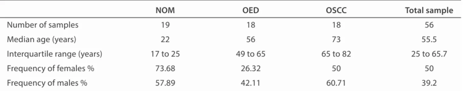
The number of samples included in the study, age and gender distribution, for each diagnosis and the totality of the samples, are shown in Table 1.
Regarding EGFR expression by extension and intensity (Figure 1, Table 2), there was a difference in the extent of EGFR expression when comparing each diagnostic group (p<0.001). In contrast, no differences were found in the intensity of detected EGFR when comparing each diagnos-tic group (p=0.533). On the other hand, when analyzing OSCC samples according to their degree of differentiation,
NOM OED OSCC Total sample
Number of samples 19 18 18 56
Median age (years) 22 56 73 55.5
Interquartile range (years) 17 to 25 49 to 65 65 to 82 25 to 65.7
Frequency of females % 73.68 26.32 50 50
Extension Intensity
2 3 4 1 2 3
26-50% 51-75% 476-100% Mild Moderate Marked
n (%) n (%) n (%) n (%) n (%) n (%)
NOM 1 (5.26) 18 (94.74) - - - - 11 (57.89) 8 (42.11)
OED - - 14 (77.78) 4 (22.22) 1 (5.56) 12 (55.56) 7 (38.89)
OSCC - - 2 (10.53) 17 (89.17) 1 (5.26) 7 (36.84) 11 (57.89)
EGFR expression (Brown) in normal oral mucosa (A) and, oral epithelial dysplasia (B:) mild (C) and marked (D). Oral squamous cell carcinoma: well differentiated (E) and poorly differentiated (F). 10x magnification.
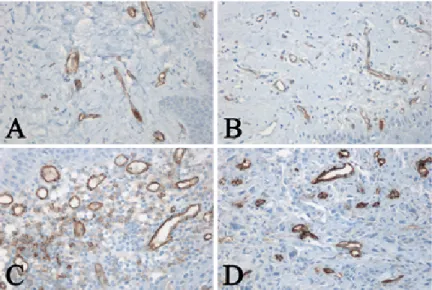
Expression (Brown) of CD31 showing blood vessels in normal oral mucosa (A), mild oral epithelial dysplasia (B), and oral squamous cell carcinoma (C) and (D). 10x magnification.
Comparison of the extent and intensity of EGFR expression in normal oral mucosa, oral epithelial dysplasia and oral squamous cell carcinoma.
no differences were found in the intensity and extent of EGFR (p=0.704 and p=0.816, respectively).
Regarding MVD (Figure 2), a mean of 21.17±4.98 ves-sels were observed in NOM, 23.40±5.77 vesves-sels in OED, and 33.92±8.39 vessels in OSCC. The MVD was different between NOM and OSCC (p<0.01) and between OED and OSCC (p<0.01). However, no differences were found between NOM and OED groups (p=0.91).
By relating MVD and the extent of EGFR expression in categories 3 and 4, at category 3 of EGFR expression there was a mean of 22.79±6.65 vessels, and at category 4 there was a mean of 31.55±8.83 vessels (p<0.001). In contrast, no differences were found between the number of vessels and the EGFR expression (p=0.351).
In the last decade, interest has increased in identifying markers that may allow prediction of malignant trans-formation of normal and dysplastic oral epithelium into an OSCC. This is due to the fact that even in spite of advances in scientific knowledge, the histopathological diagnosis remains the gold standard for making diagnos-tic and therapeudiagnos-tic decisions, which does not allow the prediction of how an oral lesion will evolve.
It was found that dysplastic lesions of the oral mucosa preceded the neoplastic epithelial pathology, which co-incides with that reported in the literature. This may be because the pathogenesis of both OED and OSCC is associated with the same carcinogenic stimuli. However, in OSCC, it is associated with a long accumulation of its effects. In the OED group, no gender bias was found. However, a higher frequency of OSCC was found among women. There is controversy as to which sex is most af-fected. This could be explained by region specific habits, such as chewing tobacco or betel nut.
Regarding evaluation of EGFR, authors such as Rössle et al. and Rajesweri et al. and the authors of this study found that in all the samples analyzed, regardless of di-agnosis, immunostaining of EGFR was always positive. However, there were variations in its intensity range. The extension of EGFR expression in the NOM group was mainly of type 3, reflecting that EGFR was expressed more in the basal, suprabasal and spinosum strata, disap-pearing in the superficial stratum, which coincides with
that described in the literature. The intensity of EGFR was mainly moderate and marked, which coincides with Rajesweri et al. These results can be explained because EGFR is expressed in proliferating epithelial cells, such as in the basal stratum, and its expression is lost in the superficial or corneous stratum where the prevalence of proliferative cells decrease. Unlike the NOM group, EGFR expression in the OED group in some samples involved even the stratum corrneum, which may be due to an increase in normal proliferative activity, although in an irregular growth pattern. Its intensity was catego-rized as “marked”, similarly to that of the NOM group, coinciding with the data reported in the literature. In the OSCC samples, it was found that in most cases the extension of EGFR covered the entire sample and its in-tensity was defined as “marked” in a high percentage. Rössle et al. and Sarkis et al. reported similar findings. From this data, it could be inferred that the extent and in-tensity found for EGFR in carcinomas would be revealing a completely uncontrolled growth of neoplastic epithelial cells and that the extent of EGFR expression is higher as malignancy increases.
In the present study, no difference was found between intensity and extent of EGFR regarding different degrees of OSCC differentiation, coinciding with Ragomir et al. This fact may indicate that EGFR is not related to the degree of differentiation of neoplastic keratinocytes. Al-though Sarkis et al. and Laimer et al., consider that both the extent and intensity of EGFR reflect an alter-ation in the regulalter-ation of cell proliferalter-ation. The results of this study suggest that the alteration of cell proliferation would be mainly represented by the number of cells af-fected and not by expression intensity.
but used different types of markers for blood vessels. How-ever, it should be noted that most of the studies, indepen-dent of the marker used, coincide with the results of this study in relation to the increase in MDV in the transition from NOM to OED to OSCC.
To our knowledge, the association between EGFR and CD expression has not been previously studied in oral samples of NOM, OED and OSCC. This study demon-strates that when the EGFR extent was greater than 50%, independent of its diagnosis, MDV increased. These re-sults suggest that EGFR could positively regulate angio-genesis, probably via secretion of growth factors. a recent publication showed that a decrease in EGFR was involved in the decrease of VEGF expression and that the activa-tion of EGFR-VEGF favored angiogenesis in
hepatocel-lular carcinoma.
Some consideration should be given to the limitations of this study, such as sample size, due to the fact that OSCC is a low prevalence pathology and the large differ-ence in the age range of subjects in the NOM group, when compared to subjects in the OED and OSCC groups. The latter could be explained by the samples of NOM, which were obtained from mucosa that included third molars.
EGFR is expressed in normal, dysplastic or neoplastic oral epithelium. However, the extent of its expression is greater as malignancy increases. MVD varies according to the diagnosis.
Fernández A, Córdova P, Badenier O, Esguep A. Epidemiolo-gical characterization of oral cancer. Literature review. J Oral Res. 2015;4(2):137–45.
Irimie AI, Braicu C, Cojocneanu-Petric R, Berindan-Neagoe I, Cam-pian RS. Novel technologies for oral squamous carcinoma biomarkers in diagnostics and prognostics. Acta Odontol Scand. 2015;73(3):161–8.
Scully C. Challenges in predicting which oral mucosal potentially malignant disease will progress to neoplasia. Oral Dis. 2014;20(1):1–5.
Veeravarmal V, Austin RD, Siddavaram N, Thiruneelakandan S, Nassar MH. Caspase-3 expression in normal oral epithelium, oral sub-mucous fibrosis and oral squamous cell carcinoma. J Oral Maxillofac Pathol. 2016;20(3):445–52.
Pitiyage G, Tilakaratne WM, Tavassoli M, Warnakulasuriya S. Molecular markers in oral epithelial dysplasia: review. J Oral Pathol Med. 2009;38(10):737–52.
Rössle M, Weber CS, Züllig L, Graf N, Jochum W, Stöckli SJ, Moch H, Huber GF. EGFR expression and copy number changes in low T-sta-ge oral squamous cell carcinomas. Histopathology. 2013;63(2):271–8.
Christensen ME, Therkildsen MH, Hansen BL, Albeck H, Han-sen GN, Bretlau P. Epidermal growth factor receptor expression on oral mucosa dysplastic epithelia and squamous cell carcinomas. Eur Arch Otorhinolaryngol. 1992;249(5):243–7.
Bruno A, Pagani A, Pulze L, Albini A, Dallaglio K, Noonan DM, Mortara L. Orchestration of angiogenesis by immune cells. Front Oncol. 2014;4:131.
Shivamallappa SM, Venkatraman NT, Shreedhar B, Mohanty L, Shenoy S. Role of angiogenesis in oral squamous cell carcinoma deve-lopment and metastasis: an immunohistochemical study. Int J Oral Sci. 2011;3(4):216–24.
Pujari RK, Vanaki SS, Puranik RS, Desai RS, Motupalli N, Ha-lawar S. Histomorphometric analysis of vascularity in normal buccal mucosa, leukoplakia, and squamous cell carcinoma of buccal mucosa. J Oral Maxillofac Pathol. 2013;17(3):334–9.
Ries J, Vairaktaris E, Agaimy A, Bechtold M, Gorecki P, Neukam FW, Nkenke E. The relevance of EGFR overexpression for the predic-tion of the malignant transformapredic-tion of oral leukoplakia. Oncol Rep. 2013;30(3):1149–56.
Sarkis SA, Abdullah BH, Abdul Majeed BA, Talabani NG.
Immu-nohistochemical expression of epidermal growth factor receptor (EGFR) in oral squamous cell carcinoma in relation to proliferation, apoptosis, angiogenesis and lymphangiogenesis. Head Neck Oncol. 2010;2:13.
Cheng SH, Liu JM, Liu QY, Luo DY, Liao BH, Li H, Wang KJ. Prognostic role of microvessel density in patients with renal cell carcino-ma: a meta-analysis. Int J Clin Exp Pathol. 2014;7(9):5855–63.
Krishnan L, Karpagaselvi K, Kumarswamy J, Sudheendra US, Santosh KV, Patil A. Inter- and intra-observer variability in three gra-ding systems for oral epithelial dysplasia. J Oral Maxillofac Pathol. 2016;20(2):261–8.
Geetha KM, Leeky M, Narayan TV, Sadhana S, Saleha J. Grading of oral epithelial dysplasia: Points to ponder. J Oral Maxillofac Pathol. 2015;19(2):198–204.
Shirako Y, Taya Y, Sato K, Chiba T, Imai K, Shimazu Y, Aoba T, Soeno Y. Heterogeneous tumor stromal microenvironments of oral squamous cell carcinoma cells in tongue and nodal metastatic lesions in a xenograft mouse model. J Oral Pathol Med. 2015;44(9):656–68.
Abdulmajeed AA, Farah CS. Can immunohistochemistry serve as an alternative to subjective histopathological diagnosis of oral epithelial dysplasia? Biomark Cancer. 2013;5:49–60.
Noda Y, Kishino M, Sato S, Hirose K, Sakai M, Fukuda Y, Mu-rakami S, Toyosawa S. Galectin-1 expression is associated with tumour immunity and prognosis in gingival squamous cell carcinoma. J Clin Pathol. 2017;70(2):126–133.
Chrun ED, Modolo F, Vieira DS, Borges ÁL, Castro RG, Daniel FI. Immunoexpression of HDAC1, HDAC2 and HAT1 in actinic cheilitis and lip squamous cell carcinoma. Oral Dis. 2017:[Epub ahead of print].
Laimer K, Spizzo G, Gastl G, Obrist P, Brunhuber T, Fong D, Bar-bieri V, Jank S, Doppler W, Rasse M, Norer B. High EGFR expression predicts poor prognosis in patients with squamous cell carcinoma of the oral cavity and oropharynx: a TMA-based immunohistochemical analy-sis. Oral Oncol. 2007;43(2):193–9.
Ziebart T, Blatt S, Günther C, Völxen N, Pabst A, Sagheb K, Kühl S, Lambrecht T. Significance of endothelial progenitor cells (EPC) for tumorigenesis of head and neck squamous cell carcinoma (HNSCC): possible marker of tumor progression and neovascularization? Clin Oral Investig. 2016;20(8):2293–300.
precursor lesions and malignant transformation--who, where, what, and when? Br J Oral Maxillofac Surg. 2015;53(9):831–5.
Niu LX, Feng ZE, Wang DC, Zhang JY, Sun ZP, Guo CB. Prognos-tic factors in mandibular gingival squamous cell carcinoma: A 10-year retrospective study. Int J Oral Maxillofac Surg. 2017;46(2):137–43.
Matsushita Y, Yanamoto S, Takahashi H, Yamada S, Naruse T, Sakamoto Y, Ikeda H, Shiraishi T, Fujita S, Ikeda T, Asahina I, Umeda M. A clinicopathological study of perineural invasion and vascular inva-sion in oral tongue squamous cell carcinoma. Int J Oral Maxillofac Surg. 2015;44(5):543–8.
Narayan TV, Shilpashree S. Meta-analysis on clinicopathologic risk factors of leukoplakias undergoing malignant transformation. J Oral Maxillofac Pathol. 2016;20(3):354–61.
Bijina BR, Ahmed J, Shenoy N, Ongole R, Shenoy S, Baliga S. De-tection of human papilloma virus in potentially malignant and malignant lesions of the oral cavity and a study of associated risk factors. South Asian J Cancer. 2016;5(4):179–81.
Dholam KP, Chouksey GC. Squamous cell carcinoma of the oral cavity and oropharynx in patients aged 18-45 years: A case-control study to evaluate the risk factors with emphasis on stress, diet, oral hy-giene, and family history. Indian J Cancer. 2016;53(2):244–51.
Rajeswari MR, Saraswathi TR. Expression of epithelial growth factor receptor in oral epithelial dysplastic lesions. J Oral Maxillofac
Pa-thol. 2012;16(2):183–8.
Ribeiro DC, Gleber-Netto FO, Sousa SF, Bernardes VD, Gui-marães-Abreu MH, Aguiar MC. Immunohistochemical expression of EGFR in oral leukoplakia: association with clinicopathological features and cellular proliferation. Med Oral Patol Oral Cir Bucal. 2012;17(5):e739–44.
Dragomir LP, Mărgăritescu C, Florescu A, Olimid AD, Dragomir M, Popescu MR. The immunoexpression of EGFR and Her2/neu in oral squamous carcinoma. Rom J Morphol Embryol. 2012;53(3):597–601.
Yadav L, Puri N, Rastogi V, Satpute P, Sharma V. Tumour An-giogenesis and Angiogenic Inhibitors: A Review. J Clin Diagn Res. 2015;9(6):XE01–5.
Sathyakumar M, Sriram G, Saraswathi T, Sivapathasundha-ram B. Immunohistochemical evaluation of mast cells and vascular endothelial proliferation in oral precancerous lesion-leukoplakia. J Oral Maxillofac Pathol. 2012;16(3):343–8.
Basnaker M, Sr S, Bnvs S. Expression of Endoglin (CD-105) and Microvessel Density in Oral Dysplasia and Squamous Cell Car-cinoma. J Clin Diagn Res. 2014;8(9):ZC91–4.