R E S E A R C H A R T I C L E
Open Access
Role of the microtubule-targeting drug vinflunine
on cell-cell adhesions in bladder epithelial tumour
cells
Luis A Aparicio
2†, Raquel Castosa
1†, Mar Haz-Conde
1, Marta Rodríguez
1, Moisés Blanco
1, Manuel Valladares
2and Angélica Figueroa
1*Abstract
Background:Vinflunine (VFL) is a microtubule-targeting drug that suppresses microtubule dynamics, showing
anti-metastatic properties bothin vitroand in living cancer cells. An increasing body of evidence underlines the influence of the microtubules dynamics on the cadherin-dependent cell-cell adhesions. E-cadherin is a marker of epithelial-to-mesenchymal transition (EMT) and a tumour suppressor; its reduced levels in carcinoma are associated with poor prognosis. In this report, we investigate the role of VFL on cell-cell adhesions in bladder epithelial tumour cells.
Methods:Human bladder epithelial tumour cell lines HT1376, 5637, SW780, T24 and UMUC3 were used to analyse
cadherin-dependent cell-cell adhesions under VFL treatment. VFL effect on growth inhibition was measured by using a MTT colorimetric cell viability assay. Western blot, immunofluorescence and transmission electron microscopy analyses were performed to assess the roles of VFL effect on cell-cell adhesions, epithelial-to-mesenchymal markers and apoptosis. The role of the proteasome in controlling cell-cell adhesion was studied using the proteasome inhibitor MG132. Results:We show that VFL induces cell death in bladder cancer cells and activates epithelial differentiation of the remaining living cells, leading to an increase of E-cadherin-dependent cell-cell adhesion and a reduction of mesenchymal markers, such as N-cadherin or vimentin. Moreover, while E-cadherin is increased, the levels of Hakai, an E3 ubiquitin-ligase for E-cadherin, were significantly reduced in presence of VFL. In 5637, this reduction on Hakai expression was blocked by MG132 proteasome inhibitor, indicating that the proteasome pathway could be one of the molecular mechanisms involved in its degradation.
Conclusions:Our findings underscore a critical function for VFL in cell-cell adhesions of epithelial bladder tumour cells, suggesting a novel molecular mechanism by which VFL may impact upon EMT and metastasis.
Keywords:Microtubule, Cell-cell contacts, E-cadherin, Vinflunine, Bladder cancer
Background
Bladder cancer is a common malignancy affecting the genitourinary system that represents the fifth most com-mon cancer in the world. Transitional cell carcinoma (TCC) represents 95% of these tumours [1]. Most blad-der cancers (70%-80%) present non-muscle invasive or superficial disease confined to the bladder mucosa (Ta)
or lamina propria (T1), and the remaining (20%-30%) are muscle-invasive at the time of diagnosis (T2-T4) [2]. Although both bladder cancers originate from urothe-lium in the urinary bladder (the epitheurothe-lium that lines the urinary tract), they have different clinical characteristics. Muscle invasive TCC of the bladder is associated with a high frequency of metastasis, resulting in poor prognosis for patients [3]. Therefore, an effective strategy for pre-venting the progression of bladder cancer is clearly needed.
Epithelial cells bind to each other, forming a strong adhesive cell layer with important barrier functions. * Correspondence:[email protected]
†Equal contributors
1Translational Cancer Research Group, Instituto de Investigación Biomédica A
Coruña (INIBIC), Complejo Hospitalario Universitario A Coruña (CHUAC), Sergas. As Xubias, 15006 A Coruña, España
Full list of author information is available at the end of the article
Cell-cell contacts comprise different types of junctions, but adherens junctions are the major cell–cell junc-tions that mediate cell recognition, adhesion, morpho-genesis, and tissue integrity. Adherens junctions are linked to the actin cytoskeleton, establishing molecular communication with other cell–cell junctions and cell–substratum adhesions, and are involved in the organization and movement of the cells within the epi-thelium and in the transmission of information to the interior of the cell. The most important mediators of cell-to-cell adhesion are the transmembrane proteins called cadherins. E- and N-cadherin were the first cad-herins identified [4]. E-cadherin is the prototype and best-characterized member of adherens junctions in mammalian epithelial cells. It contains an extracellular domain that forms homophilic interactions in a calcium-dependent man-ner and is responsible for cell-to-cell adhesions, and a cyto-plasmic domain linked to the actin cytoskeleton through its interaction with several catenins [5,6]. E-cadherin is regarded as a tumour suppressor and its loss is associ-ated with poor prognosis in carcinoma.
E-cadherin is considered a hallmark of epithelial-to-mesenchymal transition (EMT). EMT is an early step during carcinoma metastasis characterized by the loss of epithelial morphology and the acquisition of mesenchy-mal and motile characteristics, resulting from the loss of apical-basal polarity, the loss of cell–cell contacts, and the reorganization of the actin cytoskeleton [7-9]. Nu-merous studies suggest that EMT is associated with can-cer cell invasion, recurrence, progression and metastasis in various malignancies, including bladder cancer [10]. However, the EMT is a reversible transitional process, as the cells can return to their epithelial phenotype: a process that is known as mesenchymal-to-epithelial transition [11]. The loss of E-cadherin expression may also have a pivotal role in tumour progression character-ized by increased mobility and invasiveness in bladder cancer [12-14]. Indeed, several studies on the prognostic role of E-cadherin in bladder cancer have shown that its aberrant expression is associated to tumour progression and poor prognosis [15]. A key change that occurs dur-ing EMT is the "cadherin switch", in which the normal expression of E-cadherin is replaced by the abnormal ex-pression of N- or P-cadherin [16,17]. Another important marker frequently used in cells undergoing EMT during metastatic progression is vimentin. Vimentin is an inter-mediate filament protein that is also upregulated during EMT. Vimentin expression induces cell changes includ-ing mesenchymal cell shape, increased cell motility, and loss of adhesion in epithelial cells during EMT [18]. Other studies have also suggested that transcriptional and posttranscriptional regulators are involved in the control of EMT [19,20]. E-cadherin is also regulated at posttranslational level; Hakai was the first posttranslational
regulator of E-cadherin stability [19,21]. Hakai is a RING finger-type E3 ubiquitin-ligase for the E-cadherin complex that mediates E-cadherin ubiquitination, endocytosis and degradation; in consequence, it disrupts cell-cell contacts. Moreover, many articles have described the emerging bio-logical functions for Hakai protein pointing out its influ-ence on tumour progression during EMT, proliferation, and oncogenesis [21-28].
The microtubule system, a major component of the cytoskeleton, was identified as a suitable target for can-cer therapy, primarily based on their biological import-ance in coordinating chromosome segregation during mitosis. Microtubules are macromolecular filaments composed of tubulin. The clinical efficacy of the first-generation vinka alkaloid has prompted further research for novel analogues with improved clinical efficacy and safety. Such efforts have led to the development of vinflu-nine (VFL), a third-generation, semi-synthetic vinca alkal-oid that, similar to other microtubule-targeting drugs, suppresses microtubule dynamics both in vitro and in living cancer cells [29,30]. In contrast to other vinca alkaloids, VFL shows superior antitumor activity and an excellent safety profile. VFL was approved by the European Medicines Agency (EMEA) as a second-line treatment for patients with urothelial carcinoma resist-ant to first-line platinum-containing chemotherapy [31,32]. VFL has shown anti-angiogenic, anti-vascular and anti-metastatic propertiesin vitroandin vivo [33]. Some potential underlying mechanisms of the anti-angiogenic property of microtubule targeting-agents have been reviewed [34,35]. Interestingly, in endothelial cells, it was shown that microtubule-targeting agents, including VFL, may produce their anti-migratory/anti-angiogenic effects through an increase in interphase microtubule dynamics. In endothelial cells, at low and non-cytotoxic concentrations, VFL inhibits cell motility [36].
Hakai were significantly reduced by VFL treatment in all cell lines tested. Moreover, this reduction in Hakai protein levels was recovered in presence of the proteasome inhibitor MG132 in 5637 cell line, suggesting that Hakai could be, at least, partially degraded in a proteasome-dependent manner. Our data suggest that VFL may be involved in a cross-talk between microtubule networks and cell-cell adhesion sites by its function as a microtubule-targeting drug, suggesting a novel molecular mechanism by which VFL may impact upon EMT and metastasis.
Methods
Cell culture and treatments
Human bladder epithelial tumour cell lines HT1376, 5637, UMUC3, SW780 and T24 were used. HT1376 cell line was obtained from American Type Culture Collec-tions (Manassas, VA). UMUC3 and SW780 were gener-ously donated by Dr. F. Garcia (Pharmamar S.A., Madrid) and 5637 and T24 by Dr. F. Real (Spanish National Cancer Research Centre from Madrid, Spain). HT1376 and UMUC3 cells were cultured in DMEM medium (Gibco, LifeTech), 5637 was cultured in RPMI medium (Gibco, LifeTech), SW780 was cultured in Leibovitz’s medium (Gibco, LifeTech) and T24 cell line was culture in McCoy’s 5A (Gibco, LifeTech); each media was sup-plemented with 100 U/ml penicillin, 100 μg/ml strepto-mycin, 1% L-glutamine and 10% foetal bovine serum. Cultures were maintained at 37ºC with 5% CO2in a
hu-midified incubator. HT1377 cells were grown in the indicated medium additionally supplemented with non-essential aminoacids (Gibco, LifeTech). A stock solution of vinflunine was prepared in distilled water. Cells were treated with VFL at the indicated final concentrations and for the times shown. MG132 was obtained from Sigma-Aldrich (St Louis, USA) and was added to the medium at final concentration of 20μM for 2 hours.
Antibodies and reagents
Antibodies were used that recognized the cytoplasmic por-tion of E-cadherin (Invitrogen, California, USA), Hakai (Hakai-2498, kindly provided by Dr. Yasuyuki Fujita [14]), N-cadherin (Abcam, Cambridge, UK), vimentin (Cell Sig-naling Technology, Massachussetts, USA), cyclin D1 (Santa Cruz Biotechnology, Texas, USA), and glyceraldehyde-3-phosphate dehydrogenase (GAPDH) (Invitrogen, California, USA). HRP-rabbit and mouse polyclonal antibodies were from GE Healthcare (Uppsala, Sweden) and Alexa Fluor 488 secondary antibody was from Invitrogen (UK). All anti-bodies were used at dilutions of 1:1000 for Western blot analysis, except for HRP-rabbit, mouse polyclonal anti-bodies, and anti-GAPDH antibodies that were used at 1:2000, 1:2000, and 1:10000 respectively. E-cadherin anti-body (BD Bioscence, California, USA) was used for
immunofluorescence at a dilution of 1:500 and Alexa Fluor 488 secondary antibody was used at a dilution of 1:100.
Viability assay
For cytotoxicity assays, 1 × 10⁴cells were plated per well into a 96-well plate and cultured for 24 h before treat-ment with VFL for 48 h. Serial dilutions of VFL dis-solved in fresh medium were added to the cells in fresh medium. Growth inhibition of the epithelial tumour bladder cell lines was measured by using a MTT colori-metric cell viability assay kit (Sigma Aldrich, St Louis, MO) according to the manufacturer’s instructions. To measure absorbance at 570 nm, a Multiskan Plus Reader (Thermo Fisher, MA, USA) was used. The half-maximal inhibitory concentration (IC50) and the corresponding
95% confidence interval (95% CI) values were calculated from dose–response curves constructed using GraphPad Prism software. The data presented are the average of three independent experiments performed six times.
Phase contrast microscopy
For phase-contrast images, 2 × 10⁵cells were plated per well in a 6-well plate and treated with the indicated final concentrations of VFL (VFL) during 48 h. Cells were then fixed with 4% paraformaldehyde in phosphate-buffered saline (PBS) for 15 min. Phase-contrast images were acquired using a Nikon Eclipse-Ti microscope.
Protein analysis
Protein was isolated using TriPure Reagent (Roche, Germany) according to the manufacturer’s instructions. Cell lysates (20 μg of proteins) were obtained by lysing cells in a buffer containing 1% Triton X-100 (20 mM Tris/HCl pH 7.5, 150 mM NaCl and 1% Triton X-100), a protease inhibitor cocktail (Sigma Aldrich, St Louise, USA), and 50 mM PMSF. Western blot analysis was per-formed as described previously [43].
Transmission electron microscopy
formvar-coated copper mesh grids. Samples were exam-ined with a JEOL JEM 1010 transmission electron microscope at 80 kV.
Immunofluorescence and TUNEL assay
For immunofluorescence and TUNEL assay, 3 × 10⁴cells were plated in chambers slides (Millipores, USA), fixed in 4% paraformaldehyde for 10 min, and then permeabilized in 0.5% Triton X-100-phosphate buffered saline (PBS) for 15 min. Cell death was measured by using Click-it TUNEL Alexa Fluor® 594 Imaging Assay (Invitrogen, UK) according to manufacturer’s instructions followed by blocking with BSA 3% in PBS for 1 h. Incubation with E-cadherin primary antibody for 1 h was followed by incubation in Alexa-Fluor 488-conjugated secondary antibody solution for 1 h. To visualize nuclei, it was used 4',6-Diamidino-2-Phenylindole, Dihydrochloride (DAPI, LifeTech, UK).Finally, the mounting media used was ProLong Gold antifade reagent (LifeTech, USA). Epi-fluorescence images were taken in Olympus microscope.
RNA analysis
Total RNA was isolated using TriPure Reagent (Roche, Germany) according to manufacturer´s instruction. The immunoprecipitated RNA pellet was washed by follow-ing an alternative protocol described for small RNAs in RiboPure (Life Technologies, UK). The quality and quantity of the obtained RNA was determined by using
Nanodrop ND-spectrophotometer (Thermo Fisher
Scientific, MA, USA). For reverse transcription (RT), random hexamers and SuperScript first-strand Syn-thesis System for RT-PCR (Invitrogen, UK) were used. For mRNA analysis, real-time quantitative (q)
PCR analysis was performed using gene-specific
primers 5’-CGCAGACGAATTCCTATAAAGC-3’ and
5’- CCTTCTTCATCACCAGGTGG -3’ for human
Hakai and 5’-TGACCTTGATTTATTTTGCATACC-3’
and 5’-CGAGCAAGACGTTCAGTCCT-3’ for HPRT.
PCR was performed by using Light Cycler 480 SYBR Green I Master (Roche, Germany); amplification and quantification were carried out using a LightCycler 480 real-time lightcycler (Roche, Germany).
Statistical analysis
Unless indicated, all experiments were analysed by using Students t-test to evaluate differences between treat-ments at the indicated significance levels.
Results
Vinflunine induces epithelial phenotype in bladder tumour cells
VFL, a microtubule-targeting drug, is used in monother-apy for treatment of advanced or metastatic urothelial cancers in adults. Given the rising evidence of crosstalk
between microtubule networks and cell-cell adhesion sites, we sought to investigate the possible impact of VFL on cell-cell adhesions in bladder epithelial tumour cells. To this end, we first examined the effect of VFL on cell viability of HT1376, 5637, SW780, T24 and UMUC3 bladder epithelial tumour cells by using increasing con-centrations of VFL (0–100 μM) treatment for 48 h. Figure 1 shows the dose-dependent inhibition of cell growth observed: IC50= 4.677 μM for HT1376, IC50=
3.478 μM for 5637 cells, IC50= 1.734 μM for SW780,
IC50= 0.277μM for UMUC3 cells and IC50= 0.068μM
for T24, the latest cell lines showing the highest sensi-tivity to VFL. The cellular morphology following VFL treatment was analysed by phase-contrast microscopy in the indicated cell lines (Additional file 1 and Figure 2). As shown, HT1376 and 5637 showed drastic changes, with a morphology resembling that of epithelial cells, at VFL doses ranging between 1–20 μM, and tighter cell–cell contacts, as compared to control cells, which displayed a fibroblast-like morphology with decreased cell–cell con-tacts and increased numbers of membrane protrusions (Figure 2). This effect was also observed in SW780 tumor bladder cell line (data not shown). However, the fibroblas-tic morphology of UMUC3 and T24 cell lines was not af-fected by VFL treatment, showing an increased cell death, even in the presence of lower VFL concentrations (Figure 2 and data not shown). In conclusion, VFL affects the fibro-blastic phenotype in HT1376, 5637 and SW780 bladder epithelial tumour cells, but not in UMUC3 and T24 cells.
VFL effect on epithelial-to-mesenchymal transition markers
Figure 1Effect of VFL on cytotoxicity of bladder tumour cell lines.The five indicated human tumour bladder cell lines (HT1376, 5637, SW780 in upper panel; UMUC3 and T24 in bottom panel) were treated with increasing concentrations of VFL (0–100μM) for 48 h. Cell viability was determined by the MTT assay.A, HT1376. B, 5637.C,SW780.D, UMUC3E, T24. Data are the means ± SEM of three independent experiments represented by logarithmic scale, and the IC50 value and CI95% for each cell line are indicated.
VFL (μM)
5637
0 1 2 10
UMUC3
0 1 2 10
0 10 20
HT1376
2
phenotype. Therefore, our results suggest that VFL can modulate cell death and epithelial cell differentiation.
VFL has an anti-metastatic property in vitro and in vivo;in vitro invasion assays showed an inhibitory ef-fect of VFL treatment on invasion ability in a transitional cell carcinoma of the bladder. Moreover, in an orthoto-pic murine model of transitional cell carcinoma of the bladder, VFL showed potent high antitumor activity [44]. Since the initiation of metastasis requires invasion, which is enabled by EMT, we were interested in deter-mining whether VFL might regulate the levels of EMT protein markers. A key change that occurs during EMT is the“cadherin switch”, in which the normal expression
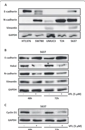
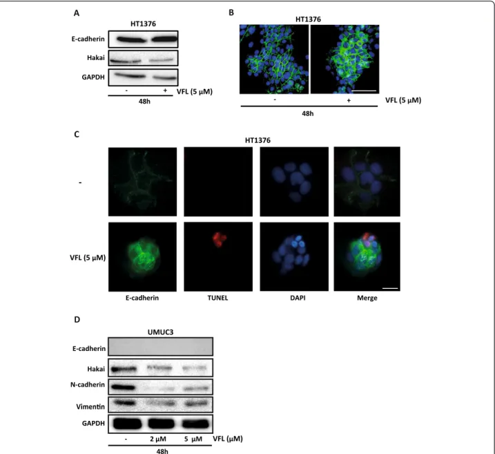
of E-cadherin is replaced by the abnormal expression of N-cadherin [16,17]. Downregulation of E-cadherin, re-sponsible for the loss of cell-cell adhesions, and upregula-tion of mesenchymal-related proteins, such as vimentin or N-cadherin, define the EMT process [9]. As shown in Figure 3B, VFL treatment (5 μM) modestly increased protein expression of E-cadherin after 48 and 72 hours in 5637 bladder tumour cells; instead, the mesenchy-mal N-cadherin marker was reduced under the treat-ment. Moreover, the E3 ubiquitin-ligase Hakai for the E-cadherin complex was significantly reduced under these conditions, suggesting that the disappearance of Hakai protein could influence the recovery of E-cadherin expression. Hakai was also proposed to be involved in the regulation of both cell–cell contacts and cell proliferation. It was suggested that cyclin D1, a member of the cyclin protein family involved in the regulation of the cell cycle progression, was one of the substrate effector proteins through which Hakai might regulate cell proliferation [25]. Indeed, VFL treatment of 5637 cells caused a reduc-tion in cyclin D1 protein levels compared to control con-ditions, while Hakai was also decreased (Figure 3C). In addition, transmission electron microscopy indicated that neighbouring VFL-treated E-cadherin expressing 5637 cells had very closely apposed cell-cell contacts compared to control cells (Figure 4). We extended this study in other bladder tumour epithelial cells. As shown in Figure 5A, in HT1376, VFL treatment modestly increases E-cadherin protein levels while Hakai is reduced; these cells do not ex-press the mesenchymal markers vimentin or N-cadherin. By immunofluorescent staining, the VFL-elevated E-cadherin was detected at cell-cell contacts in epithelial cells (Figure 5B) while a reduction of E-cadherin protein at cell-cell was observed in cells undergoing apoptosis (Figure 5C). Finally, in UMUC3 cells, which do not ex-press E-cadherin, it was shown that Hakai, vimentin,
HT1376 SW780 UMUC3 T24 5637
5637 5637
Cyclin D1
- + - +
48h 72h
GAPDH
VFL (5 M)
- + - +
48h 72h
E-cadherin
Hakai
N-cadherin
GAPDH
VFL (5 M)
GAPDH
μ μ
C A
E-cadherin
N-cadherin
B
Figure 3Epithelial-to-mesenchymal markers. A, the endogenous expression levels of the epithelial marker E-cadherin and mesenchymal markers N-cadherin and vimentin were assessed in bladder tumour cells (HT1376, SW780, UMUC3, T24 and 5637) by western blot analysis. B, the effect of 5μM VFL treatments of 5637 bladder tumour cells for 48 and 72 h on the indicated protein markers was assessed by western blot analysis.C, Cyclin D1 expression levels were assessed by Western blot analysis after treatment of 5637 bladder tumour cells for 48 and 72 h with VFL. Western blot data are representative of three independent experiments and GAPDH antibody was used as loading control for normalization.
5μM VFL Ctrl.
Cyt.
Cyt.
Cyt. Cyt.
Nucl.
Nucl.
and N-cadherin levels were reduced after 48 h of vin-flunine treatment (Figure 5D). Taken together, these data suggest that VFL causes cell death and epithelial cell differentiation in the E-cadherin-expressing cells.
VFL promotes proteasome-mediated Hakai degradation
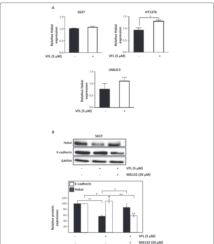
Since VFL causes a reduction in Hakai protein levels, we examined whether VFL affects Hakai mRNA levels using reverse transcription (RT) followed by real-time, quanti-tative (q) PCR. In contrast with Hakai protein levels,
Hakai mRNA levels were not downregulated by VFL treatment in 5637, HT1376 and UMUC3 (Figure 6A), suggesting that VFL lowers Hakai protein levels without decreasing Hakai mRNA abundance. Previous studies demonstrated that in all tissues, the majority of intracel-lular proteins are degraded by the ubiquitin proteasome pathway [45]. However, extracellular proteins and some cell surface proteins are taken up by endocytosis and de-graded within lysosomes. Given that Hakai is an intracel-lular protein, we investigated whether the reduced Hakai
+ VFL (5 μM)
48h
-+ VFL (5 μM)
48h GAPDH
E-cadherin
Hakai
HT1376
-Hakai E-cadherin
GAPDH
-48h
M 5 M
UMUC3
N-cadherin
VFL (μM) VFL (5 μM)
-E-cadherin TUNEL DAPI Merge
HT1376
HT1376 B
A
C
D
2 μ μ
VFL (5 μM) - + VFL (5 μM) - +
+
VFL (5 μM)
-Hakai
E-cadherin
GAPDH
VFL (5 μM) MG132 (20 μM)
+
- +
- - +
VFL (5 μM) MG132 (20 μM)
+
- +
- - +
expression expression
5637
expression
UMUC3
HT1376
expression 20 40 60 80 100
E-cadherin Hakai
120 *
* **
** **
*
5637
A
B
levels in VFL-treated cells could be affected by the in-creased degradation via proteasome of Hakai protein. We analyzed the effect of the proteasome inhibitors MG132 in VFL-treated 5637 cells compared to control conditions. As shown in Figure 6B, treatment of 5637 bladder cancer cells with VFL consistently reduced Hakai protein levels; however, the addition of MG132 inhibited this VFL-mediated-Hakai down-regulation. As expected, given that Hakai protein levels are restored by MG132 treatment, E-cadherin is reduced under these conditions. In conclusion, in 5637 cell lines Hakai reduction can be recovered by using proteasome inhibitors, MG132, further supporting the notion that Hakai down-regulation in-duced by VFL can be at least partially controlled in a proteasome-dependent mechanism.
Discussion
The transitional cell carcinomas of the bladder that in-vade muscle are associated with high frequency of me-tastasis, which is the major cause of death from cancer. Microtubules, a major component of the cytoskeleton, are one of the best established targets for cancer therapy. Indeed, the microtubule-targeting drug VFL, which sup-presses microtubule dynamicsin vitroand in vivo, is the recommended option treatment of metastatic transi-tional cell carcinoma of the urothelial tract which has progressed after treatment with platinum-containing chemotherapy. Given the increasing body of evidence supporting that microtubules regulate cadherin biology, and the well-established role of E-cadherin in the EMT during bladder cancer progression and metastasis, here we have studied the effect of VFL on E-cadherin cell-cell adhesions. Using bladder cancer cell lines, we have shown that VFL treatment induces cell death in bladder cancer cells and activates epithelial differentiation in the remaining cells, leading to increased E-cadherin-dependent cell-cell adhesions and to reduced levels of mesenchymal markers, such as N-cadherin or vimentin. Moreover, we have demonstrated that Hakai, a post-translational regula-tor of E-cadherin stability, was significantly reduced in a VFL-dependent manner in 5637 suggesting that the prote-asome pathway is at least partially involved in its dimin-ution; however, other post-translational mechanisms are waiting to be investigated. In conclusion, we have demon-strated a novel molecular mechanism of VFL to explain its anti-invasive effect.
In the last few years, differentiation therapy came out as a novel strategy for treating cancers. This ap-proach is based on the concept that cancer cells arise from tissue stem cells and share the stemness and plas-ticity with normal stem cells. Differentiation therapy aims to induce cancer cells to differentiate by treat-ment with differentiation-inducing agents [46,47]. Most of the differentiation agents can inhibit proliferation and
induce cells to differentiate and then undergo apoptosis [48,49]. It is well established that VFL blocks mitosis at the metaphase/anaphase transition, leading to apoptosis [50]. It is still necessary to elucidate how VFL could modulate epithelial cell differentiation and cell death; however, it appears to be an important therapeutic strat-egy for transitional cell carcinomas by its influence on these processes. Understanding the differentiation mecha-nisms and the fate of the treated cells may eventually lead us to gain insights into cancer therapy by differentiation.
important predictive marker for the responsiveness to microtubule-targeting VFL therapy.
Several studies have demonstrated the influence of the microtubules on cadherin-dependent cell-cell adhesions. Kitase et. al. demonstrated that RhoA is implicated dur-ing neurodetermination, where it influences cell-cell contact and cadherin levels [56]. They used the P19 cell model of neuronal differentiation to show that RhoA af-fects cadherin protein level and cell-cell contacts during neuroinduction. RhoGTPases have an important role during neurite growth, axonal guidance, and synaptogen-esis [57-59]. The cellular effects of RhoA are mediated by ROCK and seem to involve microtubules, pointing, for the first time, at the existence of a potential complex cross-talk between RhoA/ROCK, N-cadherin, and micro-tubules. The effect on cadherin level occurred in the tim-ing that corresponds to the switch of E- to N-cadherin that trigger neurodifferentiation [60]. N-cadherin level ap-pears to be critical for cell fate determination during mor-phogenesis. Advanced induction of Cdc42 had similar effects to RhoA inactivation, underscoring the importance of correct timing of RhoGTPases during neurodifferentia-tion [56].
Similar to VFL, another microtubule destabilizing agent, nocodazole, influenced cell-cell adhesions. The study reporting these findings examined the role of mi-crotubules on the transcriptional regulation of cell adhe-sion proteins, providing evidence that the microtubule cytoskeleton critically affects EMT by regulating TGF-β/ SMAD2 signaling during palatal fusion and prevents E-cadherin repression [61]. During palatal fusion, the midline epithelial seam (MES) degrades to achieve mesenchymal confluence. EMT is one of the mechanisms that function during MES degradation. TGF-β induces EMT in medial edge epithelium (MEE) by down-regulating the epithelial marker E-cadherin. Microtubule disassembly impaired palatal fusion leading to a multi-layered MES in the mid-region and inhibited palatal fusion accompanied by the development of a multi-layered MES in the mid-palatal region [62]. The authors further showed that treat-ment with nocodazole led to the accumulation of cell-cell adhesion proteins at intercellular junctions in medial edge epithelium [61]. Microtubule disruption by nocodazole triggered the aberrant accumulation of E-cadherin adhe-sion at intercellular junctions in MEE. Due to the aberrant expression of both negative (Snail and Zeb) and positive (c-MYC) E-cadherin transcriptional regulators when the TGF-β/SMAD2 signaling pathway was blocked, resulted in failure for EMT to progress. These data also support the important role of the microtubule cytoskeleton in mediat-ing TGF-β/SMAD2 signals to control E-cadherin expres-sion in MEE during palatal fuexpres-sion [61].
Several lines of evidence support that the interaction of the microtubules with cadherin affects cadherin
biology [63]. First, the junctional integrity of cadherin is perturbed by drugs such as nocodazole and VFL, which disrupt microtubules. Second, specific targeting of microtubule-binding proteins found at junctions also impairs broad aspects of cadherin biology. For ex-ample, Nezha-bound microtubules at their minus ends and tethered them to the zonula adherens. In cultured mammalian epithelial cells, depletion of either Nezha or PLEKHA7, which is responsible for recruiting Nezha to p120-ctn, disrupted the ability of cells to concentrate E-cadherin to the apical junction of the zonula adherens. The functional impact of these junctional microtubule-binding proteins further supports the idea that microtu-bules that interact with cadherin adhesions are responsible for regulating junctional integrity. Still, how microtubules influence these diverse aspects of cadherin biology is com-plex and poorly understood. Microtubules are commonly implicated in directing intracellular traffic of membrane-bound vesicles or molecular complexes; accordingly, it was postulated to influence cadherins through their intra-cellular traffic. Indeed, a number of microtubule-based motors have been identified that support intracellular transport of cadherins [64].
approaches directed against EMT can benefit cancer patients diagnosed at early stages of the disease to pre-vent invasion and dissemination, while anti-MET drugs could be potentially benefit patients with established metastasis. Still, a better understanding of the molecu-lar mechanisms of the EMT in bladder cancer is cru-cial to the development of new therapeutic modalities for this cancer [69,70].
Conclusions
Our data suggest that VFL, a microtubule-targeting drug, may be involved in a cross-talk between micro-tubule networks and cell-cell adhesions. We suggest a novel molecular mechanism by which VFL may impact upon EMT and metastasis. VFL impacts on E-cadherin-based cell-cell adhesion and influence on the EMT markers in epithelial bladder tumour cell lines. We have shown that VFL induces cell death in bladder cancer cells and activates epithelial differentiation of the remaining living cells. VFL increases E-cadherin dependent cell-cell adhesion, and reduces vimentin and N-cadherin mesenchymal markers. Moreover, the levels of the E3 ubiquitin-ligase Hakai were reduced by VFL in 5637 suggesting that proteasome-dependent mechanism could be one of the molecular mechanisms implicated in this reduc-tion. Our findings suggest the existence of a new mechanism of VFL-microtubule targeting drug to cell-cell contacts with potential functional implications in the maintenance of epithelial cell phenotype.
Additional file
Additional file 1:Figure for reviewer 3.Effect of VFL on the phenotype of 5637 and HT1376 bladder tumour cell lines. Phase-contrast microscopy images, showing larger fields from images of Figure 2, of the indicated bladder cell lines taken 48 h after treatment with the indicated concentrations of VFL compared to control conditions.
Abbreviations
EMT:Epithelial-to-mesenchymal transition; MEE: Medial edge epithelium; MET: Mesenchymal-to-epithelial transition; TCC: Transitional cell carcinoma; VFL: Vinflunine.
Competing interests
The authors declare that they have no competing interests. Authors’contributions
RC, MHC and MR contributed to experimental research. AF contributed to the design, the analysis of the results and writing the manuscript. All the authors have given the discussion, critical reading of the manuscript and final approval of the version to be published.
Acknowledgments
This work is supported by grants from Consellería de Sanidade (PS09/24), from Secretaría Xeral I + D + I, (10CSA916023PR), both from Xunta de Galicia; and from Instituto Carlos III (Acción Estratégica de Salud 2013–2016, PI13/ 00250). R.C. is the recipient of a grant from Secretaria Xeral I + D + I (10CSA916023PR), Xunta de Galicia. A.F was supported by Secretaria Xeral I + D + I (IPP.08-07), from Xunta de Galicia. Part of this work was awarded in the XIX edition of the“Rafael Hervada”from San Rafael Hospital (A Coruña, Spain).
Author details
1
Translational Cancer Research Group, Instituto de Investigación Biomédica A Coruña (INIBIC), Complejo Hospitalario Universitario A Coruña (CHUAC), Sergas. As Xubias, 15006 A Coruña, España.2Servizo de Oncología Médica, Complejo Hospitalario Universitario A Coruña (CHUAC), Sergas, A Coruña, Spain.
Received: 10 February 2014 Accepted: 28 June 2014 Published: 10 July 2014
References
1. Siegel R, Naishadham D, Jemal A:Cancer statistics, 2012.CA Cancer J Clin 2012,62(1):10–29.
2. Raghavan D, Shipley WU, Garnick MB, Russell PJ, Richie JP:Biology and management of bladder cancer.N Engl J Med1990,322(16):1129–1138. 3. Stenzl A, Cowan NC, De Santis M, Jakse G, Kuczyk MA, Merseburger AS,
Ribal MJ, Sherif A, Witjes JA:The updated EAU guidelines on muscle-invasive and metastatic bladder cancer.Eur Urol2009,55(4):815–825.
4. Nollet F, Kools P, van Roy F:Phylogenetic analysis of the cadherin superfamily allows identification of six major subfamilies besides several solitary members.J Mol Biol2000,299(3):551–572.
5. Perez-Moreno M, Jamora C, Fuchs E:Sticky business: orchestrating cellular signals at adherens junctions.Cell2003,112(4):535–548.
6. Yonemura S:Cadherin-actin interactions at adherens junctions.Curr Opin Cell Biol2011,23(5):515–522.
7. Christofori G:New signals from the invasive front.Nature2006, 441(7092):444–450.
8. Yang J, Weinberg RA:Epithelial-mesenchymal transition: at the crossroads of development and tumor metastasis.Dev Cell2008,14(6):818–829. 9. Thiery JP, Acloque H, Huang RY, Nieto MA:Epithelial-mesenchymal
transitions in development and disease.Cell2009,139(5):871–890. 10. Yun SJ, Kim WJ:Role of the Epithelial-Mesenchymal Transition in
Bladder Cancer: From Prognosis to Therapeutic Target.Korean J Urol 2013,54(10):645–650.
11. Chen J, Han Q, Pei D:EMT and MET as paradigms for cell fate switching.
J Mol Cell Biol2012,4(2):66–69.
12. Birchmeier W, Behrens J:Cadherin expression in carcinomas: role in the formation of cell junctions and the prevention of invasiveness.Biochim Biophys Acta1994,1198(1):11–26.
13. Behrens J, Vakaet L, Friis R, Winterhager E, Van Roy F, Mareel MM, Birchmeier W:Loss of epithelial differentiation and gain of invasiveness correlates with tyrosine phosphorylation of the E-cadherin/beta-catenin complex in cells transformed with a temperature-sensitive v-SRC gene.
J Cell Biol1993,120(3):757–766.
14. Perl AK, Wilgenbus P, Dahl U, Semb H, Christofori G:A causal role for E-cadherin in the transition from adenoma to carcinoma.Nature1998,392(6672):190–193. 15. Shimazui T, Schalken JA, Giroldi LA, Jansen CF, Akaza H, Koiso K, Debruyne FM,
Bringuier PP:Prognostic value of cadherin-associated molecules (alpha-, beta-, and gamma-catenins and p120cas) in bladder tumors.Cancer Res 1996,56(18):4154–4158.
16. De Wever O, Pauwels P, De Craene B, Sabbah M, Emami S, Redeuilh G, Gespach C, Bracke M, Berx G:Molecular and pathological signatures of epithelial-mesenchymal transitions at the cancer invasion front.
Histochem Cell Biol2008,130(3):481–494.
17. Yilmaz M, Christofori G:EMT, the cytoskeleton, and cancer cell invasion.
Cancer Metastasis Rev2009,28(1–2):15–33.
18. Mendez MG, Kojima S, Goldman RD:Vimentin induces changes in cell shape, motility, and adhesion during the epithelial to mesenchymal transition.FASEB J2010,24(6):1838–1851.
19. Aparicio LA, Valladares M, Blanco M, Alonso G, Figueroa A:Biological influence of Hakai in cancer: a 10-year review.Cancer Metastasis Rev2012, 31(1-2):375–386.
20. Batlle E, Sancho E, Francí C, Domínguez D, Monfar M, Baulida J, García De Herreros A:The transcription factor snail is a repressor of E-cadherin gene expression in epithelial tumour cells.Nat Cell Biol 2000,2(2):84–89.
21. Fujita Y, Krause G, Scheffner M, Zechner D, Leddy H, Behrens J, Sommer T, Birchmeier W:Hakai, a c-Cbl-like protein, ubiquitinates and induces endocytosis of the E-cadherin complex.Nat Cell Biol2002,4(3):222–231. 22. Swaminathan G, Cartwright CA:Rack1 promotes epithelial cell-cell adhesion
23. Janda E, Nevolo M, Lehmann K, Downward J, Beug H, Grieco M:Raf plus TGFbeta-dependent EMT is initiated by endocytosis and lysosomal degradation of E-cadherin.Oncogene2006,25(54):7117–7130. 24. Zhou WJ, Geng ZH, Chi S, Zhang W, Niu XF, Lan SJ, Ma L, Yang X, Wang LJ,
Ding YQ, Geng JG:Slit-Robo signaling induces malignant transformation through Hakai-mediated E-cadherin degradation during colorectal epithelial cell carcinogenesis.Cell Res2011, 21(4):609–626.
25. Figueroa A, Kotani H, Toda Y, Mazan-Mamczarz K, Mueller E, Otto A, Disch L, Norman M, Ramdasi R, Keshtgar M, Gorospe M, Fujita Y:Novel roles of hakai in cell proliferation and oncogenesis.Mol Biol Cell2009, 20(15):3533–3542.
26. Figueroa A, Fujita Y, Gorospe M:Hacking RNA: Hakai promotes tumorigenesis by enhancing the RNA-binding function of PSF.Cell Cycle 2009,8(22):3648–3651.
27. Rodríguez-Rigueiro T, Valladares-Ayerbes M, Haz-Conde M, Aparicio LA, Figueroa A:Hakai reduces cell-substratum adhesion and increases epithelial cell invasion.BMC Cancer2011,11:474.
28. Abella V, Valladares M, Rodriguez T, Haz M, Blanco M, Tarrío N, Iglesias P, Aparicio LA, Figueroa A:miR-203 Regulates Cell Proliferation through Its Influence on Hakai Expression.PLoS One2012,7(12):e52568.
29. Jordan MA, Horwitz SB, Lobert S, Correia JJ:Exploring the mechanisms of action of the novel microtubule inhibitor vinflunine.Semin Oncol2008, 35(3 Suppl 3):S6–S12.
30. Jordan MA, Wilson L:Microtubules as a target for anticancer drugs.Nat Rev Cancer2004,4(4):253–265.
31. Gerullis H, Ecke T, Eimer C, Wishahi M, Otto T:Vinflunine as second-line treatment in platin-resistant metastatic urothelial carcinoma: a review.
Anticancer Drugs2011,22(1):9–17.
32. Gerullis H:Vinflunine: a fluorinated vinca alkaloid for bladder cancer therapy.Drugs Today (Barc)2011,47(1):17–25.
33. Aparicio LM, Pulido EG, Gallego GA:Vinflunine: a new vision that may translate into antiangiogenic and antimetastatic activity.Anticancer Drugs 2012,23(1):1–11.
34. Pasquier E, Honoré S, Braguer D:Microtubule-targeting agents in angiogenesis: where do we stand?Drug Resist Updat2006,9(1–2):74–86. 35. Schwartz EL:Antivascular actions of microtubule-binding drugs.Clin
Cancer Res2009,15(8):2594–2601.
36. Honoré S, Pagano A, Gauthier G, Bourgarel-Rey V, Verdier-Pinard P, Civiletti K, Kruczynski A, Braguer D:Antiangiogenic vinflunine affects EB1 localization and microtubule targeting to adhesion sites.Mol Cancer Ther 2008,7(7):2080–2089.
37. Akhmanova A, Yap AS:Organizing junctions at the cell-cell interface.Cell 2008,135(5):791–793.
38. Akhmanova A, Stehbens SJ, Yap AS:Touch, grasp, deliver and control: functional cross-talk between microtubules and cell adhesions.Traffic 2009,10(3):268–274.
39. Stehbens SJ, Paterson AD, Crampton MS, Shewan AM, Ferguson C, Akhmanova A, Parton RG, Yap AS:Dynamic microtubules regulate the local concentration of E-cadherin at cell-cell contacts.J Cell Sci2006, 119(Pt 9):1801–1811.
40. Stehbens SJ, Akhmanova A, Yap AS:Microtubules and cadherins: a neglected partnership.Front Biosci (Landmark Ed)2009,14:3159–3167. 41. Han SP, Yap AS:The cytoskeleton and classical cadherin adhesions.
Subcell Biochem2012,60:111–135.
42. Shtutman M, Chausovsky A, Prager-Khoutorsky M, Schiefermeier N, Boguslavsky S, Kam Z, Fuchs E, Geiger B, Borisy GG, Bershadsky AD:Signaling function of alpha-catenin in microtubule regulation.Cell Cycle2008, 7(15):2377–2383.
43. Rodríguez Rigueiro T, Valladares Ayerbes M, Haz Conde M, Blanco M, Aparicio G, Fernández Puente P, Blanco FJ, Lorenzo MJ, Aparicio LA, Figueroa A:A novel procedure for protein extraction from formalin-fixed paraffin-embedded tissues.Proteomics2011,11(12):2555–2559.
44. Bonfil RD, Russo DM, Binda MM, Delgado FM, Vincenti M:Higher antitumor activity of vinflunine than vinorelbine against an orthotopic murine model of transitional cell carcinoma of the bladder.Urol Oncol2002, 7(4):159–166.
45. Rock KL, Gramm C, Rothstein L, Clark K, Stein R, Dick L, Hwang D, Goldberg AL: Inhibitors of the proteasome block the degradation of most cell proteins and the generation of peptides presented on MHC class I molecules.Cell 1994,78(5):761–771.
46. Zhang Z, Zhou Y, Qian H, Shao G, Lu X, Chen Q, Sun X, Chen D, Yin R, Zhu H, Shao Q, Xu W:Stemness and inducing differentiation of small cell lung cancer NCI-H446 cells.Cell Death Dis2013,4:e633.
47. Guichet PO, Bieche I, Teigell M, Serguera C, Rothhut B, Rigau V, Scamps F, Ripoll C, Vacher S, Taviaux S, Chevassus H, Duffau H, Mallet J, Susini A, Joubert D, Bauchet L, Hugnot JP:Cell death and neuronal differentiation of glioblastoma stem-like cells induced by neurogenic transcription factors.Glia2013,61(2):225–239.
48. Campos B, Wan F, Farhadi M, Ernst A, Zeppernick F, Tagscherer KE, Ahmadi R, Lohr J, Dictus C, Gdynia G, Combs SE, Goidts V, Helmke BM, Eckstein V, Roth W, Beckhove P, Lichter P, Unterberg A, Radlwimmer B, Herold-Mende C: Differentiation therapy exerts antitumor effects on stem-like glioma cells.Clin Cancer Res2010,16(10):2715–2728.
49. Seigel GM:Differentiation potential of human retinoblastoma cells.Curr Pharm Biotechnol2011,12(2):213–216.
50. Okouneva T, Hill BT, Wilson L, Jordan MA:The effects of vinflunine, vinorelbine, and vinblastine on centromere dynamics.Mol Cancer Ther 2003,2(5):427–436.
51. Bryan RT, Tselepis C:Cadherin switching and bladder cancer.J Urol2010, 184(2):423–431.
52. Debnath J, Brugge JS:Modelling glandular epithelial cancers in three-dimensional cultures.Nat Rev Cancer2005,5(9):675–688. 53. Deep G, Gangar S, Agarwal C, Agarwal R:Role of E-cadherin in anti-migratory
and anti-invasive efficacy of silibinin in prostate cancer cells.Cancer Prev Res (Phila)2011,4(8):1222–1232.
54. Wu K, Zeng J, Li L, Fan J, Zhang D, Xue Y, Zhu G, Yang L, Wang X, He D: Silibinin reverses epithelial-to-mesenchymal transition in metastatic prostate cancer cells by targeting transcription factors.Oncol Rep2010, 23(6):1545–1552.
55. Black PC, Brown GA, Inamoto T, Shrader M, Arora A, Siefker-Radtke AO, Adam L, Theodorescu D, Wu X, Munsell MF, Bar-Eli M, McConkey DJ, Dinney CP: Sensitivity to epidermal growth factor receptor inhibitor requires E-cadherin expression in urothelial carcinoma cells.Clin Cancer Res2008, 14(5):1478–1486.
56. Laplante I, Béliveau R, Paquin J:RhoA/ROCK and Cdc42 regulate cell-cell contact and N-cadherin protein level during neurodetermination of P19 embryonal stem cells.J Neurobiol2004,60(3):289–307.
57. Lehmann M, Fournier A, Selles-Navarro I, Dergham P, Sebok A, Leclerc N, Tigyi G, McKerracher L:Inactivation of Rho signaling pathway promotes CNS axon regeneration.J Neurosci1999,19(17):7537–7547.
58. Luo L:Rho GTPases in neuronal morphogenesis.Nat Rev Neurosci2000, 1(3):173–180.
59. Yuan XB, Jin M, Xu X, Song YQ, Wu CP, Poo MM, Duan S:Signalling and crosstalk of Rho GTPases in mediating axon guidance.Nat Cell Biol2003, 5(1):38–45.
60. Gao X, Bian W, Yang J, Tang K, Kitani H, Atsumi T, Jing N:A role of N-cadherin in neuronal differentiation of embryonic carcinoma P19 cells.Biochem Biophys Res Commun2001,284(5):1098–1103.
61. Kitase Y, Shuler CF:Microtubule disassembly prevents palatal fusion and alters regulation of the E-cadherin/catenin complex.Int J Dev Biol2013, 57(1):55–60.
62. Kitase Y, Shuler CF:Multi-layered hypertrophied MEE formation by microtubule disruption via GEF-H1/RhoA/ROCK signaling pathway.Dev Dyn2012,241(7):1169–1182.
63. Brieher WM, Yap AS:Cadherin junctions and their cytoskeleton(s).Curr Opin Cell Biol2013,25(1):39–46.
64. Meng W, Mushika Y, Ichii T, Takeichi M:Anchorage of microtubule minus ends to adherens junctions regulates epithelial cell-cell contacts.Cell 2008,135(5):948–959.
65. Brabletz T, Jung A, Reu S, Porzner M, Hlubek F, Kunz-Schughart LA, Knuechel R, Kirchner T:Variable beta-catenin expression in colorectal cancers indicates tumor progression driven by the tumor environment.Proc Natl Acad Sci U S A2001,98(18):10356–10361.
66. Chaffer CL, Brennan JP, Slavin JL, Blick T, Thompson EW, Williams ED: Mesenchymal-to-epithelial transition facilitates bladder cancer metastasis: role of fibroblast growth factor receptor-2.Cancer Res2006, 66(23):11271–11278.
68. Tsai JH, Donaher JL, Murphy DA, Chau S, Yang J:Spatiotemporal regulation of epithelial-mesenchymal transition is essential for squamous cell carcinoma metastasis.Cancer Cell2012,22(6):725–736.
69. Brabletz T:EMT and MET in metastasis: where are the cancer stem cells?
Cancer Cell2012,22(6):699–701.
70. Nieto MA, Cano A:The epithelial-mesenchymal transition under control: global programs to regulate epithelial plasticity.Semin Cancer Biol2012, 22(5–6):361–368.
doi:10.1186/1471-2407-14-507
Cite this article as:Aparicioet al.:Role of the microtubule-targeting drug vinflunine on cell-cell adhesions in bladder epithelial tumour cells.BMC Cancer201414:507.
Submit your next manuscript to BioMed Central and take full advantage of:
• Convenient online submission
• Thorough peer review
• No space constraints or color figure charges
• Immediate publication on acceptance
• Inclusion in PubMed, CAS, Scopus and Google Scholar
• Research which is freely available for redistribution