TítuloCancer incidence in heart transplant recipients with previous neoplasia history
15
0
0
Texto completo
(2) transplantation is considered only once the cancer-free interval meets the length of time for the cancer to be considered cured, usually 5 years [13-15]. Further studies are needed to determine other putative factors that may influence the post-HT recurrence of a previous tumor and the occurrence of new tumors after HT in patients with pre-HT history of malignancy. A comparison of the aforementioned patients with HT patients who had no previous tumor (NPT) might help identify these factors as well as characterize the patients. To know more about these patients to make more precise informed decisions when indicating HT, a study in patients from the Spanish Post–Heart Transplant Tumor Registry (SPHTTR) was conducted to compare HT recipients who had a previous history of neoplasm with those who had no previous history.. Materials and Methods A historical cohort study was conducted in HT recipients included in the SPHTTR from 1984 to December 31, 2010. The SPHTTR continually updates data on tumors for every patient undergoing HT at age ≥16 years in Spain since 1984, when HT was initiated in that country. The SPHTTR is a standardized database that includes 175 clinical variables with data on recipients, donors, surgery, immunosuppression and follow-up [16]. The use of a similar database by all Spanish transplant teams confers high reliability for the results [16]. This registry is one of the largest national heart transplant registries and contributes to other larger international registries, such as the International Registry for Heart and Lung Transplantation [17]. In addition, it probably has one of the largest samples of HT patients with previous tumors. The main objective of the study was to describe the profile of the HT recipients with a history of previous neoplasia and to compare their epidemiological data (age and sex), baseline and subsequent immunosuppression, and the incidence of post-HT tumors with the rest of the HT population in Spain with NPT. In addition, the possible impact of previous tumor (PT) history on overall survival (OS) compared with the OS of patients with NPT was evaluated. Patients included in the study should have survived >3 mo after transplantation. For the PT group, patients with previous nonmalignant tumors were excluded from the analysis, as were those with cardiac angiosarcoma because patients with this type of tumor are no longer transplanted due to the high recurrence rate [18]. In addition, retransplanted patients were excluded because retransplantation is a significant risk factor for cancer [19]. The types of pre-HT tumors were described for the PT group, and the types of post-HT tumors were described for both groups. Localization and histology were considered for the classification of tumor types: solid tumor (e.g. breast, prostate, colon, stomach, kidney, bladder, and lung carcinoma), melanoma, hematologic tumor (multiple myeloma, Hodgkin lymphoma, non-Hodgkin lymphoma, leukemia), nonmelanocytic skin tumor (e.g. epidermoid and basocellular carcinoma) and Kaposi sarcoma. The research protocol was approved by the institutional review board of the University 12 de Octubre Hospital (Madrid, Spain; number 13/267), and the study was conducted according to Spanish regulations. The retrospective design of the study made it difficult to locate patients (some of them already dead), and because the patients were codified in the registry (maintaining their anonymity), informed consents were not required to conduct the study. Patients gave their consent to have their data included in the registry database.. Statistical analysis Categorical variables were described by means of absolute and relative frequency tables, whereas continuous variables were described by central and dispersion measurements. The Pearson Chi-square test was used to compare the distribution of the categorical variables between the PT and NPT groups. The incidence rate of post-HT tumor and the mortality rate per 1000 person-years were assessed for each group as well as the ratios between rates (rate ratio [RR]). OS during the first 10 years after transplant was estimated by Kaplan–Meier curves for the PT and NPT groups, and the two curves were compared with the log-rank test. December 31, 2010, was considered the end of follow-up. All statistical calculations were performed using Stata version 10.1 for Windows (StataCorp, College Station, TX)..
(3) Results At the time of the study, the SPHTTR contained records for 5672 patients who had undergone HT in Spain at age ≥16 years between 1984 and the end of 2010. Of these, 1025 died within 3 mo of HT, and 86 met other exclusion criteria (Figure 1). Consequently, 4561 patients, 77 (1.7%) with PT (malignant noncardiac neoplasia) and 4484 (98.3%) with NPT, were included in this analysis.. Figure 1. Patient disposition flow chart. NPT, no previous tumor; PT, previous tumor; SPHTTR, Spanish Post–Heart Transplant Tumor Registry.. The sociodemographic and clinical characteristics of the patients are shown in Table 1. There were significantly more men in the NPT group than in the PT group (84% vs. 62%; p < 0.001), but age distribution was similar in both groups (p = 0.459). Every patient with a previous tumor had been considered cured by the multidisciplinary transplantation medical team, with a mean time since cancer diagnosis to the transplant of 8.3 years. In the PT group, 64% of previous tumors were solid tumors, 31% were hematologic and 5% were melanomas. Regarding tumor stage, 84.4% were localized tumors, and cancer spread in 12 patients (7 hematologic neoplasia and 5 solid tumors with local extension but no further dissemination)..
(4) Table 1. Sociodemographic and clinical characteristics of the HT recipients included in the analyses PT patients (n = 77) NPT patients (n = 4484) p-value Men 48 (62.3) Age (years) <45 17 (22.1) 45–54 19 (24.7) 55–64 32 (41.6) ≥65 9 (11.7) Time from neoplasia diagnosis to HT (years) 8.3 [2.2, 12.7] Indication for HT CIC 18 (23.4) Incidence of post-HT tumors 44.27 [19.89, 98.55] Rate of post-HT tumors 6 (33.3) Other cardiopathy 59 (76.6) Incidence of post-HT tumors 67.72 [45.39, 101.04] Rate of post-HT tumors 24 (40.7) Pre-HT tumor type Solid tumors 49 (63.6) Breast carcinoma 13 (16.9) Colon carcinoma 9 (11.7) Prostate carcinoma 6 (7.8) Kidney carcinoma 6 (7.8) Bladder carcinoma 5 (6.5) Stomach carcinoma 2 (2.6) Other 8 (10.4) Hematologic tumors 24 (31.2) Hodgkin lymphoma 11 (14.3) Leukemia 6 (7.8) Non-Hodgkin lymphoma 4 (5.2) Multiple myeloma 3 (4.0) Melanoma 4 (5.2) Stage Localized disease 65 (84.4) Extended disease 12 (15.6) Induction No 15 (19.5) OKT3/ATG/thymoglobulin/ 21 (27.3) Basiliximab/daclizumab 41 (53.2). 3760 (83.8) 998 (22.3) 1.339 (29.9) 1.816 (40.5) 331 (7.4) –. <0.001. 0.459. –. – – – – – –. – – – – – –. –. –. –. –. –. –. – –. – –. 1263 (28.2) 1973 (44.1) 1241 (27.7). ≤0.0001. Data expressed as n (%) for categorical variables and as mean [25th, 75th percentiles] for continuous variables. ATG, antithymocyte globulin; CIC, chemotherapy-induced cardiomyopathy; HT, heart transplant; OKT3, Orthoclone OKT3; NPT, no previous tumor; PT, previous tumor..
(5) Immunosuppressive treatment The most frequent immunosuppressive drugs used in both groups were prednisone (in almost every patient) and cyclosporine A (CsA; ≥74%) (Figure 2), which were administered mainly during the first 3 mo after HT and decreased gradually thereafter. Azathioprine (AZA) also decreased with time but seemed to be used in a larger proportion of NPT than PT patients. Mycophenolate mofetyl (MMF) decreased with time only in the PT group; use stayed at similar proportions in the NPT group. Tacrolimus (TAC) stayed in similar levels up to 2 years and seemed to be used in a higher proportion of PT than NPT patients. Muromonab CD3 (Orthoclone OKT3 [OKT3]; Janssen Biotech, Horsham, PA), antithymocyte globulin (ATG), thymoglobulin and IL-2R blockers (basiliximab and daclizumab) were typically used only during the first 3 mo after HT because they are used mostly as induction therapy. A significantly (p ≤ 0.0001) larger proportion of PT patients received induction compared with NPT patients (80.5% vs. 71.8%, respectively) (Table 1). In contrast, sirolimus and everolimus were typically used after 3 mo, even more after 2 years, with everolimus used in higher proportions of PT than NPT patients. Globally, a higher percentage of PT than NPT patients seemed to use TAC, MMF, everolimus, basiliximab and daclizumab, whereas a higher percentage of NPT than PT patients seemed to use AZA, OKT3 and thymoglobulin (Figure 2).. Figure 2. Immunosuppressive treatments. ATG, antithymocyte globulin; CsA, cyclosporine A; MMF, mycophenolate mofetil; MPS, mycophenolate sodium; NPT, no previous tumor; OKT3, Orthoclone OKT3; PT, previous tumor..
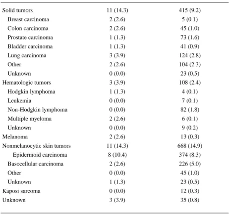
(6) Incidence of posttransplant tumors In the post-HT period, 1281 tumors developed in 914 patients (30 tumors in 25 PT patients and 1251 tumors in 889 NPT patients). The different types of tumors developed are shown in Table 2. The incidence of post-HT tumors was greater in the PT group than in the NPT group (RR 1.8, 95% confidence interval [CI] 1.2–2.6, p < 0.001) because of an excess risk among patients with a pre-HT hematologic tumor (RR 2.3, 95% CI 1.3–4.0, p = 0.04) (Table 3). When excluding the four cases considered as relapsing malignancies (one hematologic neoplasia and three solid tumors), the incidence of post-HT tumors was still greater in the PT than the NPT group (RR 1.6, 95% CI 1.1–2.4, p = 0.016) (Table 3).. Table 2. Post–heart transplant tumors PT group patients (n = 77) NPT group patients (n = 4484) Solid tumors. 11 (14.3). 415 (9.2). Breast carcinoma. 2 (2.6). 5 (0.1). Colon carcinoma. 2 (2.6). 45 (1.0). Prostate carcinoma. 1 (1.3). 73 (1.6). Bladder carcinoma. 1 (1.3). 41 (0.9). Lung carcinoma. 3 (3.9). 124 (2.8). Other. 2 (2.6). 104 (2.3). Unknown. 0 (0.0). 23 (0.5). 3 (3.9). 108 (2.4). Hodgkin lymphoma. 1 (1.3). 4 (0.1). Leukemia. 0 (0.0). 7 (0.1). Non-Hodgkin lymphoma. 0 (0.0). 82 (1.8). Multiple myeloma. 2 (2.6). 6 (0.1). Unknown. 0 (0.0). 9 (0.2). 2 (2.6). 13 (0.3). 11 (14.3). 668 (14.9). 8 (10.4). 374 (8.3). Basocellular carcinoma. 2 (2.6). 226 (5.0). Other. 0 (0.0). 45 (1.0). Unknown. 1 (1.3). 23 (0.5). Kaposi sarcoma. 0 (0.0). 12 (0.3). Unknown. 3 (3.9). 35 (0.8). Hematologic tumors. Melanoma Nonmelanocytic skin tumors Epidermoid carcinoma. Data expressed as number of tumors (%). NPT, no previous tumor; PT, previous tumor..
(7) Table 3. Incidence rates of post-HT tumors and mortality in the PT and NPT groups. At risk person-years Post-HT tumors Incidence ratea. 95% CI. RRb. 95% CI. p-valuee. Post-HT tumors 36 543.1. 1251. 34.2. 32.4 36.2. 1.0. –. –. 490.5. 30. 61.2. 42.8 87.5. 1.8 1.2. 2.6. 0.001. Solid tumor. 316.2. 14. 44.3. 26.2 74.8. 1.3 0.8. 2.2. 0.337. Hematologic tumor. 154.3. 12. 77.8. 44.2 136.9 2.3 1.3. 4.0. 0.004. NPT PT. –. PT versus NPT adjustedc. 1.9 1.3. 2.7. <0.001. PT versus NPT adjustedd. 1.8f 1.3. 2.6. 0.001. 2.4. 0.016. f. Post-HT tumors excluding relapses NPT. 36 532.7. 1250. 34.2. 32.4 36.2. 474.2. 26. 54.8. 37.3 80.5. 1.6 1.1. 36 532.7. 1696. 46.4. 44.3 48.7. 1.0. –. –. 493.9. 29. 58.7. 40.8 84.5. 1.3 0.9. 1.8. 0.209. Solid tumor. 319.7. 16. 50.1. 30.7 81.7. 1.1 0.7. 1.8. 0.764. Hematologic tumor. 154.3. 10. 64.8. 34.9 120.5 1.4 0.8. 2.6. 0.291. PT Mortality NPT PT. –. CI, confidence interval; HT, heart transplant; NPT, no previous tumor; PT, previous tumor; RR, rate ratio. a Incidence rate per 1000 person-years. b RR with respect to the NPT group. c NPT versus PT adjusted by induction. d NPT versus PT adjusted by time period. e The p-value is from the chi-square test; p-values in bold are significant. f Adjusted RRs are calculated with the Mantel-Haenszel method.. Chemotherapy-associated cardiomyopathy and posttransplant tumors Overall, 18 of the 77 patients with PT required HT because of chemotherapy-induced cardiomyopathy (CIC). No significant differences in the incidence rate or in the types of post-HT tumors were shown between those patients with CIC and those requiring HT due to other cardiopathies (RR 1.53, 95% CI 0.62–3.74, p = 0.348) (Table 1).. Induction therapy and posttransplant tumors Patients receiving induction had greater incidence of post-HT tumors than those not receiving induction (RR 1.98, 95% CI 1.71–2.28, p ≤ 0.0001) because of excess risk in patients receiving classic induction (OKT3, ATG and thymoglobulin; RR 2.20, 95% CI 1.91–2.55, p ≤ 0.0001) (Table 4). This finding was the same when looking exclusively at patients with NPT. In the PT group, it seemed there was no difference in the incidence of post-HT tumors whether or not patients received induction, but this is probably due to the small sample size..
(8) Table 4. Incidence rates of post-HT tumors in patients submitted or not to induction treatment. At risk person-years Post-HT tumors Incidence ratea. 95% CI. RRb 95% CI p-valuec. Post-HT tumors No induction. 11 217.15. 231. 20.59. 18.10 23.43 1.00 –. Induction. 25 816.40. 1050. 40.67. 19 941.48. 905. 45.38. 38.28 43.21 1.98 1.71 2.28 ≤0.0001 42.52 48.44 2.20 1.91 2.55 ≤0.0001. 5835.65. 145. 24.85. 21.11 29.24 1.21 0.98 1.49 0.076. No induction. 11 091.89. 227. 20.47. 17.97 23.31 1.00 –. Induction. 25 451.21. 1024. 40.23. 37.84 42.78 1.97 1.70 2.27 ≤0.0001. 19 731.03. 890. 45.11. 5680.91. 134. 23.59. 42.24 48.17 2.20 1.91 2.55 ≤0.0001 19.91 27.94 1.15 0.93 1.43 0.192. OKT3/ATG/thymoglobulin Basiliximab/daclizumab. –. –. Post-HT tumors in NPT patients. OKT3/ATG/thymoglobulin Basiliximab/daclizumab. –. –. Post-HT tumors in PT patients No induction. 125.26. 4. 31.93. 11.99 85.08 1.00 –. Induction. 365.19. 26. 71.2. 48.48 104.57 2.23 0.78 6.39 0.125. –. –. OKT3/ATG/thymoglobulin. 210.45. 15. 71.28. 42.97 118.23 2.23 0.74 6.72 0.143. Basiliximab/daclizumab. 154.74. 11. 71.09. 39.37 128.36 2.23 0.71 6.99 0.159. ATG, antithymocyte globulin; CI, confidence interval; HT, heart transplant; OKT3, Orthoclone OKT3; RR, rate ratio. a Incidence rate per 1000 person-years. b RR with respect to no induction. c The p-value is from the chi-square test.. When the incidence rates of post-HT tumors were adjusted by induction, PT patients receiving induction showed an RR of 1.9 more risk of post-HT tumors than NPT patients receiving induction (95% CI 1.3–2.7, p < 0.01) (Table 3).. Time period and posttransplant tumors The incidence rate of post-HT tumors was 32.77% (95% CI 29.09–36.90) when transplantation was performed between 1984 and 1992, 39.52% (95% CI 36.93–42.30) between 1993 and 2001, and 22.99% (95% CI 19.84–26.65) between during 2002 and 2010. When post-HT tumor incidence rates were adjusted by time period, PT patients had a risk of post-HT tumors that was 1.8 times greater than that of NPT patients (95% CI 1.3–2.6; p = 0.001) (Table 3).. Posttransplant mortality and OS No significant differences were observed in post-HT mortality incidence rates or hazard ratios (HRs) between the groups (Tables 3 and 5). Women had significantly lower mortality risk than men, and mortality decreased significantly in more recent periods (Table 5). Mortality HRs were adjusted by sex and time period, but again, no significant differences were observed between the PT and NPT groups (Table 5)..
(9) Table 5. Mortality by Cox regression analysis HR. 95% CI. p-value. Pre-HT tumor No Yes. 1 1.26 0.87. 1.82. 0.217. 2.04. 0.068. 0.87. <0.001. Adjusted pre-HT tumora No Yes. 1 1.41 0.98. Sex Men Women. 1 0.76 0.66. Time period 1984–1992. 1. 1993–2001. 0.79 0.70. 0.88. <0.001. 2002–2010. 0.59 0.51. 0.69. <0.001. The p-values in bold are significant. CI, confidence interval; HR, hazard ratio; HT, heart transplant. a Adjusted by sex and period year.. OS during the 10-year posttransplant period was significantly greater in the NPT than the PT group (p = 0.048) (Figure 3A), showing the following survival data for NPT and PT groups, respectively: 93% and 95% at 1 year, 79% and 74% at 5 years, and 65% and 51% at 10 years. When the survival analysis was performed after the diagnosis of the first post-HT tumor (Figure 3B), no significant survival differences were observed between the groups..
(10) Figure 3. Kaplan-Meier survival curves for patients with PT and NPT. (A) Starting right after transplantation. (B) Starting at the time of first post–heart transplant diagnosed tumor. NPT, no previous tumor; PT, previous tumor..
(11) Table 6 shows the causes of mortality in both groups. Cancer-related death was 21.3% and 32.2% in the NPT and PT groups, respectively.. Table 6. Cause of death in the PT and NPT groups PT n (%). NPT n (%). Deaths, total. 28 (36.4). 1560 (34.8). Cardiovascular. 3 (10.7). 356 (22.8). Cerebrovascular. 4 (14.3). 52 (3.3). Graft failure. 7 (25.0). 318 (20.4). Acute rejection. 4 (14.3). 112 (7.2). Chronic rejection. 3 (10.7). 139 (8.9). Other. 0 (0.0). 67 (4.3). Hemorrhage. 0 (0.0). 14 (0.9). Infection. 2 (7.1). 231 (14.8). Bacterial. 0 (0.0). 188 (12.1). Fungal. 1 (3.6). 20 (1.3). Viral. 1 (3.6). 14 (0.9). Other. 0 (0.0). 9 (0.6). 9 (32.1). 333 (21.3). Posttransplant lymphoproliferative disease. 0 (0.0). 39 (2.5). Skin. 0 (0.0). 9 (0.6). Other. 9 (32.1). 285 (18.3). Multiple organ failure. 0 (0.0). 73 (4.7). Pulmonary. 1 (3.6). 44 (2.8). Other. 2 (7.1). 139 (8.9). Malignancy. NPT, no previous tumor; PT, previous tumor.. Discussion The current study shows that the tumor incidence rate in post-HT patients with PT history was almost double that of post-HT patients with NPT history (RR 1.8, p ≤ 0.001), in agreement with previous studies showing higher incidence of de novo tumors in posttransplant patients with PT history [20, 21]. In the study by Higgins et al conducted in 6211 U.S. HT recipients, of whom 283 had pretransplant malignancies, a multivariate analysis showed that previous history of cancer had a relative risk of 1.6 (p = 0.02) for posttransplant tumors, although older age and earlier date of transplant were the two most significant risk factors. In agreement with these data, our study showed that post-HT tumor incidence rates were higher at earlier dates of transplant compared with more recent dates. It is important to note that in the current study, the tumor incidence rates in the group with previous solid tumors and in the NPT group were similar, and it was only that of the group with previous hematologic tumors that was significantly larger compared with the rate in the NPT group (RR 2.3, p ≤ 0.004). Some tumors, including hematologic tumors, are prone to genomic instability, namely, an increased tendency of tumor cells to acquire new mutations with each cell division [22, 23]. In addition, leukemia cells have been shown to release fragmented DNA derived from their genome, which may enter the nuclei of other cells and induce double-strand breaks or integrate into the chromosomal DNA, promoting genome instability of these cells [24]. All of these aspects might help explain the greater incidence of post-HT tumors in patients with such a history of cancer. In agreement with this theory, Bratsttrom et al showed that the type of previous malignancy had a great impact on cancer recurrence and mortality, with hematologic cancer showing the greater overall and cancer-specific mortality among the different pretransplant cancer types [25]. The incidence rates of tumors in the NPT group (34.2 per 1000 person-years) and the PT group (61.2 per 1000 person-years) were much higher than the rate estimated by Globocan 2008 for the overall.
(12) Spanish population (4.4 per 1000 person-years for all cancers, excluding nonmelanoma skin cancer) [26], as expected for patients submitted to transplantation. Skin cancer, especially nonmelanoma skin cancer, represented a major proportion of the post-HT tumors, in agreement with previous data, showing this cancer type as the most common to develop de novo in recipients of organ transplant overall and in recipients of HT specifically [5, 27-29]. Eleven de novo nonmelanocytic skin cancers arose in 77 patients with previous cancer history (14.3%), and the frequency of skin cancer in patients without a cancer history was similar at 14.9% (668 de novo tumors in 4484 patients). Noncutaneous solid tumors arose in 14.3% of patients with PT (three considered relapsing malignancies [two colon carcinomas, one bladder carcinoma]) and 9.3% of patients with NPT. Lung cancer was the most common one, in agreement with data from a study assessing post-HT lung cancer incidence [30] and other studies showing lung cancer as one of the most frequently developing solid tumors in HT recipients [19, 21, 31, 32]. Hematologic tumors developed in 3.9% (three of 77) of PT patients (one tumor was multiple myeloma, considered a relapsing malignancy) and 2.4% (108 of 4484) of NPT patients. One-third of the PT patients had a history of hematologic tumors, and such tumors are likely to recur [25]. Still, the frequency of recurrence was low (5.2% or 4% in 77 patients), as observed by Higgins et al [21]. The proportion of post-HT multiple myeloma was higher in the PT than the NPT group (2.6% vs. 0.1%, respectively), although the latter group had a higher proportion of men, and multiple myeloma is known to be more common in men than in women [33]. Immunosuppressive treatments affect the cancer risk of transplanted patients [29]. Drugs used for induction, other than IL-2R blockers, have been shown to increase the risk of neoplasia [34-36]. The mammalian target of rapamycin (mTOR) inhibitors sirolimus and everolimus are known to show anticancer effects in HT recipients and other solid organ recipients [37-39]. The use of induction therapy has increased in recent years, and the drugs used have changed from mainly OKT3 and ATG to daclizumab and, especially, basiliximab [16]. In addition, mTOR inhibitors have been available only recently [37-39]. The immunosuppressive regimen used in HT in Spain has evolved from OKT3 induction and maintenance with CsA, AZA and steroids to basiliximab induction and maintenance with CsA/TAC, MMF and steroids [40]. According to the changes in immunosuppressive drugs used, patients transplanted more recently showed significantly lower posttransplant tumor rates. In addition, patients receiving induction therapy showed increased post-HT tumor rates over those with no induction, due to the increased rate in the group induced by classic induction drugs (OKT3, ATG, and thymoglobulin). No such effect was observed when induction was performed with the IL-2R blockers basiliximab or daclizumab. Because most patients with previous tumors were transplanted recently (this type of patient has been considered for HT only recently), immunosuppressive treatments differed between them and patients with NPT. When incidence rates were adjusted by time period or by induction, the PT group still showed a post-HT tumor rate almost twofold greater than the NPT group. The use of antineoplastic treatments in PT patients prior to HT might also influence the incidence of post-HT treatment; however, when patients who were transplanted due to chemotherapy-induced cardiomyopathy were compared with those transplanted for other indications, no significant differences in post-HT tumor incidence rates were observed. OS was significantly better in the NPT than the PT group during the first 10-year posttransplant period, a difference that started being noticeable only by the fifth or sixth year. The similar survival data between the groups during the first 4–5 years after transplant seem to agree with a previous study conducted in Spain that showed similar actuarial survival at 1, 3, and 5 years in the PT and NPT groups [41]. In the study by Higgins et al [21], the percentage of patients free from malignancy, which was significantly lower in the PT group compared with the NPT group in the 15-year posttransplant period, did not start to show noticeable differences between groups until the fifth or sixth year. Consequently, the appearance of a survival difference between our two groups of patients only 5 years after transplant might reflect the different timing of occurrence of the first posttransplant tumors, which seem to occur earlier in the PT than the NPT group. In fact, when OS was estimated after the first tumors were diagnosed, no differences in survival were observed between the PT and NPT groups, further suggesting that the differences observed in survival from the fifth to the 10th year after transplant reflect an earlier appearance of tumors in the patients with a history of cancer. The mortality HR or instantaneous risk of dying was not significantly different between the PT and NPT groups (adjusted for sex and time period, HR 1.41 [95% CI 0.98-2.04, p = 0.068, Table 5). The study by Bratsttrom et al [25] conducted in a Swedish population-based cohort of 10 448 solid organ recipients, of whom 416 (4%) had a prior malignancy, showed an 80% increase in overall mortality (adjusted for sex, age and time period, HR 1.8 [95% CI 1.3–2.5]) among nonkidney recipients (including HT recipients) with cancer history, compared with those without such history, driven by cancer-specific death. Bratsttrom et al showed that the type of previous malignancy and the waiting time between cancer remission and transplantation had a great impact on relapses and mortality. Every PT patient in our study.
(13) had been considered cured at the time of transplantation, with a mean interval of 8.3 years from diagnosis to transplant, and in fact, relapses were very uncommon. When HT started to be performed in patients with a history of tumors, the average cancer-free interval before transplantation was almost 10 years [42]; however, the current arbitrary threshold value for considering cancer cured is 5 years, although it depends on the cancer type [13, 43]. In the study by Bratsttrom et al, 45% of patients had a waiting time ≤5 years, and those patients, as well as those with a waiting time >10 years (29%), had significantly higher cancerrelated mortality than patients with a waiting time of 5–10 years [25]. This high percentage of patients with <5 years of waiting time might explain the increased mortality observed in the PT patients from the study by Bratsttrom et al; this did not show in our PT patients. A limitation of the current study might be the different sizes of the PT and NPT groups; however, the analyses were initially performed simply, with no adjustments, minimizing the possible problems that this difference might represent. Another limitation of the study is that although both groups were homogeneous in age, they differed in proportions of men and women, time period of transplantation and use of induction; however, the analyses were adjusted by sex, time period and induction, and results similar to the initial analyses were obtained. In conclusion, after adjusting the analyses by different variables, HT patients with previous malignant noncardiac tumors, specifically those with previous hematologic tumors, showed higher post-HT tumor incidence than those with NPT and worse OS over a 10-year period, most likely due to earlier occurrence of posttransplant tumors in patients with PT. Consequently, patients with a history of previous noncardiac tumor should be carefully considered for HT. In addition, when transplantation is carried out in such patients, they should be followed more carefully, with increasing surveillance for arising tumors during at least the first 5 years after transplant.. Acknowledgments We are grateful to the researchers and staff of all the Spanish heart transplant centers that contributed data to this study and Odds for helping with study coordination. This study was partially supported by an unrestricted grant from Novartis and by the Spanish cardiovascular networks RIC (Instituto de Salud Carlos III, Ministerio de Economia y Competitividad). Other investigators involved in maintaining the Spanish Post-Heart Transplant Tumour Registry are as follows: Manuel Gómez Bueno, Javier Segovia Cubero, and Inés Sayago Silva (Hospital Universitario Puerta de Hierro, Madrid); Vicens Brossa, Eulàlia Roig, and Laura López (Hospital Santa Creu i Sant Pau, Barcelona); Luis Martínez, Ignacio SánchezLázaro, Mónica Cebrián, and Elena Marqués (Hospital Universitari i Politècnic La Fe. Valencia); María Ángeles Castel (Hospital Clínic i Provincial de Barcelona); María Jesús Paniagua Martín, Raquel Marzoa Rivas, and Eduardo Barge Caballero (Hospital Universitario A Coruña, La Coruña); José Antonio Vázquez de Prada (Hospital Universitario Marqués de Valdecilla, Santander); Iago Sousa, Adolfo Villa Arranz, and Juan Fernández-Yáñez (Hospital General Universitario Gregorio Marañón, Madrid); Marisa Sanz Julve and Ana Portoles Ocampo (Hospital Universitario Miguel Servet, Zaragoza); Miguel Angel Gómez, Pilar Escribano, and Maria José Ruiz Cano (Hospital Universitario 12 de Octubre, Madrid); Nicolás Manito Lorite and Josep Roca (Hospital Universitari de Bellvitge, Barcelona); Amada Recio Platero (Hospital Clínico Universitario de Valladolid); Montserrat Lorente (Clínica Universidad de Navarra, Pamplona); J. Manuel Sobrino Márquez and Diego Rangel Sousa (Hospital Universitario Virgen del Rocío, Sevilla); Iris Garrido Bravo (Hospital Universitario Virgen de la Arrixaca, Murcia); José Luis Lambert Rodríguez (Hospital Universitario Central de Asturias); Amador López Granados, Carmen Segura Saint-Gerons, and Juan Carlos Castillo Domínguez (Hospital Universitario Reina Sofía, Córdoba).. Disclosure The authors of this manuscript have no conflicts of interest to disclose as described by the American Journal of Transplantation..
(14) References 1. Penn I. Second malignant neoplasms associated with immunosuppressive medications. Cancer 1976; 37(2 Suppl): 1024–1032. 2. Honda H, Barloon TJ, Franken EA Jr, Garneau RA, Smith JL. Clinical and radiologic features of malignant neoplasms in organ transplant recipients: Cyclosporine-treated vs untreated patients. Am J Roentgenol 1990; 154(2): 271–274. 3. Krynitz B, Edgren G, Lindelof B, Baecklund E, Brattstrom C, Wilczek H, et al. Risk of skin cancer and other malignancies in kidney, liver, heart and lung transplant recipients 1970 to 2008–a Swedish population-based study. Int J Cancer 2013; 132(6): 1429–1438. 4. Penn I. Incidence and treatment of neoplasia after transplantation. J Heart Lung Transplant 1993; 12(6 Pt 2): S328–S336. 5. Collett D, Mumford L, Banner NR, Neuberger J, Watson C. Comparison of the incidence of malignancy in recipients of different types of organ: A UK Registry audit. Am J Transplant 2010; 10(8): 1889–1896. 6. Rinaldi M, Pellegrini C, D'Armini AM, Aiello M, Negri M, Arbustini E, et al. Neoplastic disease after heart transplantation: Single center experience. Eur J Cardiothorac Surg 2001; 19(5): 696–701. 7. Hinkamp T, Sullivan H, Bakhos M, Grieco J, Pifarre R. Orthotopic cardiac transplantation in two patients with previous malignancy. Ann Thorac Surg 1991; 51(6): 1004–1006. 8. Cruz RJ, Jr, , Ranganathan S, Mazariegos G, Soltys K, Nayyar N, Sun Q, et al. Analysis of national and singlecenter incidence and survival after liver transplantation for hepatoblastoma: New trends and future opportunities. Surgery 2013; 153(2): 150–159. 9. Mazzaferro V, Regalia E, Doci R, Andreola S, Pulvirenti A, Bozzetti F, et al. Liver transplantation for the treatment of small hepatocellular carcinomas in patients with cirrhosis. N Engl J Med 1996; 334(11): 693–699. 10. Mazzaferro V, Bhoori S, Sposito C, Bongini M, Langer M, Miceli R, et al. Milan criteria in liver transplantation for hepatocellular carcinoma: An evidence-based analysis of 15 years of experience. Liver Transpl 2011; 17(Suppl 2): S44–S57. 11. Cheung TT, Ho CL, Lo CM, Chen S, Chan SC, Chok KS et al. 11C-acetate and 18F-FDG PET/CT for clinical staging and selection of patients with hepatocellular carcinoma for liver transplantation on the basis of Milan criteria: Surgeon's perspective. J Nucl Med 2013; 54(2): 192–200. 12. Chapman JR, Sheil AG, Disney AP. Recurrence of cancer after renal transplantation. Transplant Proc 2001; 33(1–2): 1830–1831. 13. Mehra MR, Kobashigawa J, Starling R, Russell S, Uber PA, Parameshwar J, et al. Listing criteria for heart transplantation: international Society for Heart and Lung Transplantation guidelines for the care of cardiac transplant candidates–2006. J Heart Lung Transplant 2006; 25(9): 1024–1042. 14. Ward KM, Binns H, Chin C, Webber SA, Canter CE, Pahl E. Pediatric heart transplantation for anthracycline cardiomyopathy: Cancer recurrence is rare. J Heart Lung Transplant 2004: 23(9): 1040–1045. 15. Hanaway MJ, Weber S, Buell JF, Trofe J, Alloway R, Beebe T, et al. Risk for recurrence and death from preexisting cancers after transplantation. Transplant Rev 2005; 19: 151–163. 16. Almenar L, Segovia J, Crespo-Leiro MG, Palomo J, Arizon JM, Cobo M, et al. [Spanish registry on heart transplantation. 22nd official report of the Spanish Society of Cardiology Working Group on Heart Failure and Heart Transplantation (1984-2010)]. Rev Esp Cardiol 2011; 64(12): 1138–1146. 17. Stehlik J, Edwards LB, Kucheryavaya AY, Benden C, Christie JD, Dipchand AI, et al. The Registry of the International Society for Heart and Lung Transplantation: 29th official adult heart transplant report–2012. J Heart Lung Transplant 2012; 31(10): 1052–1064. 18. Crespo MG, Pulpon LA, Pradas G, Serrano S, Segovia J, Vegazo I, et al. Heart transplantation for cardiac angiosarcoma: Should its indication be questioned? J Heart Lung Transplant 1993; 12(3): 527–530. 19. Kellerman L, Neugut A, Burke B, Mancini D. Comparison of the incidence of de novo solid malignancies after heart transplantation to that in the general population. Am J Cardiol 2009; 103(4): 562–566. 20. Kauffman HM, Cherikh WS, McBride MA, Cheng YA, Delmonico FL, Hanto DW. Transplant recipients with a history of a malignancy: Risk of recurrent and de novo cancers. Transplant Rev 2005; 19: 55–64. 21. Higgins RS, Brown RN, Chang PP, Starling RC, Ewald GA, Tallaj JA, et al. A multi-institutional study of malignancies after heart transplantation and a comparison with the general United States population. J Heart Lung Transplant 2014; 33(5): 478–485. 22. Abdel-Rahman WM. Genomic instability and carcinogenesis: An update. Curr Genomics 2008; 9(8): 535–541. 23. Economopoulou P, Pappa V, Papageorgiou S, Dervenoulas J, Economopoulos T. Abnormalities of DNA repair mechanisms in common hematological malignancies. Leuk Lymphoma 2011; 52(4): 567–582. 24. Dvorakova M, Karafiat V, Pajer P, Kluzakova E, Jarkovska K, Pekova S, et al. DNA released by leukemic cells contributes to the disruption of the bone marrow microenvironment. Oncogene 2013; 32(44): 5201–5209. 25. Brattstrom C, Granath F, Edgren G, Smedby KE, Wilczek HE. Overall and Cause-Specific Mortality in Transplant Recipients with a Pretransplantation Cancer History. Transplantation 2013; 96(3): 297–305. 26. Globocan [Internet]. 2008 [cited 2013 Aug 11]. Available from: http://globocan.iarc.fr/summary_table_pop.asp?selection=184724&title=Spain&age_from=1&age_to=10&sex=0 &type=0&window=1&sort=0&submit=%A0Execute%A0SPAIN. 27. Gertz MA, Noel P, Kyle RA. Second malignancies after chemotherapy and transplantation. Crit Rev Oncol Hematol 1993; 14(2): 107–125. 28. Brewer JD, Colegio OR, Phillips PK, Roenigk RK, Jacobs MA, Van de Beek D, et al. Incidence of and risk factors for skin cancer after heart transplant. Arch Dermatol 2009; 145(12): 1391–1396..
(15) 29. Jensen P, Hansen S, Moller B, Leivestad T, Pfeffer P, Geiran O, et al. Skin cancer in kidney and heart transplant recipients and different long-term immunosuppressive therapy regimens. J Am Acad Dermatol 1999; 40(2.pt.1): 177–186. 30. Crespo-Leiro MG, Villa-Arranz A, Manito-Lorite N, Paniagua-Martin MJ, Rabago G, Almenar-Bonet L, et al. Lung cancer after heart transplantation: Results from a large multicenter registry. Am J Transplant 2011; 11(5): 1035–1040. 31. Roithmaier S, Haydon AM, Loi S, Esmore D, Griffiths A, Bergin P, et al. Incidence of malignancies in heart and/or lung transplant recipients: A single-institution experience. J Heart Lung Transplant 2007; 26(8): 845–849. 32. Jiang Y, Villeneuve PJ, Wielgosz A, Schaubel DE, Fenton SS, Mao Y. The incidence of cancer in a populationbased cohort of Canadian heart transplant recipients. Am J Transplant 2010; 10(3): 637–645. 33. Alexander DD, Mink PJ, Adami HO, Cole P, Mandel JS, Oken MM, et al. Multiple myeloma: A review of the epidemiologic literature. Int J Cancer 2007; 120(Suppl 12): 40–61. 34. Crespo-Leiro MG, Alonso-Pulpon L, Vazquez de Prada JA, Almenar L, Arizon JM, Brossa V, et al. Malignancy after heart transplantation: Incidence, prognosis and risk factors. Am J Transplant 2008; 8(5): 1031–1039. 35. Bernstein D, Baum D, Berry G, Dahl G, Weiss L, Starnes VA, et al. Neoplastic disorders after pediatric heart transplantation. Circulation 1993; 88(5 Pt 2): II230–II237. 36. Crespo-Leiro MG, Alonso-Pulpon L, Arizon JM, Almenar L, Delgado JF, Palomo J, et al. Influence of induction therapy, immunosuppressive regimen and anti-viral prophylaxis on development of lymphomas after heart transplantation: Data from the Spanish Post-Heart Transplant Tumour Registry. J Heart Lung Transplant 2007; 26(11): 1105–1109. 37. Epailly E, Albanell J, Andreassen A, Bara C, Campistol JM, Delgado JF, et al. Proliferation signal inhibitors and post-transplant malignancies in heart transplantation: Practical clinical management questions. Clin Transplant 2011; 25(5): E475–E486. 38. Rao RD, Buckner JC, Sarkaria JN. Mammalian target of rapamycin (mTOR) inhibitors as anti-cancer agents. Curr Cancer Drug Targets 2004; 4(8): 621–635. 39. Euvrard S, Morelon E, Rostaing L, Goffin E, Brocard A, Tromme I, et al. Sirolimus and secondary skin-cancer prevention in kidney transplantation. N Engl J Med 2012; 367(4): 329–339. 40. Alonso-Pulpon L, Almenar L, Crespo MG, Silva L, Segovia J, Manito N, et al. [Practice guidelines of the Spanish Society of Cardiology Cardiac and heart-lung transplants]. Rev Esp Cardiol 1999; 52(10): 821–839. 41. Fernandez-Vivancos C, Paniagua-Martin MJ, Marzoa-Rivas R, Barge-Caballero E, Grile-Cancela Z, RecioMayoral A, et al. Long-term outcome in heart transplant patients with pretransplant malignancies. Transplant Proc 2010; 42(8): 3006–3010. 42. Goldstein DJ, Seldomridge JA, Addonizio L, Rose EA, Oz MC, Michler RE. Orthotopic heart transplantation in patients with treated malignancies. Am J Cardiol 1995; 75(14): 968–971. 43. Sigurdardottir V, Bjortuft O, Eiskjaer H, Ekmehag B, Gude E, Gustafsson F, et al. Long-term follow-up of lung and heart transplant recipients with pre-transplant malignancies. J Heart Lung Transplant 2012; 31(12): 1276– 1280..
(16)
Figure


+6


Documento similar