Linoleic acid induces opening of connexin26 hemichannels through a PI3K/Akt/Ca2+ dependent pathway
11
0
0
Texto completo
(2) 1170. V. Figueroa et al. / Biochimica et Biophysica Acta 1828 (2013) 1169–1179. exogenous expression system. Low LA concentrations increased Cx46 HC-mediated currents, while high concentrations reduced them [14]. However, the molecular mechanism through which PUFAs affect Cx HC activity is unknown, as well as if LA affects HCs composed of other Cxs. Considering that Cx26 HCs are activated by AA, a by product of LA, we decided to evaluate if LA modulates the activity of Cx26 HCs expressed in HeLa cells, using the ethidium (Etd) uptake as a measure of membrane permeability mediated by HCs [15]. LA was found to increase the membrane permeability to Etd through Cx26 HCs in a concentration-dependent manner. The LA-induced increase in Cx26 HC activity was dependent on an increase in intracellular free Ca2+ concentration, and required the activation of a PI3K/Akt (Protein kinase B)-dependent pathway. LA was also found to increase Cx26 HC currents in X. laevis oocytes, indicating that the effect of LA on Cx26 HCs is independent of the cell type. Our data strongly suggest that LA affects the functional state of HCs composed of several different Cxs and the molecular mechanism by which LA is acting may be due to the activation of intracellular pathways. 2. Materials and methods 2.1. Reagents Linoleic acid (LA), methyl-linoleate, ethidium (Etd) bromide and LaCl3 were obtained from Sigma-Aldrich (St. Louis, MO, USA); Fura 2-AM and BAPTA-AM from Molecular Probes (Eugene, Oregon, USA.); Akt inhibitor VIII (Akti) and PI3K inhibitor Wortmannin (PI3Ki) from Calbiochem (Merck KGaA, Darmstadt, Germany); p38 MAP kinase inhibitor SB20 3580 (p38Ki) and polyclonal anti-Akt as well as anti-phosphorylated Akt (S473) antibodies were from Cell Signaling (Danvers, MA, USA); and anti-rabbit secondary antibody conjugated to horseradish peroxidase were from Santa Cruz Biotechnology Inc. (Santa Cruz, CA, USA). The mimetic peptide Gap26 (Sequence: N-VCYDKSFPISHVR-C) was synthesized by Beijing SBS Genetech Co. Ltd. (Beijing, China). 2.2. Cell culture HeLa-Parental cells were obtained from ATCC (CCL-2; ATCC, Rockville, MD) and HeLa cells stably transfected with rat Cx26 were kindly provided by Dr. Bruce Nicholson, Department of Biochemistry at the University of San Antonio, USA. HeLa cells stably transfected with mouse Cx32, Cx43 or Cx45 were kindly provided by Dr. Klaus Willecke from the Limus Institute, Bonn University, Germany. All cell lines were cultured in DMEM-10% fetal bovine serum (GIBCO, Invitrogen) with 100 U/ml penicillin, 100 μg/ml streptomycin sulfate and 0.5 μg/ml puromycin. The latter was used to select transfected cells. Untransfected HeLa-Parental cells were used as control. For each experiment, HeLa cells (transfected or parental) were seeded at a density of 60,000 cells/plate in a 60-mm-diameter plate, containing several #1 glass coverslips at the bottom (Nunc, Roskilde, Denmark). Cells were used 48 h after seeded. 2.3. Dye uptake HC activity was evaluated by using the Etd uptake method [15]. Briefly, a coverslip with subconfluent HeLa cells was transferred to a 30-mm dish, coated with a thin layer of vaseline so as to adhere and immobilize the coverslip. Then, cells were washed twice with a recording solution [in mM: NaCl (148); KCl (5); CaCl2 (1.8); MgCl2 (1); glucose (5); HEPES (5), pH = 7.4] containing 5 μM Etd to replace the culture medium. Basal fluorescence intensity (without LA) from selected regions was recorded for 5 min, and cells were subsequently exposed to LA in the same recording solution. To confirm that Etd uptake was mediated by HCs, a HC blocker, either La 3 + or the mimetic peptide Gap26, was added at the end of each recording [16–18]. Dye uptake was recorded in an Olympus BX51WI upright microscope,. with a 40x water immersion objective (Melville), and equipped with the image acquisition system Q Imaging, model Retiga 13001, fastcooled monochromatic digital camera (12-bit) (Qimaging, Burnaby, BC, Canada). Images were captured every 30 s (exposure time = 30 ms, gain = 0.5). Metafluor software (version 6.2R5, Universal Imaging Co., Downingtown, PA, USA) was used for off-line image analysis and fluorescence quantification. For data representation and calculation of uptake slopes, the average of three independent background fluorescence intensity measurements (FB, expressed as arbitrary units, AU) was subtracted from the fluorescence intensity in each cell (F1). Results of this calculation (F1-FB), including at least 20 cells per experiment, were averaged and plotted against time (expressed in minutes). Slopes were calculated with Microsoft Excel software and expressed as AU/min. Microscope and camera settings remained the same in all experiments. 2.4. Dye coupling The intercellular communication through GJCs was evaluated in the presence of 200 μM La 3 + in the extracellular solution to avoid possible cellular leakage via HCs from the microinjected cell; dye coupling was assessed in the absence as in presence of 100 μM LA. Cells were grown on coverslips and allowed to reach ∼80% confluence and then a single cell was iontophoretically microinjected with a glass micropipette filled with a solution containing 75 mM Etd and the number of cells to which the dye was transferred in 2 min was counted. The fluorescent cells were observed using an inverted microscope equipped with xenon arc lamp illumination and a filter appropriate for Etd fluorescence (XF34; Omega Optical, Inc., Brattleboro, VT, USA). In all experiments dye coupling was tested by injecting a minimum of 10 cells. 2.5. Western blots analyses Cell cultures were rinsed twice with PBS (pH 7.4) and harvested by scraping with a rubber policeman in ice cold solution containing a protease and phosphatase inhibitor cocktail (Sigma). Proteins were measured in aliquots of cell lysates with the Bio-Rad protein assay (Bio-Rad, Richmond, CA). Pelleted cells were resuspended in 60 μl of the protease and phosphatase inhibitor solution (Thermo Scientific, Pierce, Rockford, IL, USA) placed on ice, and lysed by sonication (Ultrasonic cell disrupter, Microson, Ultrasons, Annemasse, France). Subsequently, samples were analyzed by immunoblotting. Briefly, aliquots of cell lysates (100 μg of protein) were resuspended in Laemli's sample buffer, separated on 12% SDS-PAGE, and electrotransferred to nitrocellulose sheets. Nonspecific protein binding was blocked by incubation of nitrocellulose sheets in 5% nonfat milk in TBS, 1% Tween-20 buffer for 60 min. Blots were then incubated with primary polyclonal anti-Akt or anti-phospho Akt (S473) sera overnight at 4 °C, followed by five 20-min TBS, 1% Tween-20 buffer washes (each primary antibody was diluted in 5% nonfat milk in TBS, 1% Tween-20 buffer at 1:1000 dilution). They were then incubated with goat anti-rabbit secondary antibody, conjugated to horseradish peroxidase (1:5000 in 5% nonfat milk in TBS, 0.1% Tween-20). Finally, immunoreactivity was detected by ECL using the SuperSignal kit (Pierce, Rockford, IL) according to the manufacturer's instructions. 2.6. Cell surface biotinylation Proteins located at the cell surface were biotinylated as described previously [19]. Cell cultures grown in 100 mm dishes, were washed three times with ice-cold Hank's saline solution, pH 8.0, and 4 ml of sulfo-NHS-SS-biotin (0.5 mg/ml) was added followed by incubation for 30 min at 4°C. Cells were washed three times with ice-cold saline plus 15 mM glycine, pH 8.0, to quench unreacted biotin. Then, they were harvested in saline solution containing a cocktail of protease (200 μg/ml.
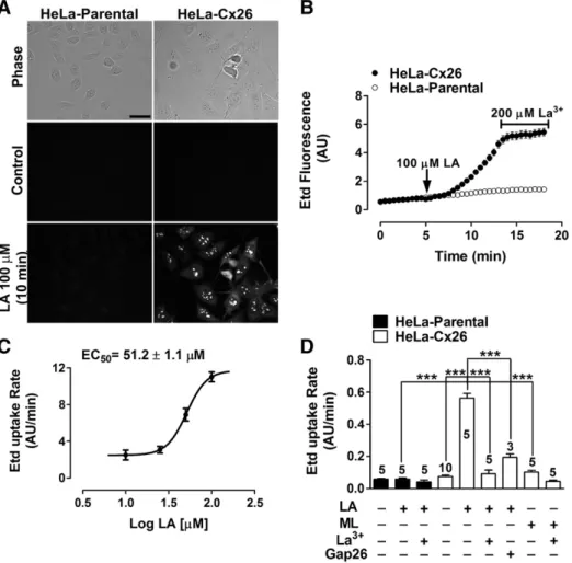
(3) V. Figueroa et al. / Biochimica et Biophysica Acta 1828 (2013) 1169–1179. trypsin inhibitor from soybean, 6.4 mM benzamidine, 7.6 mM εaminocaproic acid, 20 mM EDTA, 3.2 mM phenylmethanesulfonyl fluoride, 6.1 μM aprotinin, 20 μM leupeptin) and phosphatase inhibitors (100 mM Na2P2O7 × 10 H2O, 100 mM NaF). Then, an excess of immobilized NeutrAvidin was added (1 ml of NeutrAvidin per 3 mg of biotinylated protein), and the mixture was incubated for 1 h at 4°C. Then, 1 ml of washing buffer (saline solution, pH 7.2 plus 0.1% SDS and 1% Nonidet P-40) was added. The mixture was centrifuged for 2 min at 14,000 rpm at 4°C and the supernatant was removed and discarded. After removing the supernatant, 40 μl of saline solution (pH 2.8), plus 0.1 M glycine were added to release the proteins from biotin. The pellet was resuspended, and centrifuged at 14,000 rpm for 2 min at 4°C. The supernatant was placed in an Eppendorf tube (1.5 ml), and the pH was adjusted immediately by adding 10 μl of 1 M Tris, pH 7.5. Relative levels of proteins were evaluated by Western blot analysis as described above.. 2.7. Intracellular Ca 2+ signal measurement Cells plated on glass coverslips were loaded with 5 μM Fura-2-AM, in DMEM, without serum for 30 min at 37°C, and then washed three times in recording solution [in mM: NaCl (148); KCl (5); CaCl2 (1.8); MgCl2 (1); glucose (5); HEPES (5), pH= 7.4]. The experimental protocol for intracellular Ca2+ signal measurements involved data acquisition every 3 s. Fluorescence intensity was measured at excitation wavelengths of 340 and 380 nm (the emission wavelength was 510 nm), by using an Olympus BX 51W1I upright microscope and imaging system described above. METAFLUOR software (Universal Imaging, Downingtown, PA) was used for image acquisition and analysis. Analysis involved determining the pixel values assigned to each cell. The average pixel value allocated to each cell was calculated from the excitation wavelength, and then corrected for background. The ratio was obtained after dividing the 340-nm by the 380-nm fluorescence image on a pixel-by-pixel base (R= F340 nm/F380 nm). Some experiments were conducted in recording solution without extracellular Ca2+ and Mg 2+. For experiments with BAPTA, which is a Ca2+ chelator, cells were simultaneously pre-incubated for 30 min with BAPTA-AM (5 μM) and Fura 2-AM (5 μM).. 2.8. Cx26 cRNA preparation and injection into X. laevis oocytes The plasmid pOocyte-Cx26, containing human Cx26 cDNA, was kindly provided by Dr. Guillermo Altenberg (Texas Tech University Health Sciences Center, Lubbock, Texas). cRNA was prepared as follows: cDNA was cut with Sal I enzyme (New England Biolabs, MA, USA) for 3 h at 37°C. Then, cut cDNA was purified with the kit gel extractor (QIAGEN) following the manufacturer's recommendations. cRNA was obtained by mixing the purified cDNA with the enzyme mix, which was provided by the mMessenger mMachine kit (AMBION). The resulting mixture was incubated for 3 h at 37°C in a heated bath. Then, cRNA was precipitated by adding 30 μl of RNAse/DNAse free H2O, plus 30 μl of LiCl solution [mMessenger mMachine kit (AMBION)], and stored at −80°C. The next day, cRNA was precipitated by centrifugation at 15,000 rpm at 4°C for 15 min. Supernatant was discharged and 1 ml of 70% ethanol was used to clean the cRNA. After centrifugation at 15,000 rpm at 4°C for 15 min, ethanol was carefully discharged and cRNA was resuspended in H2O, aliquoted and stored at −80°C. Each oocyte was injected with 12.5 ng of antisense Cx38 oligonucleotide, so as to avoid the expression of endogenous Cx38, plus 50 ng of cRNA coding for Cx26. After cRNA injection, oocytes were maintained in Barth's solution (containing in mM: 88 NaCl, 1 KCl, 5 CaCl2, 0.8 MgCl2, 10 HEPES/NaOH, pH 7.4) supplemented with 0.1 mg/ml gentamycin and 20 units/ml of penicillin-streptomycin each for 24– 48 h, in order to reach a good expression level.. 1171. 2.9. Electrophysiological recordings in oocytes Whole cell recordings of transfected X. laevis oocytes were carried out as previously described [14]. In brief, oocytes expressing Cx26 were placed in the recording chamber and positioned on a stereomicroscope. The chamber was superfused with ND96 solution (in mM: 96 NaCl, 2 KCl, 1.8 CaCl2, and 5 HEPES/NaOH, pH 7.4), and recording micropipettes were filled with 3 M KCl. All experiments were performed at room temperature. For data acquisition and off-line analysis, we used a patch-clamp amplifier for oocytes (Warner Instruments, Hamden, CT, model OC-725C), which was connected to a digital-to-analog converter (Molecular Devices, model DigiData 1440A). The pClamp 10 software was used to acquire and analyze the recordings. Currents were recorded following 15-s rectangular voltage pulses, ranging from −50 mV to +60 mV, in 10-mV steps, with a holding potential of −60 mV and 10-s intervals between pulses. The I–V relationship was calculated from the current values at the end of each depolarizing pulse. Then, normalized conductance values were calculated and the Boltzmann equation was fit to the data. The fatty acid was dissolved in ND96 and carefully added to final concentrations ranging from 0.1 to 1000 μM. The recordings were performed after 3 min of incubation with the fatty acid. 2.10. Statistics For each group of data, results are expressed as means ± SEM and n is the number of independent experiments or the number of cells, as indicated. Data sets were compared by one-way analysis of variance (ANOVA) followed by a Tukey's post-test or paired Student's t test, as appropriate. Differences were considered significant at p≤0.05. The analyses were performed with GraphPad Prism 5 software for Windows (1992–2007, GraphPad Software). 3. Results 3.1. LA increases Etd uptake rate of HeLa-Cx26 cells in a concentrationdependent manner Recently, LA was demonstrated to induce a biphasic effect on the functional state of HCs formed by Cx46 in X. laevis oocytes, used as an exogenous expression system. Cx46 HC currents are enhanced by low (b 10 μM) and reduced by high (>100 μM) LA concentrations [14]. In this work, we explored the possible effect of LA on properties of Cx26 HCs expressed in both HeLa cells and X. laevis oocytes. All recordings were performed in the presence of physiological extracellular Ca2+/Mg2+ concentrations. Both HeLa-Parental and -Cx26 did not show significant Etd uptake under resting conditions (Fig. 1A, control), which is consistent with absence of Cx26 HCs and the low open probability of Cx26 HCs in the presence of extracellular divalent cations, respectively. However, in HeLa-Cx26, but not HeLa-Parental cells, an increased Etd uptake after the application of 100 μM LA for 10 min was observed (Fig. 1A). In time-lapse experiments, the application of 100 μM LA induced a progressively increase in Etd uptake after a lag of about 2–3 min, after which Etd uptake was relatively high and constant (Fig. 1B). The LAinduced increase of Etd uptake was not observed in HeLa-Parental cells (Fig. 1B) and the HC blocker La3+ (200 μM) significantly inhibited Etd uptake in Hela-Cx26 cells (Fig. 1B). The LA–induced increase in Etd uptake rate of HeLa-Cx26 cells was concentration-dependent (Fig. 1C) with an EC50 of around 51.2 ± 1.1 μM. Pre-incubation with the mimetic peptide Gap26 (200 μM) [20] strongly reduced the Etd uptake rate induced by LA (Fig. 1D). Methyl linoleate (ML) has a similar structure to LA, except that it has a methyl instead of the acid group. ML (100 μM) applied to the bath solution did not affect the Etd uptake rate of HeLa-Cx26 cells (Fig. 1D), indicating that the carboxyl group of LA is critical to induce the increase of HCs activity. HeLa-Cx26.
(4) 1172. V. Figueroa et al. / Biochimica et Biophysica Acta 1828 (2013) 1169–1179. Fig. 1. LA increases ethidium (Edt) uptake in HeLa- Cx26 but not in HeLa-Parental cells. A: Representative fluorescent fields of HeLa-Parental or HeLa-Cx26 cells incubated for 10 min in saline solution containing 5 μM Etd under control conditions, or after exposure to 100 μM LA. Scale bar, 40 μm. B: Representative time-lapse experiments showing Etd uptake in HeLa-Cx26 and HeLa-Parental cells under the control condition (first 5 min) and after applying 100 μM LA (following 15 min). Cells were treated with 200 μM La3+, a Cx HC blocker, during the last 5 min. In the presence of physiological extracellular concentrations of divalent cations, measurements were taken every 30 s as fluorescence emission intensity of Etd bound to DNA and referred to as Florescence intensity expressed in arbitrary units (AU). Each value corresponds to the mean ± SEM of at least 20 cells. C: Etd uptake induced by different LA concentrations in HeLa-Cx26 cells. Each point represents the mean ± SEM of at least 3 independent experiments, as the one shown in B. Values were normalized considering basal response in the absence of LA and adjusted to a Hill equation, EC50 = 51.2 ± 1.1 μM, R2 = 0.95. D: Etd uptake rates measured under control conditions and after exposure to 100 μM methyl-linoleate (ML), 100 μM LA, pre-incubated with 200 μM La3+ or 200 μM Gap26 mimetic peptide, both blockers of Cx HCs, and then followed by LA or ML treatment of Hela-Parental or HeLa-Cx26 cells. Data are presented as mean ± SEM, with the number of independent experiments indicated in each bar. ***p b 0.001. Values recorded in at least 20 cells per experiment were included.. and HeLa-Parental cells treated with the vehicle (ethanol) did not show any change in the Etd uptake (data not shown). Additionally, we found that LA increases Etd uptake in HeLa cells transfected with Cx32, Cx43 or Cx45 (see Supplementary Fig. 1) and it was completely blocked by 200 μM La 3+; further suggesting that LA increases the activity of HCs formed by other Cxs. In contrast, the dye-coupling between HeLa-Cx26 cells was drastically reduced by 100 μM LA (see Supplementary Fig. 2). According increased HCs activity and reduction of intercellular coupling trough GJCs, have been previously reported in astrocytes and HeLa expressing Cx43 or Cx26 [13,16,17,21]. We also tested the effect of three additional HC blockers, ßglycyrrhetinic acid (ßGA), octanol (Oct) and carbenoxolone (CBX), on the Etd uptake induced by LA in HeLa-Cx26 cells. Neither of these HC blockers reduced the Etd uptake induced by LA (Supplementary Fig. 3). In contrast, ßGA (50 μM) and Oct (50 μM) induced a slight (but not significant) increase (Supplementary Fig. 3A and B), whereas the same concentration of CBX significantly increased the Etd uptake induced by 100 μM LA (Supplementary Fig. 3A and B), suggesting that LA interferes with the mechanism by which these blockers exert their expected inhibitory effects. We use a divalent cation free solution to open HCs and test the effectiveness of each. blocker, as shown in Supplementary Fig. 3. HeLa-Cx26 did not show significant Etd uptake in the presence of extracellular divalent cations (Supplementary Fig. 3C and D, Ca 2 +/Mg 2 +), which was consistent with the lack of Cx26 HCs expression and the low open probability of Cx26 HCs in the presence of extracellular divalent cations, respectively. An increased Etd uptake was observed in HeLa-Cx26, after replacing the bath solution by a Ca 2 +/Mg 2 +-free solution (Supplementary Fig. 3C and D, Ca 2 +/Mg 2 + free). No further increase of Etd uptake was observed in HeLa-Cx26 after addition of 50 μM of each blocker in Ca 2 +/Mg 2 +-free solution (Supplementary Fig. 3C and D), demonstrating that these inhibitors, exert the expected effect on the HCs activity induced by a divalent cation free solution. 3.2. The increase in membrane permeability to Etd induced by LA is mediated by a PI3K/Akt-dependent pathway Proinflammatory conditions are known to induce HC opening in astrocytes via a p38 mitogen-activated protein kinase-dependent pathway [16,21]. Since LA can activate several kinases, including Akt and p38 kinase [22,23], we decided to use a pharmacological approach to study the possible role of these intracellular pathways in the Etd uptake rate increase of HeLa-Cx26 cells treated with LA..
(5) V. Figueroa et al. / Biochimica et Biophysica Acta 1828 (2013) 1169–1179. 1173. Fig. 2. The LA-induced Etd uptake in HeLa-Cx26 is mediated by a PI3K/Akt- but not by a p38 MAPK-dependent pathway. A: Representative time-lapse experiment of Etd uptake in HeLa-Cx26 induced by LA under control conditions, or pre-incubated for 30 min with 10 μM Akti, 1 μM PI3Ki or 10 μM p38Ki, inhibitors of Akt, PI3K and p38 kinase, respectively. Each point represents the mean ± SEM of the fluorescence intensity of at least 20 cells per experiment. B: Etd uptake rates relative (%) to the control conditions (100 μM LA) or after pre-incubation with 10 μM Akti, 1 μM PI3Ki, 10 μM p38Ki or 200 μM La3+ prior to LA treatment. Each bar represents the mean ± SEM of independent experiments including at least 20 cells, as showed in A. The number of independent experiments is indicated in each bar. ***p b 0.001, **p b 0.01. C: This panel shows a representative Western blot of three independent experiments showing that LA induces phosphorylation of Akt on S473. Confluent Hela-Cx26 cultures in 60 mm dishes were or not preincubated for 30 min with Akti prior to LA treatment or vehicle. Then, they were exposed to 100 μM LA for 2, 5 and 10 min, or to 10 nM insulin for 30 min, as positive control.. Thus, we tested the effect of SB202190, a p38 MAP kinase inhibitor (p38Ki), wortmannin (PI3Ki), a PI3K inhibitor and Akt inhibitor VIII (Akti), a cell-permeable Akt1/Akt2 inhibitor, on the Etd uptake rate of HeLa-Cx26 cells treated with 100 μM LA. The Etd uptake induced. by LA was inhibited in cells pre-incubated for 30 min with 1 μM PI3Ki or 10 μM Akti, whereas pre-incubation with 10 μM p38Ki had no effect (Fig. 2A and B). The analyzes of dye uptake experiments show that both PI3K and AKT blockers importantly inhibited the. Fig. 3. LA increases the intracellular Ca2+ signal in HeLa-Cx26 cells: Representative experiments showing pseudo color images of Ca2+ signals in HeLa-Cx26 cells loaded with Fura-2 (changes in intracellular Ca2+ signals are expressed as the Fura-2 fluorescence ratio F340/F380) at A: 2 min, B: 7 min, and C: 25 min after exposure to 100 μM LA D: Representative experiment showing the individual Ca2+ signal changes over time in 29 HeLa-Cx26 cells, shown in A, B and C, after treatment with 100 μM LA. The Fura-2 fluorescence ratio changes were measured every 3 s in the presence of physiological extracellular divalent cation concentrations..
(6) 1174. V. Figueroa et al. / Biochimica et Biophysica Acta 1828 (2013) 1169–1179. Fig. 4. The LA-induced rise in intracellular Ca2+ signal in HeLa-Cx26 cells is sensitive to HC blockade and depends on intra and extracellular Ca2+. HeLa-Cx26 and HeLa-Parental cells were loaded with Fura-2 and then treated with 100 μM LA. Representative traces of Ca2+ signal changes induced by LA are shown (values correspond to mean ± SEM of at least 20 cells) in the presence of physiological concentrations of extracellular divalent cations (except in C). A: HeLa-Cx26 cells. B: HeLa-Parental cells. C: HeLa-Cx26 cells in divalent cation-free solution. D and E: HeLa-Cx26 cells pre-incubated for 10 min with 200 μM La3+, or pre-loaded with BAPTA (5 μM) and then treated with 100 μM LA, respectively. F: The amplitude of the transient increase in intracellular Ca2+ signal induced by LA was reduced to basal level by free divalent cation solution, La3+ or BAPTA. Values represent mean ± SEM. The number of independent experiments is indicated in each bar. *p b 0.05, **p b 0.01, ***p b 0.001.. rate of Etd uptake (Fig. 2B), indicating that PI3K and AKT are activated by LA and both are involved in LA-induced HC opening. It is well known that Akt acts downstream of PI3K and its activation requires phosphorylation at S473 [24]. Insulin is a well-known activator of Akt in several cell types [25]). In addition, HeLa cells are known to express insulin receptors [26]. Therefore, we used insulin as a positive control of LA-induced Akt activation by phosphorylation. Accordingly, a rise in phosphorylation in this amino acid residue was detected by western blot analyses of cells treated with 100 nM insulin. This response was completely inhibited by Akti (Fig. 2C, 3rd and 4th lanes), as it is known to occur in cells expressing insulin-sensitive receptors [27]. In Hela-Cx26 cells exposed to 100 μM LA for 2, 5 and 10 min, the increase in Akt phosphorylation in S473 was higher than in control cells. The maximal effect was observed at 5 min, and decreased at 10 min post-treatment (Fig. 2C). Because increases in HC activity can be explained by rises in the number of HCs located in the plasma membrane [19], we studied whether LA through Akt/PI3k may affect the abundance of HCs at the cell membrane. To achieve this purpose, cell surface proteins were. biotinylated and isolated and levels of Cx26 were determined by Western blot analyses. It was found that 100 μM LA does not change the levels of Cx26 at 2, 5 or 10 min (Fig. 2D). Therefore, the LAinduced increase in Cx26 HC activity is not due to a rise of HCs at the plasma membrane.. 3.3. LA increases the intracellular Ca 2+ signal in HeLa-Cx26 cells In myocyctes and stem cells, two cell types that express Cxs, LA increases the free intracellular Ca 2+ concentration ([Ca 2+]i) [28,29]; in myocytes, this rise occurs through a La 3+-sensitive pathway [22,30]. Regarding to HCs, the elevation in [Ca 2 +]i to about 500 nM has been shown to increase the activity of Cx HCs [13,31,32]. Therefore, we decided to study if LA affects the Ca 2+ signal in HeLa-Cx26 cells. The intracellular Ca 2+ signal (ratio 340/380) was monitored in HeLa-Cx26 cells loaded with the Ca 2+ indicator Fura-2 (Fig. 3A–C). LA (100 μM) induced a fast and transient increase in Ca 2+ signal during the first 5 min after treatment (indicated with the number 1 in.
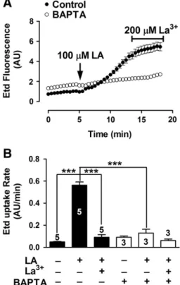
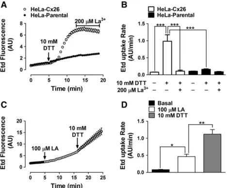
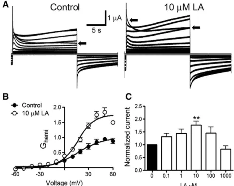
(7) V. Figueroa et al. / Biochimica et Biophysica Acta 1828 (2013) 1169–1179. 1175. Fig. 3D), which was followed by a slow and progressive increase after about 7.5 min (indicated with the number 2 in Fig. 3D). 3.4. The LA-induced increase in intracellular Ca 2+ signal in HeLa-Cx26 cells requires Ca 2+ inflow via HCs Recent studies in HeLa cells transfected with mCx32 or mCx43, and in purified human Cx26 HCs reconstituted in liposomes as well as in X. laevis oocytes expressing human Cx26, have shown that HCs formed by these Cxs are permeable to Ca 2+ [33–36]. To test the possible involvement of HCs in the Ca 2+ signal increments induced by LA, we conducted a series of experiments in which the extracellular Ca 2+ was excluded, or the intracellular Ca 2+ was chelated. In the presence of physiological concentrations of divalent cations in the extracellular solution, LA induced a fast and transient rise followed by a progressive increase in Ca2+ signal (Fig. 4A). as illustrated in Fig. 3D (1). The fast and transient rise in Ca2+ signal was also detected in HeLa-Parental cells, however the second sustained increase was not observed (Fig. 4B), suggesting that the latter is mediated by HCs. The LA-induced rises of the Ca 2 + signal in HeLa-Cx26 cells were also drastically reduced in cells bathed with extracellular solution nominally free of divalent cations (Fig. 4C); while the amplitude of the transient rise (1) was drastically reduced, the progressive increase (2) was absent (Fig. 4F). Pre-incubation with La 3 + (200 μM) for 5 min in a saline solution containing physiological Ca 2 +/Mg 2 + concentration abolished completely both Ca 2 + signals (1 and 2) induced by LA (Fig. 4D). Similarly, LA did not affect the Ca 2 + signal in cells preloaded with BAPTA (Fig. 4E). The results are summarized in the graph presented in Fig. 4F. Moreover, cells loaded with BAPTA in saline solution containing physiological Ca 2 +/Mg 2 + concentration did not show increase in membrane permeability upon treatment with LA (Fig. 5) and 200 μM La 3 + blocked the dye uptake in control but not in BAPTA loaded cells (Fig. 5), suggesting that both Ca 2 + and Etd cross the cell membrane through the same pathway. 3.5. The LA-induced increase in intracellular Ca 2+ signal in Hela-Cx26 cells is directly related to the opening of Cx26 HCs To elucidate whether the effect of LA on the Ca 2+ signal is related to Cx26 HC opening, intracellular Ca2+ signal changes and Etd uptake in Hela-Cx26 cells were simultaneously monitored. We used Fura-2 loaded HeLa-Cx26 cells and extracellular saline solution containing physiological Ca2+/Mg2+ concentrations as well as 5 μM Etd (Fig. 6A). After the application of 100 μM LA, the Ca2+ signal showed a rapid onset with almost no delay (Fig. 6B). Before the maximal rise of the first transient rise of Ca2+ signal (1) the Etd uptake increased slowly (from mb= 0.012 to m1= 0.159 AU/min) (Fig. 6B). However, ~3 min later the dye uptake showed a more pronounced increase (m3 =0.352 AU/ min) (Fig. 6B), whereas the Ca2+ signal showed a slow and progressive increase (2). The application of 200 μM La3+ at minute 26 reduced drastically both the Etd uptake and Ca2+ signal rise (Fig. 6B). 3.6. Dithiothreitol increases the dye uptake and enhances it in LA-treated HeLa-Cx26 cells The open probability of some HCs as those formed by Cx43 is affected by redox changes [16,19]. Moreover, LA is metabolized to AA, a substrate of cyclooxygenases and lipooxgenases that generate superoxide anions [37]. Thus, we tested if the LA effect on Cx26 HC activity was due to changes in redox state. To this end, HeLa-Cx26 cells under control conditions were exposed to 10 mM DTT (Fig. 7A). After ~ 1 min of DTT addition to the bath solution an important increase of dye uptake rate occurred (from 0.077 ± 0.004 to 0.985 ± 0.190 AU/min), which was blocked by 200 μM La + 3 (0.106 ± 0.031 AU/min) (Fig. 7B). Moreover, the effect of DTT was additive to that of LA (Fig. 7C); LA (100 μM) increased the dye. Fig. 5. BAPTA prevents LA-induced Etd uptake in HeLa-Cx26 cells. A: Representative time-lapse experiment showing the LA-induced Etd uptake in HeLa-Cx26 cells under control conditions or pre-loaded with BAPTA (5 μM); each point represents the mean ± SEM of the fluorescence intensity of at least 20 cells per experiment. B: Etd uptake rates of HeLa-Cx26 cells in the presence of LA under control conditions, or HeLa-Cx26 cells pre-loaded with BAPTA and subsequently treated or not with 200 μM La3+ are shown. Each bar represents the mean ± SEM. The number of independent experiments is indicated within each bar. ***p b 0.001.. uptake rate (from 0.080 ± 0.009 to 0.465 ± 0.066 AU/min, Fig. 7D) and the posterior addition of 10 mM DTT induced a new increase (1.121 ± 0.133 AU/min). However, 10 mM DTT did not significantly change the dye uptake rate in parental cells (from 0.104 ± 0.005 to 0.160 ± 0.015 AU/min, Fig. 7B). 3.7. LA increases Cx26 HC currents in X. laevis oocytes Finally, we tested whether the effect of LA over Cx26 HCs is cell-dependent. To study this possibility X. laevis oocytes expressing Cx26 were used. Forty eight hours after Cx26 cRNA injection, the oocytes were exposed to different LA concentrations (0.1–1000 μM) for 3 min and whole cell membrane currents were measured. Holding potential was maintained at − 60 mV. Under control conditions, transfected oocytes showed higher instantaneous and steadystate membrane currents than oocytes not injected with Cx26 cRNA (not shown) after depolarization pulses (10 mV, 15 s) ranging from − 60 to + 60 mV. In transfected oocytes the appearance of prominent outward currents at voltages above 0 mV was evident. Around +20 mV the oocytes expressing Cx26 HCs showed a fast increase of whole cell currents followed by a relaxation of the outward currents, a second increase (slower than the first one) was observed after the relaxation. However, at +60 mV the second increase was much less evident. Similar recordings have been previously reported [36,38]. After each depolarization pulses, the membrane potential was maintained at −60 mV for 10 s. A sustained tail current was also observed when the membrane potential (after depolarization) returned to the holding potential (− 60 mV) (Fig. 8A, control). The bath application of 10 μM LA increased (176 ± 15%, n = 8) both the maximum current and tail current (Fig. 8A, 10 μM LA). The current increases were more evident at voltages over 0 mV (Fig. 8B)..
(8) 1176. V. Figueroa et al. / Biochimica et Biophysica Acta 1828 (2013) 1169–1179. Fig. 6. The LA-induced rise in intracellular Ca2+ signal is directly related to Cx26 HC opening. HeLa-Cx26 cells pre-loaded with Fura-2 were used. The Ca2+ signal was evaluated for 5 min under control conditions in an extracellular solution containing physiological concentrations of Ca2+/Mg2+ and 5 μM Etd. Then, cells were treated with 100 μM LA. A: Representative experiments showing pseudo color images of Ca2+ signals in HeLa-Cx26 cells loaded with Fura-2 (changes in Ca2+ signal are expressed as the fura-2 fluorescence ratio F340/F380) and pictures of Etd-stained nucleic acids, as observed in HeLa-Cx26 cells after 30 min application of 100 μM LA. B: Representative simultaneous records showing changes in Fura-2 fluorescence ratio and Etd uptake versus time in HeLa-Cx26 cells, in response to 100 μM LA. Each point represents the mean ± SEM of the fura-2 fluorescence ratio F340: F380, and fluorescence emission intensity of Etd bound to DNA of 30 cells. The basal Etd uptake rates and the successive changes in slope during the experiment are indicated as mb and m1–3, respectively.. Quantification of the maximum current (recorded after the initial maximal current) at + 60 mV (Fig. 8A and B, arrow) revealed that concentrations of LA ranging between 0.1 – 100 μM increased the steady-state current. However, such increase was statistically different from control values only at 10 μM. Boltzmann analysis of HC conductance (Ghemi) showed that both V50 and voltage sensitivity are not modified after 3 min of 10 μM LA addition. Thus, the V50 under control conditions was 16.2 ± 3.5 mV and after LA was 16.8± 1.7 mV (p > 0.05). Whereas, the voltage sensitivity (A) under control conditions was 0.06± 0.02 and after LA was 0.04± 0.02 (p > 0.05). But the Imax (maximal hemichannel current) increased from 1.01 ±0.04 to 1.76 ±0.04 after treatment with LA. Therefore, this LA concentration did not change the voltage sensitivity of Cx26 HCs, but it increased either the number of hemichannel ready to open, or increase the open probability of available HCs in the plasma membrane and/or increase the unitary conductance of Cx26 HCs. The increase in membrane current induced by 100 μM was less prominent than that induced by 10 μM. Furthermore, 1000 μM LA slightly decreased the membrane current induced by voltage, but the difference was not statistically different from control values.. Thus, the concentration-response relationship of LA on membrane current had a bell shape. 4. Discussion In the present work, LA was found to increase the activity of Cx26 HCs as evaluated by the Etd uptake method. We ascribe the LA effect on cellular Etd uptake to Cx26 HC opening, because it is not observed in HeLa-Parental cells, levels of Cx26 in the cell membrane in HeLa-Cx26 cells are not affected and the effect is rapidly abrogated by two HC blockers, La 3+ and the mimetic peptide Gap26. Opening of Cx26 HCs is mediated by an Akt and PI3K-dependent signaling pathway and increases the intracellular Ca +2 concentration. LA also increases Cx26 HC currents in Xenopus oocytes and dye uptake in HeLa-Cx32, -Cx43 and –Cx45, suggesting that its effect is cell and Cx type independent. In contrast, ML, a derivative of LA in which the carboxylic acid is replaced by a methyl group, fails to increase the membrane permeability to Etd. The Akt activity is enhanced through a PI3K-dependent signaling pathway [39] and Akt becomes phosphorylated at residues Thr 308.
(9) V. Figueroa et al. / Biochimica et Biophysica Acta 1828 (2013) 1169–1179. and Ser 473, which are required for full Akt kinase activity [39]. Here, we show that LA induces Akt phosphorylation on Ser 473 in a time-dependent manner and its maximal phosphorylation (5 min after LA treatment) ocurrs before the maximal increase in Etd uptake (~7.5 min after LA treatment), suggesting that Akt phosphorylation precedes HC opening. In support to our results, HeLa cells express a free fatty acid receptor, but to our knowledge it remains uncharacterized, and could mediate activation of a PI3K/Akt-dependent pathway. However, it is clear that LA, through Akt activation, increases Cx26 HC activity probably via an increase in open probability or in Etd permeability since Cx26 levels present in the cell membrane are not modified. These changes might be the result of a cytoplasmic regulator of Cx26 HCs or a direct consequence of Cx26 phosphorylation. However, it remains controversial if Cx26 is a phosphoprotein [40,41]. We also found that LA enhances the intracellular Ca2+ signal and this response is temporally associated with the increase in Etd uptake, suggesting a reciprocal dependency. In support to this interpretation, the first and transient intracellular Ca2+ increase induced by LA is significantly reduced but not completely abrogated in HeLa-Cx26 cells bathed with divalent cation-free solution, suggesting the involvement of a mechanism independent of Ca 2+ influx. However, inhibition of PLC with 10 μM U73122 applied 30 min before LA treatment prevented neither the first nor the second Ca +2 peak, ruling out the involvement of intracellular calcium stores (data not shown). Thus, we speculate that the first peak is due to Ca +2 influx through a La 3+ sensitive ion channel (i.e., P2X and/TRP channels). This influx might be mediated by the gradient of Ca +2 still present between the intracellular and extracellular compartment of cells bathed with nominal Ca +2-free solutions (usually ~10 μM Ca2+ in the extracellular milieu and 100 nM in the intracellular space). Accordingly, the first rise in Ca 2+ signal is still evident in HeLaParental cells, which does not express Cx26 HCs. Moreover, the first transient increase in Ca2+ signal appears to be essential to enhance Cx26 HC activity, since intracellular BAPTA prevented both the rise in. 1177. Ca 2+ signal and opening of the Etd permeable membrane pathway. In contrast, the second and sustained intracellular Ca 2+ increase was completely abrogated by omission of extracellular Ca 2+ signal in HeLa-Cx26 cells and did not occur in HeLa-Parental cells, indicating that the second increase in Ca2+ signal is due to Ca2+ influx through Cx26 HCs. It was recently shown that HCs formed by Cxs 26, 32 or 43 are Ca2+ permeable and provide a cell membrane route for Ca2+ influx [31,33–36], which is consistent with our results. It remains unknown why La3+ blocked the first LA-induced Ca 2+ signal increase. A possible mechanism could involve inhibition of the LA interaction with its membrane receptor. Alternatively, early activation of Cx26 HCs might be essential to activate a possible feed-forward mechanism. However, clarification of this issue requires further investigation. We also demonstrated that HCs formed by human Cx26, or mouse Cxs 43 or 45 are also activated by LA, suggesting that Ca 2+ homeostasis could be affected in different cell types and species. It has been described rises of intracellular Ca2+ signal in response to LA in other cell types such as endothelial cells and myocytes [22,30,42,43]. Particularly in myocytes, LA, palmitic acid and AA increase the intracellular Ca2+ signal through a pathway inhibited by 100 μM La3+ [30], also known to block Cx HCs. Mammalian myocytes express Cxs 40, 43 and 45 [28], which also form functional HCs [44,45]. Since LA also increases the activity of Cx43 and Cx45 HCs. It is possible that the Ca 2+ rises induced by LA in myocytes are due to opening of HCs formed by cardiac Cxs. We also found that LA reduces gap junctional communication, which is reminiscent of the opposite effect of pro-inflammatory agents on HCs and GJCs [13,16,17,21]. DTT, a \SH group reducing compound, increases the activity of Cx43 HCs [19] and reduces their open probability in cells under oxidative stress [19,21]. To this end, Cx26 presents a cysteine residue in 218 position located in the carboxyl terminus [46] and in the present study DTT was found to increase the activity of Cx26 HC under control conditions. Moreover, in HeLa-Cx26 cells treated with LA, known to. Fig. 7. DTT, a sulfhydryl group reducing agent, increases the Etd uptake in HeLa-Cx26 cells and its effect is additive to that of LA. A. Representative time-lapse experiment of Etd uptake in HeLa-Cx26 and HeLa-Parental cells treated with 10 mM DTT followed by bath application of 200 μM La3+. Each point represents the mean ± SEM of the fluorescence intensities of a least 20 cells per experiment. B. Etd uptake rates of HeLa-Cx26 and HeLa-Parental cells after treatment with 10 mM DTT; each bar represents the mean ± SEM of 3 independent experiments. ***p b 0.001. C. Representative time-lapse experiment of Etd uptake in HeLa-Cx26 cells induced by 100 μM LA followed by exposure to 10 mM DTT. Each point represents the mean ± SEM of the fluorescence intensities of a least 20 cells per experiment. D. Etd uptake rates of HeLa-Cx26 induced by 100 μM LA and by 100 μM LA followed by 10 mM DTT. Each bar represents the mean of Etd uptake rates ± SEM of the 3 independent experiments. *pb 0.05, **p b 0.01..
(10) 1178. V. Figueroa et al. / Biochimica et Biophysica Acta 1828 (2013) 1169–1179. Fig. 8. LA increases Cx26 HC currents. A: Representative whole-cell current records from Xenopus oocytes previously (48 h before) injected with Cx26 cRNA under control conditions (left) or treated with 10 μM LA (right). Oocytes were depolarized from −60 mV to +60 mV in steps of 10 mV for 15 s. The current generated at +60 mV is indicated by an arrow in control and 10 μM LA treated oocytes B: Normalized HC mediated membrane conductance of oocytes under control condition (black circles) and after the addition of 10 μM LA (white circles). Currents were measured at the end of each voltage step (arrows panel A and B). C: Normalized currents at +60 mV in the presence of different LA concentrations. Data are presented as mean ± SEM. Each bar, n = 6.. induce oxidative stress in different cell types including HeLa cells [47,48], DTT enhanced the Etd uptake in a way additive to the LA, indicating that oxidation of \SH groups does not explain the LA effect on Cx26 HCs. The effect of DTT on Cx26 HCs of LA-treated cells also rules out the possible involvement of oxidant stress in the activation of PI3K and Akt. Unexpectedly, lipophilic GJC blockers, BGA, CBX and Oct, did not reduce the increase in Etd uptake induced by LA. In contrast, all of them tended to increase the Etd uptake in LA-treated cells, which was particularly evident for CBX. Given the lipophilic nature of these compounds, we hypothesize, without excluding other possible explanation, that LA might induce a conformational change of Cx26 HCs, initiated in a cytoplasmic domain of the protein subunit, via a Ca2+/PI3K/Akt-dependent pathway. This putative change would modify the BGA, CBX and Oct binding sites likely to be located in a hydrophobic domain of Cx26 HCs. This would also implicate that the binding sites of these three liposoluble compounds are similar and possibly common. Moreover, the proposed conformational change might not significantly affect extracellular hydrophilic domains of Cx26 because the HCs remained sensitive to La3+ and Gap26, two hydrosoluble HC blockers. Our findings support the presence of a new HC control mechanism triggered by LA and mediated by an Akt/PI3K-dependent pathway and therefore, it could also shed light on the role of HCs in phenomena where fatty acids and/or Akt/PI3K are involved.. 5. Conclusions In the present work, LA increased the activity of Cx26 HCs expressed in HeLa cells and in Xenopus oocytes. LA also enhanced the activity of HCs formed by Cxs 32, 43 or 45. The effect of LA on Cx26 HCs was mediated by a transient rise of intracellular free Ca2+ signal and activation of an intracellular transduction pathway involving PI3K and Akt protein kinases. The delayed and progressive increase in intracellular Ca 2 + was mediated by Ca 2 + influx through Cx26 HCs. DTT, a membrane permeant reducing agent, also increased the activity of Cx26. HCs under control conditions and its effect was additive to that of LA. Moreover, it was found that LA drastically reduces the permeability of gap junctions to Etd. Acknowledgements We would like to thank Ms. Teresa Vergara and Ms. Paola Fernández for their technical support. This work was partially funded by Fondecyt projects 1120214 (to MAR), 1111033 (to JCS), Chilean Science Millennium Institute (P09-022-F; to JCS, VAF and ADM) and Conicyt 24100132 (to VF), and the Anillo ACT-71 project (to JCS) as well as Fondef project DO7I1086 (to JCS). The data of this paper are from a thesis submitted in partial fulfillment of the requirements for the degree of Doctor in Sciences, Mention in Neuroscience (V. Figueroa) at University of Valparaiso, Valparaiso, Chile. Appendix A. Supplementary data Supplementary data to this article can be found online at http://dx. doi.org/10.1016/j.bbamem.2012.12.006. References [1] G.S. Goldberg, V. Valiunas, P.R. Brink, Selective permeability of gap junction channels, Biochim. Biophys. Acta 1662 (2004) 96–101. [2] J.C. Sáez, M.A. Retamal, D. Basilio, F.F. Bukauskas, M.V. Bennett, Connexin-based gap junction hemichannels: gating mechanisms, Biochim. Biophys. Acta 1711 (2005) 215–224. [3] J.C. Sáez, K.A. Schalper, M.A. Retamal, J.A. Orellana, K.F. Shoji, M.V. Bennett, Cell membrane permeabilization via connexin hemichannels in living and dying cells, Exp. Cell Res. 316 (2010) 2377–2389. [4] A. Chandrasekhar, A.K. Bera, Hemichannels: permeants and their effect on development, physiology and death, Cell Biochem. Funct. 30 (2012) 89–100. [5] K. Arita, M. Akiyama, Y. Tsuji, J.R. McMillan, R.A. Eady, H. Shimizu, Changes in gap junction distribution and connexin expression pattern during human fetal skin development, J. Histochem. Cytochem. 50 (2002) 1493–1500. [6] K. Kammen-Jolly, H. Ichiki, A.W. Scholtz, M. Gsenger, A. Kreczy, A. Schrott-Fischer, Connexin 26 in human fetal development of the inner ear, Hear. Res. 160 (2001) 15–21..
(11) V. Figueroa et al. / Biochimica et Biophysica Acta 1828 (2013) 1169–1179 [7] A.D. Martínez, R. Acuña, V. Figueroa, J. Maripillan, B. Nicholson, Gap-junction channels dysfunction in deafness and hearing loss, Antioxid. Redox Signal. 11 (2009) 309–322. [8] G. Richard, Connexin disorders of the skin, Clin. Dermatol. 23 (2005) 23–32. [9] U.N. Das, Essential fatty acids: biochemistry, physiology and pathology, Biotechnol. J. 1 (2006) 420–439. [10] K.A. Youdim, A. Martin, J.A. Joseph, Essential fatty acids and the brain: possible health implications, Int. J. Dev. Neurosci. 18 (2000) 383–399. [11] K. Dlugosova, L. Okruhlicova, M. Mitasikova, R. Sotnikova, I. Bernatova, P. Weismann, J. Slezak, N. Tribulova, Modulation of connexin-43 by omega-3 fatty acids in the aorta of old spontaneously hypertensive rats, J. Physiol. Pharmacol. 60 (2009) 63–69. [12] H. Nojima, Y. Ohba, Y. Kita, Oleamide derivatives are prototypical anti-metastasis drugs that act by inhibiting Connexin 26, Curr. Drug Saf. 2 (2007) 204–211. [13] E. De Vuyst, E. Decrock, M. De Bock, H. Yamasaki, C.C. Naus, W.H. Evans, L. Leybaert, Connexin hemichannels and gap junction channels are differentially influenced by lipopolysaccharide and basic fibroblast growth factor, Mol. Biol. Cell 18 (2007) 34–46. [14] M.A. Retamal, F. Evangelista-Martínez, C.G. Leon-Paravic, G.A. Altenberg, L. Reuss, Biphasic effect of linoleic acid on connexin 46 hemichannels, Pflugers Arch. 461 (2011) 635–643. [15] K.A. Schalper, N. Palacios-Prado, J.A. Orellana, J.C. Sáez, Currently used methods for identification and characterization of hemichannels, Cell Commun. Adhes. 15 (2008) 207–218. [16] M.A. Retamal, N. Froger, N. Palacios-Prado, P. Ezan, P.J. Sáez, J.C. Sáez, C. Giaume, Cx43 hemichannels and gap junction channels in astrocytes are regulated oppositely by proinflammatory cytokines released from activated microglia, J. Neurosci. 27 (2007) 13781–13792. [17] J.E. Contreras, H.A. Sánchez, E.A. Eugenin, D. Speidel, M. Theis, K. Willecke, F.F. Bukauskas, M.V. Bennett, J.C. Sáez, Metabolic inhibition induces opening of unapposed connexin 43 gap junction hemichannels and reduces gap junctional communication in cortical astrocytes in culture, Proc. Natl. Acad. Sci. U. S. A. 99 (2002) 495–500. [18] C.S. Wright, M.A. van Steensel, M.B. Hodgins, P.E. Martin, Connexin mimetic peptides improve cell migration rates of human epidermal keratinocytes and dermal fibroblasts in vitro, Wound Repair Regen. 17 (2009) 240–249. [19] M.A. Retamal, C.J. Cortés, L. Reuss, M.V. Bennett, J.C. Sáez, S-nitrosylation and permeation through connexin 43 hemichannels in astrocytes: induction by oxidant stress and reversal by reducing agents, Proc. Natl. Acad. Sci. U. S. A. 103 (2006) 4475–4480. [20] W.H. Evans, L. Leybaert, Mimetic peptides as blockers of connexin channel-facilitated intercellular communication, Cell Commun. Adhes. 14 (2007) 265–273. [21] J.A. Orellana, D.E. Hernández, P. Ezan, V. Velarde, M.V. Bennett, C. Giaume, J.C. Sáez, Hypoxia in high glucose followed by reoxygenation in normal glucose reduces the viability of cortical astrocytes through increased permeability of connexin 43 hemichannels, Glia 58 (2010) 329–343. [22] M.H. Kim, M.O. Kim, Y.H. Kim, J.S. Kim, H.J. Han, Linoleic acid induces mouse embryonic stem cell proliferation via Ca2+/PKC, PI3K/Akt, and MAPKs, Cell. Physiol. Biochem. 23 (2009) 53–64. [23] Y. Zheng, E.J. Lim, L. Wang, E.J. Smart, M. Toborek, B. Hennig, Role of caveolin-1 in EGCG-mediated protection against linoleic-acid-induced endothelial cell activation, J. Nutr. Biochem. 20 (2009) 202–209. [24] W.L. Yang, C.Y. Wu, J. Wu, H.K. Lin, Regulation of Akt signaling activation by ubiquitination, Cell Cycle 9 (2010) 487–497. [25] Z.Y. Jiang, Q.L. Zhou, K.A. Coleman, M. Chouinard, Q. Boese, M.P. Czech, Insulin signaling through Akt/protein kinase B analyzed by small interfering RNA-mediated gene silencing, Proc. Natl. Acad. Sci. U. S. A. 100 (2003) 7569–7574. [26] J.E. Ayala, J.N. Boustead, S.C. Chapman, C.A. Svitek, J.K. Oeser, R.B. Robey, R.M. O'Brien, Insulin-mediated activation of activator protein-1 through the mitogen-activated protein kinase pathway stimulates collagenase-1 gene transcription in the MES 13 mesangial cell line, J. Mol. Endocrinol. 33 (2004) 263–280. [27] H.C. Dan, A.S. Baldwin, Differential involvement of IkappaB kinases alpha and beta in cytokine- and insulin-induced mammalian target of rapamycin activation determined by Akt, J. Immunol. 180 (2008) 7582–7589.. 1179. [28] B.J. Darrow, J.G. Laing, P.D. Lampe, J.E. Saffitz, E.C. Beyer, Expression of multiple connexins in cultured neonatal rat ventricular myocytes, Circ. Res. 76 (1995) 381–387. [29] P. Worsdorfer, S. Maxeiner, C. Markopoulos, G. Kirfel, V. Wulf, T. Auth, S. Urschel, J. von Maltzahn, K. Willecke, Connexin expression and functional analysis of gap junctional communication in mouse embryonic stem cells, Stem Cells 26 (2008) 431–439. [30] K.M. Fang, A.S. Lee, M.J. Su, C.L. Lin, C.L. Chien, M.L. Wu, Free fatty acids act as endogenous ionophores, resulting in Na+ and Ca2+ influx and myocyte apoptosis, Cardiovasc. Res. 78 (2008) 533–545. [31] M. De Bock, N. Wang, M. Bol, E. Decrock, R. Ponsaerts, G. Bultynck, G. Dupont, L. Leybaert, Connexin 43 hemichannels contribute to cytoplasmic Ca2+ oscillations by providing a bimodal Ca2+-dependent Ca2+ entry pathway, J. Biol. Chem. 287 (2012) 12250–12266. [32] E. De Vuyst, N. Wang, E. Decrock, M. De Bock, M. Vinken, M. Van Moorhem, C. Lai, M. Culot, V. Rogiers, R. Cecchelli, C.C. Naus, W.H. Evans, L. Leybaert, Ca(2+) regulation of connexin 43 hemichannels in C6 glioma and glial cells, Cell Calcium 46 (2009) 176–187. [33] M.C. Fiori, V. Figueroa, M.E. Zoghbi, J.C. Sáez, L. Reuss, G.A. Altenberg, Permeation of Calcium through Purified Connexin 26 Hemichannels, J. Biol. Chem. 287 (2012) 40826–40834. [34] K.A. Schalper, H.A. Sánchez, S.C. Lee, G.A. Altenberg, M.H. Nathanson, J.C. Sáez, Connexin 43 hemichannels mediate the Ca2+ influx induced by extracellular alkalinization, Am. J. Physiol. Cell Physiol. 299 (2010) C1504–C1515. [35] H.A. Sánchez, J.A. Orellana, V.K. Verselis, J.C. Sáez, Metabolic inhibition increases activity of connexin-32 hemichannels permeable to Ca2+ in transfected HeLa cells, Am. J. Physiol. Cell Physiol. 297 (2009) C665–C678. [36] H.A. Sánchez, G. Mese, M. Srinivas, T.W. White, V.K. Verselis, Differentially altered Ca2+ regulation and Ca2+ permeability in Cx26 hemichannels formed by the A40V and G45E mutations that cause keratitis ichthyosis deafness syndrome, J. Gen. Physiol. 136 (2010) 47–62. [37] A.D. Martínez, J.C. Sáez, Arachidonic acid-induced dye uncoupling in rat cortical astrocytes is mediated by arachidonic acid by products, Brain Res. 816 (1999) 411–423. [38] H. Ripps, H. Qian, J. Zakevicius, Properties of connexin26 hemichannels expressed in Xenopus oocytes, Cell. Mol. Neurobiol. 24 (2004) 647–665. [39] P.J. Coffer, J. Jin, J.R. Woodgett, Protein kinase B (c-Akt): a multifunctional mediator of phosphatidylinositol 3-kinase activation, Biochem. J. 335 (Pt 1) (1998) 1–13. [40] O. Traub, J. Look, R. Dermietzel, F. Brummer, D. Hülser, K. Willecke, Comparative characterization of the 21-kD and 26-kD gap junction proteins in murine liver and cultured hepatocytes, J. Cell Biol. 108 (1989) 1039–1051. [41] D. Locke, S. Bian, H. Li, A.L. Harris, Post-translational modifications of connexin26 revealed by mass spectrometry, Biochem. J. 424 (2009) 385–398. [42] B. Hennig, M. Toborek, S. Joshi-Barve, S.W. Barger, S. Barve, M.P. Mattson, C.J. McClain, Linoleic acid activates nuclear transcription factor-kappa B (NF-kappa B) and induces NF-kappa B-dependent transcription in cultured endothelial cells, Am. J. Clin. Nutr. 63 (1996) 322–328. [43] V. Saraswathi, G. Wu, M. Toborek, B. Hennig, Linoleic acid-induced endothelial activation: role of calcium and peroxynitrite signaling, J. Lipid Res. 45 (2004) 794–804. [44] K. Shintani-Ishida, K. Uemura, K. Yoshida, Hemichannels in cardiomyocytes open transiently during ischemia and contribute to reperfusion injury following brief ischemia, Am. J. Physiol. Heart Circ. Physiol. 293 (2007) H1714–H1720. [45] S.A. John, R. Kondo, S.Y. Wang, J.I. Goldhaber, J.N. Weiss, Connexin-43 hemichannels opened by metabolic inhibition, J. Biol. Chem. 274 (1999) 236–240. [46] J.T. Zhang, B.J. Nicholson, Sequence and tissue distribution of a second protein of hepatic gap junctions, Cx26, as deduced from its cDNA, J. Cell Biol. 109 (1989) 3391–3401. [47] P. Sangeetha Sagar, U.N. Das, R. Koratkar, G. Ramesh, M. Padma, G. Sravan Kumar, Cytotoxic action of cis-unsaturated fatty acids on human cervical carcinoma (HeLa) cells: relationship to free radicals and lipid peroxidation and its modulation by calmodulin antagonists, Cancer Lett. 63 (1992) 189–198. [48] M. Toborek, S.W. Barger, M.P. Mattson, S. Barve, C.J. McClain, B. Hennig, Linoleic acid and TNF-alpha cross-amplify oxidative injury and dysfunction of endothelial cells, J. Lipid Res. 37 (1996) 123–135..
(12)
Figure

+2
Documento similar