ESICM LIVES 2016: part two
200
0
0
Texto completo
(2) Intensive Care Medicine Experimental 2016, 4(Suppl 1):30. carbon dioxide 30 [27–35] mmHg and median temperature 37.1 [36.8-37.3] °C. After removal of artefacts, the mean monitoring time was 22 h08 (8 h54). All patients had impaired cerebral autoregulation during their monitoring time. The mean IAR index was 17 (9.5) %. During H0H6 and H18H24, the majority of our patients; respectively 53 and 71 % had an IAR index > 10 %. Conclusion According to our data, patients with septic shock had impaired cerebral autoregulation within the first 24 hours of their admission in the ICU. In our patients, we described a variability of distribution of impaired autoregulation according to time. References Schramm P, Klein KU, Falkenberg L, et al. Impaired cerebrovascular autoregulation in patients with severe sepsis and sepsis-associated delirium. Crit Care 2012; 16: R181. Aries MJH, Czosnyka M, Budohoski KP, et al. Continuous determination of optimal cerebral perfusion pressure in traumatic brain injury. Crit. Care Med. 2012.. Fig. 1 (abstract 392). IAR index by 6 hours period. A393 Changes in muscle thickness throughout hospitalisation after traumatic brain injury L. Chapple1, A. Deane1,2, L. Williams3, R. Strickland2, K. Lange4, D. Heyland5, M. Chapman1,2 1 University of Adelaide, Discipline of Acute Care Medicine, Adelaide, Australia; 2Royal Adelaide Hospital, Intensive Care, Adelaide, Australia; 3 Griffith University, Menzies Health Institute of Queensland, Gold Coast, Australia; 4University of Adelaide, Discipline of Medicine, Adelaide, Australia; 5Kingston General Hospital, Clinical Evaluation Research Unit, Kingston, Canada Correspondence: L. Chapple - University of Adelaide, Discipline of Acute Care Medicine, Adelaide, Australia Intensive Care Medicine Experimental 2016, 4(Suppl 1):A393 Introduction Patients with a traumatic brain injury (TBI) remain in hospital for extended periods. It is likely that muscle mass in these patients diminishes over the hospital admission, yet this has not previously been quantified. Ultrasonography may provide a useful measure of changes in muscle size over the hospital stay. Objectives To quantify changes in ultrasound-derived quadriceps muscle layer thickness (QMLT) and establish the feasibility of repeated ultrasound examinations throughout the entire hospitalisation of patients with a TBI. Methods Adult patients with a moderate-severe TBI (Glasgow Coma Scale 3–12) consecutively admitted to the intensive care unit (ICU) at a single trauma referral centre over 12 months were eligible following informed consent. Ultrasounds of QMLT at the midpoint and two-thirds between the anterior superior iliac spine and top of the patella were conducted weekly during admission. Data were censored at 3-months and are mean (SD) unless otherwise stated. Results Thirty-three patients [45.4 (16.6) years; 88 % male; 55 % severe TBI; Trauma Injury Severity Score 0.5 (0.3); median APACHE II. Page 206 of 607. 18 (IQR: 14–22); admission body mass index 27.2 (6.5) kg/m2] were studied. Primary injury cause was vehicular (58 %) and 67 % of TBIs were multi-trauma. Median length of ICU and ward-based stay was 13.4 [IQR: 6.5-17.9] and 31.8 [9.4-52.4] days, respectively. At 3-months all patients were alive and four remained in hospital. A total of 123 ultrasounds were taken; 30 (24 %) in ICU and 93 (76 %) on the acute ward, equal to 3.7 ultrasounds per patient. Ultrasounds were not possible at 21 % of time-points in hospital due to patient agitation and 61 % at 3-month follow-up due to inability to attend appointment in person. Twenty-eight (85 %) patients had >1 ultrasound taken during their admission, for which the mean baseline measure was 1.78 (0.72) cm and within patient standard deviation 0.3 cm. Nineteen patients (58 %) had >2 ultrasounds during the hospital admission with a baseline measure of 1.78 (0.68) and proximate measure to discharge of 1.53 (0.52) cm. There was a 6.2 (35.8) % decrease between the baseline and proximate discharge measures. QMLT decreased over the first four weeks of hospital admission [week 1 (n = 22): 2.01 (0.84), week 2 (n = 24): 1.66 (0.59), week 3 (n = 19): 1.50 (0.57), week 4 (n = 13): 1.40 (0.38)]. At 3-months the QMLT was 2.00 (0.7) cm (n = 22), with a decrease from baseline of 3.2 (26.0) %. Conclusions This is the first study to describe longitudinal change in muscle size over the hospital admission in patients with a moderatesevere TBI. Whilst technically feasible, issues with compliance and missing data and intra-individual variation between measurements are challenges when conducting regular ultrasound measures in this population. Muscle thickness reduced throughout hospitalisation, but improved towards baseline by 3-months.. A394 Acute impairment of saccadic eye movement is associated with cerebral infarction after aneurysmal subarachnoid haemorrhage M.J. Rowland1,2, P. Garry1,2, J. Westbrook1,2, R. Corkill2, C.A. Antoniades1,2, K.T. Pattinson1,2 1 University of Oxford, Nuffield Department of Clinical Neurosciences, Oxford, UK; 2Oxford University Hospitals NHS Foundation Trust, Neurosciences Intensive Care Unit, Oxford, UK Correspondence: M.J. Rowland - Oxford University Hospitals NHS Foundation Trust, Neurosciences Intensive Care Unit, Oxford, UK Intensive Care Medicine Experimental 2016, 4(Suppl 1):A394 Introduction Cerebral infarction due to delayed cerebral ischaemia (DCI) remains a significant cause of morbidity and mortality following aneurysmal subarachnoid haemorrhage (SAH). Damage to the brain in the first 72 hours (“early brain injury”) is likely to play a key pathophysiological role but remains difficult to quantify objectively and noninvasively at the bedside- especially in those patients who do not require sedation or ventilation. Current diagnostic modalities used in routine clinical practice are either invasive, require ionising radiation or contrast or have a high learning curve and user variability. Objectives We sought to determine whether saccadic eye movements are impaired following SAH and whether measurement of saccadic latency (SL) in the acute period post-aneurysm rupture is associated with the likelihood of developing DCI in patients after SAH. Methods 24 male/female patients (mean age 53.32, range 31–70) with World Federation of Neurosurgeons (WFNS) grade I and II aneurysmal SAH, treated endovascularly within 72 hours of rupture were recruited. DCI and DCI-related cerebral infarction were defined as per consensus guidelines.1 Saccadometry data was collected at three time points: in the first 72 hours, between days 5 and 10 and at three months post-SAH. Data from 10 healthy age/gender matched controls was collected on one occasion for comparison. Results Age-adjusted median SL in patients was significantly prolonged in the first 72 hours post-SAH when compared to controls (188.7, 95 % CI = (176.9, 202.2)ms v 160.7, 95 % CI = (145.6, 179.4)ms, p = 0.0054 t-test). By 3 months post-SAH, there was no significant difference in median SL compared to controls (188.7, 95 % CI = (176.9, 202.2)ms v 180.0, 95 % CI = (165.1, 197.8)ms, p = 0.4175 t-test). Patients diagnosed with cerebral infarction due to DCI had a significantly higher age-adjusted median SL in the first 72 hours than those without infarction (240.6, 95 % CI = (216.7, 270.3)ms v 204.1, 95 %.
(3) Intensive Care Medicine Experimental 2016, 4(Suppl 1):30. CI = (190.7, 219.5)ms, p = 0.0157 t-test). This difference was more pronounced during days 5–10 post-SAH - the peak incidence for DCI (303.7, 95 % CI = (266.7 352.7)ms v 207.6, 95 % CI = (193.7, 223.6)ms, p < 0.0001 t-test). A binary generalised linear model showed that median SL in the first 72 hours was the only significant predictor of cerebral infarction after SAH. Conclusions This is the first study to use saccadometry to measure eye movements in patients with SAH during the acute period post-rupture. Results show that median saccadic latency in the first 72 hours following SAH is an independent predictor of the risk of developing cerebral infarction due to DCI. Saccadometry may act as a potential objective biomarker of early brain injury to guide the need for intensive care admission and treatment. References 1. Vergouwen MDI et al. 2010, Stroke;(41):2391–2395 Grant acknowledgment MRC (UK).. A395 The incidence of spreading depolarisations in ischaemic brain injury using non-invasive near-infrared spectroscopy G. Fatania1, A.J. Strong2 1 King's College Hospital, Department of Neurosurgery, London, UK; 2 King's College London, Institute of Psychiatry, Psychology and Neuroscience, Department of Clinical Neuroscience, London, UK Correspondence: G. Fatania - King's College Hospital, Department of Neurosurgery, London, UK Intensive Care Medicine Experimental 2016, 4(Suppl 1):A395 Introduction Spreading depolarisations (SD) occur spontaneously in ischaemic cortex and are implicated in the evolution of the ischaemic penumbra. Subsequent enlargement of the ischaemic lesion causes worse neurological outcomes. Monitoring for SDs is solely performed through invasive electrocorticography (ECoG) which is located when a patient requires emergency surgery. SDs can cause significant haemodynamic changes which may be amenable to non-invasive monitoring. Objectives This study seeks to develop such a technique using nearinfrared spectroscopy (NIRS). NIRS allows surrogate measure of cerebral blood flow by inferring changes in the concentration of oxyhaemoglobin and deoxyhaemoglobin. Methods 5 ischaemic brain injury patients requiring emergency neurosurgery (4 retrospective, 1 prospective) recruited to the COSBID study at King's College Hospital (and monitored with ECoG) underwent concomitant NIRS monitoring. The NIRS data was analysed for significant haemodynamic response to SDs. Results In total, three ECoG-confirmed SDs occurred during NIRS monitoring, each in a different patient. Two acute (one minute) hypoperfusion responses were seen after an SD in one patient. One acute (one minute) hyperperfusion response was seen after an SD in one patient. Three subacute (20 minute) hypoperfusion responses were seen across two patients. Conclusion This preliminary data is promising that NIRS can be used to assess the haemodynamic responses secondary to SDs. More data is required so that the responses can be characterised adequately which could lead to a novel non-invasive monitoring technique.. A396 Predicting intracranial pressure and brain tissue oxygen crises in patients with severe traumatic brain injury R.B. Myers1, C. Lazaridis2, C.M. Jermaine1, C.S. Robertson3, C.G. Rusin4 1 Rice University, Computer Science, Houston, TX, USA; 2Baylor College of Medicine, Neurology, Neurocritical Care, Houston, TX, USA; 3Baylor College of Medicine, Neurosurgery, Houston, TX, USA; 4Baylor College of Medicine, Pediatric Cardiology, Houston, TX, USA Correspondence: C. Lazaridis - Baylor College of Medicine, Neurology, Neurocritical Care, Houston, TX, USA Intensive Care Medicine Experimental 2016, 4(Suppl 1):A396. Page 207 of 607. Introduction Two central physiologic targets in the management of severe traumatic brain injury (TBI) are the intracranial pressure (ICP) and the partial brain tissue oxygen tension (PbtO2). Intracranial pressure and PbtO2 thresholds are incorporated into step-tiered clinical protocols and guidelines however by the time treatment is provided, it may be too late. The ability to predict the onset of these “crisis” events would potentially provide clinicians with valuable time to attempt aborting the episode and/or to appropriately manage it. Combining statistical machine learning with physiologic and clinical insight allows the construction of robust quantitative models to predict future events such as elevated ICP or low PbtO2. Such models provide targets for evidence-based individualized treatment in real time. Objectives Develop computer algorithms that can recognize physiologic patterns in TBI patients that occur in advance of intracranial pressure ICP and PbtO2 crises. The automated early detection of crisis precursors can provide clinicians with time to intervene in order to prevent or mitigate secondary brain injury. Methods A retrospective study was conducted from prospectively collected physiologic data. Intracranial pressure and PbtO2 crisis events were defined as ICP ≥ 20 mmHg lasting at least 15 minutes and PbtO2 values < 10 mmHg for at least 10 minutes, respectively. The physiologic data preceding each crisis event were used to identify precursors associated with crisis onset. Multivariate classification models were applied to recorded data in 30-minute epochs of time to predict crises between 15 and 360 minutes in the future. Our cohort consisted of 817 severe TBI subjects admitted to the neurosurgical intensive care unit of Ben Taub General Hospital in Houston, Texas. Results Our algorithm can predict the onset of an ICP crisis with 30 minutes advance warning and an AUC of 0.86 using only ICP measurements and time since last crisis. An analogous algorithm can predict the start of PbtO2 crises with 30 minutes advanced warning and an AUC of 0.91. Conclusions We report here novel algorithms that provide accurate and timely predictions of intracranial hypertension and tissue hypoxia crises in patients with severe TBI. Almost all of the information needed to predict the onset of these events is contained within the signal of interest and the time since last crisis. These predictive algorithms offer clinicians the opportunity to prepare and potentially prevent secondary brain injury insults. Grant acknowledgment Supported by a training fellowship from the Keck Center of the Gulf Coast Consortia, on Rice University´s NLM Training Program in Biomedical Informatics (grant number T15LM007093) and by the NSF under grant number 0964526.. A397 Early EEG for outcome prediction of postanoxic coma: validation of undisputable predictive value and cost-effectiveness analysis J. Hofmeijer1,2, L. Sondag2, M.C. Tjepkema-Cloostermans3, A. Beishuizen4, F.H. Bosch5, M.J.A.M. van Putten1,3 1 University of Twente, Clinical Neurophysiology, Enschede, Netherlands; 2 Rijnstate Hospital, Neurology, Arnhem, Netherlands; 3Medical Spectrum Twente, Clinical Neurophysiology, Enschede, Netherlands; 4Medical Spectrum Twente, Intensive Care, Enschede, Netherlands; 5Rijnstate Hospital, Intensive Care, Arnhem, Netherlands Correspondence: J. Hofmeijer - Rijnstate Hospital, Neurology, Arnhem, Netherlands Intensive Care Medicine Experimental 2016, 4(Suppl 1):A397 Introduction Early identification of patients without potential for recovery of brain function may prevent inappropriate treatment of comatose patients after cardiac arrest. We recently showed that evolution of the EEG pattern within the first 24 hours after cardiac arrest robustly contributes to multimodal prediction of either poor or good outcome [1]. Objectives We aim to confirm our results and present a costeffectiveness analysis. Methods 432 consecutive comatose patients after cardiac arrest were included in a prospective cohort study on two intensive care units..
(4) Intensive Care Medicine Experimental 2016, 4(Suppl 1):30. Continuous EEG was measured during the first three days. EEGs were visually classified as unfavorable (isoelectric, low-voltage, burst-suppression-with-identical-bursts), intermediate, or favorable (continuous patterns), at 12, 24, 48, and 72 hours by two reviewers, independently. Outcome was dichotomized as good (CPC score 1 or 2) or poor (CPC score 3, 4, or 5) at six months. EEG parameters were related to outcome using logistic regression analysis. Costeffectiveness in the hospital is currently estimated by decision tree analysis. Results Poor outcome occurred 54 % of included patients. Single parameters unequivocally predicting poor outcome in the first 277 patients were an unfavorable EEG pattern at 24 hours, absent pupillary light responses at 48 hours, and absent SSEPs at 72 hours. Together, these had a specificity of 100 % and a sensitivity of 50 %. Favorable EEG patterns at 12 hours were strongly associated with good outcome. EEG beyond 24 hours had no additional predictive value. For the remaining 155 patients, EEG analyses are ongoing. Data on cost-effectiveness, based on the assumption of EEG based treatment discontinuation, will be presented. Conclusions EEG within 24 hours is a robust contributor to prediction of poor or good outcome and should be included in guidelines for treatment of comatose patients after cardiac arrest. Reference 1. Hofmeijer et al. Neurology 2015;85:137–43.. A398 Cerebral energy dysfunction and hyperemia during the early brain injury phase following aneurysmal subarachnoid hemorrhage L. Carteron, C. Patet, D. Solari, M. Oddo CHUV, Department of Intensive Care Medicine, Neuroscience Critical Care Research Group, Lausanne, Switzerland Correspondence: L. Carteron - CHUV, Department of Intensive Care Medicine, Neuroscience Critical Care Research Group, Lausanne, Switzerland Intensive Care Medicine Experimental 2016, 4(Suppl 1):A398 Introduction Mechanisms of early brain injury (EBI) following aneurysmal subarachnoid hemorrhage (SAH) are poorly understood. Using brain perfusion CT (pCT) and cerebral microdialysis (CMD), we examined the relationship of cerebral energy metabolism with brain perfusion during the EBI phase after SAH in humans. Methods Prospective observational cohort of poor-grade SAH patients monitored with cerebral microdialysis (CMD) who were resuscitated according to current guidelines. Data from brain pCT (performed 44 ± 25 hrs from ictus) were matched to CMD epochs displaying cerebral energy dysfunction, defined by a CMD lactate/pyruvate ratio > 40 and/or lactate > 4 mmol/L. Results A total of 19 pCT and 127 hours of CMD samples (15 patients) were analyzed. The majority (14/19) of pCT were associated with cerebral energy dysfunction, despite main cerebral physiologic variables were within normal range (intracranial pressure 14.2 ± 7.2 mmHg, PbtO2 25 ± 10 mmHg). Energy dysfunction was associated with simultaneous normal (n = 9; 28–65 mL/100 g/min) or hyperemic cerebral blood flow (n = 5; 69–85 mL/100 g/min). Energy dysfunction also correlated with concomitant pathological elevations of glutamate and glycerol, that were more pronounced in hyperemic than normal pCT (LPR 54 ± 12 vs. 42 ± 7 and glycerol 157 ± 76 vs. 95 ± 41 μmol/L, both p < 0.01; glutamate 38 ± 52 vs. 26 ± 24 μmol/L, p = 0.18, t-test for comparisons between groups). Conclusions EBI after SAH is associated with profound cerebral metabolic and cellular distress, despite normal-hyperemic brain perfusion. Our findings reveal alternative pathophysiological mechanisms to ischemia in the EBI phase of SAH in humans and support therapeutic strategies targeted to energy dysfunction in this setting.. Page 208 of 607. GRANTS Supported by research grants from the Swiss National Science Foundation (SNSF), the Novartis Foundation for Biomedical Research, the Société Française d'Anesthésie et de Réanimation (SFAR) and the “Fondation des Gueules Cassées”. A399 Optic nerve sheath diameter evaluated by transorbital sonography in healthy volunteers from Pakistan M.A. Ali Aga Khan University Hospital, Anaesthesiology, Karachi, Pakistan Intensive Care Medicine Experimental 2016, 4(Suppl 1):A399 Introduction Raised intracranial pressure is a common manifestation of severe brain injury. Rapid diagnosis and timely intervention is required to prevent secondary brain damage and death.An accurate, reliable, noninvasive, point-of-care monitoring device to identify presence of intracranial hypertension would be helpful in situations where there is clinical suspicion for intracranial hypertension. Bedside ocular ultrasound is an emerging noninvasive technique to measure optic nerve sheath diameter. Knowledge of the normal range of optic nerve sheath diameter in a healthy population is essential to interpret this measurement as a marker of raised intracranial pressure in clinical practice. Objectives To evaluate normal optic nerve sheath diameter in healthy volunteers in Pakistan. Methods Hundred healthy volunteers of Pakistani origin, aged more than 18 years were recruited in the study. The ultrasound probe was placed on the superior and lateral aspect of the orbit against the upper eyelid with the eye closed. For each subject, the primary investigator performed three measurements on each eye. The measurements of each eye were then averaged to yield a mean optic nerve sheath diameter (ONSD). Results are presented as mean ± standard deviation (SD). Statistical analysis was performed with SPSS software version 19. Mann Whitney U test was used to compare unpaired variables between genders and Wilcoxon matched pairs signed rank test to compare left and right eyes. Results The median ONSD of right eye was 4.84 mm and 95 % of individuals had mean ONSD in the range 4.84-4.97 mm while the median ONSD of left eye was 4.86 mm and 95 % of individuals had mean ONSD in the range 4.85-4.96 mm. There was no difference among the 3 repeated measures of ONSD in each eye. There was no relationship between ONSD with age, gender and measurement taken between left and right eyes. Conclusions 95 % of healthy Pakistani adults have an ONSD less than 4.82 mm. ONSD more than 4.82 mm in this population should be considered abnormal and may reflect raised intracranial pressure. Note: This abstract has been previously published and is available at [3]. It is included here as a complete record of the abstracts from the conference. References 1. Brain Trauma Foundation, American Association of Neurological Surgeons, Congress of Neurological Surgeons, Joint Section on Neurotrauma, Critical Care, AANS/CNS. Guidelines for the management of severe traumatic brain injury. VI. Indications for intracranial pressure monitoring. J Neurotrauma. 2007; 24(Suppl 1):S37-44. 2. Morgenstern LB, Hemphill JC III, Anderson C, Becker K, Broderick JP, Connolly ES Jr, Greenberg SM, Huang JN, Mac-Donald RL, Messe´ SR, Mitchell PH, Selim M, Tamargo RJ, American Heart Association Stroke Council and Council on Cardiovascular Nursing. Guidelines for the management of spontaneous intracerebral hemorrhage: a guideline for healthcare professionals from the American Heart Association/American Stroke Association. Stroke. 2010;41:2108–29. 3. Ashgar A, Hasmi M, Hussain A (2015) Optic nerve sheath diameter evaluated by transorbital sonography in health volunteers from Pakistan. Anaesthesia, Pain & Intensive Care 19(3):p282..
(5) Intensive Care Medicine Experimental 2016, 4(Suppl 1):30. A400 Heart rate variability and multimodal brain monitoring before and after decompressive craniectomy in traumatic brain injury C. Dias1,2, R. Almeida3,4,5, A. Vaz-Ferreira6, J. Silva6, E. Monteiro1,2, A. Cerejo7,8, A.P. Rocha4,9 1 Centro Hospitalar São João, Intensive Care, Porto, Portugal; 2Faculty of Medicine, University of Porto, Medicine, Porto, Portugal; 3Faculdade de Ciências, Universidade do Porto, Departamento de Matemática, Porto, Portugal; 4Centro de Matemática, Universidade do Porto, Porto, Portugal; 5 The Biomedical Research Networking Center in Bioengineering, Biomaterials and Nanomedicine, Zaragoza, Spain; 6Centro Hospitalar São João, Porto, Portugal; 7Centro Hospitalar São João, Neurosurgery, Porto, Portugal; 8Faculty of Medicine, University of Porto, Clinical Neurosciences, Porto, Portugal; 9Faculdade de Ciências, Universidade do Porto, Porto, Portugal Correspondence: C. Dias - Faculty of Medicine, University of Porto, Medicine, Porto, Portugal Intensive Care Medicine Experimental 2016, 4(Suppl 1):A400 Introduction Autonomic control and cerebral autoregulation (CAR) may be disturbed after traumatic brain injury (TBI) and become severely impaired due to intracranial hypertension (IH). Decompressive craniectomy (DC) after TBI is a tiered therapy for patients with IH, but the links between autonomic derangement, intracranial pressure (ICP), impaired cerebral autoregulation and outcome remain poorly explored. Objectives We aimed to evaluate the relationship between heart rate variability (HRV), as a surrogate of autonomic control, ICP and CAR before and after DC. Methods We retrospectively studied 9 adult TBI patients, admitted to the Neurocritical Care Unit at Hospital São João, Porto that were submitted to primary or secondary DC and had completed monitoring records 12 h before and 24 h after surgery. Patients were monitored with continuous ECG, ICP, cerebral perfusion pressure, cerebral autoregulation with pressure reactivity and pressure-volume compensatory reserve index (RAP). Automatic delineation of ECG signal was performed using a wavelet-based approach and HRV analysis in time and frequency domain was done according to the guidelines for both periods selected. Results A total of 48 consecutive TBI patients submitted to DC were screened, but we had to exclude 39 patients because of incomplete monitoring data. The median age was 27 (IQR13), 7 were men, median GCS was 6 (IQR3), median GOS was 4 (IQR2) and hospital mortality was 22 %. DC led to a significantly decrease in ICP maximum value (p = 0.011), CPP (p = 0.038) and RAP (p = 0.008) after surgery. There were no significant differences between values of PRx and HRV variables both in time and frequency domain. However, we found statistically significant correlations between HRV nonparametric variables and multimodal brain monitoring variables (Fig. 2). Before DC normalized low frequency (LFn), high frequency (HFn) and LF/HF ratio are positively correlated with ICP maximum (ICPmax) value, (respectively Rs = 0.5,p = 0.000; Rs = 0.3,p = 0.005;Rs = 0.4,p = 0.0001). After DC the correlation becomes negative but non-significant. Total power (Tp) has a positive and significant correlation with CPP before DC (Rs = 0.5,p = 0.000). RAP is negatively correlated with Tp and HF after DC (Rs = −0.4,p = 0.000;Rs = −0.5,p = 0.000). PRx increases after DC and we could not find any important correlation with HRV. Conclusions Further studies are warranted to better understand the brain changes and autonomic distress associated with high intracranial pressure and decompressive craniectomy. References 1. HRV: Standards of measurement, physiological interpretation, and clinical use. Task Force of the European Soc. of Cardiology and the North American Soc. of Pacing and Electrophysiology. Eur Heart J 1996;17(3):354–81.. Page 209 of 607. 2. Sykora M, et al. Autonomic Impairment in Severe TBI: A Multimodal Neuromonitoring Study. Crit Care Med 2016. 3. Kolias AG, et al. Decompressive craniectomy following TBI: developing the evidence base. Br J Neurosurg 2016;30(2):246–50.. Fig. 2 (abstract A400). Monitoring time frame analysis before and after DC. Fig. 3 (abstract A400). Multimodal Brain Monitoring before and afterDC. A401 Value of noninvasive ultrasonographic techniques in assessing increased intracranial pressure in patients with moderate to severe traumatic brain injury A.A. Elsayed1, A.M. Abougabal2, B.N. Beshey1, K.M. Alzahaby1 1 Faculty of Medicine, Alexandria University, Critical Care Medicine Department, Alexandria, Egypt; 2Faculty of Medicine, Alexandria University, Radiology Department, Alexandria, Egypt Correspondence: K.M. Alzahaby - Faculty of Medicine, Alexandria University, Critical Care Medicine Department, Alexandria, Egypt Intensive Care Medicine Experimental 2016, 4(Suppl 1):A401 Introduction: The idea of a non-invasive method of measuring intracranial pressure (ICP) is captivating, as disadvantages seen in relation to the invasive methods of ICP measuring, that is, hemorrhage, infection, and costly expenses are avoidable. Objectives: Determine the value of ultrasonographic transcranial Doppler (TCD) and optic nerve sheath diameter(ONSD)in assessing increased ICP and outcome of moderate to severe traumatic brain injury patients..
(6) Intensive Care Medicine Experimental 2016, 4(Suppl 1):30. Methods: 40 patients with moderate to severe traumatic brain injury(GCS ≤ 13) admitted to a university teaching hospital were enrolled. ONSD and TCD measurements were performed daily for 7 days. TCD was performed on both middle cerebral arteries(MCA), recording peak systolic (sFV), end diastolic(dFV)and mean timeaveraged(mFV)blood flow velocities in cm/s. The Gosling and King's pulsatility index(PI) was calculated:PI = sFV-dFV/mFV. Marshall and Rotterdam head CT neuroimaging scales were recorded on admission, 48 hours and 5 to 7 days later. Glasgow Outcome Scale(GOS)was assessed six months after discharge for survivors. Results: The PI significantly increased in days 2 and 3 then it showed a non-significant decrease in day 4 and 5, all compared to baseline value. This was followed by a significant decrease in days 6 and 7. Comparing the PI's daily reading with the previous day was significant, increasing from day 1 to day 2, and from day 2 to day 3 then decreasing from day 3 till day 7(p < 0.001). The PI showed a significant direct correlation the Marshall and Rotterdam Scales on days 3 and 7, and Rotterdam Scale on day 7. A significant direct correlation was demonstrated between the ICU LOS and the PI determined on days 4 to7. No significant correlation was found between the mean ONSD and the PI measured on days 1 to 5, but a significant direct correlation was demonstrated between the two parameters on day 6 and day 7. The sFV, dFV and mFV were significantly higher on days 2 to 4 in survivors than in non-survivors. While the PI on day 2 and 3 was significantly lower in survivors than in non-survivors. On day 2, a mFV of ≤27.31 cm/sec was 94.44 % accurate in detecting non-survivors. On day 3, a mFV of ≤6.62 cm/sec was 100 % accurate in detecting non-survivors. On day 2, a PI of >1.26 was 89.47 % accurate in detecting nonsurvivors. On day 3, a PI of >1.87 was 100 % accurate in detecting non-survivors. Conclusions: There was a significant day-to-day changes in the ONSD and PI in TBI patients making serial estimation of clinically/ radiologically diagnosed raised ICP possible in the absence of invasive ICP measurement.The correlation between PI and CT imaging of the head together with its cost-effectiveness, safety to the patient and permanent availability makes the sonographic approach highly beneficial. TCD derived parameters predicted survival in TBI patients.TCD derived PI correlated directly with ICU length of stay.. Page 210 of 607. Fig. 5 (abstract A401). ROC curve for mFV to determine its ability to pred. Fig. 6 (abstract A401). Correlation between ONSD and PI on day 7. A402 Cerebral near-infrared spectroscopy in adults on veno-arterial extracorporeal membrane oxygenation S. Pozzebon, A. Blandino Ortiz, S. Cristallini, O. Lheureux, A. Brasseur, J.-L. Vincent, J. Creteur, F.S. Taccone Erasme University Hospital, Université Libre de Bruxelles, Department of Intensive Care, Brussels, Belgium Correspondence: S. Pozzebon - Erasme University Hospital, Université Libre de Bruxelles, Department of Intensive Care, Brussels, Belgium Intensive Care Medicine Experimental 2016, 4(Suppl 1):A402. Fig. 4 (abstract A401). ROC curve for PI to determine its ability to predi. Introduction: Veno-arterial extracorporeal membrane oxygenation (VA-ECMO) is increasingly used to treat severe cardio-pulmonary failure. Among the possible complications, central nervous system (CNS) events, such as stroke or hemorrhage, are associated with long-term neurologic morbidity and increased mortality. Additionally, peripheral VA-ECMO can result in delivery of hypoxic blood to the brain. The use of cerebral near-infrared spectroscopy (NIRS) can detect cerebral hypoperfusion non-invasively..
(7) Intensive Care Medicine Experimental 2016, 4(Suppl 1):30. Methods: We reviewed our institutional VA-ECMO database (n = 159) from November 2008 to December 2015 and identified those patients who were monitored with cerebral NIRS. Sensors were placed on the patients´ foreheads using the Foresight device (CAS Medical Systems, Inc, Branford, USA). Regional saturation (rSO2) was recorded and analysed by calculating the time below different thresholds (60 %, 55 % and 50 %). Results: A total of 39 patients (age: 54 [48–72] years) had cerebral NIRS monitoring during VA-ECMO for cardiogenic shock (n = 19), refractory cardiac arrest (n = 14) and post heart/lung transplantation (n = 6). ECMO was applied for 6 [3–10] days and NIRS monitoring for 3 [2–4] days. Nine (23 %) patients developed an ischemic stroke (8/9 in the anterior circulation) and 6 (15 %) differential hypoxia -lower PaO2 in the upper body than in the lower body, because of normal cardiac output with severe impairment of pulmonary function- during the first 3 days of monitoring. Hospital mortality was 22/39 (56 %). Twenty-seven (69 %) patients had a drop of rSO2 < 60 % for at least 5 % of the NIRS monitoring period, resulting in hemodynamic interventions, which involved increasing pressure, oxygenation, and/or ECMO flow. In the 3/6 patients with differential hypoxia, these interventions were unsuccessful and a veno-arterial-venous ECMO was implemented. Patients developing ischemic stroke had a higher differential right-left rSO2 (11[6–12]% vs. 6 [4–7]%; p = 0.004) than others. Survivors had a shorter rSO2 time < 60 % than non-survivors (6.6[1.2-24.5]% vs. 43 [12–60]%; p = 0.007). Conclusions: Cerebral NIRS monitoring may be helpful in patients undergoing VA-ECMO to detect cerebrovascular events or brain hypoperfusion/reduced oxygen delivery. In these patients cerebral hypoperfusion is associated with a poor outcome.. A403 Cerebral regional tissue oxygenation in patients with neurocardiac injury after subarachnoid hemorrhage M. Hravnak1, K. Yousef1, Y. Chang2, E. Crago1, R.M. Friedlander2 1 University of Pittsburgh, School of Nursing; Department of Acute & Tertiary Care, Pittsburgh, Pennsylvania, PA, USA; 2University of Pittsburgh, School of Medicine: Department of Neurosurgery, Pittsburgh, PA, USA Correspondence: M. Hravnak - University of Pittsburgh, School of Nursing; Department of Acute & Tertiary Care, Pittsburgh, Pennsylvania, PA, USA Intensive Care Medicine Experimental 2016, 4(Suppl 1):A403 Introduction: Although it has been demonstrated previously that patients with aneurysmal subarachnoid hemorrhage (aSAH) may also experience neurocardiac injury, the impact of this complication on cerebral and peripheral tissue perfusion is not well understood. Objectives: We aimed to determine if neurocardiac injury as defined by increased levels of cardiac troponin I (cTnI) was associated with changes in regional cerebral (CrSO2) and peripheral (PrSO2) tissue oxygen saturation as measured with continuous near-infrared spectroscopy (NIRS) across days 1–3 after aSAH. Methods: Longitudinal prospective analysis of 13 patients with aSAH. Inclusion criteria: age 21–75 years, spontaneous aneurysm rupture, Fisher grade >1 and/or Hunt and Hess grade >2, and with NIRS data. Exclusion: traumatic SAH, and recent myocardial dysfunction. cTnI was measured at least daily. Continuous non-invasive CrSO2 and PrSO2 commenced immediately following study enrollment. CrSO2 was acquired utilizing two separate sensors affixed to the left and right forehead, while PrSO2 was obtained utilizing a single sensor attached to either the left or right thenar eminence. Continuous CrSO2 and PrSO2 values were recorded every 5 milliseconds and then averaged on hourly intervals for statistical analyses. The daily peak values of cTnI were used in the analysis as a time-varying variable. The hourly averaged CrSO2 and PrSO2 values were also treated as time varying variables. Generalized estimating equation (GEE) was used to. Page 211 of 607. evaluate the association between cTnI and both right and left CrSO2, as well as PrSO2, where cTnI was used as the independent variable and CrSO2/PrSO2 as dependent variables, while controlling for age and gender. Results: The 13 patients in the analysis had a mean age of 56.6 years, SD = 8.9, and were predominantly female (85 %). The GEE modeling revealed that higher peak cTnI was significantly associated with lower PrSO2 and higher left-sided CrSO2. Higher cTnI was also associated with a trend toward lower CrSO2 on the right side, but that was not statistically significant (Fig. 7). Conclusions: Neurocardiac injury after aSAH as defined by higher levels of cTnI is associated with impaired peripheral perfusion, but its impact on cerebral perfusion is variable. Further investigation in a larger sample is needed to see if site of the aneurysm, cerebral vascular autoregulation, or vasospasm may play a role. Grant acknowledgment NIH R01NR014221. Fig. 7 (abstract A403).. A404 Study of the incidence of convulsive and non-convulsive seizures in the acute phase of ischemic cerebrovascular stroke S.A. Abdelmonem1, S.A. Tahon2, T.A. Helmy1, H.S. Meligy1 1 University of Alexandria, Criticalcare, Alexandria, Egypt; 2University of Alexandria, Neurology, Alexandria, Egypt Correspondence: S.A. Abdelmonem - University of Alexandria, Criticalcare, Alexandria, Egypt Intensive Care Medicine Experimental 2016, 4(Suppl 1):A404 Introduction: Stroke is a major health problem including both ischemic and hemorrhagic types. Although a long-recognized clinical phenomenon, there remain many questions regarding the epidemiology of seizures and epilepsy after ischemic stroke, their effect on outcome, and their treatment [1]. Objectives: This pilot study assesses the incidence of seizures in acute ischemic cerebrovascular disease. Methods: The study was carried out on all patients presented within the first 24 of ischemic cerebrovascular stroke admitted to the units of Critical Care Medicine, Alexandria Main University Hospital, during a period of 6 months. EEG was performed in the first 24 of presentation, at the end of the first and second week of admission and if the level of consciousness deteriorated at any time during the acute phase of cerebral infarction and not explained by CT findings or any metabolic derangement..
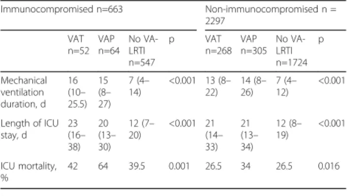
(8) Intensive Care Medicine Experimental 2016, 4(Suppl 1):30. Results: Among all the study group, the incidence of overall seizures was 20 %, of them, incidence of non-convulsive seizures were 13.3 % compared to 6.6 % of the patients showed convulsive seizures. Most seizures occurred on the first 24 hours of ischemia (66.7 %) compared to those occurring at the end of first week (25 %) and those at the end of the second week (8.3 %). Conclusions: Ischemic stroke is considered as a risk factor for the development of seizures and status epilepticus both convulsive and non-convulsive types especially during the first twenty-four hours. References 1- WHO. Stroke 1989. Recommendations on stroke prevention, and therapy. Report of the WHO Task Force on stroke and other cerebrovascular disorders. Stroke; a journal of cerebral circulation 1989; 20:1407–31 Grant acknowledgment Thanks to all members of the Department of Critical Care Medicine, University of Alexandria. Contemporary issues in infection and sepsis I A405 Does plasma from septic patients influence tumor cell growth? F. Puig1, I. Dunn-Siegrist1, J. Pugin1,2 1 University of Geneva, Department of Microbiology and Molecular Medicine (MIMOL), Geneva, Switzerland; 2Geneva University Hospital, Division of Intensive Care, Geneva, Switzerland Correspondence: F. Puig - University of Geneva, Department of Microbiology and Molecular Medicine (MIMOL), Geneva, Switzerland Intensive Care Medicine Experimental 2016, 4(Suppl 1):A405 Introduction: Sepsis is associated with immune suppression, including depressed cellular immunity. A decrease in surveillance by immune cells could be one of the mechanisms explaining the growing perception that patients surviving sepsis are at increased risk of developing cancers. However, other mechanisms may be involved in this increased rate of tumors in sepsis survivors, involving humoral factors. Sepsis is also associated with marked up- or down-regulation of various soluble mediators affecting cell growth. We hypothesized that some of these mediators may promote the proliferation of tumors. Objective: To identify and isolate this (those) plasma factor(s) enhancing tumor cell growth. Methods: Heparinized human blood samples were collected from patients with septic shock and in healthy donors. Human adenocarcinoma epithelial cells from lung (A549), colon (SW620), breast (Hs578T), and liver (HepG2) and human monocytic leukemia cells (THP-1 and HL-60) were grown to 70 % confluence and incubated with 5 - 15 % septic or healthy plasma, or control media. After 24 hours, cell proliferation was evaluated by MTT Cell Proliferation Assay. Septic and healthy plasma fractionation was performed using classical biochemical techniques. Results: Septic plasma supported significantly higher epithelial cell proliferation than plasma from healthy subjects (+48 % in A549, +59 % in SW620, +41 % in Hs578T, and +29 % in HepG2 cells with 15 % plasma). Interestingly, this effect was not observed in tumoral cells of monocytic origin. Adding healthy plasma to septic plasma decreased the tumor epithelial cell proliferation effect seen with septic plasma alone in a dose-dependent manner. Initial plasma fractionation studies suggest that the proliferation factor(s) in septic plasma remains in an immunoglobulin- and albumin-free plasma fraction, is(are) not precipitable by 50 % ammonium sulfate, and is(are) a trypsin-sensitive protein(s) > 50 kDa.. Page 212 of 607. Conclusions: The identification of such plasma protein(s) affecting tumor cell growth could be of great interest in the fields of sepsis and cancer biology. Grant acknowledgment Unrestricted research grant from Novartis Research Foundation (15B094). A406 High Flow Nasal Cannula (HFNC) as an alternative to noninvasive ventilation (NIV) in acute respiratory failure (ARF) in immunosuppressed patients - an Indian post liver transplant experience S. Gupta, D. Govil, S. Srinivasan, S.J. Patel, J.K. N, A. Gupta, D.S. Tomar, M. Shafi, R. Harne, D.P. Arora, N. Talwar, S. Mazumdar Medanta - The Medicity, Gurgaon, India Correspondence: S. Gupta - Medanta - The Medicity, Gurgaon, India Intensive Care Medicine Experimental 2016, 4(Suppl 1):A406 Introduction: In immunocompromised patients, acute respiratory failure (ARF) is associated with high mortality [1] and many studies have confirmed the utility of prolonged non invasive ventilation (NIV) in such condition. Lately High Flow Nasal Cannula (HFNC) has been used in ARF in various patient subsets but not specifically in post transplant patients. Objectives: To compare HFNC vs NIV as the modality to manage ARF in postoperative hypoxemia in post liver transplant patients. Methods: This was a pilot study conducted in a Liver Transplant intensive care unit (ICU) of a tertiary care hospital in India. We randomly assigned 20 consecutive post transplant patients who developed respiratory failure in the post-operative period to either HFNC group (n = 10) or to NIV group (n = 10). The HFNC was initiated at a flow rate of 60 l/min whereas NIV was set at EPAP of 5 cm and IPAP at 10 cm. Both the device setting and oxygen titration was done according to arterial blood gas (ABG) analysis. Apart from ABG analysis, we assessed the COMFORT scale as well as the RASS and CAM-ICU scale of the patient and also assessed the total nutritional deficit at the end of 48-hr therapy duration. The need for invasive mechanical ventilation (IMV) was also noted for both the groups. Results: None of the patients in the HFNC group required need for IMV whereas 2 patients in NIV group had to be intubated. Patients in the HFNC group had better average PaO2 as compared to NIV (98.2 vs 72.6 mm Hg) respectively. The patients in the NIV group received more sedation with dexmedetomidine as compared to HFNC group (22 vs 10 hrs) respectively. The patients in the HFNC group were more comfortable as compared to NIV group and two patients in the NIV group developed delirium for which they required IMV. The average RASS score was 0 to +1 in the HFNC group whereas it ranged from −2 to +2 in the NIV group. All patients were fed either orally or enterally but the NIV group consumed less feeding due to the inability to feed orally and apprehension of aspiration due to aerophagia when fed enterally. The NIV group received 52 % less calories as compared to HFNC group in 48-hr period. Conclusions: HFNC is may be an excellent armamentarium for managing hyperemic respiratory failure in immunocompromised patients with reduced risk of intubation, more comfortable to the patients and little interruption in providing adequate nutrition. References 1. Lemaire V, Mokart D, Mayaux J, Lambert J, Rabbat A, Demoule A, Azoulay E. The effects of a 2-h trial of high-flow oxygen by nasal cannula versus venturi mask in immunocompromised patients with hypoxemic acute respiratory failure: a multicentre randomized trial. Crit Care 2015 Nov 2;19:380.
(9) Intensive Care Medicine Experimental 2016, 4(Suppl 1):30. Fig. 8 (abstract A406). HFNC vs NIV. A407 Intra-abdominal hypertension increases the frequency of ventilator associated pneumonia E.E. Papakrivou, D. Makris, E. Manoulakas, B. Tsolaki, B. Karadodas, E. Zakynthinos University of Thessaly School of Medicine, Department of Critical Care Medicine, Larisa, Greece Correspondence: E.E. Papakrivou - University of Thessaly School of Medicine, Department of Critical Care Medicine, Larisa, Greece Intensive Care Medicine Experimental 2016, 4(Suppl 1):A407 Introduction: To study the effect of intra-abdominal hypertension (IAH) on the frequency of ventilator associated pneumonia (VAP) in critical care patients with risk factors for IAH. Methods: This one-center prospective study was conducted in the ICU of the University Hospital of Larissa, Greece. Consecutive critical care patients were recruited if they presented risk factors for IAH. Patients were evaluated systematically for IAH and VAP for a 28-day period. Results: Twenty-four out of 59 (41 %) patients presented IAH and 28 (47.4 %) presented VAP; seventeen (70.83 %) patients presented VAP following IAH. Multivariate analysis showed that VAP [1.18(1.10-1.22) (p = 0.004)], COPD [1.28(1.09-1.86) (p = 0.001)] and the use of antacids [9.54(2.74-33.19) (p = 0.016)] were independently associated with IAP. Conclusion: IAH may have an adverse impact on the frequency of VAP in critically ill patients with risk factors for IAH. A408 Azithromycin modulates inflammatory response in a murine model of Pseudomonas aeruginosa severe infection I. Palacios Garcia, A. Diaz Martin, V. Sanchez Encinares, M. Pachón Ibañez, J. Garnacho Montero, G. Labrador, T. Cebrero Cangueiro Hospital Virgen del Rocio, Sevilla, Spain Correspondence: I. Palacios Garcia - Hospital Virgen del Rocio, Sevilla, Spain Intensive Care Medicine Experimental 2016, 4(Suppl 1):A408 Objectives: Macrolides, apart from its antibiotic properties, are able to modulate the inflammatory response: inhibit production and secretion of pro-inflammatory cytokines (IL-1, IL-6, IL-8 and TNF-α), increase levels of anti-inflammatory cytokines (IL-10) and inhibit secretion of nitric oxide (NO). Our working hypothesis is that the use of azithromycin (AZM) with ceftazidime (CFZ) in a mouse model of severe sepsis by P. aeruginosa, modulates the inflammatory response.. Page 213 of 607. Methods: lethal sepsis model mouse with a clinical P. aeruginosa strain (Pa4) is characterized. i) CFZ (dose 100 mg / kg / ip / 12 h), ii) AZM (30 mg / kg / ip / 24 h): To study the inflammatory response, the following treatment groups (n = 15) for 72 hours were performed iii) COMB: CFZ + AZM, iv) control of infected animals and untreated group (CON) and v) group of uninfected mice. TNF-α determinations, IL-6, IL-10 and nitrite / nitrate (NOx) routine employed as a surrogate marker of NO metabolism in mouse plasma by ELISA (commercially available kits) ratio were performed. Results: We compare the TNF-α, IL-10 and NOx plasma concentrations, in each group (4 and 8 hours post-treatment) related to those obtained in the CON group. The COMB (AZM + CFZ) group showed lower plasma concentrations of TNF-α (pg/ml) than AZM and CFZ groups: [CON: 1477–716; COMB: 720–567; AZM: 1911–1663; CFZ: 793–666]. Plasma concentrations of IL-10 (pg/ml) were higher in the COMB and AZM groups than in the CFZ one: [CON: 1868–1761; COMB: 1541– 2035; AZM: 1860–2002; CFZ: 1898–1886]. NOx concentrations (M) observed were lower in the COMB group than in AZM and CFZ ones: [CON: 76–47; COMB: 59–28; AZM: 61–51; CFZ: 35–42]. Conclusions: These results suggest the immunomodulatory capability of AZM as an adjunct treatment to appropriate antibiotic. Further studies are needed to infer these findings to human setting. Grant acknowledgment This project (PI10/01563) was funded by the "Health Research Fund" Health Institute Carlos III. A409 Intravenous glutamine increases risk of death in severe sepsis V. Poulose1, J. Koh1, J.W. Kam2 1 Changi General Hospital, Respiratory & Critical Care Medicine, Singapore, Singapore; 2Changi General Hospital, Clinical Trials & Research Unit, Singapore, Singapore Correspondence: V. Poulose - Changi General Hospital, Respiratory & Critical Care Medicine, Singapore, Singapore Intensive Care Medicine Experimental 2016, 4(Suppl 1):A409 Introduction: Intravenous glutamine can have beneficial effects on critically ill patients by preserving gut barrier and improving immune function. Objectives: We wanted to prove the benefit of intravenous glutamine in patients admitted to medical intensive care unit (MICU) with severe sepsis and receiving enteral nutrition. Methods: Randomized, single center, double -blind, placebo-controlled, pilot study on patients admitted to the MICU who met the criteria for severe sepsis. In the intervention arm, intravenous glutamine was given for 5 days at a dose of 0.5 g/kg body weight/day. All patients were fed enterally as per the MICU feeding protocol. The primary outcomes were 28-day mortality and the occurrence of new infections. We also looked at severity scores (SOFA), ICU length of stay (LOS), hospital LOS and duration of mechanical ventilation. Results: Thirty nine patients were randomized to receive glutamine (n = 19) or placebo (n = 20). The glutamine group had less disease severity than placebo (median SOFA score 8 versus 11, p =0.038). There was no difference in 28-day mortality between the glutamine and placebo groups (42 % vs 15 %, p = 0.06). When adjusted for disease severity, the glutamine arm had 5.6 times higher death rates (95 % CI 1.1-30.2, p = 0.044). The glutamine group had lesser incidence of new infections (0 % vs 30 %, p = 0.02). There was no difference in ICU LOS, hospital LOS or the duration of mechanical ventilation. Conclusions: Intravenous glutamine increases mortality risk in ICU patients with severe sepsis, although it reduces risk of new infections Grant acknowledgement SingHealth Foundation Research Grant.
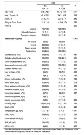
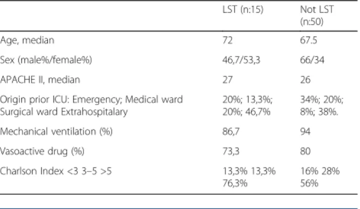
(10) Intensive Care Medicine Experimental 2016, 4(Suppl 1):30. A410 Sequential vitamin d measurement in patients with septic shock: could vitamin D levels be suppressed in septic shock? H. Yeter1, A. Kara2, O. Aktepe3, A. Topeli4 1 Hacettepe University, Internal Medicine, Ankara, Turkey; 2Hacettepe University, Intensive Care Medicine, Ankara, Turkey; 3Hacettepe University, Internal medicine, Ankara, Turkey; 4Hacettepe University, Intensive care medicine, Ankara, Turkey Correspondence: H. Yeter - Hacettepe University, Internal Medicine, Ankara, Turkey Intensive Care Medicine Experimental 2016, 4(Suppl 1):A410 Objective: Sepsis is characterized by dysregulated immune response to infection leading to organ dysfunction. Vitamin D plays a pivot role in the immune system and low levels of vitamin D have been shown to be associated with worse outcome in septic patients. In this study we tested vitamin D levels sequentially and aimed to define the vitamin D response in septic shock. Methods: Between September 2014 and January 2016, 41patients with septic shock were included in the study. Patients were excluded if they had a disease affecting calcium and vitamin D metabolism such as malignancy, chronic kidney disease, parathyroid disorders, pancreatitis, tumor lysis syndrome, rhabdomyolysis, renal tubular disorders and pregnancy. We measured vitamin D levels in day 1 and day 5after diagnosis of septic shock. Results: The median (min-max) age of 41 patients was 67 (19–88). 21of these patients were male. The median APACHE II score was 28(11–45). Day 1 and day 5 median vitamin D levels were 6.8 ng/ml(1–30) and 12 (2–29) ng/ml, respectively. Baseline corrected calcium and ionized calcium levels were 9.04 mg/dl (4.70-11.50) and 1.09 mmol/L (0.80-1.22). 21of the 41 septic shock patients died within 28 day. The APACHE II scores were similar between the survivor and non-survivor groups [27.5 (11–40), 30 (11–45), p = 0.13]. Baseline median corrected calcium and ionized calcium levels of survivors vs. non-survivors were 9.01 mg/dl (8.310.0) vs 9.16 mg/dl (4.7-11.5) and 1.07 mmol/L (0.90-1.18) vs. 1.10 mmol/ L (0.80-1.22). Day 1 median vitamin D levels of survivors and nonsurvivors were 8.7 ng/ml(4.3-30.4) and5.3 ng/ml (1.0-21.7), respectively(p = 0.047). Day 5 vitamin D levels were not statistically different between survivors and non-survivors (n = 17; 12.3 ng/ml and n = 7; 5.7 ng/ml, p = 0.37).Vitamin D levels increased in the survivor group from 8.7 ng/ml in day 1 to 12.4 ng/m lin day 5, however the difference was not statistically different (p = 0.18). Vitamin D levels did not change in the non-survivor group from day 1 to day 5 (5.3 ng/ml to 5.7 ng/ml, p = 0.89).Kaplan Meier survival analysis revealed that patients with vitamin D levels ≥ 6.8 ng/ml (median value) had increased 28-day survival as compared to patients with low vitamin D levels (<6.8 ng/ml) (long rank test p = 0.012). Conclusion: Our study showed that vitamin D response might be different between surviving and non-surviving patients during the course of septic shock. Surviving patients had higher day 1vitamin D levels as compared to non-surviving patients. Increase in vitamin D level from day 1 to 5 suggests a clinically significant albeit statistically insignificant association of vitamin D and survival, in line with high vitamin D levels in baseline comparisons of the groups, favoring survivors, due to the limited sample size. Further studies in larger groups are clearly warranted. A411 Central line associated blood stream infections in the obese and overweight critically ill patients (preliminary data) I. Tsolakoglou1, G. Intas2, P. Stergiannis3, A.A. Kolaros4, E. Chalari5, E. Athanasiadou6, A. Martika1, G. Fildisis7 1 General Hospital of Thessaloniki Agios Pavlos, ICU, Thessaloniki, Greece; 2 General Hospital of Nikaia-Pireus AG. Panteleimon, Actinotherapy, Pireus, Greece; 3General Hospital of Athens “Agioi Anargyroi, ICU, Athens, Greece; 4 General Hospital of Thessaloniki Theageneio, ICU, Thessaloniki, Greece; 5 General Hospital of Nikaia-Pireus AG. Panteleimon, Anesthesiology, Pireus, Greece; 6General Hospital of Thessaloniki Agios Pavlos, Nursing Department Management, Thessaloniki, Greece, 7University of Athens, Faculty of Nursing, Critical Care Directorate, Athens, Greece Correspondence: I. Tsolakoglou - General Hospital of Thessaloniki Agios Pavlos, ICU, Thessaloniki, Greece Intensive Care Medicine Experimental 2016, 4(Suppl 1):A411. Page 214 of 607. Introduction: Central-Line-Associated Bloodstream Infections (CLABSI) have been studied extensively in ICU patients. There are no data in the literature regarding a potential association between obesity and CLABSI. Objectives: To test the hypothesis that CLABSI depends on the presence of obesity in critically ill patients. Methods: We conducted an 18-month observational study on 576 critically ill patients, in three general ICUs in Greece. All patients had inserted a triple-lumen catheter in a central vein (internal jugular, femoral or subclavian). Body Mass Index (BMI) was determined by a dietitian on ICU admission. BMI was categorized a priori as < 18.5 (underweight), 18.5-24.9 (normal weight), 25–29.9 (overweight), and >30 (obese). CLABSI was diagnosed by examining the catheter's tip and a blood sample. Multivariate logistic regression analysis was used to estimate the association between BMI groups and CLABSI. Results: From the 576 critically ill patients, (258 men, 318 women) mean aged 62.3 ± 18.4 years, 28 (4.9 %) were underweight, 220 (38.2 %) normal weight, 234 (40.6 %) overweight and 94 (16.3 %) obese. CLABSI was diagnosed in 156 (27.1 %) patients. Overweight and obese patients had significant higher CLABSI rates than the other patients (p < 0.05). Obese patients had significantly less survival rates (p < 0.05). Patient's data according to the BMI category are shown on Table 1. Additional adjustment for obesity-central line catheter association for the presence of CLABSI attenuates the obesity- central line catheter association: underweight CLABSI OR = 1.79 (95 % CI 1.54-2.03; p = 0.006); normal weight CLABSI OR = 1.86 (95 % CI 1.80-1.93; p = 0.003); overweight CLABSI OR = 1.63 (95 % CI 1.541.72; p = 0.001); obese- CLABSI OR = 1.64 (95 % CI 1.50-1.78, p = 0.001). Conclusions: Obesity appears to be associated with the presence of CLABSI in critically ill patients. This could be partially attributed to the more efforts made by physicians to insert the catheter in the obese patients than the other patients. REFERENCE(S) Tagliabue C, Principi N, Giavoli C, Esposito S. Obesity: impact of infections and response to vaccines. Eur J Clin Microbiol Infect Dis. 2016 Mar;35(3):325–31. Table 1 (abstract A411). Patient's characteristics according to BMI categor Underweight Normal weight (N=28) (N=220). Overweight Obese (N=234) (N=94). p. Apache II score. 23.7±7.7. 20.5±6.9. 23.2±6.9. 26.8±5.7. 0.001. Femoral vein catheter insertion, n (%). 10 (35.7%). 30 (13.6%). 28 (11.9%). 14 (14.9%). 0.025. Subclavian vein catheter insertion, n (%). 10 (35.7%). 142 (64.5%). 156 (65%). 54 (61.4%). 0.025. Jugular vein catheter insertion, n (%). 8 (28.6%). 48 (21.9%). 56 (21.1%). 20 (23.7%). 0.025. N of attempts for catheter insertion. 1.3±0.6. 1.1±0.2. 2.1±0.6. 3.3±1.2. 0.028. CLABSI, n (%). 6 (21.4%). 30 (13.6%). 86 (36.8%). 34 (36.2%). 0.001. ICU LOS. 11.4±11.8. 15.1±10.7. 23.1±16.5. 24.4±10.4. 0.001. Hospital LOS. 16.2±14.1. 21.1±10.6. 28.9±12.3. 29.3±15.4. 0.001. Survival, n (%). 16 (42.9%). 148 (67.3%). 94 (40.2%). 8 (8.5%). 0.001. A412 Reactive oxygen species (ROS) production and leukocyte immunoglobulin-like receptors (LILR) expression by immune cells in pleural fluid (PL) and blood (BL) in critical care septic patients V. Faivre1, C. Mengelle1, B. Favier2, D. Payen1,3 1 Université Paris Diderot, Sorbonne Paris Cité, INSERM UMR1160, Paris, France; 2Université Paris Sud / CEA, Inserm U1184, Fontenay Aux Roses, France; 3APHP, Lariboisiere University Hospital, Surgical ICU, Paris, France Correspondence: V. Faivre - Université Paris Diderot, Sorbonne Paris Cité, INSERM UMR1160, Paris, France Intensive Care Medicine Experimental 2016, 4(Suppl 1):A412 Introduction: Sepsis induces a hyperinflammatory phase with a concomitant immunosuppression that predominates after initial phase.
(11) Intensive Care Medicine Experimental 2016, 4(Suppl 1):30. and exposes to a failure of controlling primary infection, or to an increased risk of secondary infections. Blood monocyte (Mo) HLA-DR expression (MHC Class II) seems to characterize well innate immunodepression. The LILR expression on immune cells interacting with HLA class I induces a functional inhibition of neutrophils (PMN) as shown by the expression of LILR B2 modified during sepsis1. Objectives: To investigate the expression of LILR subtypes on blood Mo CD16- (classical Mo) and Mo CD16+ (non-classical patroller) as on PMNs and eosinophils (Eo) vs expression on Pl cells, more related to lung tissue inflammation2, during sepsis. Methods: In patients having severe sepsis or septic shock: 1/(flow cytometry)LILR A2, LILR B1, LILR B2, LILR B3, LILR B4 and HLA I on Mo CD16-, Mo CD16+, PMN (CD16+ Granulocytes (Gr)) and CD16- Gr in Bl and Pl. 2/ (chemiluminescence3) Spontaneous and stimulated ROS production. Measurements done at the 1st collection of PL (T1) and a week after (T2) when possible. Statistical analysis: non parametric tests. Ethical committee: CPP Ile De France VII, n° PP 15–010. Results: 11 patients were enrolled, vs 7 healthy volunteers (HV, Bl only). Compared to HV, in Bl, HLA-DR expression decreased in patients on Mo CD16- (p < 0,05) with a higher ROS production (p < 0,05). HLA-DR expression was higher on Mo CD16+. All LILR expressions were significantly higher on Mo CD16-, Mo CD16+, PMNs, except for LILRB1, with a decrease in their ligand HLA-I (p < 0,05). On Gr CD16-, LILR B1 only was increased. Comparison between Bl and Pl showed no differences except for LILRB3 expression being higher on PMNs with a higher HLADR expression on Mo CD16-. All LILR expressions decreased between T1 and T2 (p < 0,05) regardless the patients prognosis. Conclusions: The increased LILR expression in Bl myeloid cells during sepsis was similar in Pl cells, with a decrease along time without relation with prognosis. LILR expression could be used both in Mo and PMNs in association with Bl Mo HLA-DR to characterize immunodepression and the potential therapy for immunomodulation. References 1. Baudhuin J et al. PNAS 2013, 110: 17957–17962. 2. Marie C et al. Am. J. Respir. Crit. Care Med. 1997, 156: 1515–1522. 3. Lukaszewicz et al. Ann Crit Care 2012; 2: 10. Grant acknowledgement Fondation pour la Recherche Médicale, Commissariat à l'Energie Atomique, Ministère de l'Enseignement Supérieur et de la Recherche.. A413 The impact of red blood cell (RBC) transfusion on sphingosine-1phosphate (S1P) levels of critically ill patients A. Poppe1, M.S. Winkler1, E. Mudersbach2, J. Schreiber3, M.-L. Wruck1, E. Schwedhelm2, S. Kluge3, C. Zöllner1 1 University Medical Center Hamburg-Eppendorf, Department of Anaesthesiology, Hamburg, Germany; 2University Medical Center Hamburg-Eppendorf, Institute of Clinical Pharmacology and Toxicology, Hamburg, Germany; 3University Medical Center Hamburg-Eppendorf, Department of Intensive Care Medicine, Hamburg, Germany Correspondence: A. Poppe - University Medical Center HamburgEppendorf, Department of Anaesthesiology, Hamburg, Germany Intensive Care Medicine Experimental 2016, 4(Suppl 1):A413 Introduction: Sphingosine-1-phosphate (S1P) is a G-protein coupled signaling lipid. In particular S1P has been shown to reduce sepsis induced endothelial leakage and cytokine release from immune cells and to regulate vascular tone. Among others these processes are responsible for the high mortality of critically ill patients. Therefore S1P has been studied in critically ill patients; lately low S1P concentrations have been associated with sepsis severity by our group [1]. S1P is mainly released by haematopoietic cells such as erythrocytes (EC) and thrombocytes (TC) [2]. EC produce and secrete S1P continuously, whereas TC produce S1P but release S1P only upon activation [3]. Objective: We were interested if S1P concentration in blood of critically ill patients may be influenced by red blood cell (RBC) transfusion. Methods: S1P concentration in serum and EDTA plasma of 28 critically ill patients was measured before and after transfusion of one. Page 215 of 607. RBC preservation at several times (pre and 30 min, 180 min, 24 h post). S1P was also measured in the transfused RBC preservation. Differences between transfusion of “fresh” (RBC ≤ 15d) and “old“(RBC > 15d) RBC were analysed. Clinical data like SOFA scores, haemoglobin (HB) and haematocrit (Hct) values of all patients were monitored repeatedly. Results: S1P concentration was higher in RBC preservations compared to serum and plasma levels in patients (P < 0,05). Serum S1P levels were higher compared to plasma S1P levels (p < 0,05), though serum and plasma levels correlated positively during time. S1P concentration in plasma declined 30 min after transfusion (p < 0,05) and increased to pre levels within 24 h. Serum levels did not change. Comparing “fresh” and “old” blood preservations, S1P concentration was significantly higher in “fresh” RBC (P < 0,05). However in patients, blood S1P values did not differ if “fresh” or “old” RBCs have been transfused. Conclusions: RBC preservations contain high amounts of S1P compared to patient´s endogenous S1P. S1P concentration in RBC preservations correlates negatively with duration of storage. Transfusion of RBC influences plasma S1P negatively within 30 min. S1P declines immediately after transfusion and increases thereafter during the next 24 h. Serum S1P levels are not changed by RBC transfusion. However alteration of plasma S1P levels by RBC transfusion had no influence on the clinical outcome of our patients. References 1. Winkler, M.S., et al., Decreased serum concentrations of sphingosine-1phosphate in sepsis. Crit Care, 2015. 19: p. 372. 2. Hanel, P., P. Andreani, and M.H. Graler, Erythrocytes store and release sphingosine 1-phosphate in blood. FASEB J, 2007. 21(4): p. 1202–9. 3. Yatomi, Y., et al., Sphingosine-1-phosphate: a platelet-activating sphingolipid released from agonist-stimulated human platelets. Blood, 1995. 86(1): p. 193–202.. A414 Intensive care adult and pediatric patients at risk of mortality: inflammatory - immunity and metabolic profiles T. Tavladaki1, A.M. Spanaki1, H. Dimitriou2, E. Kondili3, C. Choulaki4, E. Meleti2, D. Kafetzopoulos4, D. Georgopoulos3, G. Briassoulis1 1 University of Crete, Medical School, University Hospital, PICU, Heraklion, Greece; 2University of Crete, Medical School, Paediatric Haematology Oncology, Heraklion, Greece; 3University of Crete, Medical School, University Hospital, ICU, Heraklion, Greece; 4Institute of Molecular Biology and Biotechnology (IMBB), Foundation for Research and Technology Hellas (FORTH), Heraklion, Greece Correspondence: T. Tavladaki - University of Crete, Medical School, University Hospital, PICU, Heraklion, Greece Intensive Care Medicine Experimental 2016, 4(Suppl 1):A414 Introduction: Biomarkers influenced by tissue utilization, clearance, and metabolic derangements have not been shown to be able to identify complex cellular abnormalities of critical illness pathobiology.1 Currently there are no comparative data on metabolic and innate immunity - inflammatory changes in adult (A) and pediatric (P) patients with earlyonset sepsis or systemic inflammatory response syndrome (SIRS). Objectives: To determine the «at risk of mortality» profiles in A and P intensive care unit (ICU) patients in comparison to ICU-survivors and healthy (H) controls. To assess possible associations of heat shock proteins (HSP) and Resistin and Adiponectin hormone changes or oxygen consumption (VO2), dioxide production (VCO2), energy expenditure (EE) and metabolic pattern alterations with mortality in the early phase of critical illness. Methods: Seventy-eight adults (S/22; SIRS/23; H/33) and 67 children (S/18; SIRS/23; H/26) mechanically ventilated were included in the study. Blood samples were obtained within 24-hours upon admission. Mean Fluorescence Intensity (MFI) for HSP expression in monocytes (m) or neutrophils (n) was determined by Flow Cytometry. Restistin, Adiponectin and extracellular (e) HSP72 were measured using ELISA and energy-expenditure (EE) by E-COVX. Genomic DNA was extracted with PureLink Genomic DNA kit to detect HSP72 SNPs..
Figure
+7
Documento similar
Therefore, this study aimed to evaluate the effect of the mother kangaroo method on mothers of preterm infants and its influence on maternal stress and anxiety in the first days
Clinical information system (CIS); Intensive care unit (ICU); Predicted body weight (PBW); Acute respiratory distress syndrome (ARDS); Tidal volume (TV); Mechanical ventilation
Effects of early high-volume continuous venovenous hemofiltration on survival and recovery of renal function in intensive care patients with acute renal failure:
Pneumonia versus Graft Dysfunction as the cause of acute respiratory failure after Lung Transplant: A 4-year multicenter prospective study in 153 adults requiring.. intensive
Severe community-acquired pneumonia: validation of the Infectious Diseases Society of America/American Thoracic Society guidelines to predict an intensive care unit admission..
First, compared to the intensive users, the non-intensive users experienced: (1) significantly more anxiety, more scepticism, and more inefficacy in terms of technostress; and (2)
AF, atrial fibrillation; CABG, coronary artery bypass graft surgery; DHA, docosahexaenoic acid; EPA, eicosapentaenoic acid; ICU, intensive care unit; PUFA, omega-3 polyunsaturated
A tenor de las consecuencias del síndrome de burnout, se evidencia que es un problema generalizado en múltiples profesiones que afecta a la satisfacción laboral,
