Novel aspects of the apolipoprotein E receptor family: Regulation and functional role of their proteolytic processing
31
0
0
Texto completo
(2) 114. Proteolytic processing of ApoE receptors. (Fortini, 2009). The ectodomain proteolytic processing of many receptors is produced by the action of zinc-dependent metalloproteinase family. A subgroup within this family is constituted by the endopeptidases, among which are the ADAMs (a disintegrin and metalloproteinase) and MMPs (matrix metalloproteinases). ADAMs are integral membrane proteins that possess disintegrin and metalloproteinase activities (Huovila et al., 2005). This family of proteins has more than 40 members, 13 of which have enzymatic activity and are involved in embryonic development and in several biologic processes in the adult (Edwards et al., 2008). ADAM knockout mice (ADAM 10, 17 or 19) exhibit embryonic or early postnatal lethality, therefore the study of these metalloproteinases is important (Huovila et al., 2005; Edwards et al., 2008). These enzymes participate in signaling processes, and they are regulated by genetic and post-translational mechanisms (Box1). On the other hand, MMPs are also related to the shedding of various proteins. Many of them are secreted into the extracellular medium, resulting in the proteolytic processing of cell adhesion proteins, growth factor precursors proteins, inmunomodulators and receptors (Hayashida et al., 2010). For example MMP-2 sheds syndecan-2 (Fears et al., 2006), a cell adhesion molecule, and MMP-3 can shed Fas ligand (Matsuno et al., 2001) and also the Heparin-binding EGF-like growth factor (HB-EGF) (Suzuki et al., 1997). There are integral and associated membranes MMPs (Massova et al., 1998). The membrane Type 1-Matrix Metalloproteinase MT1-MMP is crucial during development, since mice deficient in this protease have problems in the. remodeling of skeletal and extraskeletal connective tissues triggering early death (Holmbeck et al., 1999). MT1-MMP degrades various components of the extracellular matrix, including collagen I, II and III (Ohuchi et al., 1997), and also acts as a sheddase, processing the ectodomain of hyaluronan receptor CD44 (Kajita et al., 2001), syndecan-1 (Endo et al., 2003) and tumor necrosis factor-related activation-induced cytokine (Schlöndorff et al., 2001), among others (Karsdal et al., 2002; Mu et al., 2002). Many studies of the molecular and cellular determinants of proteolytic processing have used the amyloid precursor protein (APP) because of its relevance in Alzheimer’s disease (AD) and the Notch/Delta system because of the central role it plays in human disease and in determining cell fate and pattern formation during development. APP can be processed by two pathways. In the amyloidogenic pathway, BACE1 cleaves APP, which releases sβAPP and produces the membrane-bound fragment βCTF (Vassar et al., 1999; Wakabayashi and De Strooper, 2008; Lichtenthaler et al., 2011). βCTF is further processed by the γ-secretase complex (Wakabayashi and De Strooper, 2008), producing Aβ peptides and the intracellular cytoplasmic domain of APP (AICD). In the second pathway (non-amyloidogenic), the first cut is catalyzed by an α-secretase (possibly ADAM 10 or 17) (Buxbaum et al., 1998; Kuhn et al., 2010; Jorissen et al., 2010), producing an sAPPα and the corresponding CTFα. Depending on the activation system, other proteases may be involved (Delarasse et al., 2011).The γ-secretase complex processes CTFα, generating the non-amyloidogenic peptide p3 and the AICD. The comprehensive study of the. Box 1 Many proteases belong to the ADAMs family, and 13 of them are proteolytically active and are expressed in human tissues (Edwards et al., 2008). ADAMs have many substrates (Edwards et al., 2008) and several mechanisms regulate their activity: 1) Genetic regulation This mechanism includes the increase in ADAMs gene expression induced by cytokines or in cancer, and the existence of alternative splicing and inducible promoters that are present in several of the genes encoding these proteinases (Murphy, 2009). 2) Post-translational regulation and trafficking The mature forms of ADAMs that have proteolytic activity require the proteolytic processing of their prodomain to function properly. For example, ADAM 12, 17 and 33 undergo pro-domain maturation, but the peptide still interacts with the mature ADAM and, consequently, regulates protease activity. Location. ADAMs are located in perinuclear regions, but they are also found in lesser amounts at the cell surface, it being the location of choice for processing many of their substrates, such as adhesion proteins and surface receptors (Reiss et al., 2006; Murphy, 2009). However, active forms of ADAMs are also found in intracellular compartments (Shirakabe et al., 2001; Yokozeki et al., 2007); for example, ADAMs 10/17 induces shedding of APP in the trans-Golgi network (TGN) (Skovronsky et al., 2000). ADAMs and lipid rafts. The active forms of ADAM17 and ADAM19 are associated with lipid rafts (Wakatsuki et al., 2004; Tellier et al., 2006; Thiel and Carpenter, 2006; Takaguri et al., 2011), and it is in this microdomain where ADAM17 exerts its proteolytic activity on TNFα, TNFR I and II (Tellier et al., 2006) and the ligand-induced shedding of ErbB4 (Thiel and Carpenter, 2006). In contrast, other studies postulate that ADAM 10 and ADAM 17 are not associated with lipid rafts (Kojro et al., 2001; von Tresckow et al., 2004; Zimina et al., 2005; Gil et al., 2007; Harris et al., 2009), but many of their substrates are partitioned in these membrane domains (Tellier et al., 2006). Consequently, the shedding of these proteins is spatially regulated. Therefore, cholesterol depletion that disrupts lipid rafts increases the processing of some ADAMs substrates, such as TNFα, TNFR I and II (Tellier et al., 2006), IL-6R (Matthews et al., 2003), CD3 (von Tresckow et al., 2004), collagen XVII (Zimina et al., 2005) and APP (Kojro et al., 2001). 3) Intracellular signaling regulation The cytoplasmic region of ADAMs have motifs that interact with cytosolic protein domains, including SH3, SH2, PDZ, WW, EVH1 and 14-3-3 (Huovila et al., 2005). The cytoplasmic domains are phosphorylated at Tyr, Ser and Thr residues. Phorbol esters have been widely used to activate ADAMs and induce the shedding of their substrates (Huovila et al., 2005). Oxidative stress, UV and intracellular calcium concentrations are also involved in ADAMs regulation (Huovila et al., 2005; Murphy, 2009). Among the cellular mediators involved in these processes are the following: protein kinase C (PKC), the mitogen-activated protein (MAP) kinases p38 and ERK, which also modulate constitutive shedding, and the activation of G proteincoupled receptors (GPCR) (Murphy, 2009). Intracellular signaling affects the cellular localization of ADAM 12, ADAM 10 and ADAM 17 (Peiretti et al., 2003; Sundberg et al., 2004 Soond et al., 2005; Marcello et al., 2007),and the phosphorylation of ADAMs (Izumi et al., 1998; Diaz-Rodríguez et al., 2002; Zheng et al., 2002; Fan et al., 2003; Soond et al., 2005; Zhang et al., 2006; Lemjabbar-Alaoui et al., 2011), which are processes that in some cases, might be involved in regulating their own proteolytic activity (Diaz-Rodríguez et al., 2002)..
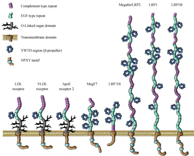
(3) Jorge A. LARIOS and Maria-Paz MARZOLO. mechanisms involved in APP processing and trafficking has allowed the discovery of many relevant aspects of the cell biology of these processes that can applied to the proteolytic processing of other substrates. In the Notch/Delta system, signaling depends on the proteolytic processing of the Notch receptor in three sequential steps (Fortini, 2009). The first cut is mediated by furin and it occurs during biosynthesis of the protein, probably in the Golgi apparatus (Blaumueller et al., 1997; Logeat et al., 1998). Once the receptor reaches the plasma membrane and is in contact with a ligand presented by a neighboring cell, the receptor is processed by metalloproteinases of the ADAM family, resulting in a membrane Notchassociated C-terminal fragment (NCTF) that is a substrate of the γ-secretase complex. The NICD that is produced by the action of γ-secretase translocates to the nucleus to regulate gene transcription (Fortini, 2009). The participation of the γ-secretase complex in various physiologic processes, such as the Notch signaling pathway, is the rationale for avoiding the use of inhibitors that selectively block the activity of this. 115. complex to treat AD. The γ-secretase complex that performs RIP consists of four essential transmembrane proteins, specifically, presenilin (has aspartyl protease activity), nicastrin, Aph-1 and Pen-2, which form a complex with a 1∶1∶1∶1 stoichiometry. There is preferential processing of substrates that have an N terminus that faces the extracellular environment and are no more than 50 aminoacids from the transmembrane domain. The protease responsible for proteolytic processing of the intramembrane region of the substrate has less specificity because there is no clear-cut consensus cleavage site, and multiple cleavage sites can be found on the same substrate (Wakabayashi and De Strooper, 2008). Among the recently recognized substrates of the γ-secretase complex are members of LDLR or the apolipoprotein-E receptor family. Members of this receptor family include LDLR, LRP1, LRP1b, megalin/LRP2, ApoER2, VLDLR and LRP5/6 (Fig. 1). These proteins and their ligands play important roles in development, in biologic processes in adults and are associated with diverse pathologies (Herz and. Figure 1 Schematic representation of members of the LDLR family. Members of the LDLR family share common structural motifs, including ligand binding repeats, epidermal growth factor repeats, YWTD spacer domains, a single transmembrane domain, and a short cytoplasmic domain containing at least one conserved NPXY endocytic motif that binds to adaptor proteins to regulate internalization and recycling. The same motif may play a role in the recruitment of signaling adaptor proteins. The LRP5/6 receptors are a subclass of this family that lacks some structural features, such as the ligand binding repeats and the NPXY cytosolic motifs..
(4) 116. Proteolytic processing of ApoE receptors. Willnow, 1994; Willnow, 1999; Willnow et al., 1999; Marzolo and Farfán, 2011). These receptors are type I membrane glycoproteins and have structural similarities, including a large extracellular domain, a transmembrane domain and a relatively short cytoplasmic tail. They share common features that include the following: 1) ligandbinding complement-type repeats, 2) epidermal growth factor (EGF) receptor-like repeats, 3) YWTD β-propeller domains, 4) a cytoplasmic tail that contains at least one Asn-Pro-X-Tyr (NPXY) motif, which is normally responsible for receptor interactions with intracellular adaptor proteins (Table 1) regulating their coupling to specific trafficking and/or signal transduction pathways. In addition, almost all of the receptors require the assistance of endoplasmic reticulum chaperones, such as receptor-associated protein (RAP) (Willnow et al., 1996c; Czekay et al., 1997; Bu and Schwartz, 1998; Bu and Marzolo, 2000) and mesoderm development (MESD) (Culi and Mann, 2003; Hsieh et al., 2003; Culi et al., 2004; Lighthouse et al., 2010) for the correct folding of the extracellular domains. Members of the apolipoprotein E family are classically recognized as endocytic receptors that mediate the internalization and degradation of a variety of ligands, and LDLR is the first described and prototypic member responsible for the catabolism of low-density lipoproteins (Goldstein et al., 1985; Brown and Goldstein, 1986). In addition to the role of these receptors in endocytosis, especially in the case of LRP5/6 and ApoER2, ligand binding triggers signaling pathways that have relevant physiologic functions in development and also in the adult life (Stockinger et al., 2000; Beffert et al., 2002; Assadi et al., 2003; Bock and Herz, 2003; Beffert et al., 2005; Li and Bu, 2005; Simó et al., 2006; Sentürk et al., 2011). In this review, we discuss the evidence that supports the proteolytic processing of these receptors, how the processing Table 1 Receptor APOER2. VLDLR. LRP1. is regulated and potential functional roles for the proteolytic fragments that are produced in the different cell systems in which these membrane proteins are found.. LRP1: an endocytic and signaling receptor Structure and function of LRP1 LDL receptor-related protein 1 (LRP1) is a large endocytic receptor (Fig. 1) that is widely expressed in several tissues and systems, such as the liver (hepatocytes and macrophages), vascular tissue, adipose tissue, the central nervous system (blood-brain barrier (BBB), neurons and glial cells) and the immune system (Lillis et al., 2008). This glycoprotein, which is synthesized as a polypeptide chain of more than 600 kDa, was initially discovered as the receptor that endocytoses the plasma protease inhibitor α2-macroglobulin (α2M) (Moestrup and Gliemann, 1989; Kristensen et al., 1990; Strickland et al., 1990), tissue type plasminogen (tPA) (Bu et al., 1992b; Bu et al., 1993; Bu et al., 1994) and apolipoprotein E, being responsible for chylomicron remnant clearance (Beisiegel et al., 1989). Currently, LRP1 is known to interact with at least 30 different ligands, including apolipoproteins, proteases, protease inhibitors, extracellular matrix proteins, and membrane proteins such as APP (Kounnas et al., 1995; Knauer et al., 1996; Pietrzik et al., 2004) and the urokinase-type plasminogen receptor (uPAR) (Nykjaer et al., 1992; Lillis et al., 2008) indicating LRP1 participation in many biologic processes. Because genetically modified mice that do not express LRP1 die during gestation, this receptor is critical for development (Herz et al., 1992a). The extracellular region of LRP1 (N-terminal) contains four ligand-binding repeats, the second and fourth of which exhibit stronger interactions with ligands (Neels et al., 1999).. LDLRs intracellular adaptors Intercellular interacting proteins. Reference. *JIP1. Stockinger et al., 2000. *JIP2. Stockinger et al., 2000. *PSD95. Gotthardt et al., 2000. *Talin homolog. Gotthardt et al., 2000. *APC subunit 10. Gotthardt et al., 2000. *ICAP-1. Gotthardt et al., 2000. *X11/. He et al., 2007. Dab1. Trommsdorff et al., 1999. Fe65. Hoe et al., 2006. SNX17. Stockinger et al., 2002. Dab2. Cuitino et al., 2005. ARH. Jones et al., 2003. SNX17. Stockinger et al., 2002. Dab1. Trommsdorff et al., 1999. ARH. Jones et al., 2003. JIP1. Stockinger et al., 2000. JIP2. Stockinger et al., 2000.
(5) Jorge A. LARIOS and Maria-Paz MARZOLO. 117. (Continued) Receptor. Intercellular interacting proteins. Reference. Dab1. Trommsdorff et al., 1998. PSD95. May et al., 2004, Gotthardt et al., 2000 Martin et al., 2008. Mint2 (X11-like). Gotthardt et al., 2000. Fe65. Trommsdorff et al., 1998. ARH. Jones et al., 2003. SNX17. Stockinger et al., 2002 van Kerkhof et al. 2005, Betts et al., 2008. LRP1b. Megalin. LRP5/6. Homolog of PIP 4, 5 kinase. Gotthardt et al., 2000. CED-6/GULP. Su et al., 2002. ShcA. Barnes et al., 2001. Na channel 3. Gotthardt et al., 2000. APC subunit 10. Gotthardt et al., 2000. Cbl. Takayama et al., 2004. Shp2. Betts et al., 2008. PICK1. Shiroshima et al., 2009. RanBPM. Shiroshima et al., 2009. PKB. An et al., 2008. CSK. Guttman et al., 2009. PI3K p85. Guttman et al., 2009. CamKII. Guttman et al., 2009. PLC. Guttman et al., 2009. JIP1b. Shiroshima et al., 2009. JIP2. Shiroshima et al., 2009. PICK1. Shiroshima et al., 2009. RanBPM. Shiroshima et al., 2009. SNTG2. Shiroshima et al., 2009. Grb7. Shiroshima et al., 2009. PSD95. Marschang et al., 2004. AIP. Marschang et al., 2004. JIP1. Gotthardt et al., 2000. JIP2. Gotthardt et al., 2000. PSD95. Gotthardt et al., 2000. PIP4,5 kinase homolog. Gotthardt et al., 2000. Mint2 (X11-like). Gotthardt et al., 2000. Dab1. Gotthardt et al., 2000. ICAP-1. Gotthardt et al., 2000. ARH. Jones et al., 2003; Nagai et al., 2003. MegBP. Petersen et al., 2003. Dab2. Oleinikov et al., 2000. Fe65. Alvira-Botero et al., 2010. PKB. Caruso-Neves et al., 2006. ANKRA. Rader et al., 2000. MAGI-1. Patrie et al., 2001. GIPC. Lou et al., 2002. Axin. Mao et al., 2001; MacDonald et al., 2011. Frat1. Hay et al., 2005. GSK3. Mi et al., 2005. The cytoplasmic regions of the members of the LDLR protein family that undergo proteolytic processing interact with various intracellular adaptors. These interactions allow the performance or regulation of multiple cellular and physiologic functions, including receptor trafficking, processing, signaling and scaffolding..
(6) 118. Proteolytic processing of ApoE receptors. Figure 2 Transmembrane and cytoplasmic regions of LDLR-family receptors. The amino-acid sequences and lengths of the transmembrane (italics) and cytoplasmic regions of the members of LDLR family that undergo proteolytic processing. Phosphorylation motifs (ser/thr) and interaction with intracellular proteins (Table 1) are highlighted: NPXY (red), PPPSP (purple) and the motif recognized by PDZ domains (orange). The spliced isoform ApoER2Δpro does not have the proline-rich region (underlined) that confers on ApoER2 the ability to interact with multiple adaptors (I).. The intracellular region of LRP1 is made up of 100 amino acids (Fig. 2) and contains two NPXY motifs and two dileucine, which interact with intracellular proteins. The NPXY motif was first identified in the LDL receptor, where it is essential for clathrin-mediated internalization (Bansal and Gierasch, 1991). The proximal NPXY motif in LRP1 binds to sorting nexin 17 (SNX17) and is involved in receptor recycling (van Kerkhof et al., 2005). The distal NPXY motif shares the Y residue with the YATL motif that is involved in rapid clathrin-dependent internalization (Li et al., 2000a). This last motif has been implicated in the interaction with phosphortyrosine binding (PTB) domain-containing intracellular signaling proteins such as Disabled 1 (Dab1) (Trommsdorff et al., 1998) and the APP adaptor Fe65 (discussed below) (Pietrzik et al., 2004), among several others (Table 1). In addition, this motif is tyrosine phosphorylated in response to stimulation with platelet-derived growth factor (PDGF) (Boucher et al., 2002; Loukinova et al., 2002),. connective tissue growth factor (CTGF) (Yang et al., 2004) and in v-Src transformed mouse fibroblasts (Barnes et al., 2003). Phosphorylation of the distal NPXY motif would facilitate the phosphorylation of the proximal NPXY motif (Betts et al., 2008), affecting the receptor’s interaction with cytosolic proteins (Guttman et al., 2009). The expression of LRP1 in the liver allows the clearance of various proteins that are present in the plasma. In liver tissue, LRP1 is the key receptor of α2M, facilitating its endocytosis with proteases that are attached to it. Furthermore, LRP1 allows plasma clearance of serine proteases inhibitors (Nykjaer et al., 1992; Kounnas et al., 1996; Kasza et al., 1997), clotting factor VIII (Lenting et al., 1999; Saenko et al., 1999; Bovenschen et al., 2005), chylomicron remnants (Rohlmann et al., 1998) and matrix metalloproteinases (MMPs) (Emonard et al., 2005). In atherosclerotic dysfunction, LRP1plays a protective role that is associated with its expression in vascular smooth muscle cells (Boucher et al.,.
(7) Jorge A. LARIOS and Maria-Paz MARZOLO. 2003), liver (Espirito Santo et al., 2004), macrophages (Hu et al., 2006; Overton et al., 2007) and adipocytes (Hofmann et al., 2007). For example, in vascular smooth muscle cells, LRP1 expression decreases the activation of the signaling pathway that is triggered by PDGF (Boucher et al., 2003), which normally stimulates the secretion of extracellular matrix proteins (Pompili et al., 1995) and blood vessel wall thickening after damage (Abe et al., 1997). The BBB regulates the passage of molecules into the central nervous system. The permeability of the BBB can be upregulated in hypoxic conditions (Baker et al., 1971; Garcia et al., 1978). tPA and apolipoprotein E, regulate the permeability of the BBB in a LRP1-dependent manner (Nishitsuji et al., 2011; Yepes et al., 2003). LRP1 plays an important role in neurons because it promotes long-term potentiation (LTP) and either survival (Fuentealba et al., 2009) or apoptosis (Hashimoto et al., 2000; An et al., 2008). In addition, LRP1 interacts with APP (Kounnas et al., 1995; Knauer et al., 1996; Pietrzik et al., 2004) promoting the amyloidogenic pathway (Ulery et al., 2000; Pietrzik et al., 2002). This effect is also seen in vivo in LRP1-overexpressing PDAPP mice (model of AD), which exhibit an increased generation of soluble Aβ and memory deficits (Zerbinatti et al., 2004; Bu et al., 2006). During cell migration, the remodeling of the cytoskeleton and the extracellular matrix is necessary and involves changes in membrane protease content, such as MMPs. There is evidence showing that the participation of LRP1 in cell migration involves its ability to internalize proteases and protease inhibitors (Yang et al., 2001; Emonard et al., 2004; Emonard et al., 2005) and to regulate their expression (Fayard et al., 2009; Song et al., 2009; Jung et al., 2010). In addition LRP1 signaling (Orr et al., 2002; Orr et al., 2003; Zhu and Hui, 2003; Barker et al., 2004) and the ability of LRP1 to stabilize integrins (Czekay et al., 2003; Salicioni et al., 2003) are involved in cell migration. LRP1 is a calreticulin coreceptor that interacts with the extracellular matrix protein thrombospondin-1, which is known for its anti-adhesive properties (Murphy-Ullrich and Höök, 1989; Greenwood and Murphy-Ullrich, 1998). This interaction activates a trimeric G protein-dependent signal transduction pathway that induces reorganization of the actin cytoskeleton, which leads to focal adhesion disassembly and the stimulation of cell motility (Orr et al., 2002, 2003; Barker et al., 2004). The interaction between LRP1 and its ligands tPA, uPA and plasminogen activator inhibitor-1 (PAI-1) (Bu et al., 1992b, 1993, 1994) is associated with the migration of cells, such as smooth muscle cells (Okada et al., 1996) and mouse embryonic fibroblasts (MEF) (Okada et al., 1996; Weaver et al., 1997). Given the importance of LRP1 in cell migration, it has also been associated with cancer cells (Li et al., 1997, 1998a, 2000; Chazaud et al., 2002; Li et al., 2002; Dedieu et al., 2008; Song and Bu, 2009), where LRP1 interacts with several extracellular matrix proteases that regulate its clearance (Selvais et al., 2011) and expression (Song et al., 2009). Proteolytic. 119. processing of the receptor (discussed below) regulates all of these processes (Selvais et al., 2011). LRP1 plays an important role in inflammation and phagocytosis, increasing the activity of phagocytes and allowing the ingestion of apoptotic cells (Ogden et al., 2001; Vandivier et al., 2002; Gardai et al., 2005). Using the same mechanism, LRP1 facilitates the removal of degraded myelin that is present in the extracellular medium (Gaultier et al., 2009). The association of LRP1 with the innate immune response (Binder et al., 2000; Basu et al., 2001; Cao et al., 2006; Lillis et al., 2008) is also evidenced as a receptor for heat-shock proteins (Binder et al., 2000; Basu et al., 2001; Binder and Srivastava, 2004; Facciponte et al., 2005), viral peptides (Congote, 2007), and the clearance of apoptotic debris (Tarr and Eggleton, 2005). Regulation of LRP1 shedding and the role of sLRP1 During its biosynthesis and trafficking, LRP1 is processed by furin, which generates two non-covalently bound subunits, a 515 kDa (α) subunit and a membrane-bound 85 kDa subunit, which is also known as the β-subunit (Willnow et al., 1996b). The first evidence that indicated LRP1 shedding was the identification of a soluble LRP1 fragment (sLRP1) in plasma (Quinn et al., 1997). Interestingly, this fragment was associated with ligands, such as α2M, tPA, PAI-1 and the ER chaperone RAP. Among the LRP1 fragments, were a 500 kDa furin-processed fragment and a 67 kDa, which corresponds to part of the 85 kDa β-chain. In the human choriocarcinoma cell line BeWo, sLRP1 is detected after 2 h following a metabolic pulse of 35S-methionine. In these cells this fragment is shed and is produced at the plasma membrane by a process that is not stimulated by phorbol esters but is blocked by metalloproteinases inhibitors (Quinn et al., 1999). Interestingly, sLRP1 is composed of α-chain that is noncovalently bound to a portion of the extracellular part of the β-chain (Quinn et al., 1999). Therefore, similar to Notch processing, the furin-mediated first cut probably precedes the second and possibly regulatory processing step. LRP1 processing is induced by PKC activators (PMA) (May et al., 2002), which occurs with other proteins that undergo shedding (Caporaso et al., 1992; Garton et al., 2001; Kanning et al., 2003). PKC-induced shedding is not related to the phosphorylation of LRP1 itself, and this activity is likely to directly or indirectly activate metalloproteinases of the ADAM family (Merlos-Suárez et al., 2001; Jacobsen et al., 2010; Killock and Ivetić, 2010). There is evidence indicating that ADAM 10 and 17 are involved in the generation of sLRP1 and that this fragment is present in the brain and cerebral spinal fluid of older patients, suggesting that clearance of the fragment is impaired and/or its production is increased (Liu et al., 2009). The addition of Aβ peptide to cells expressing LRP1 in vitro increases shedding of the receptor and suggests that in AD and older patients, the reduction of total LRP1 could be due in part to an.
(8) 120. Proteolytic processing of ApoE receptors. increase in processing (Liu et al., 2009). Concordant with LRP1 participation in innate immunity and inflammation (Binder et al., 2000; Basu et al., 2001; Cao et al., 2006; Lillis et al., 2008), both lipopolysaccharide (LPS) - and interferon-γ-induced LRP1 shedding from the β-chain (Gorovoy et al., 2010) releases sLRP1 and produces a 25 kDa CTF (Zurhove et al., 2008). This effect is seen in vitro in cultured macrophages cell lines and in vivo in mice (Gorovoy et al., 2010). LPS activates Toll-like receptor 4 (Miller et al., 2005) and triggers PKC-mediated ADAM 17 activation (Gorovoy et al., 2010). Increased levels of sLRP1 are seen in plasma from patients with rheumatoid arthritis and systemic lupus erythematosus. Purified sLRP1 from human plasma activates signaling pathways, such as p38 MAPK and JNK in macrophage cell lines, and sLRP1 transiently activates the NFkB pathway and the expression of inflammatory cytokines, such as TNF-α and IL-10 (Gorovoy et al., 2010). This activation indicates that soluble fragments may be relevant in cell-to-cell communication (Fig. 3) that in the case of sLRP1 potentiates LPS-induced inflammatory responses. In contrast to the inflammatory role of sLRP1 in macrophages, injured sciatic nerve from mice exhibit an increase in the shedding of LRP1, but in this case the. sLRP1α has anti-inflammatory properties, decreasing TNFα expression and p38 activation (Gaultier et al., 2008). Interestingly, a control mechanism that is dependent on the production of LRP1-ICD and γ-secretase activity is important for limiting the inflammatory response. LRP1-ICD binds to the interferon regulatory factor 3, promoting its degradation and reducing the expression of inflammatory genes (Zurhove et al., 2008). As previously mentioned, among the LRP1 ligands are several proteases and proteases-inhibitors (Herz et al., 1992b; Kounnas et al., 1993; Andreasen et al., 1994; Bu et al., 1994; Herz and Willnow, 1994; Strickland et al., 1995; Kounnas et al., 1996; Stefansson et al., 1996; Knauer et al., 1997), including metalloproteinases (MMPs) such as MMP2, MMP3 and MMP9 (Hahn-Dantona et al., 2001; Yang et al., 2001; Strickland and Ranganathan, 2003; Mantuano et al., 2008) that are degraded after LRP1-mediated internalization. Through the regulation of the availability of these ligands, LRP1 regulates the dynamics of the extracellular matrix, modulating the clearance of proteases and consequently, cell signaling, adhesion and migration. This protease system regulates LRP1 shedding via a feedback mechanism. This observation is particularly interesting for cancer cells. Figure 3 Overview of proteolytic processing. Schematic representation of the proteolytic processing of a membrane protein. Initially, membrane proteins are proteolytically processed and release a soluble extracellular fragment in a process that is called “shedding.” Among the proteases that carry out shedding are matrix metalloproteinases, ADAMs and BACE. Next, the protein fragment that remains associated with the membrane is proteolytically processed by the γ-secretase complex and releases a soluble intracellular fragment (ICD). Proteolytic processing can be constitutive, but it can also be regulated by different factors, such as extracellular and intracellular ligands (1) association of the membrane protein with lipid rafts (2) and/or endocytic trafficking (3) the resulting fragments (CTFs and ICDs) may have the ability to signal which may affect various signaling pathways through their interaction with adaptors, or in the case of ICD may migrate to the nucleus, where it can affect gene expression. The soluble fragments can also modulate the availability of some ligands and function, for example, as dominant negative forms of the receptors..
(9) Jorge A. LARIOS and Maria-Paz MARZOLO. that express high levels of membrane-associated MMPs. MT1-MMP activates MMP2, and this activation correlates with an increase in the invasive capacity of the cancer cells (Seiki, 2003). MT1-,2-,3- and 4-MMPs also induce the shedding of the LRP1 α-chain, avoiding the role of the receptor in the clearance of MMPs (Rozanov et al., 2003). In addition, under conditions that affect pulmonary function, such as in patients suffering from acute respiratory distress syndrome, the increased shedding of LRP1 that is detected by the presence of sLRP1 is associated with high levels of MMP2, MMP9 and lamininin the bronchoalveolar fluids (Wygrecka et al., 2011). This observation indicates that the presence of intact LRP1 is required for the appropriate degradation of these MMPs and the maintenance of the basal membrane. Further more, damage to a blood vessel wall induces an increase in the expression of MT1-MMP, which is related to the dedifferentiation of contractile vascular smooth muscle cells (VSMC) to a proliferative and invasive phenotype and results in vascular disorders and damage. The upregulation of MT1-MMP induces an increase in the proteolytic processing of LRP1, which, together with the PDGF receptor-β (PDGFR-β) pathway, induces a reduction in VSMC contractile proteins and promotes the proliferative phenotype (Lehti et al., 2009). In other systems, such as the endometrium, LRP1 shedding is likely to be mediated by ADAM12 and is under the control of steroidal hormones. LRP1 mRNA expression is constant during the menstrual cycle; however, LRP1 protein levels decrease because of proteolytic processing. In the presence of progesterone and estradiol, the release of the sLRP1 515-kDaα-chain and of 55-kDa truncated form of the β-chain subunit is inhibited (Selvais et al., 2009). At the end of the cycle, the hormone levels drop and this reduction activates LRP1 processing. Next, MMP2 and MMP9 accumulate, allowing the detachment of the endometrial layer through menstrual bleeding (Selvais et al., 2009). The interaction of LRP1 with tPA (Bu et al., 1992a, b; Bu et al., 1993) regulates the systemic degradation of this thrombolytic enzyme. In addition, this interaction promotes LTP in the hippocampus (Zhuo et al., 2000) and increases the synthesis of MMP9 and MMP3 (Wang et al., 2003; Suzuki et al., 2009; Zhang et al., 2009). By regulating the expression of MMPs, LRP1/tPA regulates the BBB permeability and is associated with cerebral bleeding and brain injury (Yepes et al., 2003; Suzuki et al., 2009). tPA is also involved in the processing of LRP1, inducing the release of sLRP1in cultured astrocytes. This processing would require LRP1 itself because the process is blocked by exogenous RAP. In vivo, this processing occurs under conditions of cerebral ischemia and is associated with an increase in detachment astrocytic end-feet processes and edema (Polavarapu et al., 2007). Interestingly, depending on the cells, the interaction of tPA with LRP1 results in the differential activation of AKT; in astrocytes, AKT binds to the cytoplasmic domain of LRP1 and is activated. The phosphorylation of AKT in this case. 121. relates to astrocyte detachment and edema (An et al., 2008). In contrast, the binding of tPA to LRP1 in neurons under hypoxic conditions (experimentally induced by middle cerebral artery occlusion in mice), is related to a decrease in AKT activity (An et al., 2008), neuronal apoptosis, caspase 3cleavage, and an increase in γ-secretase activity, which correlates with the processing of LRP1-CTF and the translocation of LRP-ICD to the nucleus (Polavarapu et al., 2008). Related to its role as a signaling protein and a receptor for extracellular matrix proteins, including the proteases mentioned above, LRP1 participates in cancer. However, the role of LRP1 in this pathology is controversial and appears to be highly dependent on the cell context. As mentioned before, LRP1 regulates the degradation and expression of proteinases, and LRP1 expression has been positively and negatively associated with tumor development and progression (Li et al., 1997; Li et al., 1998b; Li et al., 2000; Chazaud et al., 2002; Li et al., 2002; Dedieu et al., 2008; Song and Bu, 2009). Certain cancer cell lines also manage LRP1 processing differently. For example, in the HT1080 human fibrosarcoma cell line, the association of LRP1 with lipid rafts decreases the production of the soluble β-chain fragment and the release of the intact α-chain in a reaction that is catalyzed by ADAM12 and MT1-MMP (Selvais et al., 2011). HT1080 cells exhibit different phenotypes, an epithelial-like and a fibroblast-like phenotype that differ in their plasma membrane cholesterol content. In the epithelial phenotype, there is less processing of LRP1, and this effect is reversed upon depletion of cholesterol. In contrast, the fibroblast-like cells contain lower levels of plasma membrane cholesterol and accumulate MMP2 and MMP9 in the cell medium along with an increase in LRP1 processing (Selvais et al., 2011). The studies in HT1080 cells indicate that LRP1 plays a protective role against degradation of the extracellular matrix in some cancer cells; however, as a signaling protein, LRP1 also induces the expression of its own ligands MMP2 and MMP9, which correlates with the invasive phenotype of cancer cells (Song et al., 2009). LRP1 binds growth factors, such as PDGF-BB (Loukinova et al., 2002; Boucher et al., 2003) and TGFβ (Huang et al., 2003; Tseng et al., 2004), and regulates their corresponding signaling pathways and physiologic functions. Therefore, LRP1 shedding has the potential to interfere with these events, for example, if the sLRP1 binds and sequesters the signaling ligands (Fig. 3). One published report indicates that LRP1 is processed by BACE (von Arnim et al., 2004), the secretase that processes APP at the β-site, and generates βCTF and sβAPP. The interaction between LRP1 and BACE, as demonstrated by coimmunoprecipitation and FRET-FLIM (fluorescence resonance energy transfer - fluorescence lifetime imaging microscopy) experiments, was determined in different cell lines of neuronal origin. Interestingly, this interaction is independent of the LRP1 ectodomain, occurs predominantly in the endosomal compartment and is regulated by cholesterol.
(10) 122. levels. In vitro, BACE generates a sLRP1 that is derived from the β-chain and a 25 kDa LRP1-CTF. BACE expression correlates with the detection of LRP1-ICD, the product of the second processing step that is mediated by the action of γ-secretase (von Arnim et al., 2004). The physiological relevance of this processing it is unknown. Gamma-secretase-mediated processing of LRP1-CTF and the role of LRP1-ICD After LRP1 shedding, the 25 kDa membrane-associated β-chain fragment (LRP1-CTF) is further processed by the γ-secretase complex, thereby producing an intracellular fragment or ICD. The generation of this fragment was first shown and its nuclear translocation was inferred using a reporter gene (luciferase) under the control of the Gal4-Vp16 promoter that was fused to the carboxyl-terminal of the LRP1 tail (May et al., 2002). In addition, the 12 kDa LRP1-ICD that was detected in HEK293 cells expressing LRP1 disappears when the γ-secretase complex is inhibited by DAPT, a γ-secretase inhibitor, and its degradation is controlled by the proteasome (May et al., 2002). In vivo, in mice with a middle cerebral artery occlusion (MCAO), it is possible to find nuclear staining for LRP1-ICD in neurons. located in the ischemic area of the brain. The localization of LRP1-ICD in neurons is dependent on γ-secretase activity and on the tPA/ LRP1 interaction, and it is related to neuronal apoptosis, suggesting a role for the receptor ICD in this process (Polavarapu et al., 2008) (Fig. 3). In addition to the examples described here, the functional relevance of the fragments generated after shedding, either the LRP1-CTF or LRP1-ICD, is not evident. One simplistic view is to consider the processing steps of LRP1 to be part of a pathway to down regulate the receptor. Another possibility is to consider the different partners that bind to the LRP1 cytosolic domain and the functions they perform in the cell (Table 1). One of the cytosolic proteins that bind to both NPXY motifs in LRP1 is Fe65 (Pietrzik et al., 2004). Fe65, Fe65L and Fe65L2 are multidomain adaptor proteins that form multiprotein complexes. Fe65 is highly expressed in the nervous system, is involved in neuronal development and FE65/Fe65L double-KO mice have cortical dysplasia (Guénette et al., 2006). Fe65 has three modules that allow it to interact with different proteins and, therefore, determine its multiple functions. Through its PTB domain 1, Fe65 interacts with the NPXY motifs of LRP1 (Trommsdorff et al., 1998; Pietrzik et al., 2004), megalin/LRP2 (Alvira-Botero et al., 2010) and ApoER2 (Hoe et al., 2006a) (Table 1) and to the transcription factors CP2/LSF/LBP1 (Zambrano et al., 1998) and Tip60 (Cao and Südhof, 2001). FE65 interacts with APP through its PTB2 domain and with MENA (mammalian homolog of Drosophila-enabled) and the nucleosome assembly factor SET (McLoughlin and Miller, 2008) through its WW domain. Therefore, the function of Fe65 is related to gene expression regulation, cell migration and neuronal. Proteolytic processing of ApoE receptors. growth cone formation. The overexpression of Fe65L1 drives the LRP1 receptor to the degradation pathway (Guénette et al., 2002). Regarding the function of the LRP1 fragments, the binding of LRP1-CTF to Fe65 through its second NPXY motif could regulate APP processing (Pietrzik et al., 2002). The LRP1-ICD fragment could regulate the AICD-mediated Fe65 activation of transcriptional activity (Yang et al., 2006) and/or because LRP1 binds to the same PTB domain as Tip60 and CP2/LSF, LRP1-ICD may affect the intrinsic transcriptional activity of Fe65.. Megalin/LRP2, a scavenger receptor with relevant physiologic roles Tissue expression and the roles of megalin/LRP2 Megalin/LRP2 is a 600 kDa glycoprotein that acts mainly at the cell surface as an endocytic receptor for several ligands (Barth and Argraves, 2001; Marzolo and Farfán, 2011; Moestrup and Verroust, 2001). Megalin is composed of a large extracellular domain consisting of four cysteine-rich complement-type ligand binding repeats that are separated by β-propeller domains (Saito et al., 1994) made up of YWTD repeats that are flanked by EGF-like modules (Fig. 1). In addition, megalin contains one transmembrane domain that allows the receptor to associate with membrane domains that are rich in cholesterol and glycosphingolipids (Marzolo et al., 2003) and to be a substrate for the γ-secretase complex (Zou et al., 2004; Biemesderfer, 2006) (see below). Human megalin contains a 209-amino-acid cytoplasmic domain that has motifs that regulate receptor trafficking and function. Among the cytoplasmic motifs are three NPXY motifs that are important for receptor internalization and binding to adaptors, such as Dab2 (Oleinikov et al., 2000; Morris et al., 2002b; Gallagher et al., 2004), ARH (Nagai et al., 2003) and signaling proteins (May et al., 2003a). The cytoplasmic tail of megalin contains two proline-rich sequences and a PDZ binding motif that are responsible for the receptor’s interaction with scaffold and signaling proteins, such as GIPC/synectin, megalin-binding protein (MegBP), ANKRA, myosin VI, SKIP, Dab2, APPL1 and GSK3 (Rader et al., 2000; Patrie et al., 2001; Lou et al., 2002; Larsson et al., 2003; Petersen et al., 2003; Naccache et al., 2006; Erdmann et al., 2007; Marzolo and Farfán, 2011) (Table 1). Megalin is unique among the receptors of the LRP family (with the exception of the receptors LRP5 and 6, see below), in that it is constitutively phosphorylated by GSK3 on a PPPSP motif in a distal proline-rich domain of its cytoplasmic tail (Yuseff et al., 2007). The PPPSP motif and the PDZ binding motif are conserved in megalin receptors from different species (Marzolo and Farfán, 2011) (Fig. 2). Megalin is predominantly expressed in epithelial tissues, specifically at the apical surface of the epithelial cell (Cui et al., 1996; Morales et al., 1996; Willnow et al., 1996a;.
(11) Jorge A. LARIOS and Maria-Paz MARZOLO. Nielsen et al., 1998; Zheng et al., 1998; Hermo et al., 1999; Mizuta et al., 1999; Marzolo et al., 2003). During development, megalin is expressed in the neuroepithelium (Willnow et al., 1996a; Spoelgen et al., 2005) and in a subpopulation of neural progenitors in the spinal cord of embryonic mouse (Wicher et al., 2005). In the adult nervous system, the receptor is located in the choroid plexus (Chun et al., 1999; Carro et al., 2005), in the ependymal cells of the lateral ventricles (Gajera et al., 2010) and in the spinal cord (Wicher et al., 2006). In addition, the retinal ganglion cells (Fitzgerald et al., 2007), cerebellar granule neurons (Ambjørn et al., 2008), the inner ear (Mizuta et al., 1999; König et al., 2007) and the eye (Lundgren et al., 1997; Assémat et al., 2005; Fisher and Howie, 2006) express megalin. At the periphery, megalin is present in several organs, including the kidney, lung, thyroid (Willnow et al., 1996a; Lundgren. et al., 1997; Marinò et al., 1999), epydidymis (Hermo et al., 1999), mammary gland (Lundgren et al., 1997; Rowling et al., 2006) and gallbladder (Erranz et al., 2004; Tsaroucha et al., 2008). The physiologic functions of megalin have been determined using different animal models that are deficient in this receptor (Leheste et al., 1999; Willnow et al., 1996a; Leheste et al., 2003; Rubera et al., 2004; Spoelgen et al., 2005; Gajera et al., 2010). Megalin KO mice exhibit a very low survival rate (1 in 50), because they die immediately after birth from respiratory insufficiency (Willnow et al., 1996a). A physiologically critical role for megalinis is in the kidney, where it mediates the recapture of filtered molecules in the proximal tubule (PT) (Cui et al., 1996; Christensen et al., 1998; Moestrup et al., 1998; Christensen et al., 1999; Leheste et al., 1999; Leheste et al., 2003). Megalin also plays a role in the recapture of lysosomal enzymes and lysosomal biogenesis (Nielsen et al., 2007). The KO mice for this receptor exhibits ultrastructural abnormalities in the endosomal compartments of PT epithelial cells, including the absence of apical recycling endosomes, dense tubules and other endocytic structures, such as clathrin coated pits and vesicles (Leheste et al., 1999). In addition, the KO mice exhibit several malformations in their brains, which is a syndrome known as holoprosencephaly (Willnow et al., 1996a). Because of its function as a receptor for signaling proteins and morphogens, such as BMP4 and Shh (McCarthy et al., 2002; Spoelgen et al., 2005), the receptor is relevant in the development of the ventral telencephalon and of the mouse spinal cord (Wicher and Aldskogius, 2008). The role of megalin in controlling BMP4 levels and activity has a consequence in adult brain neurogenesis (Gajera et al., 2010). The expression of megalin in the central and peripheral nervous systems and its ability to bind ligands, such as metallothionein (Klassen et al., 2004; Wolff et al., 2006; Fitzgerald et al., 2007; Ambjørn et al., 2008; Chung et al., 2008; Pedersen et al., 2010;) and transthyretin (Fleming et al., 2009), suggest that the receptor is involved in processes of neuronal survival and regeneration. Megalin binds and internalizes several ligands, promoting. 123. their degradation or recovery (Marzolo and Farfán, 2011). Among these ligands are apolipoproteins (Hammad et al., 2000; Christoffersen et al., 2006), albumin (Cui et al., 1996), vitamin D, B12 and A complexes with their corresponding vitamin binding proteins (Christensen et al., 1999) and signaling molecules, such as angiotensin II and (1-7) (Gonzalez-Villalobos et al., 2004; Gonzalez-Villalobos et al., 2005), leptin (Hama et al., 2004), clusterin/apoJ (Zlokovic et al., 1996; Hammad et al., 1997) and insulin (Orlando et al., 1998). The renal functions associated with these ligands are crucial for understanding the phenotype of the megalin knockout mice, which suffer from extensive proteinuria and rickets (Nykjaer et al., 1999; Leheste et al., 2003). In humans, many pathology-associated conditions, including diabetic nephropathy, hypertension and genetic diseases, such as Dent disease and Lowe Syndrome, exhibit megalin dysfunctions (Marzolo and Farfán, 2011). Evidence of megalin/LRP2 processing The expression of megalin in the kidney and its presence in the urine indicate the possibility of proteolytic processing. In the early1990s, Birk and colleagues (Birk et al., 1991) found a high molecular weight hydrophobic polypeptide in urine, which they thereafter identified as megalin. This protein was shed, along with other membrane proteins from the proximal tubule (PT) from different areas of the brush border. Using antibodies against the N-terminal or the C-terminal of megalin, the full-length receptor was shown to be at the base of the microvilli and in the coated pit regions. Truncated forms of megalin, detected only by N-terminal antibodies, are present in the areas where the full-length protein is present and over the microvilli (Bachinsky et al., 1993). The presence of soluble megalin in the urine is common but in conditions such as Lowe Syndrome, in which patients exhibit proteinuria, there is a significant reduction of megalin in the urine (Norden et al., 2002), thereby suggesting that the recycling and/or the processing of the protein is affected. Megalin studies using primary cultured PTCs have been difficult because the expression of the receptor is progressively lost. Some PT cell lines express megalin, and indications of megalin processing have been found in these cells. In a ratderived PT cell line, IRPT, an antibody that recognizes the amino-terminal of megalin but not its carboxyl-terminal, detects fragments of approximately 200 and 400 kDa in the culture medium. The full-length megalin protein is detected in the cell lysate by the antibody that recognizes the carboxylterminal of the receptor (Jung et al., 1998). The in vivo and in vitro evidence indicates that megalin undergoes proteolytic processing and has prompted the investigation of the mechanisms and roles of this proteolytic processing using different PTCs. The incubation of opossum kidney proximal tubule (OKP) cells with PKC stimulators, which have been linked to the activation of α-secretase, results in the generation of 35–40 kDa megalin fragments that are recog-.
(12) 124. nized by an antibody against the carboxy-terminal domain of the receptor. Similar results are obtained when the γ-secretase complex is inhibited, demonstrating that megalin also undergoes RIP (Zou et al., 2004). Megalin processing has a constitutive component; however, although many of its ligands, such as insulin, angiotensin, sonic hedgehog and BMP, activate their own signaling receptors, it is not known which ligands also regulate megalin processing. There is evidence that the insulin-signaling pathway activates ADAM 10 and 17 metalloproteinases in kidney cells, stimulating the shedding of the type I membrane protein Klotho (Chen et al., 2007). Interestingly, some Klotho functions overlap with and/or antagonize megalin functions, for example, the homeostasis of vitamin D, calcium and phosphate (Lanske and Razzaque, 2007; Kuro-o, 2008). To be precise, 25-OH vitamin D enters PTC as a complex with the vitamin D-binding protein (DBP), through megalin endocitosis (Nykjaer et al., 1999), thereby allowing the activation to 1,25-(OH)2 Vitamin D by the enzyme 1α-hydroxylase (CYP27B1). Adding DBP to OKP cells induces a low level of megalin CTF-accumulation (MCTF), suggesting that this ligand may regulate megalin processing (Zou et al., 2004). Therefore, it is plausible that similar or related mechanisms or ligands (for example, insulin and/or DBP) control megalin and Klotho proteolysis. In contrast to the known roles for the Notch and APP ICDs, a role for the final product generated by proteolytic processing has been difficult to determine for the LDLR family, partly because it is difficult to detect this cytosolic fragment. In the case of megalin, cytoplasmic domain fragments of 30 kDa and 35–40 kDa, possibly corresponding to MICD and MCTF, respectively, have been found in LLC-PK1 epithelial cells (a PTC derived from pig) and non-epithelial cells (spinal cord oligodendrocytes from mouse) both in the nucleus and in membrane fractions (Wicher et al., 2006). Additionally, nuclear staining of megalin has been shown in primary cultures of human gallbladder epithelial cells using an antibody that recognizes the cytoplasmic domain of the receptor (Marzolo and Farfán, 2011). Indeed, many megalin fragments are detected in extracts of gallbladder epithelial cells (Erranz et al., 2004), suggesting the active proteolytic processing of the receptor in this tissue. A putative role for MICD has been examined in cell culture (Li et al., 2008) and in transgenic mice (Christ et al., 2010), and the results have been controversial. When MCTF or MICD is expressed in OKP cells the endogenous expression of megalin and of the sodium-proton Exchange protein 3 (NHE3) decreases. This reduction is not observed when γ-secretase is blocked, suggesting that MICD mediates the inhibition of megalin and NHE3 expression. In addition, MCTF processing takes place in the endosomal compartment (Li et al., 2008). In this work, the detection of a double-tagged MICD was not possible in transfected cells, and the analysis was carried out using an antibody that recognizes the cytoplasmic domain of the receptor, which does not. Proteolytic processing of ApoE receptors. distinguish the endogenous megalin from the transfected MCTF and MICD receptor fragments. Conversely, a possible role for MICD in the in vivo regulation of megalin expression and function in the kidney was analyzed in a transgenic model mouse expressing one megalin and one MICD allele (Christ et al., 2010). In these animals, megalin protein levels were unchanged, and the endocytic function of renal PT was similar to the control animals. However, it is important to note that the construct used in this report contained three Flag epitopes that could affect the nuclear expression of MICD, which was not detected in the nuclei of the PT cells. The nuclear presence of MICD is considered important for this fragment to play a role in the regulation of gene expression, as has been determined for other proteins that experience proteolytic processing, such as Notch (Fortini, 2002). The roles of MCTF and MICD are potentially related to the functions of proteins that associate with the cytoplasmic domain of megalin (Table 1). As mentioned above, several adaptor proteins that regulate membrane trafficking, cytoskeleton association and/or signaling, including Dab2, APPL1, GSK3, GIPC, MegBP and AKT (Caruso-Neves et al., 2006), recognize and bind motifs in the cytoplasmic tail of megalin. For example, Dab2 is an endocytic adaptor protein that mediates the clathrin dependent internalization of LDLR and ApoER2 (Cuitino et al., 2005; Morris and Cooper, 2001), which interacts with myosin VI and with the actin cytoskeleton (Morris et al., 2002a). Dab2 is a signaling protein because it plays a role in the activation of the TGFβ signaling pathway (Hocevar et al., 2001) and in the inhibition of the canonical Wnt-signaling pathway (Hocevar et al., 2003). This adaptor protein plays a role in development (Morris et al., 2002b; Yang et al., 2007) and in cancer, where it acts as a tumor suppressor protein (Xu et al.,1998; Zhou and Hsieh, 2001), and there is evidence suggesting that it can translocate to the nucleus (Cho et al., 2000). Conversely, APPL1 is a signaling protein that associates with endosomes and regulates several signaling pathways including those for neurotrophins through TrkA (Lin et al., 2006), EGFR (Miaczynska et al., 2004) and adiponectin (Mao et al., 2006). APPL1 is found in the nucleus, where it is thought to act as a transcription regulator (Pilecka et al., 2007). The activity of these adaptor proteins could be regulated by the localization of the megalin fragment, i.e., in the plasma membrane or endosome for MCTF and in the cytosolic or nuclear fraction for MICD, and this, in turn, may regulate trafficking and other physiologically important signaling pathways (Fig. 3).. ApoER2, a signaling receptor with an essential role in the CNS Functional characteristics of ApoER2 ApoER2 is a type I membrane protein (Fig. 1) that is.
(13) Jorge A. LARIOS and Maria-Paz MARZOLO. expressed in neurons, during CNS development and in adult neurons, placenta, endothelial cells, platelets and certain epithelial tissues, such as the choroid plexus, the epididymis and enterocytes (Kim et al., 1996; Novak et al., 1996; Stockinger et al., 1998; Riddell et al., 1999; García-Miranda et al., 2010). Considering that LDLR knockout mice exhibit normal cholesterol homeostasis in the brain (Osono et al., 1995), it was initially thought that ApoER2 participated in SNC cholesterol lipoprotein homeostasis. However, later studies showed that the main function of this receptor is to trigger a signal transduction pathway following binding of reelin (Darcangelo et al., 1999), which is an extracellular matrix protein that is secreted in the developing brain and in the adult brain (Ogawa et al., 1995; Del Río et al., 1997; Bar and Goffinet, 1999). Along with VLDLR, another member of the LDLR family that binds reelin, ApoER2/reelin participates in brain development by regulating neuronal migration and correct positioning in the cortex, the hippocampus and the cerebellum (Hiesberger et al., 1999; Trommsdorff et al., 1999). These features of brain development are significantly altered in VLDLR/ApoER2 doubleknockout mice (Trommsdorff et al., 1999), which is similar to the phenotype of reelin-deficient mice (Ogawa et al., 1995; Ogawa, 2000). The ApoER2/reelin system also plays a role in synaptic plasticity, dendritic branching, maturation and surface expression of NMDA-R, neuronal survival, neurogenesis in the adult brain and in neuroregeneration (Beffert et al., 2005; Beffert et al., 2006; Pujadas et al., 2010). ApoER2 is expressed as several splice variants, one of which includes exon 18 that allows the expression of a 59-amino-acid insertion in the cytoplasmic domain of the human receptor (Fig. 2). This variant is not required for the role of reelin during development, but it is required for reelin-mediated activation and tyrosine-phosphorylation of NMDA-R and LTP stimulation (Beffert et al., 2005, 2006; Hoe et al., 2006b). ApoER2 knockout mice exhibit a long delay in the late phase of the LTP, which is related to the stability and maintenance of synapses in the central nervous system (Weeber et al., 2002). Reelin binding to ApoER2 induces the recruitment of the Dab1adaptor protein to the endocytic NPXY motif in the receptor, where it is phosphorylated by the SRC-family kinase Fyn (Keshvara et al., 2001; Arnaud et al., 2003; Ballif et al., 2003; Bock and Herz, 2003; Beffert et al., 2006). SRCs are activated by reelin following binding to plasma membrane ephrins type b (Sentürk et al., 2011). Following Dab1 phosphorylation (Rice et al., 1998), ApoER2/reelin activates a variety of proteins that are involved in different signaling pathways, including PI3K (Bock et al., 2003), LimK1 and n-cofilin that are involved in the leading processes of migrating neurons (Chai et al., 2009a, 2009b), and CRKL and the small GTPase Rap1 (Ballif et al., 2004), which induce actin cytoskeleton rearrangements.. 125. ApoER2 proteolytic processing and its relationship with receptor trafficking and signaling The first evidence indicating that ApoER2 experiences proteolytic processing came from the studies of Herz’s group. (May et al., 2003b). ApoER2, which is present in hippocampal neurons in embryonic rat brains, was found as a full-length protein and as two smaller 25 kDa and 18 kDa fragments corresponding to the CTFs of the two differentially spliced variants of the receptor, with the larger one containing the 59-amino-acid cytoplasmic domain insert (May et al., 2003b). Interestingly, when ApoER2-expressing cells are treated with DAPT, the CTFs levels increase (May et al., 2003b; Hoe and Rebeck, 2005; Fuentealba et al., 2007), indicating that γ-secretase is responsible for the ApoER2-CTF RIP that produces the corresponding intracellular domain or ICD. In contrast, the α-secretase protein inhibitor TIMP3 decreases the level of ApoER2-soluble extracellular fragments and CTFs, thereby suggesting an active role for α-secretase in the first step of ApoER2 processing at the plasma membrane (Hoe et al., 2007). As discussed for the other LDLR family receptors, it is important to assign a functional role for proteolytic processing of the receptors and to determine how processing is regulated. Many studies have demonstrated the regulatory roles of extracellular ligands and intracellular inter-actors in ApoER2 processing. ApoE, F-spondin, reelin and APP are extracellular ligands for ApoER2 (Hoe and Rebeck, 2005; Hoe et al., 2005; Hoe et al., 2006c; Fuentealba et al., 2007). Each of these ligands induces the accumulation of the receptor CTF, which is an indication of ectodomain shedding (Hoe and Rebeck, 2005; Hoe et al., 2005, 2006c). F-spondin is an extracellular matrix protein that is involved in neuronal development (Higashijima et al., 1997; Feinstein et al., 1999; Young et al., 2004) and regeneration (Burstyn-Cohen et al., 1998).This ligand binds to ApoER2 and APP, linking them at the membrane, which results in a decrease in the APP amyloidogenic processing that generates Aβ and causes an increase in the amount of APP fragment processed by α-secretase (Hoe et al., 2005). Reelin also stimulates ApoER2 processing (Hoe et al., 2006c; Duit et al., 2010). However, the mechanism involved in the induction of ApoER2 processing is not dependent on the signaling pathway induced by reelin, as demonstrated by experiments using COS7 and 3T3 fibroblasts that lack endogenous expression of the crucial signaling protein Dab1 (Hoe and Rebeck, 2005; Duit et al., 2010). An alternate possibility is that this ApoER2 processing occurs following ligand-induced receptor clustering that promotes ApoER2 association to lipid rafts (see below). A number of adaptor proteins bind to the ApoER2 cytoplasmic domain and regulate its trafficking and signaling (Table 1). Some of these proteins, including JIP1/2 (Stockinger et al., 2000), PSD95 (Herz and Chen, 2006) and the adaptor protein X11α/βMint (He et al., 2007), bind to the exon 19-variant of ApoER2, specifically to the insert encoded.
(14) 126. by exon 18 in human ApoER2. Similar to other members of the receptor family, ApoER2 is internalized by a clathrinmediated pathway that depends on the integrity of its unique NPXY motif and on the presence of the adaptor protein Dab2 (Cuitino et al., 2005). Other adaptors, including the signaling proteins Dab1 and Fe65, also bind to NPXY (Hoe et al., 2006a; Hoe et al., 2006c; Marzolo and Bu, 2009). The analysis of Dab1 is interesting in that its function is related to the signaling pathway triggered by reelin, but it also has the potential to block ApoER2 internalization. On the other hand, Dab1 also binds to the distal NPXY motif in the cytosolic tail of LRP1 (Trommsdorff et al., 1998) and to the NPXY motif of APP (Homayouni et al., 1999), and it interacts directly with Fe65 to destabilize the FE65-mediated APP and LRP1 complex (Kwon et al., 2010). Considering its association to ApoER2, Dab1 may play a role in the receptor processing. In the absence of reelin, the overexpression of Dab1 increases the surface level and processing of ApoER2 and APP (Hoe et al., 2006c), resulting in an increase in the α-secretase processing of APP and the production of ApoER2-CTF. Therefore, as discussed previously, the extracellular ligand (reelin) alone or overexpression of the critical intracellular signaling adaptor (Dab1) alone induces the receptor processing. However, the possibility that the reelin signaling pathway regulates processing is still an open question of more physiologic relevance. If the different signaling pathways that are activated by this ligand are considered (see section “Functional characteristics of ApoER2”), reelin could also regulate the proteolytic processing mediated by ADAMs (Box1). It has been shown that the kinase Fyn, which is activated in the reelin pathway and phosphorylates Dab1 (Arnaud et al., 2003) and ApoER2 (Hoe et al., 2008), also affects the trafficking and processing of the receptor. Even in cells that lack Dab1 and express a constitutively active form of Fyn, ApoER2 accumulates at the cell surface, and there is an increase in the production of the receptor CTF. In vivo, in Fyn-KO mice, there is a reduction in ApoER2-CTF and the appearances of a smaller of 20 kDa fragment that could represent the isoform that lacks the 59-amino-acid insert (Hoe et al., 2008). Another adaptor that binds to the ApoER2 endocytic motif is Fe65 (Hoe et al., 2006a). In vitro, the overexpression of FE65 is linked to an increase in the cell surface level and processing of ApoER2, which is reflected in the accumulation of the receptor’s CTF and the secretion of its soluble ectodomain (sApoER2). These observations probably explain why Fe65 KO mice exhibit a decreased processing of ApoER2 (Hoe et al., 2006a). It is unclear if the ICD of ApoER2 interacts with FE65 in the nucleus; indeed, there is no evidence showing the presence of this fragment in the nucleus, although in an artificial system when the receptor was fused to Gal4, translocation of the ApoER2 cytoplasmic domain to the nucleus was inferred (May et al., 2003b). The overexpression of many of the adaptor proteins that bind to the ApoER2 tail in transfected cell lines expressing. Proteolytic processing of ApoE receptors. the receptor or in hippocampal neurons shows that almost all of them stimulate receptor shedding but simultaneously increase receptor levels at the cell surface. At first glance, these results suggest that processing requires the receptor to be present at the plasma membrane. Thus, by competing with the endocytic machinery, the adaptor proteins binding to the NPXY motif would indirectly stimulate processing in a manner that does not decrease the cell surface expression of the receptor. However, the expression of a receptor containing a mutated NPXY motif that normally stays longer at the cell surface (Cuitino et al., 2005) but is unable to recruit Dab1 and Fe65 does not show an increase in its processing (Hoe et al., 2006c). This finding indicates that the mere presence of ApoER2 at the cell surface is not sufficient to trigger its processing. Indeed, it is not completely clear where the different processing steps take place in the receptor’s intracellular itinerary (Fig. 3). An significant proportion of ApoER2 resides in cholesterol- and glycosphingolipid- rich membrane domains or lipid rafts (Riddell et al., 2001; Cuitino et al., 2005), and this property is probably due to the O-glycosylation that some variants of the receptor exhibit (Duit et al., 2010); however, receptor internalization is not altered when lipid rafts are affected (Cuitino et al., 2005). Following reelin binding, ApoER2 is internalized using the clathrin-mediated pathway and enters the lysosomal degradation pathway. In contrast, a VLDLR isoform that lacks O-glycosylation and does not associate with lipid rafts recycles to the plasma membrane following reelin signaling and internalization (Duit et al., 2010). In ApoER2 proteolytic processing, the role of O-glycosylation and raft association is still unclear. The first report showing ApoER2 processing indicated that the O-glycosylation was associated with an inhibition of this process (May et al., 2003b). However, a recent report using different chimeric receptors containing either the O-glycosylated ApoER2 ectodomain or the VLDLR ectodomain that lacks O-glycosylation, suggests that O-glycosylation and lipid raft association increase the processing of ApoER2 (Duit et al., 2010), which is a phenomenon that is not detected in the VLDLR isoform that lacks O-glycosylation. The authors suggest that γ-secretase resides in lipid rafts, and this possibility would explain in part the preferential processing toward the lipidraft-associated proteins. However, the γ-secretase complex normally cuts its substrates (the receptor CTF in this case) once they are generated and it is the first cut, mediated by metalloproteinases, that is regulated. If we consider the published literature, there is no evidence suggesting that ApoER2 is processed by BACE, which is an enzyme that resides in lipid rafts; most of the evidence indicates that the first cut, or shedding, is mediated by α-secretases which are more frequently found excluded from rafts. Other problems with the conclusions of Duit et al. include that they did not study the ApoER2 isoform that naturally lacks the O-glycosylation domain and the VLDLR that naturally contains this domain to establish that.
(15) Jorge A. LARIOS and Maria-Paz MARZOLO. O-glycosylation, lipid rafts association and proteolytic processing are linked events. The proteolytic processing of the receptor could be of physiologic relevance; however, it is still not completely clear if this is true. Nevertheless, in the case of one of the splice variants of the receptor that includes a furin processing sequence, it has been shown that the receptor fragment that corresponds to a part of the extracellular domain is released and inhibits reelin signaling in primary neurons (Koch et al., 2002). In this case, and in others cases in which the ectodomain is released into the extracellular environment, the fragments can act in a dominant-negative fashion in the regulation of reelin signaling and in the diverse roles this ligand plays during embryonic brain development and in the adult brain. In the case of the ApoER2-CTF, this fragment could potentially serve as a scaffold platform for signaling processes taking place, for example, in the endosomal compartment that could be completed following γ-secretase processing. In addition, ApoER2-CTF could regulate the role of ApoER2-ICD and/or compete for the binding of adaptor proteins to the cytoplasmic tails of other receptors, such as LRP1 and APP. The binding of ApoER2-ICD to the cytoplasmic tails of these proteins has not been detected; however, as discussed previously and considering its association with different cytoplasmic proteins, it has the potential to modulate the function of these proteins (Fig. 3). For example, FE65 binding to AICD has been associated with the regulation of expression of cytoskeletal proteins, the regulation of neuronal cytoskeletal dynamics (Müller et al., 2006) and the expression of GSK3 and neuronal apoptosis (Kim et al., 2003). Therefore, if ApoER2-ICD competes with AICD, it could potentially regulate these and other roles of Fe65.. Proteolytic processing of other receptors of the family In this review, the discussion has centered on LRP1, megalin/LRP2 and ApoER2 members of the LDLR receptor family. However, it is important to mention that other receptors also experience shedding and RIP and these events could be of functional relevance. The tumor suppressor LRP1B LRP1B or LRP1-DIT (Deleted in Tumor) is a receptor that has a high level of homology with LRP1 (Fig. 1), including its ectodomain processing by furin (Liu et al., 2001), is inactivated in several tumors (Nakagawa et al., 2006; Liu et al., 2000; Lu et al., 2010). LRP1B exhibits some interesting features that distinguish it from LRP1, including its slow rate of endocytosis (Liu et al., 2001; Knisely et al., 2007), which usually localizes LRP1B at the cell surface, and its restricted tissue expression (Li et al., 2005; Haas et al., 2011). LRP1B. 127. binds to a common repertoire of ligands of LRP1 (Liu et al., 2001; Li et al., 2002; Cam et al., 2004; Haas et al., 2011), indicating that they could possibly have opposite roles. In accordance with this theory, LRP1B inhibits Aβ formation (Cam et al., 2004), uPAR regeneration and cell migration (Li et al., 2002), and LRP1B attenuates smooth muscle cell migration (Seki et al., 2005). Regarding the tumor suppressor function of the receptor, the ectodomain shedding of LRP1B and production of an LRP1B-ICD was shown to control negatively the anchorage-independent growth of the human neuroglioma cell line, H4 (Liu et al., 2006). As mentioned for other members of the receptor family, part of the function of the LRP1B ICD-containing fragment could be mediated by protein partners that recognize it (Table 1), and by the phosphorylation of the tail by PKCα (Shiroshima et al., 2009). It will be interesting to determine how much of the tumor suppressor function of the receptor depends upon the shedding and RIP of LRP1B, especially considering that the role of the sLRP1B during mouse development is critical and sLRP1B can replace the loss of the complete receptor (Dietrich et al., 2010; Marschang et al., 2004). LRP5/6 and the Wnt/β-catenin signaling pathway A subclass of receptors within the LDLR family is composed of LRP5 and LRP6. These receptors are involved in the signaling pathway triggered by Wnt ligands, which is also referred to as the Wnt/canonical or the Wnt/β-catenin pathway. The role of this pathway has relevance during development because it participates in gastrulation (Christian et al., 1991; Parr and McMahon, 1994), neural crest development (Tamai et al., 2000), neuronal differentiation (Toledo et al., 2008; Wodarz and Nusse, 1998; Cajánek et al., 2009) and organogenesis (Vainio et al., 1999; Gessert and Kühl, 2010; Niu et al., 2010). In addition, the Wnt/β-catenin signaling pathway participates in stem cell maintenance (Zeng and Nusse, 2010), neurogenesis (Kalani et al., 2008) and neuronal survival and function (Cerpa et al., 2009). Alterations in this pathway have consequences in several pathologies, including certain types of cancer, such as colorectal tumors (Behrens and Lustig, 2004), osteoporosis (Agholme and Aspenberg, 2011) and AD (De Ferrari and Inestrosa, 2000; Inestrosa et al., 2002; Fuentealba et al., 2004; Inestrosa et al., 2007). The Wnt/β-catenin pathway is activated when Wnt ligands bind to the seven-transmembrane receptor Frizzled (Fzl) and to the coreceptors LRP5 or LRP6 (Pinson et al., 2000; Tamai et al., 2000; Wehrli et al., 2000). In the absence of Wnt, the cytosolic levels of β-catenin are notably low. This low level of β-catenin is a result of its interaction with the so-called “destruction complex,” which is composed of the tumor suppressor APC, the scaffold proteins Axin and dishevelled (Dvl) and the kinases casein kinase I C γ (CKIγ) and GSK3β. In this complex, β-catenin is sequentially phosphorylated by CKIγ and GSK3β, it is ubiquitylated and is finally degraded in the proteosome (Aberle et al., 1997;.
Figure


Documento similar
Thus for firms with a given level of capacity, the technological variations can be represented by g ( ) θ which can be indexed by the vertical position of the tip of the
In any case, the work of (Bashashati et al. [3]) deeply explains the different methods and signal processing algorithms used in the different steps, such as pre-processing,
Factors such as the food matrix and the processing conditions can influence the development of reactions to foods by altering the way in which the proteins are degraded
Among the topics that are addressed by single-parents is children's agency and children's role in the construction of their single- parent family project and how it can be made
Complex wounds can be frustrating for both the health care providers and patients but the early identification of risk factors and suitable treatment plan can improve the chances
With network slicing, Virtual Network Functions (VNFs) can be instantiated at different locations of the infrastructure, choos- ing their optimal placement based on parameters such
1.6 Recombinant production in E.coli and characterization of the proteolytic activity of different constructs of the Catalytic Domain of USP28 and USP25 with
The package makes it easy to detect different types of outliers (magnitude, shape, and amplitude) in functional data, and some of the implemented methods can be applied to
