Tungstate-Targeting of BK
αβ
1
Channels
Tunes ERK Phosphorylation and Cell
Proliferation in Human Vascular Smooth
Muscle
Ana Isabel Fernández-Mariño1¤, Pilar Cidad2, Delia Zafra3, Laura Nocito3, Jorge Domínguez3, Aida Oliván-Viguera4, Ralf Köhler4, José R. López-López2,
María Teresa Pérez-García2, Miguel Ángel Valverde1, Joan J. Guinovart3, José M. Fernández-Fernández1*
1Laboratori de Fisiologia Molecular i Canalopaties, Departament de Ciències Experimentals i de la Salut, Universitat Pompeu Fabra, Barcelona, Spain,2Departamento de Bioquímica y Biología Molecular y Fisiología and Instituto de Biología y Genética Molecular (IBGM), Universidad de Valladolid and Consejo Superior de Investigaciones Científicas (CSIC), Valladolid, Spain,3Institute for Research in Biomedicine (IRB Barcelona) and Department of Biochemistry and Molecular Biology, University of Barcelona, and Centro de Investigación Biomédica en Red de Diabetes y Enfermedades Metabólicas (CIBERDEM), Barcelona, Spain,4Aragon Institute of Health Sciences I+CS/IIS and Fundación Agencia Aragonesa para la Investigación y Desarrollo (ARAID), Zaragoza, Spain
¤ Current address: Department of Neuroscience, University of Wisconsin, Madison, Wisconsin, United States of America
Abstract
Despite the substantial knowledge on the antidiabetic, antiobesity and antihypertensive ac-tions of tungstate, information on its primary target/s is scarce. Tungstate activates both the ERK1/2 pathway and the vascular voltage- and Ca2+-dependent large-conductance BKαβ1
potassium channel, which modulates vascular smooth muscle cell (VSMC) proliferation and function, respectively. Here, we have assessed the possible involvement of BKαβ1
chan-nels in the tungstate-induced ERK phosphorylation and its relevance for VSMC prolifera-tion. Western blot analysis in HEK cell lines showed that expression of vascular BKαβ1
channels potentiates the tungstate-induced ERK1/2 phosphorylation in a Gi/o
protein-dependent manner. Tungstate activated BKαβ1channels upstream of G proteins as
chan-nel activation was not altered by the inhibition of G proteins with GDPβS or pertussis toxin. Moreover, analysis of Gi/oprotein activation measuring the FRET among heterologously
ex-pressed Giprotein subunits suggested that tungstate-targeting of BKαβ1channels
pro-motes G protein activation. Single channel recordings on VSMCs from wild-type andβ1
-knockout mice indicated that the presence of the regulatoryβ1subunit was essential for the
tungstate-mediated activation of BK channels in VSMCs. Moreover, the specific BK channel blocker iberiotoxin lowered tungstate-induced ERK phosphorylation by 55% and partially re-verted (by 51%) the tungstate-produced reduction of platelet-derived growth factor (PDGF)-induced proliferation in human VSMCs. Our observations indicate that tungstate-targeting of BKαβ1channels promotes activation of PTX-sensitive Giproteins to enhance the
a11111
OPEN ACCESS
Citation:Fernández-Mariño AI, Cidad P, Zafra D, Nocito L, Domínguez J, Oliván-Viguera A, et al. (2015) Tungstate-Targeting of BKαβ1Channels Tunes ERK Phosphorylation and Cell Proliferation in Human Vascular Smooth Muscle. PLoS ONE 10(2): e0118148. doi:10.1371/journal.pone.0118148
Academic Editor:Diego Alvarez de la Rosa, Universidad de La Laguna, SPAIN
Received:September 5, 2014
Accepted:January 5, 2015
Published:February 6, 2015
Copyright:© 1969 Fernández-Mariño et al. This is an open access article distributed under the terms of theCreative Commons Attribution License, which permits unrestricted use, distribution, and reproduction in any medium, provided the original author and source are credited.
Data Availability Statement:All relevant data are within the paper and its Supporting Information files.
Funding:This work was supported by grants from the Spanish Ministry of Economy and
tungstate-induced phosphorylation of ERK, and inhibits PDGF-stimulated cell proliferation in human vascular smooth muscle.
Introduction
Tungstate has antidiabetic and antiobesity actions in several animal models: 1) tungstate
treat-ment normalizes hepatic carbohydrate metabolism [1,2]; 2) stimulates insulin secretion and
regenerates pancreaticβ-cell population [3]; 3) mimics the effect of insulin on hepatocytes (but
in an insulin receptor independent manner) by increasing glycogen synthesis and deposition
[4]; 4) increases the production and translocation of the insulin-regulated glucose transporter
GLUT4 in muscle [5]; 5) favors thermogenesis and lipid oxidation in adipose tissue [6]; and 6)
modulates hypothalamic gene expression by activation of the leptin-signaling pathway
respon-sible for the regulation of food intake and energy expenditure [7]. In addition, tungstate also
re-duces blood pressure in experimental animal models of both hypertension [8,9] and metabolic
syndrome [10].
Despite this knowledge on tungstate effects, our understanding of the underlying molecular mechanisms is incomplete. In this respect, it has been suggested that activation of several kinases (extracellular signal-regulated kinases (ERK) 1/2 and JAK2) by tungstate can lead to some of its
antidiabetic and antiobesity actions [4,5,7]. Indeed, tungstate stimulates ERK phosphorylation
in different cell types, including CHO cells, Leydig cells, neurons, and hepatocytes, leading to the
phosphorylation (inactivation) of the glycogen synthase kinase-3βthat in turn modulates cell
function [4,11,12]. Although the nature of tungstate targets upstream of ERK phosphorylation
is not fully known, the involvement of a non-canonical pathway that requires pertussis toxin
(PTX)-sensitive Gi/oproteins has been proposed recently, at least in CHO and liver cells [13].
Tungstate’s antihypertensive actions seem to be achieved by inhibition of endothelial
xan-thine oxidase [8] and by activation of the large-conductance voltage- and Ca2+-activated K+
(BK) channel in vascular smooth muscle [14]. These BK channels are mostly formed by
tetra-mers of the pore-formingαsubunit (encoded by a single gene,KCNMA1) along with the
regu-latoryβ1subunit (encoded by theKCNMB1gene). This accessoryβ1subunit favors BK
channel activation by voltage and Ca2+[15–17]. BK channels are pivotal in the regulation of
ar-terial tone, where they facilitate a negative feedback mechanism which opposes the
vasocon-striction driven by Ca2+entry through voltage-gated L-type Ca2+channels. Activation of
L-type Ca2+channels by membrane depolarization not only leads to vascular smooth muscle
con-traction but also promotes the opening of ryanodine receptors in the sarcoplasmic reticulum.
BK channels are activated in vascular smooth muscle by local and transient Ca2+increases
(“Ca2+sparks”) caused by the opening of a cluster of ryanodine receptors in the sarcoplasmic
reticulum membrane adjacent to the cell membrane. The efflux of K+through BK channels is
sufficient to hyperpolarize the membrane potential limiting membrane depolarization, Ca2+
in-flux via voltage-operated L-type Ca2+channels, and smooth muscle contraction [18]. Because
of its action on BK channel function, the presence of the regulatoryβ1promotes this negative
impact on vascular resistance [19,20]. Moreover, the presence ofβ β1is required for channel
modulation by a series compounds [21–25], in particular, tungstate. Still, the putative binding
site of tungstate has been mapped to the pore-formingαchannel subunit, in close proximity to
the Mg2+binding site [14,26].
It has been suggested that K+channels serve as upstream modulators of the ERK pathway [27,
28]. As a general biophysical principle, cell membrane hyperpolarization caused by K+channel
RK). The funders had no role in study design, data collection and analysis, decision to publish, or preparation of the manuscript.
activation increases the driving force for Ca2+entry, and the resulting increase in intracellular
Ca2+concentration enhances cell proliferation by stimulating Ca2+-sensitive signaling steps.
However, additional roles of K+channels as direct transducers of intracellular signals, beyond
their ion-conducting function and hyperpolarizing action, are emerging [29]. For example, it has
been suggested that the intermediate-conductance Ca2+-dependent K+(IK1) channel (also
known as KCa3.1 or SK4, and encoded by theKCNN4gene) can promote cell proliferation
inde-pendent of K+conductance, by direct interaction with ERK1/2 and JNK signaling pathways [30].
Phenotypic modulation of vascular smooth muscle cells (VSMCs) from a contractile pheno-type toward a proliferative phenopheno-type and concomitant vascular remodeling is a hallmark of vascular pathologies such as hypertension, hyperlipidemia or neointima formation after
bal-loon catheter intervention [31,32]. At the molecular level, such phenotypic remodeling has
been linked to changes in the expression of many genes including K+channels such as KCa3.1
[32] and voltage-gated K+channels belonging to the KV1/5 subfamilies [33,34]. Although the
activation of ERK1/2 has been involved in the proliferation of VSMCs induced by growth
fac-tors such as platelet-derived growth factor (PDGF) [35], dedifferentiated VSMCs gradually
re-gain contractile functions in a process mediated by PTX-sensitive G proteins (in particular,
Gβγdimmers) that also relies on ERK pathway activation [36,37]. Such controversial role of
the ERK pathway in VSMC phenotypic modulation can be explained at least in part by
differ-ences in both strength and duration of ERK1/2 phosphorylation [34,35].
Given that tungstate promotes the activity of both the ERK1/2 pathway (in a Gi/o
protein-dependent manner) and the BKαβ1channel, we hypothesized and found that BK channel plays
a mechanistic regulatory role in the tungstate-induced ERK phosphorylation that is relevant for VSMC proliferation.
Materials and Methods
Reagents
Sodium tungstate, Pertussis Toxin (PTX), GDPβS, Noradrenaline (NA) and EGF were from Sigma-Aldrich. Iberiotoxin (IbTX) was from Alomone Labs Ltd. (Jerusalem, Israel). Tissue cul-ture media and supplements were from Sigma and Invitrogen. Fetal bovine serum (FBS) was from Gibco. Phospho-ERK antibody (1:1,000 in BSA blocking solution) was purchased from Cell Signaling TECHNOLOGY. Monoclonal mouse anti-β-Actin 1:10,000 (ab8226, Abcam, Cambridge, UK), was used in vascular smooth muscle cells (VSMCs) Western blots as loading
control. For PTX experiments, transfected cells were incubated with 100–500 ng/ml PTX in
culture medium, at 37°C and for 24–28 hours before electrophysiological recording or Western
blot assays.
cDNA constructs
Humanβ1subunit (KCNMB1) of the BK channel (cloned into pcDNA3) was a gift from Dr.
Ligia Toro (University of California—Los Angeles, Los Angeles, California, USA). Humanα
subunit (KCNMA1) of the BK channel (cloned into pcDNA3) was supplied by Dr. Ramón
Latorre, (Centro de Neurociencias de Valparaíso, Valparaíso, Chile). Rat Gαsubunit (tagged
with YFP), human Gβsubunit (tagged with CFP) and adrenergicα2Areceptor (α2A-AR) (all
cloned into pcDNA3) were kindly supplied by Dr. Moritz Büneman (Department of
Pharma-cology and ToxiPharma-cology, University of Würzburg—Germany).
Human VSMCs collection and culture
were obtained from donors at the Clinic Hospitals of Barcelona and Valladolid. Vessels were
placed in a Dulbecco’s modified Eagle’s medium (DMEM), for cell isolation. Samples were
re-ceived within 24 hours after intervention and kept at 4°C. Cultured vascular smooth muscle cells (VSMCs) were obtained from cell outgrowth of vessels explant as described elsewhere
[33]. Briefly, VSMCs were isolated from the medial layer of the vessel kept in DMEM after
manual removal of both adventitia and endothelial layers under a dissection microscope. Once
cleaned, the muscle layer was cut in 1 mm2pieces that were seeded in 35 mm Petri dishes
treat-ed with 2% gelatin (Type B from bovine skin, Sigma) or collagen (6 well multidish collagen, Thermo Scientific), in DMEM supplemented with 20% FBS, penicillin-streptomycin
(100 U/ml each), 5μg/ml fungizone, and 2 mM L-glutamine (Lonza) at 37°C in a 5% CO2
hu-midified atmosphere. Migration and proliferation of VSMCs from the explants were evident
within 10–15 days. Confluent cells were trypsinized and seeded at 1/3 density and VSMCs
were subjected to several (up to 8) passages in control DMEM supplemented with 5% FBS,
penicillin-streptomycin (100 U/ml each), 5μg/ml fungizone, 2 mM L-glutamine, 5μg/ml
Insu-lin, 1 ng/ml bFGF and 5 ng/ml EGF.
Transfection of HEK293 cell lines
For electrophysiological analysis, HEK293 cells were transfected using polyethylenimine
ExGen 500 (Fermentas Inc., Hanover, MD, USA), following the manufacturer’s instructions
[seven equivalents of polyethylenimine per 3.3μg of cDNAs (cloned into pcDNA3 vector)
ex-pressing the human BKαsubunit together with the humanβ1subunit (1:2 ratio) and the
trans-fection reporter pEGFPN1].
For FRET studies, HEK293 cells, HEKαcells (expressing constitutively the humanαsubunit
of the BK channel) and HEKαβ1cells (constitutively expressing the bovineαandβ1subunit of
the BK channel) were transiently transfected with rat Gα-YFP (YFP inserted between position
91 and 92), human CFP-Gβfusion protein (CFP fused to the N-terminus) and the Gγsubunit
using polyethylenimine ExGen 500 (Fermentas Inc., Hanover, MD, USA). For control FRET
experiments, the cDNA corresponding toα2A-AR was also co-expressed in HEK293 cells.
Western blot analysis
The different HEK293 cell lines were grown to confluence and then deprived of FBS overnight. For the study of ERK1/2 phosphorylation in human renal VSMCs, 20,000 cells were seeded on 12 mm diameter plates in control medium and after 24 hours, cells were made quiescent with serum-free medium for 48 hours. The different treatments (Tungstate, PDGF, IGF, EGF or tungstate + IbTX) were performed in serum-free medium. Plates were flash frozen in liquid ni-trogen and processed for protein extract preparation. Protein concentration was measured
using the BCA Protein Assay (Pierce, USA). Proteins were separated by SDS—PAGE loading
20μg of total protein per lane, transferred to nitrocellulose membranes (Schleicher and Schuell,
Dassel, Germany) and immunoblotted with selected antibodies. Proteins were visualized using an enhanced chemiluminescence detection system (GE Healthcare, UK) or with the VersaDoc 4000 Image System (BioRad) with chemiluminescence reagents (SuperSignal West Femto Chemiluminescent Substrate, Pierce Biotechnology). The relative amount of protein was calcu-lated by densitometric analysis of bands and, in the case of VSMCs, normalized to their
corre-spondingβ-actin signals using the Fiji (Image J) software.
Electrophysiology
Equation 1Inside-out BK currents were recorded in macropatches from EGFP-positive HEK
MOand were filled with a solution containing (in mM): 140 KCl, 1.2 MgCl2, 0.15 CaCl2, 5
EGTA, 10 HEPES (300 mOsm/l, pH 7.35). 0 Ca2+solution (nominal 0 Ca2+) bathing the
cyto-plasmic face of the membrane patch, to which 1 mM WO42-was added for 5–10 minutes after
a series of control recordings, contained (in mM): 140 KCl, 0.7 Mg2+, 5 EGTA, 10 HEPES
(300 mOsm/l and pH 7.25). We compensated series resistance to 80–90% because of the large
magnitude of BK currents. When used, GDPβS was added to the 0 Ca2+bath solution 5
min-utes before starting the electrophysiological experiments. For current-activation studies,
mem-brane macropatches were clamped at 0 mV, pulsed for 150 milliseconds from—100 mV to
+200 mV in 10 mV steps, and repolarized to—80 mV for 20 milliseconds. Experiments were
performed at room temperature (22–26°C). Relative conductance was determined by
measur-ing tail current amplitudes at—80 mV. For each patch the conductance-voltage (G-V)
relation-ship was fitted with the Boltzmann equation:
G
Gmax¼
1
1þexp VV1=2 act kact
where G is the value of the instantaneous tail current at each test voltage, Gmax is the
maxi-mum obtained tail current, V is the test voltage applied to the membrane, V1/2 actis the voltage
for half-maximal current activation, and kact(an index of the minimum number of elementary
charges that move through the electricfield to gate the channel) is the slope factor of the
Boltzmann term. V1/2 actis a convenient parameter to study the effect of BK channel
modula-tors since it is directly related to the energy required to open the channel. Single G-V curves from a set of patches at each experimental condition were averaged to obtain the shown G-V curves.
For single channel recordings, myocytes were obtained by enzymatic digestion after surgical
extraction of the aortic artery from previously euthanatized +/+ (wild-type) and−/−BKβ1(β1
-knockout) C57BL/6 mice (sacrificed by cervical dislocation). Aortic arteries were digested en-zymatically with a mixture of papain, bovine serum albumin (BSA) and collagenase (from
Sigma) solved in Hank solution containing (in mM): 125 NaCl, 5.36 KCl, 5 NaHCO3, 0.34
NaHPO4, 0.44 KHPO4, 10 glucose, 1.45 sucrose and 10 HEPES (pH adjusted to 7.4 with
NaOH 10N). After mechanical dispersion in 100 nM Ca2+recording solution, containing (in
mM): 140 KCl, 5 EGTA, 10 HEPES, 0.7 MgCl2, 2.4 CaCl2, (pH adjusted to 7.2–7.3 with
TRIS-BASE and 300 mOsm/l), vascular myocytes were seeded in plastic dishes (35 mm).
Electrophysiological studies were performed in the first 8 hours after the extraction of arteries. Single-channel currents were recorded from inside-out macropatches clamped at 0 mV, pulsed for 30 seconds at different depolarizing voltages using a gap-free protocol. Borosilicate glass
patch pipettes had 2–3 MOof resistance and were filled with a solution containing (in mM):
140 KCl, 1.2 MgCl2, 5 EGTA, 10 HEPES (300 mOsm/l, pH 7.35). All experiments were
per-formed at room temperature with the 100 nM Ca2+free recording bath solution. Tungstate
(1 mM) was added to the bath solution with a fast perfusion system after a series of control recordings.
Measurements of KCa3.1 currents in 3T3 murine fibroblasts were performed as described
previously [38]. In brief, membrane currents were recorded in the standard whole-cell mode
by using an EPC10-USB patch-clamp amplifier (HEKA, Germany) and voltage ramps ranging from-100 mV to +100 mV (duration 1 sec). The KCl-pipette solution-suitable as intracellular
solution for KCachannel recording and activation- contained (in mM): 140 KCl, 1 MgCl2, 2
EGTA, 1.71 CaCl2(1μM [Ca2+]free) and 5 HEPES (adjusted to pH 7.2 with KOH). The bath
solution contained (in mM): 140 NaCl, 5 KCl, 1 MgCl2, 1 CaCl2, 10 glucose, and 10 HEPES
The pClamp8, PatchMaster and FitMaster softwares were used for pulse generation, data acquisition, and subsequent analysis. Currents were recorded at 10 kHz and low-pass-filtered at 1 kHz.
FRET experiments
Ratiometric FRET was obtained by excitation (458 nm) of the CFP-GβCFP and emission
(514 nm) of Gα-YFP YFP in a Leica TCS SP5 confocal microscope equipped with a 63x Oil
ob-jective and analyzed using the ImageJ software. R0 (the distance at which 50% energy transfer
takes place) for the donor-acceptor CFP/YFP FRET pair has been reported to be 4.92 nm [39].
All FRET experiments were carried at room temperature and the cells were bathed in a solution
containing (in mM): 140 NaCl, 1 MgCl2, 1.2 CaCl2, 10 HEPES, 5 glucose, 0.5 EGTA (300
mOsm/l and pH 7.2–7.3). Noradrenaline (NA) was added to this bath solution in order to
de-termine agonist-induced changes in FRET from HEK293 cells transfected with cDNAs
encod-ing Gα-YFP, CFP-Gβ, Gγandα2A-AR.
Proliferation assays
Human renal VSMCs at passages 3–8 were seeded onto poly-l-lysine coated coverslips placed
in 12 mm wells at a density of 20,000 cells/well. Cells were maintained in control medium for 24 hours and synchronized in serum free (SF) medium during 48 hours. Then, PDGF (20 ng/ml) was added for an additional 24 hours period. Tungstate and/or the BK channel blocker IbTX were added one hour before and remained present during PDGF stimulation. The percentage of cells at the S phase was quantified using EdU (5-ethynyl-2´-deoxyuridine) incorporation for another 6 hours with a commercial kit (Click-iT EdU Imaging Cell Prolifera-tion Assay, Invitrogen). Finally, cells were incubated with Hoechst before mounting with Vec-tashield (Vector Laboratories Inc.,Burlingame, CA). EdU incorporation was visualized with an immunofluorescence microscopy (Nikon) at the corresponding wavelength depending on the Alexa Fluor used and was expressed as the percentage of the total cell number stained with Hoechst. In each experiment, this percentage was an average of 10 to 20 high power fields per coverslip. Triplicates were made for each condition.
Ethics Statement
Protocols to obtain human renal arteries from donors at the Clinic Hospitals of Barcelona and Valladolid, were approved by the Human Investigation Ethics Committee of the Hospitals.
These samples belong to the COLMAH collection of the HERACLES network (http://www.
redheracles.net/plataformas/en_coleccion-muestras-arteriales-humanas.html).
Murine aortic arteries were obtained from mice sacrificed by cervical dislocation. Animal
protocols forin-vitrostudies were performed in agreement with ARRIVE guidelines and the
Spanish legislation on protection of animals, specifically approved by the local Institutional Animal Care and Use Committee (Comité Ético de Experimentación Animal del Consorci Parc de Recerca Biomèdica de Barcelona, including Universitat Pompeu Fabra) (Approval ID:
JMC-07–1001P2) and conformed to the Directive 2010/63/EU of the European Parliament.
Statistics
Data are presented as means ± S.E.M. For statistical comparison of two and more groups we
used the Student’sttest, Mann-Whitney U-test, One Way Analysis of Variance (ANOVA)
fol-lowed by Tukeypost hoctest, or Nonparametric ANOVA (Kruskal-Wallis Test) followed by
Results
BK
αβ1
channels play a role in the G
i/oprotein-dependent ERK1/2
phosphorylation induced by tungstate
We have previously reported that tungstate only promoted the activation of heterologously
ex-pressed BK channels in the presence of regulatoryβ1orβ4subunits [14]. In order to evaluate
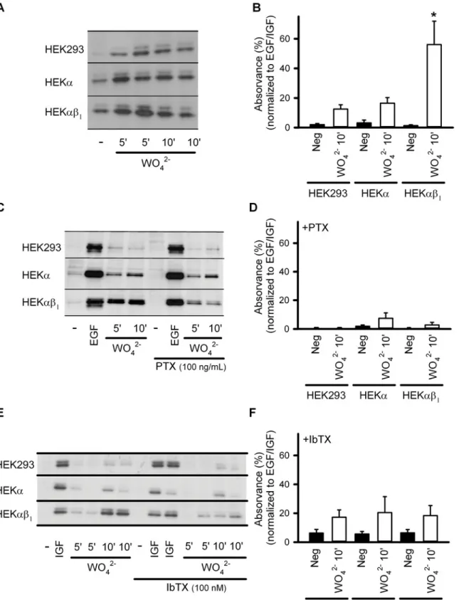
the possible role of BK channels in the tungstate-mediated activation of the ERK pathway, we analyzed ERK1/2 phosphorylation using phospho-specific antibodies. Western blot analysis of phosphorylated ERK1/2 was carried out in tungstate (1 mM)-treated non-transfected HEK293
cells, HEKαcells (constitutively expressing the humanαsubunit of the BK channel), and
HEKαβ1cells (constitutively expressing the bovineαandβ1subunits of the BK channel).
Tungstate increased the phosphorylation of ERK1/2 in all HEK293 cell lines, although to a
sig-nificantly higher level in HEKαβ1cells (P<0.001) (Fig. 1A, and 1B). Such enhanced ERK
phosphorylation induced by tungstate in HEKαβ1cells was prevented by pretreatment with
ei-ther the Gi/oprotein inhibitor pertussis toxin (PTX, 100 ng/ml) (Fig. 1C, 1D) or the specific BK
channel blocker iberiotoxin (IbTX, 100 nM) (Fig. 1E, 1F). Both toxins were without effect on
the EGF/IGF-induced ERK phosphorylation that we used as control for ERK activation.
BK
αβ1
channels function upstream of G
i/oprotein activation induced by
tungstate
Since the BKαβ1channel-dependent enhancement of tungstate-induced ERK phosphorylation
was prevented by inhibition of Gi/oproteins, we next evaluated whether the BK channel
operat-ed either upstream or downstream of the G protein signaling. For this purpose, we analyzoperat-ed
the effect of tungstate on heterologously expressed BKαβ1channels in the presence of GDPβS
(500μM) that locked the Gαprotein subunit in its inactive (GDP-bound) state or after
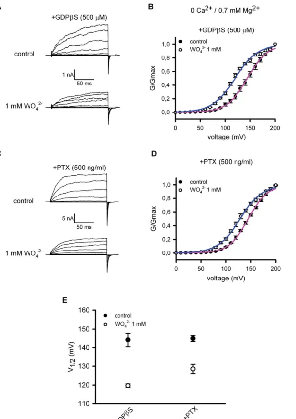
pre-in-cubation of the transfected cells with PTX (500 ng/ml).Fig. 2 (A, C)shows representative
BKαβ1currents, recorded before (control) and after the addition of 1 mM tungstate (WO42-) to
a nominally Ca2+-free bath (intracellular) solution containing 0.7 mM Mg2+(an intracellular
cation required for the tungstate-induced activation of BK channels [14]). Changes in BKαβ1
channel activity were determined by plotting the G-V relationships of the measured BK tail
currents, before and after exposure to tungstate (Fig. 2B, 2D). For each G-V curve, the voltage
for channel half-activation (V1/2 act) was calculated (Fig. 2E). We found that even in the
pres-ence of G-protein inhibitors, tungstate still decreased by ~16–25 mV the V1/2 actfor BKαβ1
channels (P<0.01, paired t-test), favouring channel activation by voltage. This action of
tung-state on BKαβ1channels was similar to the one we have previously reported (a decrease
of V1/2 actby ~22 mV), under identical experimental conditions but without interfering with
the activation of G proteins [14]. Thus, these results ruled out an involvement of G proteins in
the tungstate-induced activation of BKαβ1channels. Instead, the data suggested that the
BK-mediated and Gi/o-dependent phosphorylation of ERK1/2 induced by tungstate involved the
activation of Gi/oproteins downstream of tungstate interaction with the BK channel. To further
test this idea, we evaluated whether Giprotein activation by tungstate was related to the
pres-ence of BKαβ1channels. G protein activation was evaluated by measuring the Fluorescence
Resonance Energy Transfer (FRET) betweenαiandβsubunits of the heterotrimeric G protein
tagged with the yellow fluorescent protein (YFP) and the cyan fluorescent protein (CFP), re-spectively. It has been reported that G protein subunits undergo a molecular rearrangement during activation (rather than a complete dissociation). Thus, when the CFP was fused to the
N-terminus of the Gβsubunit, activation of the G protein following stimulation of Gi
in FRET (measured as an elevation of the ratio between YPF and CFP fluorescence emission (FYFP/FCFP)) [40].
Gαi-YFP and CFP-Gβsubunits (along with the Gγsubunit) were heterologously expressed
in HEKαand HEKαβ1cells and FRET measurements were carried out before and after the
ad-dition of tungstate. Changes of FRET in response to noradrenaline (NA) in HEK293 cells
co-expressing the above mentioned G protein subunit constructs along withα2A-ARs, were used
as positive control. Cells with a reinforced membrane fluorescence pattern (Fig. 3A), indicating
the co-localization of the expressed fluorescent G protein subunits in the cellular membrane, were selected for FRET measurements. Addition of tungstate (1 mM) only increased FRET in
HEKαβ1but not in HEKαcells expressing Gβi-YFP and CFP-Gβsubunits (Fig. 3B(magenta
and blue traces, respectively) and3C). As previously reported [40], addition of NA (1μM) to
HEK293 cells co-transfected with the cDNAs of G protein subunits and theα2A-AR, resulted
in an increase in FRET (elevated FYFP/FCFPratio) (Fig. 3C). No increase in FRET after addition
of NA was seen when the cDNA of theα2A-AR was omitted in the cell transfection process
(Fig. 3C). The tungstate-induced elevation of the FRET signal in HEKαβcells was about 56%
of that produced by NA in HEK293 cells co-expressing theα2A-AR (Fig. 3C). Furthermore, as
observed for the enhanced tungstate-induced phosphorylation of ERK found in HEKαβ1cells
(Fig. 1E, 1F), the increase in FRET among G protein subunits after tungstate application was
abolished by pre-incubating the HEKαβ1cells with IbTX (Fig. 3B(dark purple trace) and3C).
Together, these data suggested that tungstate-targeting of the BKαβ1channel promoted Gi
protein activation.
Tungstate effect on Ca
2+-activated K
+channels is specific for BK
channels
We evaluated whether the tungstate effect on BK channel activity applied also to other distantly
related Ca2+/calmodulin-gated K+channels, in particular the KCa3.1 (IK1) channel that has
been previously related to the direct activation of the ERK signaling pathway.S1 Fig. shows
that pre-activated (by 1μM intracellular Ca2+) KCa3.1 currents recorded from murine 3T3
fi-broblasts were insensitive to 1 mM tungstate, even after current potentiation by the KCa3.1
channel activator SKA-31 [41].
Tungstate-induced activation of native BK channels in vascular smooth
muscle cells depends on the expression of the regulatory BK channel
β1
subunit
We have previously reported that the positive action of tungstate on BKαβ1channel activity,
found in a heterologous expression system, was likely to be responsible of the tungstate-in-duced vasodilatation of mouse arteries pre-contracted with endothelin-1. This conclusion was based on two experimental evidences: tungstate-induced vasorelaxation depended on the
ex-pression of the regulatoryβ1subunit (vasorelaxation was lost inβ1-knockout arterial rings)
and it was not related to the reported tungstate-induced inhibition of the endothelial xanthine indicated) in the absence of toxins. (B) Average normalized relative density (phosphorylated versus total ERK) in the absence of toxins. (C) Representative Western blots obtained from HEK293, HEKαand HEKαβ1cells for ERK1/2 phosphorylation levels, without treatment (-), after treatment with 100 ng/ml EGF
(during 10 minutes) (EGF) or after treatment with 1 mM tungstate (WO42-) (during 5 and 10 minutes, as indicated) in the absence (left) or presence of PTX
(right). (D) Average normalized relative density (phosphorylated versus total ERK) in the presence of PTX. (E) Representative Western blots obtained from HEK293, HEKαand HEKαβ1cells for ERK1/2 phosphorylation levels, without treatment (-), after treatment with 100 ng/ml IGF (during 10 minutes) (IGF) or
after treatment with 1 mM tungstate (WO42-) (during 5 and 10 minutes, as indicated) in the absence (left) or presence of IbTX (right). (F) Average normalized
relative density (phosphorylated versus total ERK) in the presence of IbTX. n = 4–12 in each experimental group.*P<0.05 when compared to the other HEK cell lines (Kruskal-Wallis test followed by Dunnpost hoctest). SeeMethodsfor further details.
Fig 2. Tungstate-mediated activation of BKαβ1channels is independent of G proteins.Representative currents recorded from excised inside-out macropatches obtained from HEK293 cells expressing the BKαβ1channels in the presence of 500μM GDPβS (added to the bath solution) (A) or from
transfected HEK293 cells pre-incubated with PTX (500 ng/ml, 24 hours) (C). Currents were recorded at cytosolic 0 Ca2+and 0.7 mM Mg2+before (control, top
panels) and 5–10 minutes after cytosolic application of 1 mM tungstate (WO42-, bottom panel). The voltage protocol was as described in the Methods. (B), (D)
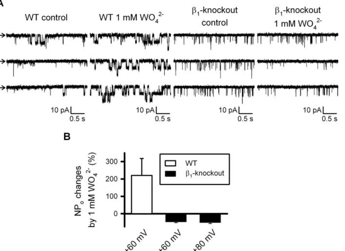
oxidase (XO) [8] as it was not replicated by the XO blocker allopurinol [14]. We have now studied directly the effect of tungstate on BK channels endogenously expressed in murine vas-cular smooth muscle cells (VSMCs). Single-channel recordings showed that BK channel open
probability (NPo) increased in response to 1 mM tungstate only in inside-out patches obtained
from VSMCs of wild-type mice but not in those fromβ1- knockout mouse VSMCs (Fig. 4).
Tungstate promotes ERK1/2 phosphorylation and reduces
PDGF-stimulated cell proliferation in human vascular smooth muscle in a BK
channel-dependent manner
We next analyzed whether tungstate also promoted ERK phosphorylation in human VSMCs
and its relationship with the BK channel. Similar to that seen in HEKαβ1cells (constitutively
expressing the bovineαandβ1subunits of the BK channel), treatment of human VSMCs with
tungstate (1 mM for 10 minutes) promoted the phosphorylation of ERK1/2 up to slightly
lower levels than PDGF treatmentalthough this difference did not reach statistical significance
(P>0.05, ANOVA followed by Tukeypost hoctest). Such tungstate-induced ERK
phosphory-lation in human VSMCs was significantly reduced by pretreatment with the specific BK
chan-nel blocker iberiotoxin (IbTX, 100 nM) (Fig. 5A and 5B). As Gβγ-stimulated ERK1/2
phosphorylation has been reported to lead to differentiation of VSMCs to a contractile
pheno-type [36,37], we also studied the effect of tungstate on PDGF-induced human VSMC
prolifera-tion. For this analysis, we lowered tungstate to 100μM because we have previously reported
that at this lower concentration, tungstate still promoted voltage-dependent activation of
BKαβ1channels and induced vasorelaxation in endothelin-1-pre-contracted mouse arteries
[14]. Besides, such tungstate concentration is only slightly higher than the tungstate levels
mea-sured in the plasma of both experimentally treated rodent models and humans (~5–20μM)
[2,42]. We found that 100μM tungstate strongly reduced the proliferative action of PDGF (the
effect was similar when using 1 mM tungstate). IbTX prevented significantly this inhibition.
IbTX alone had no significant effect on PDGF-induced VSMC proliferation (Fig. 5C).
Discussion
Tungstate is a transition metal that exerts antidiabetic [2,43], antiobesity [6] and
antihyperten-sive actions [8–10] in several experimental animal models. Despite considerable knowledge on
the pharmacological and metabolic effects of tungstate, little information exists regarding its molecular mechanisms of action. In this sense, it is known that tungstate triggers intracellular signaling pathways related to the activation of extracellular signal-regulated kinases (ERK) in
several cell types [4,11,12]. This signaling action of tungstate mimics the effect of insulin in
hepatocytes, by increasing glycogen deposition but in an insulin receptor-independent manner. conductance to the Boltzmann equation (seeMethods). (E) Voltage for half maximal activation (V1/2 act) of BKαβ1channels before (control, filled circles) and
after addition of tungstate (1 mM WO42-, open circles) obtained for the indicated experimental conditions (+GDPβS, n = 4; +PTX, n = 6). No significant
difference was found in the decrease on V1/2 actinduced by tungstate when comparing both treatments: 24±4 mV (n = 4) for GDPβS treatmentversus16±3
mV (n = 6) for PTX treatment (P = 0.11, Mann-Whitney U-test). Besides the substantial difference in the duration of both treatments to inhibit G proteins (24– 28 hours for PTX treatment and minutes for GDPβS treatment), no significant difference was found among them regarding V1/2 actbefore the addition of
tungstate (control situation for GDPβS treatment: V1/2 act= 144±4 mV (n = 4)versuscontrol situation for PTX treatment: V1/2 act= 145±2 mV (n = 6); P>
0.99, Mann-Whitney U-test). Furthermore, these V1/2 actcontrol values (before tungstate application) were similar to the ones previously reported by us under
identical experimental conditions but without interfering with the activation of G proteins: 139±2 mV (n = 7) [14] and 147±2 mV (n = 7) [26] (P = 0.1, ANOVA). Note that, as previously reported, 1 mM tungstate also reduced substantially the K+current amplitude in the absence of cytosolic Ca2+(A, C), an
effect that, contrary to the tungstate-induced reduction of V1/2 act, has been shown to occur either in the absence or presence of Mg2+and in the absence or
presence of the different regulatoryβsubunits (β1-β4) [14]. For each experimental condition V1/2 actand kactvalues were (in mV): 144±4 and 20±0.3 (control
situation for GDPβS treatment, n = 4); 120±1 and 22±1 (after WO42-addition for GDPβS treatment, n = 4); 145±2 and 19±0.3 (control situation for PTX,
n = 6); 129±2 and 21±1 (after WO42-addition for PTX treatment, n = 6). No significant differences were found among kactvalues (P = 0.07, ANOVA).
Fig 3. Tungstate-induced activation of heterologously expressed Gi/o proteins is mediated by BKαβ1channels.(A) Example of the reinforced membrane fluorescence pattern and emission levels from CFP channel (up-left), YFP (FRET channel) (up-right) and the merge channels (bottom-left) from HEK293 heterologously expressing Gαi-YFP and CFP-Gβ. Fluorescence microscopy images were recorded by using confocal microscopy (for more details
seeMethods). FRET signal was determined by using donor ratiometric parameters (458/514) after excitation in the CFP frequency and registering in the YFP emission frequency. (B) Representative FRET changes from HEK293, HEKαor HEKαβ1cells transfected with the cDNAs of G protein subunits, in response
to 1 mM tungstate (either in the absence or presence of IbTX), as indicated. (C), Average FRET changes for the different experimental conditions illustrated in B (n = 5–9).*P<0.05 (Kruskal-Wallis test followed by Dunnpost hoctest).
Tungstate activates PTX-sensitive Giproteins that in turn activate the small GTPase Ras to produce the phosphorylation of ERK, the subsequent phosphorylation of p90rsk and glycogen
synthase kinase-3β, and the activation of glycogen synthase leading to glycogen deposition
[4,13]. The antihypertensive effect of tungstate has been linked to both the inhibition of the
endothelial xanthine oxidase [8] and the activation of the large conductance voltage- and
Ca2+-activated K+(BK) channel at the vascular smooth muscle cells (VSMCs) [14]. Vascular
BK channels are formed by the pore-formingα(KCNMA1) and the regulatoryβ1(KCNMB1)
subunits. Indeed, we reported previously that tungstate only activates BK channels containing
either theβ1or theβ4(but noβ2orβ3) subunits [14,26].
Here, we provide evidence that tungstate-targeting ofβ1subunit-containing BK channels
promotes the activation of PTX-sensitive Giproteins to enhance the tungstate-induced
phos-phorylation of ERK (Fig. 6). First, we observed significant higher levels (~40–44%) of ERK
phosphorylation after tungstate treatment in HEK293 cells expressing both BK channelαand
β1subunits (HEKαβ1cells) than in cells that did not express BK channels (HEK293 cells) or
ex-pressed the BK pore-formingαsubunit alone (HEKαcells). Second, the fact that such
Fig 4. Tungstate-induced increase in the open probability of BK channels endogenously expressed in freshly isolated vascular myocytes requires the BK channelβ1subunit.(A) Representative recordings obtained from inside-out patches clamped at + 60mV from wild-type (WT) andβ1- knockout
freshly isolated mouse vascular myocytes, before (control) and after exposure to 1 mM tungstate (1 mM WO42-), as stated. Arrows indicates the closed state
level. (B) Average changes (in %) of BK channel open probability (NPo) induced by 1 mM tungstate on WT andβ1- knockout mouse vascular myocytes at the
indicated voltage membrane. Significant differences were found among WT (n = 7) andβ1- knockout (n = 9), P<0.001 (Student’s t-test). Further
depolarization to +80 mV ofβ1- knockout inside-out patches did not change the response to tungstate found at +60 mV.
enhancement of the tungstate-induced activation of the ERK pathway found in HEKαβ1cells is
prevented by either PTX or IbTX, supports the involvement of both Gi/oproteins and BK
chan-nels in this signaling process. Third, Gi/oproteins are not upstream of the tungstate-induced
ac-tivation of BKαβ1channels, since tungstate-induced changes in BK channel activity remains
unaltered even in the presence of the G protein inhibitors PTX or GDPβS. This observation is in agreement with previous electrophysiological data and comparative structural analysis sug-gesting that tungstate modulates BK channel activity by direct binding to a site located at the
BKαsubunit (around residues of the voltage sensor and the RCK1 domains that coordinate
the binding of Mg2+) [14], with the required participation ofβ1subunit extracellular loop
resi-dues that stabilize the active configuration of BK channel voltage sensor [26]. And fourth,
BKαβ1channels seem to be upstream in the tungstate-induced, Gi/oprotein-mediated ERK
expression and phosphorylation were quantified by densitometry of the corresponding Western blot signal and normalized to beta-actin. The ratio p-ERK/beta-actin in control conditions was taken as 1, so that fold-changes relative to control are shown. (C) VSMCs were serum starved for 48 hours and then incubated 30 hours with 20 ng/ml PDGF alone or in combination with the indicated compounds. During the last 6 hours of incubation, EdU was added to the media to detect the number of cells entering S-phase. Mean±S.E.M. of 6–9 determinations from at least four different cultures.***P<0.001 (ANOVA followed by Tukeypost hoc test).
doi:10.1371/journal.pone.0118148.g005
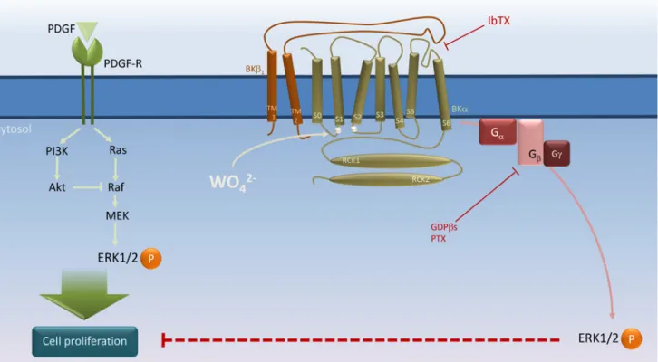
Fig 6. Schematic representation of BKαβ1channel’s role in tungstate-induced, Giprotein-mediated ERK phosphorylation and its interaction with PDGF-stimulated cell proliferation in human vascular smooth muscle.PDGF stimulates both the Ras/Raf/MEK/ERK and the phosphatidylinositol 3-kinase (PI3K)/Akt mitogenic signaling pathways to induce vascular smooth muscle cell (VSMC) proliferation [35]. Tungstate (WO42-) binding to the BK
channelαsubunit at a site involving residues of the Voltage Sensor Domain (silver circles in the S0–S1 and S2–S3 linkers) [14,26] promotes activation of the voltage sensor and channel gating in aβ1subunit-dependent manner, and subsequently, activation of PTX-sensitive G proteins to induce ERK
phosphorylation and inhibition of PDGF-stimulated VSMC proliferation. Binding of iberiotoxin (IbTX) to the external mouth of the channel in close proximity to theβ1extracellular loop, which plays an important role in the modulation of voltage sensor activation and gating of the BK channel [21,46–53], may impair
the conformational changes in the BKαβ1channel produced by tungstate to prevent the coupling of channel gating to G protein activation without the need of
K+conduction. For further details, please seeDiscussion.
phosphorylation pathway. Thus, tungstate only activated heterologously expressed Giproteins
(indicated by an increase in FRET among Gαi-YFP and CFP-Gβsubunits) in HEKαβ1cells,
but no in HEKαcells, an effect that was prevented by the blockade of BKαβ1channels with
IbTX. Altogether, these results suggest that BKαβ1channels might well act as tungstate
recep-tors to trigger the activation of the ERK pathway. Among Ca2+-activated K+channels, such
tungstate receptor function seems to be specific for BKαβ1channels: we observed that the IK1
channel, which has been related to the direct activation of the ERK signaling pathway [30], is
tungstate-insensitive.
Given that the BKβ1channel subunit is primarily found in VSMCs, where it improves
channel function for a better fine-tuned and efficient negative feedback on vascular tone [19,
20], the functional relationship between BKβ1channels and the Gi/oprotein-ERK signaling
cas-cade might also have physiological and/or pathological relevance in the vascular beds. It is well established that phenotypic remodeling of VSMCs, involving changes from contractile toward a more proliferative phenotype caused by mitogen-stimulated gene expression, occurs during
the pathogenesis of hypertension, atherosclerosis or vascular restenosis [31,32]. Therefore,
treatments that oppose such remodeling and promote VSMC re-differentiation can be of help in the management of pathological vascular remodeling. Growth factors such as platelet-de-rived growth factor (PDGF), by acting on specific tyrosine kinase receptors, stimulate several mitogenic signaling pathways that lead to VSMC proliferation: (i) phospholipase C (PLC)
iso-forms that increase the cytosolic Ca2+levels and activate protein kinase C, (ii) the Ras/Raf/
MEK/ERK pathway, and (iii) the phosphatidylinositol (PI) 3-kinase/Akt pathway [35]. These
same intracellular signaling routes promote VSMC differentiation in response to serum
com-ponents such as thrombin, acting via G-protein coupled receptors and Gi/oβγdimmers [35–
37]. The opposite effects produced by these two kinds of agonists regarding the modulation of
VSMC phenotype seem to be due to differences in both the intensity of the signals and their
ki-netic patterns, along with the cross-talk between pathways [35]. Thus, upon stimulation of
ty-rosine kinase receptors there is a rapid and strong phase of ERK phosphorylation and a robust PI 3-kinase-dependent and sustained Akt phosphorylation, which prevents the generation of a
late ERK activation phase by inhibiting Raf kinase activity and suppressing MEK (Fig. 6) [35].
Interestingly, it has been reported that growth factor-dependent mitogenesis relies on two dis-tinct signaling phases. The first phase involves MEK/ERK and c-Myc activation to make cells go through the initial phase of the G0 to S cell cycle interval. Such first phase is required to en-gage the components necessary for cell cycle progression during a second phase that depends
exclusively on PI 3-kinase lipid products [44]. However, after agonist binding to GPCR and
G protein activation, Gβγinduces not only the rapid and strong phase of ERK phosphorylation
but also a late phase that is required for changes in gene expression leading to VSMC differenti-ation. Such second phase of ERK activation can occur because, in this case, Akt
phosphoryla-tion is weak and transient and fails to inhibit Raf kinase activity [35].
Here we show that the open probability of endogenous BK channels in murine VSMCs is
in-creased by tungstate in aβ1-dependent manner. This result is consistent with our previous data
showing that tungstate activates BK channels containing either theβ1or theβ4(but notβ2or
β3) subunits in a heterologous expression system, and that tungstate-induced relaxation of
mouse pre-contracted blood vessels depends on the expression of the regulatory BKβ1subunit
[14]. Functional BK channels have been also recorded from the cultured human renal VSMCs
used in this study (data not shown). In addition, expression studies in proliferating VSMCs
show that they retain expression of both BKαand BKβ1mRNA, presenting an increase of
BKαβ4mRNA [45]. We have now found that targeting of vascular BK channels by tungstate
in part, by BK channels (Fig. 6). However, further research is required in order to establish
whether the activation of the Gi/oprotein/ERK pathway lies behind such antiproliferative
ac-tion of tungstate, by allowing the late phase of ERK phosphorylaac-tion. In this sense, it has been shown that abrogation of the PI 3-kinase/Akt signaling changes the PDGF-induced
prolifer-ative VSMC phenotype toward enhanced expression of contractile proteins [35].
The question of how the targeting of BKαβ1channels by tungstate leads to the activation of
Gi/oproteins (and the subsequent ERK phosphorylation and inhibition of VSMC proliferation
stimulated by PDGF), needs to be answered by future studies. Nonetheless, the fact that this signaling process is abolished in the presence of the BK channel blocker IbTX, might initially
suggest that the conduction of K+ions through the tungstate-activated channel is involved in
G protein activation. IbTX shares a high sequence identity (around 68%) with charybdotoxin
(ChTX) and a similar mechanism for BK channel blockade has been suggested [46]. The exact
interaction site for the toxins in BK channels is not clear but the involvement of residues
around the outer mouth pore of the channel has been proposed [47]. In agreement, the crystal
structure of a mutant form of the voltage-gated K+channel KV1.2 in complex with ChTX
indi-cates that the toxin binds to the extracellular pore entryway and interacts directly with K+
in-side the selectivity filter [48]. Furthermore, some residues in the extracellular loop of the BK
regulatoryβ1subunit are responsible of the high BK channel affinity for ChTX [21,49]. These
β1loop amino acids are in close proximity to the external mouth and, perhaps, the selectivity
filter and gate of the channel [49–51]. Besides, different studies indicate that theβ1extracellular
loop plays an important role in the modulation of voltage sensor activation and gating of the
BK channel [52,53]. Therefore, toxin binding to the BKαβ1channel may also modify the
struc-tural changes related with the activation of the voltage sensor and channel gating. Then, toxin
impairment of the conformational changes in the BKαβ1channel produced by tungstate may
affect the coupling of channel gating to G protein activation without the need of K+conduction
(Fig. 6). In support of this hypothesis, we have found that two distinct concentrations of
tung-state (100μM and 1 mM), which increase and reduce (respectively) K+flux through vascular
BKαβ1channels, equally inhibit PDGF-induced cell proliferation in human vascular smooth
muscle (Fig. 5C). Indeed, 1 mM tungstate has a dual effect on heterologously expressed BKαβ1
channels: it reduces (by ~ 30–40%) the amplitude of K+currents (an effect that is equal for BK
channels containing or not the different regulatoryβsubunits,β1-β4) and favors channel
acti-vation by voltage (i.e. decreases V1/2 actby ~ 20 mV) in a Mg2+- andβ1subunit-dependent
manner [14]. From previous studies of tungstate effects on murine arterial contractility, and
given the key role of BKαβ1channels in this process [18–20], it can be inferred that the negative
action of 1 mM tungstate on vascular BK current amplitude outweighs its positive effect on the voltage-dependent activation of the channel. Thus, a net reduction of vascular BK currents can
explain the constriction of arterial rings from both wild-type (WT) andβ1-knockout mice
in-duced by 1 mM tungstate [14]. On the contrary, at micromolar levels (100μM) tungstate still
promotes voltage-dependent activation of the vascular (β1subunit-containing) BK channel
without reducing K+current amplitude. The resulting net increase in the activity of vascular
BKαβ1channels explains why 100μM tungstate only promotes relaxation of WT but notβ1
-knockout mouse arteries pre-contracted with endothelin-1 [14].
In the same line of thought, it has been shown that activation of the ERK signaling pathway
mediated by IK1 channels does not require the capability of this channel to conduct K+ions
[30]. There are evidences that support the existence of a direct protein-protein crosstalk among
BK channels and some G-protein coupled receptors, such asμ-opioid [54] or thromboxane A2
receptors [55]. In addition, it has been shown that BKαchannel subunit can directly interact
In summary, our data provide evidence to consider BK channels as another member of the growing list of channels directly involved in the transduction of intracellular signals, beyond
their ion-conducting function [29,30]. Our results further highlight the relevance of BK
chan-nel function in VSMCs, apart from its well known role in reducing cell contractility to promote vasorelaxation, arising also as a potential target for the modulation of the growth factor-in-duced proliferative phenotype associated with certain vascular pathologies. In this sense, the
functional interaction between tungstate and vascular BKαβ1channels become an interesting
starting point to develop new antihypertensive therapies [14,57].
Supporting Information
S1 Fig. Tungstate has no effect on KCa3.1 (IK1) channel activity.(A) Whole-cell recordings
of KCa3.1 in murine 3T3 fibroblasts. KCa3.1 currents were pre-activated by infusion of 1μM
Ca2+via the patch-pipette as previously described [38]. Pre-activated currents (control) were
not changed by 1 mM tungstate (1 mM WO42-). (B) Average data showing pre-activated
KCa3.1 current densities before and after tungstate application (P>0.99, Mann-Whitney
U-test; n = 4). (C) Average changes (in %) of pre-activated KCa3.1 currents in response to 1 mM
tungstate (1 mM WO42-) in the absence or presence of KCa3.1 activator SKA-31, as indicated.
The SKA-31-potentiated KCa3.1 current was likewise insensitive to 1 mM tungstate (P = 0.56,
Mann-Whitney U-test). Numbers in brackets indicate the number of cells tested in each experimental condition.
(DOCX)
Acknowledgments
We thank Dr. R. Latorre and Dr. L. Toro for providing the cDNAS for the human BK channel
αandβ1subunits, respectively. We also thank Dr. M. Büneman for the gift of the cDNAs
en-coding rat Gα-YFP, human Gβ-CFP, and and adrenergicα2Areceptor (α2A-AR).
Author Contributions
Conceived and designed the experiments: AIFM PC DZ LN JD AOV RK JRLL MTPG MAV JG JMFF. Performed the experiments: AIFM PC DZ LN JD AOV JMFF. Analyzed the data: AIFM PC DZ LN JD AOV RK JRLL MTPG JMFF. Wrote the paper: AIFM PC DZ LN JD AOV RK JRLL MTPG MAV JG JMFF. Conducted the study, wrote and finalized the manu-script: JMFF.
References
1. BarberàA, Rodríguez-Gil JE, Guinovart JJ (1994) Insulin-like actions of tungstate in diabetic rats. Nor-malization of hepatic glucose metabolism. J Biol Chem 269: 20047–20053. PMID:8051090
2. BarberàA, Gomis RR, Prats N, Rodríguez-Gil JE, Domingo M, et al. (2001) Tungstate is an effective antidiabetic agent in streptozotocin-induced diabetic rats: a long-term study. Diabetologia 44: 507–513. PMID:11357483
3. BarberàA, Fernández-Álvarez J, Truc A, Gomis R, Guinovart JJ (1997) Effects of tungstate in neonatal-ly streptozotocin-induced diabetic rats: mechanism leading to normalization of gneonatal-lycaemia. Diabetologia 40: 143–149. PMID:9049473
4. Domínguez JE, Muñoz MC, Zafra D, Sánchez-Pérez I, Baqué S, et al. (2003) The antidiabetic agent so-dium tungstate activates glycogen synthesis through an insulin receptor-independent pathway. J Biol Chem 278: 42785–42794. PMID:12925525
6. Claret M, Corominola H, Canals I, Saura J, Barceló-Batllori S, et al. (2005) Tungstate decreases weight gain and adiposity in obese rats through increased thermogenesis and lipid oxidation. Endocrinology 146: 4362–4369. PMID:16002523
7. Amigo-Correig M, Barcelo-Batllori S, Piquer S, Soty M, Pujadas G, et al. (2011) Sodium tungstate regu-lates food intake and body weight through activation of the hypothalamic leptin pathway. Diabetes Obes Metab 13: 235–242. doi:10.1111/j.1463-1326.2010.01339.xPMID:21205112
8. Suzuki H, Delano FA, Parks DA, Jamshidi N, Granger DN, et al. (1998) Xanthine oxidase activity asso-ciated with arterial blood pressure in spontaneously hypertensive rats. Proc Natl Acad Sci U S A 95: 4754–4759. PMID:9539811
9. Swei A, Lacy F, Delano FA, Parks DA, Schmid-Schonbein GW (1999) A mechanism of oxygen free rad-ical production in the Dahl hypertensive rat. Microcirculation 6: 179–187. PMID:10501091
10. Peredo HA, Zabalza M, Mayer MA, Carranza A, Puyo AM (2010) Sodium tungstate and vanadyl sulfate effects on blood pressure and vascular prostanoids production in fructose-overloaded rats. Clin Exp Hypertens 32: 453–457. doi:10.3109/10641961003686443PMID:21029009
11. Ballester J, Domínguez J, Muñoz MC, Sensat M, Rigau T, et al. (2005) Tungstate treatment improves Leydig cell function in streptozotocin-diabetic rats. J Androl 26: 706–715. PMID:16291965
12. Gómez-Ramos A, Domínguez J, Zafra D, Corominola H, Gomis R, et al. (2006) Sodium tungstate de-creases the phosphorylation of tau through GSK3 inactivation. J Neurosci Res 83: 264–273. PMID:
16397900
13. Zafra D, Nocito L, Domínguez J, Guinovart JJ (2013) Sodium tungstate activates glycogen synthesis through a non-canonical mechanism involving G-proteins. FEBS Lett 587: 291–296. doi:10.1016/j. febslet.2012.11.034PMID:23260418
14. Fernández-Mariño AI, Porras-González C, González-Rodríguez P, Selent J, Pastor M, et al. (2012) Tungstate activates BK channels in aβsubunit- and Mg2+-dependent manner: relevance for arterial
va-sodilatation. Cardiovasc Res 95: 29–38. doi:10.1093/cvr/cvs139PMID:22473360
15. Cox DH, Aldrich RW (2000) Role of the beta1 subunit in large-conductance Ca2+-activated K+channel gating energetics. Mechanisms of enhanced Ca2+sensitivity. J Gen Physiol 116: 411–432. PMID:
10962017
16. Bao L, Cox DH (2005) Gating and ionic currents reveal how the BKCachannel’s Ca2+sensitivity is
en-hanced by itsβ1subunit. J Gen Physiol 126: 393–412. PMID:16186565
17. Contreras GF, Neely A, Álvarez O, Gonzalez C, Latorre R (2012) Modulation of BK channel voltage gat-ing by different auxiliaryβsubunits. Proc Natl Acad Sci U S A 109: 18991–18996. doi:10.1073/pnas. 1216953109PMID:23112204
18. Jaggar JH, Porter VA, Lederer WJ, Nelson MT (2000) Calcium sparks in smooth muscle. Am J Physiol Cell Physiol 278: C235–C256. PMID:10666018
19. Brenner R, Perez GJ, Bonev AD, Eckman DM, Kosek JC, et al. (2000) Vasoregulation by theβ1subunit
of the Ca2+-activated K+channel. Nature 407: 870–876. PMID:11057658
20. Tanaka Y, Koike K, Alioua A, Shigenobu K, Stefani E et al. (2004)β1-subunit of MaxiK channel in
smooth muscle: a key molecule which tunes muscle mechanical activity. J Pharmacol Sci 94: 339–347. PMID:15107573
21. Hanner M, Schmalhofer WA, Munujos P, Knaus HG, Kaczorowski GJ, et al. (1997) Theβsubunit of the high-conductance Ca2+-activated K+channel contributes to the high-affinity receptor for charybdotoxin.
Proc Natl Acad Sci U S A 94: 2853–2858. PMID:9096310
22. McManus OB, Helms LM, Pallanck L, Ganetzky B, Swanson R, et al. (1995) Functional role of theβ subunit of high conductance Ca2+- activated K+channels. Neuron 14: 645–650. PMID:7695911
23. Valverde MA, Rojas P, Amigo J, Cosmelli D, Orio P, et al. (1999) Acute activation of Maxi-K channels (hSlo) by estradiol binding to theβsubunit. Science 285: 1929–1931. PMID:10489376
24. Dick GM, Sanders KM (2001) (Xeno)estrogen sensitivity of smooth muscle BK channels conferred by the regulatoryβ1subunit: a study ofβ1knockout mice. J Biol Chem 276: 44835–44840. PMID:
11590153
25. Bukiya AN, Liu J, Toro L, Dopico AM (2007)β1(KCNMB1) subunits mediate lithocholate activation of
large-conductance Ca2+-activated K+channels and dilation in small, resistance-size arteries. Mol Phar-macol 72: 359–369. PMID:17468198
26. Fernández-Mariño AI, Valverde MA, Fernández-Fernández JM (2014) BK channel activation by tungstate requires theβ1subunit extracellular loop residues essential to modulate voltage sensor function and
channel gating. Pflügers Arch 466: 1365–75. doi:10.1523/JNEUROSCI.2953-14.2015PMID:25609622 27. Xu D, Wang L, Dai W, Lu L (1999) A requirement for K+-channel activity in growth factor-mediated
28. Guo TB, Lu J, Li T, Lu Z, Xu G, et al. (2005) Insulin-activated, K+-channel-sensitive Akt pathway is
pri-mary mediator of ML-1 cell proliferation. Am J Physiol Cell Physiol 289: C257–C263. PMID:15800056 29. Kaczmarek LK (2006) Non-conducting functions of voltage-gated ion channels. Nat Rev Neurosci 7:
761–771. PMID:16988652
30. Millership JE, Devor DC, Hamilton KL, Balut CM, Bruce JI, et al. (2011) Ca2+-activated K+channels in-crease cell proliferation independent of K+conductance. Am J Physiol Cell Physiol 300: C792–C802.
doi:10.1152/ajpcell.00274.2010PMID:21123738
31. Schwartz SM, Ross R (1984) Cellular proliferation in atherosclerosis and hypertension. Prog Cardio-vasc Dis 26: 355–372. PMID:6371894
32. Köhler R, Wulff H, Eichler I, Kneifel M, Neumann D, et al. (2003) Blockade of the intermediate-conductance calcium-activated potassium channel as a new therapeutic strategy for restenosis. Circulation 108: 1119–1125. PMID:12939222
33. Miguel-Velado E, Moreno-Domínguez A, Colinas O, Cidad P, Heras M, et al. (2005) Contribution of Kv channels to phenotypic remodeling of human uterine artery smooth muscle cells. Circ Res 97: 1280–1287. PMID:16269658
34. Cidad P, Miguel-Velado E, Ruiz-McDavitt C, Alonso E, Jiménez-Pérez L, et al. (2014) Kv1.3 channels modulate human vascular smooth muscle cells proliferation independently of mTOR signaling pathway. Pflügers Arch doi:10.1007/s00424-014-1607-yPMID:25609622
35. Reusch HP, Zimmermann S, Schaefer M, Paul M, Moelling K (2001) Regulation of Raf by Akt Controls Growth and Differentiation in Vascular Smooth Muscle Cells. J Biol Chem 276: 33630–33637. PMID:
11443134
36. Reusch HP, Schaefer M, Plum C, Schultz G, Paul M (2001) Gβγmediate differentiation of vascular smooth muscle cells. J Biol Chem 276: 19540–19547. PMID:11279222
37. Schauwienold D, Plum C, Helbing T, Voigt P, Bobbert T, et al. (2003) ERK1/2-dependent contractile protein expression in vascular smooth muscle cells. Hypertension 41: 546–552. PMID:12623957 38. Oliván-Viguera A, Valero MS, Murillo MD, Wulff H, García-Otín AL, et al. (2013) Novel phenolic
inhibi-tors of small/intermediate-conductance Ca2+-activated K+channels, K
Ca3.1 and KCa2.3. PLoS One 8:
e58614. doi:10.1371/journal.pone.0058614PMID:23516517
39. Patterson GH, Piston DW, Barisas BG (2000) Förster distances between green fluorescent protein pairs. Anal Biochem 284: 438–440. PMID:10964438
40. Bünemann M, Frank M, Lohse MJ (2003) Giprotein activation in intact cells involves subunit
rearrange-ment rather than dissociation. Proc Natl Acad Sci U S A 100: 16077–16082. PMID:14673086 41. Sankaranarayanan A, Raman G, Busch C, Schultz T, Zimin PI, et al. (2009)
Naphtho[1,2-d]thiazol-2-ylamine (SKA-31), a new activator of KCa2 and KCa3.1 potassium channels, potentiates the
endotheli-um-derived hyperpolarizing factor response and lowers blood pressure. Mol Pharmacol 75: 281–295. doi:10.1124/mol.108.051425PMID:18955585
42. Hanzu F, Gomis R, Coves MJ, Viaplana J, Palomo M, et al. (2010) Proof-of-concept trial on the efficacy of sodium tungstate in human obesity. Diabetes Obes Metab 12: 1013–1018. doi: 10.1111/j.1463-1326.2010.01293.xPMID:20880348
43. Muñoz MC, BarberàA, Domínguez J, Fernández-Álvarez J, Gomis R, et al. (2001) Effects of tungstate, a new potential oral antidiabetic agent, in Zucker diabetic fatty rats. Diabetes 50: 131–138. PMID:
11147778
44. Jones SM, Kazlauskas A (2001) Growth-factor-dependent mitogenesis requires two distinct phases of signalling. Nat Cell Biol 3: 165–172. PMID:11175749
45. Cidad P, Moreno-Domínguez A, Novensa L, Roque M, Barquin L, et al. (2010) Characterization of ion channels involved in the proliferative response of femoral artery smooth muscle cells. Arterioscler Thromb Vasc Biol. 30: 1203–1211. doi:10.1161/ATVBAHA.110.205187PMID:20299686 46. Giangiacomo KM, Garcia ML, McManus OB (1992) Mechanism of iberiotoxin block of the
large-conductance calcium-activated potassium channel from bovine aortic smooth muscle. Biochemistry 31: 6719–6727. PMID:1379069
47. MacKinnon R, Miller C (1988) Mechanism of charybdotoxin block of the high-conductance, Ca2+-activated K+channel. J Gen Physiol 91: 335–349. PMID:2454283
48. Banerjee A, Lee A, Campbell E, MacKinnon R (2013) Structure of a pore-blocking toxin in complex with a eukaryotic voltage-dependent K+channel. eLife 2: e00594. doi:10.7554/eLife.00594PMID:
23705070
50. Piskorowski RA, Aldrich RW (2006) Relationship between pore occupancy and gating in BK potassium channels. J Gen Physiol 127: 557–576. PMID:16636204
51. Chen X, Aldrich RW (2011) Charge substitution for a deep-pore residue reveals structural dynamics during BK channel gating. J Gen Physiol 138: 137–154. doi:10.1085/jgp.201110632PMID:21746846 52. Fernández-Fernández JM, Tomás M, Vázquez E, Orio P, Latorre R, et al. (2004) Gain-of-function
muta-tion in the KCNMB1 potassium channel subunit is associated with low prevalence of diastolic hyperten-sion. J Clin Invest 113: 1032–1039. PMID:15057310
53. Gruslova A, Semenov I, Wang B (2012) An extracellular domain of the accessoryβ1subunit is required
for modulating BK channel voltage sensor and gate. J Gen Physiol 139:57–67. doi:10.1085/jgp. 201110698PMID:22155735
54. Twitchell WA, Rane SG (1994) Nucleotide-independent modulation of Ca2+-dependent K+channel cur-rent by aμ-type opioid receptor. Mol Pharmacol 46: 793–798. PMID:7969064
55. Li M, Tanaka Y, Alioua A, Wu Y, Lu R, et al. (2010) Thromboxane A2 receptor and MaxiK-channel inti-mate interaction supports channel trans-inhibition independent of G-protein activation. Proc Natl Acad Sci U S A 107: 19096–19101. doi:10.1073/pnas.1002685107PMID:20959415
56. Zhou XB, Wulfsen I, Lutz S, Utku E, Sausbier U, et al. (2008) M2 muscarinic receptors induce airway smooth muscle activation via a dual, Gβγ-mediated inhibition of large conductance Ca2+-activated K+ channel activity. J Biol Chem 283: 21036–21044. doi:10.1074/jbc.M800447200PMID:18524769 57. Köhler R (2012) Heavy metal to lower the pressure? Cardiovasc Res 95: 3–4. doi:10.1093/cvr/cvs169