TítuloCarotenoid accumulation in Haematococcus pluvialis in mixotrophic growth
11
0
0
Texto completo
(2) Introduction The. fresh-water. microalga,. Haematococcus. pluvialis. (Chlorophyceae),. accumulates the orange-red pigment astaxanthin (3,30 -dihydroxy-β,β-carotene-4,40 dione), and other related carotenoids and their esters (Grung et al. 1992, Yuan & Chen 1998). These carotenoids are the main source of colour in fins, skin and flesh of the wild rainbow trout as well as other kinds of salmonid fish (Marusich & Bauerfeind 1981). Astaxanthin accumulation by this microalga is related to the formation of the palmella and aplanospore stages of the life cycle of the microalga (Elliot 1934), usually induced by stress conditions. Different studies have been carried out on the nutrient and culture condition requirements of Haematococcus. Accumulation of astaxanthin is obtained under Nstarvation, high light intensity and with agents which prevent cell division without impairing the ability of the alga to assimilate carbon (Boussiba & Vonshak 1991, Harker et al. 1996, Kobayashi et al. 1997, Fábregas et al. 1998, Boussiba 2000). However, the main problem of N-starvation is the decrease in growth rate. The effect of nitrate concentration on Haematococcus growth rate and carotenoid production throughout the culture was assayed in order to determine the optimum conditions for mass culture and astaxanthin production. Previous studies on the nutrition of H. pluvialis have shown that acetate is an important carbon source, enhancing both growth and carotenogenesis (Borowitzka et al. 1991, Kobayashi et al. 1993). However, there is little information about the effect of acetate concentration on accumulation of secondary carotenoids in Haematococcus. In the present study, carotenoid formation was studied under different acetate concentrations and also using another carbon source (malonate), to find the optimal concentrations for mass culture and maximum production of astaxanthin and other carotenoids of biotechnological interest. Due to the great interest in knowing the carotenoid composition during culture and in the different stages of the life cycle of H. pluvialis, a HPLC system was developed for the separation and identification of the pigments in a single chromatographic run..
(3) Materials and methods Alga strain and culture conditions Haematococcus pluvialis (strain 34/7) from CCAP (FBA Ambleside Cumbria, UK) was cultured in modified Bold’s Basal Medium supplemented with trace elements solution (Fábregas et al. 1984). To determine the suitable nitrate concentration to obtain the best growth and ketocarotenoids production a range of nitrate concentrations was tested: 0, 0.15, 0.25, 0.5, 0.75 and 1 g l−1 NaNO3. For subsequent experiments, a NaNO3concentration of 0.15 g l−1 was chosen. To test the effect of acetate concentration on cell growth and pigment synthesis, sodium acetate was added in the culture medium at concentrations of 0, 0.25, 0.5, 1 and 2% (w/v), adjusting the pH to 7. In the other carbon source experiment, sodium acetate was replaced with sodium malonate at the same concentrations and culture conditions. Cultures were carried out in aerated mini-reactors containing 400 ml medium and maintained at 18 ± 1 ºC and 68.25 µmol photon m−2 s−1, with a dark:light cycle of 12:12 h. All cultures were carried out in triplicate. Algal densities were determined by daily counting triplicate samples in a haematocytometer. To obtain growth velocity, doublings/day were calculated: doublings/day = ln N(n) − lnN(i)/ ln 2(tn − ti) where ti and tn are the initial and final time of the logarithmic phase, both expressed in days, and N(i) and N(n) are the initial and final cellular densities, respectively. Pigment extraction For pigment spectrophotometric analysis, 10 ml samples were centrifuged at 3000 g for 15 min. Cell pigments were extracted with DMSO, preheated to 55 ºC and vortexshaken for 30 s (Sedmak et al. 1990). The extracts were centrifuged again and chlorophylls a and b and total carotenoids were spectrophotometrically determined by recording the absorbance at 665, 649 and 480 nm, respectively, and using the equations of Wellburn (1994). HPLC method Carotenoids from the different experiments were qualitatively and quantitatively analysed. The pigment extracts in acetone were separated on a Hewlett Packard HPLC.
(4) equipped with a photodiode array detector. A reversed-phase 250 × 4 mm Hypersil C18 (5 µm) column (Hewlett Packard) was used. The elution gradient was run as follows with eluent A (water), eluent B (methanol), eluent C (acetone): 0 min 9% A, 76% B, 15% C; 9 min 5% A, 45% B, 50% C; 15 min 4% A, 38 % B, 58 % C; 17 min 3% A, 27% B, 70% C; 22 min 3% A, 27% B, 70% C; 25 min 100% C; 26 min 100% C. The flow rate was 1 ml min−1. The detection wavelengths for integration were 444 and 476 nm. β-Carotene (Sigma) and astaxanthin, canthaxanthin and echinenone (F. Hoffman La Roche Ltd) were used as standards to calculate the concentrations of the other carotenoids. Data are given as mean values ± standard error of the means.. Results Growth with different concentrations of nitrate Haematococcus cultures with different NaNO3 concentrations showed no differences in growth until the eleventh day, when the cultures with 0.15 g l−1 NaNO3 l −1. had a decrease in growth rate. Cultures under nitrogen-free conditions showed the. lowest growth rate (Table 1). The ratio between chlorophyll a and total carotenoids was about 4 when nitrate was present in the culture medium. However, this ratio dropped quickly under nitrate deficiency (Figure 1). In nitrogen-free cultures this drop was observed on the first day of culture and in cultures with 0.15 and 0.25 g NaNO3 l−1, the drop occurred after the ninth and eleventh day, respectively. The decrease of this relation showed a nitrogen deficiency in the cultures before growth had stopped. While NaNO3 remained available for the cultures, chlorophyll and total carotenoid concentrations remained at constant levels until there was N-deficiency; beyond this point, a rapid chlorophyll decline and carotenoid increase occurred throughout the further development of these cultures. Therefore, the total carotenoid content of cells reached a maximum value in cultures under nitrogen deficiency while the chlorophylls reached a minimum value..
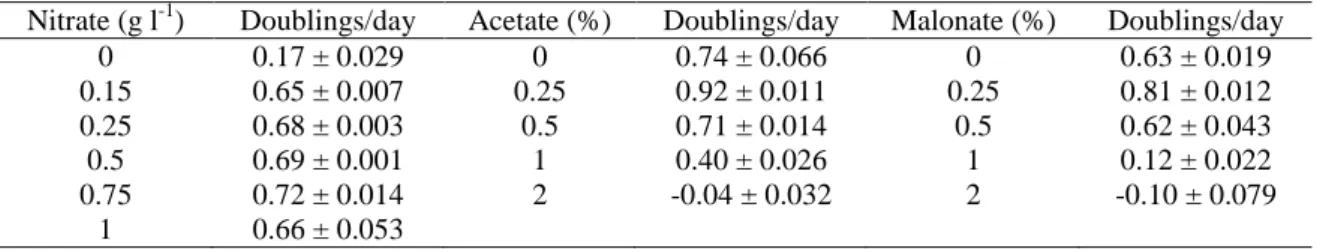
(5) The HPLC analysis at the last day of culture (Table 2) showed that the ketocarotenoids were only detected in cultures with 0, 0.15 and 0.25 g NaNO3 l−1, corresponding to the cultures with a chlorophyll a/total carotenoids ratio below 4 (Figure 1). The secondary carotenoids were mainly synthesised as astaxanthin esters, which amount was higher than 80% of the total carotenoids (Table 2). Canthaxanthin and echinenone were also detected but at lower concentrations. Lutein, the most abundant carotenoid in green cells of H. pluvialis, decreased considerably in red cultures under conditions of nitrogen deficiency. The HPLC method used resolves, in a short time one-step separation, a wide variety of carotenoids, including the separation of echinenone from the astaxanthin esters peaks which could not be obtained with other methods.. Fig. 1. Chlorophyll a/total carotenoids ratio in cultures of H. pluvialis with different concentrations of NaNO3: • gl−1; ○ 0.15 g l−1; ■ 0.25 g l−1; □ 0.5 g l−1; ▲ 0.75 g l−1; △ 1gl−1.. When the ratio chl a/total carotenoids was about 4, corresponding to cultures with 0.5, 0.75 and 1 g NaNO3 l−1, (Figure 1), the pigment profile showed the typical pattern of major carotenoids of green algae, β- carotene, lutein, violaxanthin and neoxanthin.. Table 1. Growth rates of H. pluvialis cultured with different concentrations of nitrate, acetate and malonate. Nitrate (g l-1) 0 0.15 0.25 0.5 0.75 1. Doublings/day 0.17 ± 0.029 0.65 ± 0.007 0.68 ± 0.003 0.69 ± 0.001 0.72 ± 0.014 0.66 ± 0.053. Acetate (%) 0 0.25 0.5 1 2. Doublings/day 0.74 ± 0.066 0.92 ± 0.011 0.71 ± 0.014 0.40 ± 0.026 -0.04 ± 0.032. Malonate (%) 0 0.25 0.5 1 2. Doublings/day 0.63 ± 0.019 0.81 ± 0.012 0.62 ± 0.043 0.12 ± 0.022 -0.10 ± 0.079.
(6) Mixotrophic growth with acetate Growth rates of Haematococcus pluvialis were enhanced by the addition of 0.25% (w/v) acetate with respect to control cultures without this compound, but a concentration of acetate higher than 0.5% caused growth inhibition (Table 1). The supplementation of acetate to the cultures of Haematococcus provoked the formation of cyst cells, which was closely associated with a concomitant increase in the astaxanthin concentration (Figure 2). In control cultures, there were few variations in the cellular concentration of total carotenoids thoughout the culture, but with increasing amounts of acetate there was an increase in the accumulation of total carotenoids (Figure 2). The addition of acetate to the medium had a strong effect on the cellular accumulation of carotenoids at the end of the stationary phase (Figure 2). Acetate also affected the amount of chlorophyll present. Maximum chlorophyll content per cell was detected in cultures without acetate in the medium. Total carotenoid concentration was almost double in cultures with 0.25 and 0.5% (w/v) acetate compared with control cultures (Figure 2). Acetate enhanced the accumulation of total cellular carotenoids, with values up to three times higher than in autotrophic control cultures. However, the major accumulation occurred in cultures with 2% acetate, which presented a strong growth inhibition accompanied by cell encystment. Therefore, maximum total carotenoid yield was obtained in cultures with low acetate concentrations (0.25, 0.5 and 1% w/v). An increase in acetate concentration in the medium produced lower cell and carotenoid yield due to growth inhibition. The HPLC analysis showed that acetate mainly induced the accumulation of astaxanthin esters (almost 90% of total carotenoids), as well as a strong primary carotenoids reduction (Table 2); lutein concentration was more than three times lower in cultures with 2% acetate than in control cultures..
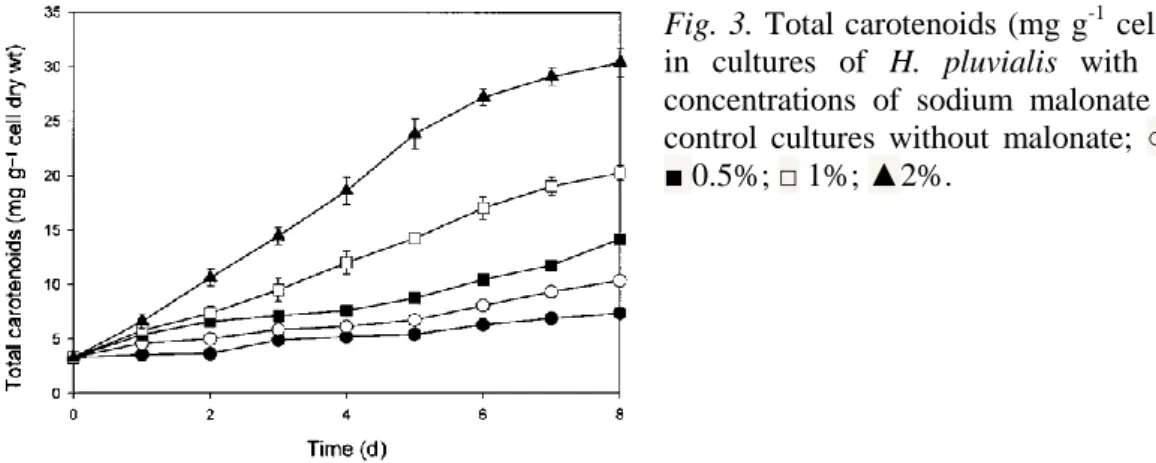
(7) Fig. 2. Total carotenoids (mg g-1 cell dry wt) in cultures of H.pluvialis with different concentrations of sodium acetate (w/v): ●control cultures without acetate; ○, 0.25%; ■ 0.5%; □ 1%; ▲ 2%.. Mixotrophic growth with malonate Over the range of concentrations tested, malonate enhanced the growth of Haematococcus pluvialis only at a concentration of 0.25% (w/v); higher malonate concentrations caused a slight or total growth inhibition (Table 1). However, an increase in cell size was microscopically observed under high malonate concentrations. The total carotenoid content increased in malonatestressed algae compared with control cultures (Figure 3). Maximum carotenoid concentration was obtained at the higher malonate concentration assayed, in cultures with 2% (w/v) malonate. In these cultures up to five times more carotenoids were accumulated than in cultures without this compound, but cell density was lower because cell division was totally inhibited. Both chlorophyll and total carotenoid yields reached the maximum value in cultures with 0.25% (w/v) malonate, because higher concentrations (≥ 0.5% w/v) produced a decrease in biomass production with respect to control cultures. The HPLC analysis of the cultures with malonate (Table 2) showed that the major compounds accumulated were the astaxanthin esters, as ocurred in cultures with acetate, and that the concentration of primary carotenoids was lower than in cultures without this carbon source..
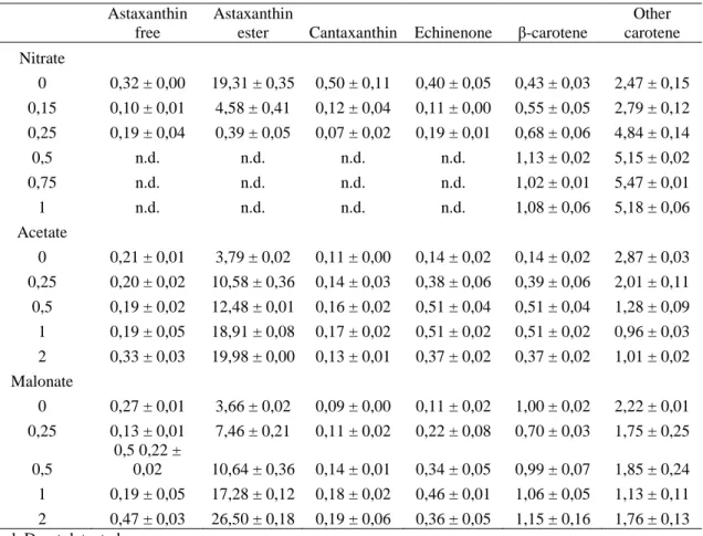
(8) Table 2. Quantitative carotenoid composition (in mg g-1 of biomass dry wt), obtained by HPLC analysis, of H. pluvialis cells at the stationary phase in cultures with different concentrations of nitrate (g l-1), acetate or malonate (% w/v). Astaxanthin free. Astaxanthin ester Cantaxanthin. Echinenone. β-carotene. Other carotene. 0. 0,32 ± 0,00. 19,31 ± 0,35. 0,50 ± 0,11. 0,40 ± 0,05. 0,43 ± 0,03. 2,47 ± 0,15. 0,15. 0,10 ± 0,01. 4,58 ± 0,41. 0,12 ± 0,04. 0,11 ± 0,00. 0,55 ± 0,05. 2,79 ± 0,12. 0,25. 0,19 ± 0,04. 0,39 ± 0,05. 0,07 ± 0,02. 0,19 ± 0,01. 0,68 ± 0,06. 4,84 ± 0,14. 0,5. n.d.. n.d.. n.d.. n.d.. 1,13 ± 0,02. 5,15 ± 0,02. 0,75. n.d.. n.d.. n.d.. n.d.. 1,02 ± 0,01. 5,47 ± 0,01. 1. n.d.. n.d.. n.d.. n.d.. 1,08 ± 0,06. 5,18 ± 0,06. 0. 0,21 ± 0,01. 3,79 ± 0,02. 0,11 ± 0,00. 0,14 ± 0,02. 0,14 ± 0,02. 2,87 ± 0,03. 0,25. 0,20 ± 0,02. 10,58 ± 0,36. 0,14 ± 0,03. 0,38 ± 0,06. 0,39 ± 0,06. 2,01 ± 0,11. 0,5. 0,19 ± 0,02. 12,48 ± 0,01. 0,16 ± 0,02. 0,51 ± 0,04. 0,51 ± 0,04. 1,28 ± 0,09. 1. 0,19 ± 0,05. 18,91 ± 0,08. 0,17 ± 0,02. 0,51 ± 0,02. 0,51 ± 0,02. 0,96 ± 0,03. 2. 0,33 ± 0,03. 19,98 ± 0,00. 0,13 ± 0,01. 0,37 ± 0,02. 0,37 ± 0,02. 1,01 ± 0,02. 0. 0,27 ± 0,01. 3,66 ± 0,02. 0,09 ± 0,00. 0,11 ± 0,02. 1,00 ± 0,02. 2,22 ± 0,01. 0,25. 7,46 ± 0,21. 0,11 ± 0,02. 0,22 ± 0,08. 0,70 ± 0,03. 1,75 ± 0,25. 0,5. 0,13 ± 0,01 0,5 0,22 ± 0,02. 10,64 ± 0,36. 0,14 ± 0,01. 0,34 ± 0,05. 0,99 ± 0,07. 1,85 ± 0,24. 1. 0,19 ± 0,05. 17,28 ± 0,12. 0,18 ± 0,02. 0,46 ± 0,01. 1,06 ± 0,05. 1,13 ± 0,11. 2 0,47 ± 0,03 n.d. D not detected.. 26,50 ± 0,18. 0,19 ± 0,06. 0,36 ± 0,05. 1,15 ± 0,16. 1,76 ± 0,13. Nitrate. Acetate. Malonate. Discussion Nitrate concentration plays a very important role in the cell division rate and in the accumulation of secondary carotenoids of Haematococcus pluvialis (Boussiba & Vonshak 1991). This suggests that the synthesis of astaxanthin requires nitrogen, and most likely reflects the need for continuous synthesis of protein in order to support the massive accumulation of the pigment. However, the source of this nitrogen could not be in the culture media, but possibly in a nitrogen intracellular store as RuBisCo, since it has been reported that RuBisCo supports cell survival, and even growth for several hours under nitrogen starvation (García-Ferris et al. 1996)..
(9) Fig. 3. Total carotenoids (mg g-1 cell dry wt) in cultures of H. pluvialis with different concentrations of sodium malonate (w/v) ● control cultures without malonate; ○ 0.25%; ■ 0.5%; □ 1%; ▲2%.. Nitrogen starvation is an effective way to enhance astaxanthin accumulation in Haematococcus (Table 2) as in other green microalgae (Orosa et al. 2000), but cell density is low due to the cessation of cell division. One solution to this problem could be the use of a low nitrate concentration, so that in a few days the nitrate present in the medium would be exhausted, but allowing to obtain higher cell density. The optimum nitrate concentration to avoid this problem seems to be 0.15 g l−1. The ratio chlorophyll a/total carotenoids could be a good indicator of the physiological state of the culture. In favourable growth conditions this value was about 4; this value indicated nutrient replete conditions (Figure 1); however, a value below 3 implied limitation of growth by reduced nutrient supply or another kind of stress, sharply decreasing this value when nutrient supply was reduced or another kind of stress occurred. Thus, it is possible to check easily the N status of the cell, which is very important since N availability is probably the main factor affecting astaxanthin accumulation. As observed previously by Droop (1954) and Borowitzka et al. (1991), acetate appeared to be an important carbon source, enhancing both growth and carotenogenesis at small quantities (Tables 1 and 2, Figure 2). However, the effect of acetate was concentration-dependent, higher concentrations inhibiting growth but markedly increasing astaxanthin content per cell (Table 2). Acetate addition in excess may generate a relative shortage of nitrogen inducing cyst formation and astaxanthin accumulation triggered by a high carbon/nitrogen (C/N) ratio (Kakizono et al. 1992). The algal cells seem to decrease their nitrogen uptake and begin to use the cellular nitrogen as in typical N-deficiency, although there is nitrogen in the culture medium..
(10) The same effect has been observed in cultures with malonate. The effect is stronger, cells accumulating more astaxanthin in less time than in cultures without this compound, or than in cultures with acetate. Even at the lower malonate concentrations assayed, the astaxanthin yield is at least twice higher than in control cultures (Table 2). Malonate is toxic for cells at high concentrations, however, at low concentrations it may enhance growth as a carbon source. In spite of the growth inhibition at higher acetate or malonate concentrations, it is important to note the strong effect on the stimulation of astaxanthin synthesis and accumulation being astaxanthin concentration more than four times higher in cultures with acetate or malonate than in cultures without these compounds (Table 2). A similar effect was observed in Phaffia rhodozyma, where the addition of another carotenoid pathway precursor (mevalonic acid) had a strong effect on the accumulation of carotenoids (Calo et al. 1995).. Acknowledgements The authors thank Dr Manuel Zapata for his scientific support in the functional analysis of cell pigments by HPLC. This work was supported by a grant from Xunta de Galicia XUGA10301B96.. References Borowitzka MA, Huisman JM, Osborn A (1991) Culture of the astaxanthin-producing green algaHaematococcus pluvialis. 1. Effects of nutrients on growth and cell type. J. Appl. Phycol. 3: 295-304. Boussiba S (2000) Carotenogenesis in the green algae Haematococcus pluvialis: cellular physiology and stress response. Physiol. Plant 108: 111-117. Boussiba S, Vonshak A (1991) Astaxanthin accumulation in the green alga Haematococcus pluvialis. Plant Cell Physiol. 32: 1077-1082. Calo P, de Miguel T, Velázquez JB, Villa TG (1995) Mevalonic acid increases trans-astaxanthin and carotenoid biosynthesis in Phaffia rhodozyma. Biotechnol. Lett. 17: 575-578. Droop MR (1954) Conditions governing haematochrome formation in Haematococcus pluvialis. Arch. Mikrobiol. 20: 391-391..
(11) Elliot AM (1934) Morphology and Protistenk. 82: 250-272.. life history of Haematococcus pluvialis. Arch.. Fábregas J, Abalde J, Herrero C, Cabezas B, Veiga M (1984) Growth of the marine microalgaTetraselmis suecica in batch cultures with different salinities and nutrient concentrations.Aquaculture 42: 207-215. Fábregas J, Domínguez A, García-Álvarez D, Lamela T, Otero A (1998) Induction of astaxanthin accumulation by nitrogen and magnesium deficiencies in Haematococcus pluvialis. Biotechnol. Lett. 20: 623-626. García-Ferris C, de los Ríos A, Ascaso C, Moreno J (1996) Correlated biochemical and ultrastructural changes in nitrogen-starved Euglena gracilis. J. Phycol. 32: 953-963. Grung M, D'souza FML, Borowitzka M, Liaaen-Jensen S (1992) Algal carotenoids 51. Secondary carotenoids, 2. Haematococcus pluvialis aplanospores as a source of (3S, 3's)astaxanthin esters. J. Appl. Phycol. 4: 165-171. Harker M, Tsavalos AJ, Young AJ (1996) Factors responsible for astaxanthin formation in the chlorophyte Haematococcus pluvialis. Bioresour. Technol. 55: 207-214. Kakizono T, Kobayashi M, Nagai S (1992) Effect of carbon/nitrogen ratio on encystment accompanied with astaxanthin formation in a green alga, Haematococcus pluvialis. J. Ferment. Bioeng. 74: 403-405. Kobayashi M, Kakizono T, Nagal S (1993) Enhanced carotenoid biosynthesis by oxidative stress in acetate-induced cyst cells of a green unicellular alga, Haematococcus pluvialis. Appl. Environ. Microbiol. 59: 867-873. Kobayashi M, Kurimura Y, Tsuji Y (1997) Light-independent, astaxanthin production by the green microalga Haematococcus pluvialis under salt stress. Biotechnol. Lett. 19: 507-509. Marusich WL, Bauerfeind JC (1981) Oxycarotenoids in poultry feed. In: Bauerfeind JC, ed.Carotenoids as Colorants and Retinol Precursors. New York: Academic Press, pp. 320-462. Orosa M, Torres E, Fidalgo P, Abalde J (2000) Production and analysis of secondary carotenoids in green algae. J. Appl. Phycol. 12: 553-556. Sedmak JJ, Weerasinghe DK, Jolly SO (1990) Extraction and quantification of astaxanthin fromPhaffia rhodozyma. Biotechnol. Tech. 4: 107-112. Sommer TR, Potts WT, Morrissy NM (1991) Utilization of microalgal astaxanthin by rainbow trout (Oncorhynchus mykiss). Aquaculture 94: 79-88. Wellburn AR (1994) The spectral determination of chlorophylls a and b, as well as total carotenoids, using various solvents with spectrophotometers of different resolution. J. Plant Physiol. 144: 307-313. Yuan JP, Chen F (1998) Chromatographic separation and purification of trans-astaxanthin from the extracts of Haematococcus pluvialis. J. Agric. Food Chem. 3371-3375..
(12)
Figure

Documento similar