TítuloInfluence of aging on proliferation, pluripotency, immunogenic profiles from bone marrow mesenchymal stem cells
251
0
0
Texto completo
(2)
(3) Dra. Mª del Carmen Arufe Gonda, PDI no Dpto. de Medicina da Facultade de Ciencias da Saude da Universidade da Coruña. CERTIFICA: Que a presente memoria de tesis titulada: “INFLUENCIA DO ENVELLECEMENTO NOS PERFIS DE PROLIFERACIÓN, PLURIPOTENCIA E POTENCIAL INMUNOXÉNICO DAS CÉLULAS NAIS MESEQUIMAIS DE MEDULA OSEA” presentada por D. Juan Antonio Fafián Labora para optar o grado de Doutor, foi realizada baixo a miña dirección no Dpto. de Medicina da Facultade de Ciencias de Saude da Universidade da Coruña e cumpre tódolos requisitos de orixinalidade e rigor científico necesarios para a súa defensa.. digitalmente por NOMBRE NOMBRE ARUFE Firmado ARUFE GONDA MARIA CARMEN NIF 36116727B de reconocimiento (DN): GONDA MARIA Nombre c=ES, o=FNMT, ou=FNMT Clase 2 ou=501090073, cn=NOMBRE CARMEN - NIF CA, ARUFE GONDA MARIA CARMEN NIF 36116727B 36116727B Fecha: 2016.09.22 11:27:34 +02'00'. Asdo. Mª del Carmen Arufe Gonda. En A Coruña, 1 de Setembro de 2016.
(4) Yo, Juan Antonio Fafián Labora, estudiante de doctorado de Ciencias de la Salud RD 99/2011, deposito mi tesis doctoral titulada INFLUENCE OF AGING ON PROLIFERATION, PLURIPOTENCY AND IMMUNOGENIC PROFILES OF BONE MARROW MESENCHYMAL STEM CELLS. A Coruña, 22 de Septiembre de 2016. Asdo. Don. Juan Antonio Fafián Labora Estudiante de Doctorado Ciencias de la Salud.
(5)
(6) ABSTRACT.
(7)
(8) ABSTRACT Mesenchymal stem cells (MSCs) are highly relevant for regeneration of mesoderm tissues such as bone and cartilage. The promising role of MSCs in cell-based therapies and tissue engineering appears to be limited due to a decline of their regenerative potential with increasing donor age. In this research we have studied and treated to understand how aging influences in proliferation and pluripotency capacities from these cells and also into their immunogenic potential. Six age groups from bone marrow mesenchymal stem cells of Wistar rats were studied (newborn, infant, young, pre-pubertal, pubertal and adult). Quantitative proteomic assay was performance by iTRAQ-8-plex and the proteins statistically significant modulated were grouped in pluripotency, proliferative and metabolism processes. Proliferation makers, CD117 and Ki67 were measure by flow cytometry assay. Real time polymerase chain reaction analysis of pluripotency markers Rex1, Oct4, Sox2 and Nanog were done. Biological differentiation was realized using specific mediums for 14 days to induce osteogenesis, adipogenesis and chondrogenesis and differentiated cells were analysed using histochemical techniques. Enzymatic analysis of several enzymes as L-lactate dehydrogenase and glucose-6-phosphate isomerase were done to validate iTRAQ data. To deeply study these differences we have analyzed by Next Generation Sequencing six age groups from bone marrow mesenchymal stem cells. A total of 9628 genes presented differences of expression among age groups and those genes were grouped into metabolic pathways. We focused our research in young, pre-pubertal and adult groups which presented the highest amount of genes differentially expressed related with inflammation mediated by chemokine and cytokine signalling pathway when compared with newborn group which was used as a control. Afterwards, extracellular vesicles from those groups were isolated and characterized by nanoparticle tracking analysis and flow cytometry and several micro-RNAs were checked by qRT-PCR because of their relationship with the pathway of interest. Since miR-21-5p was statistically significant highest in extracellular vesicles from mesenchymal stem cells of pre-pubertal group, we realized a functional experiment inhibiting it expression and investigating the modulation of Toll-Like Receptor 4 and their link to damage-associated molecular patterns. Aging affects proliferation, pluripotency and immunogenic profiles of bone marrow mesenchymal stem cells. Also its affects production, content of pro-inflammatory miRs and affectivity of bone marrow mesenchymal stem cell-derived extracellular vesicles. These findings are important to the understanding about influence of the aging on mesenchymal stem cells and to advance in the development EV-based therapies..
(9) RESUMEN Las células madre mesenquimales (CMMs) tiene una gran relevancia en la regeneración de tejidos mesenquimales como hueso y cartílago. El prometedor papel de las CMMs en terapia celular e ingeniería tisular parece estar limitado debido a la pérdida de potencial de regeneración con el incremento de la edad del donante. En esta investigación hemos tratado de entender como el envejecimiento influye en la capacidad de proliferación y pluripotencia en estas células y también en su potencial inmunogénico. CMMs de médula ósea procedentes de ratas Wistar de seis estadios de edad (neonato, infantil, juvenil, pre-pubertal, pubertal e adulto) fueron usadas en este estudio. Se llevo a cabo un ensayo proteómico cuantitativo usando iTRAQ 8-plex y las proteínas estadísticamente moduladas fueron agrupadas en tres procesos: pluripotencia, proliferación y metabolismo energético. Se midieron mediante citometría de flujo los marcadores de proliferación CD117 y Ki67. El análisis de los marcadores de pluripotencia Rex1, Oct4, Sox2 y Nanog usando reacción en cadena de la polimerasa a tiempo real. Evaluación biológica mediante diferenciaciones dirigidas usando medios específicos de osteogénesis, adipogénesis y condrogenénesis durante 14 días, las células diferenciadas fueron analizadas usando técnicas histoquímicas. También se realizaron ensayos enzimáticos de varias enzimas como L-lactato deshidrogenasa y glucosa-6-fosfato isomerasa para validar los datos obtenidos del iTRAQ. Para profundizar en el estudio de las diferencias obtenidas a nivel proteómico hemos analizado el transcriptoma de los seis grupos de edad de CMMs de médula ósea usando Next Generation Sequencing. Un total de 9628 genes se encontraron modulados significativamente entre los grupos de edad y estos fueron agrupados en rutas metabólicas. Encontramos en los grupos juvenil, prepubertal y adulto una gran cantidad de genes diferencialmente expresados relacionados con inflamación mediada por la ruta de señalización de quimiocinas y citoquinas comparados con el grupo control. Además, las vesículas extracelulares de estos grupos de edad fueron aisladas y caracterizadas usando el análisis de tráfico de nanopartículas y citometría de flujo y la expresión de varios micro-ARNs relacionados con la ruta de interés, se evaluaron por qPCR-RT. El miR-21-5p fue estadísticamente significativamente alto en vesículas extracelular de CMMs del grupo pre-pubertal, mediante experimentos funcionales inhibiendo su expresión , investigamos la modulación del receptor tipo Toll 4 y los patrones moleculares asociados al daño. El envejecimiento afecta al perfil de proliferación, pluripotencia e inmunogénico de CMMs de médula ósea, También afecta la producción, contenido de micro-ARNs proinflamatorios y la efectividad de las vesículas extracelulares procedentes de células madre mesenquimales de médula ósea. Estos descubrimientos son importantes para entender la influencia del envejecimiento en las células madre mesenquimales y el avance en el desarrollo de terapias basadas en vesículas extracelulares de las mismas..
(10) RESUMO As células nai mesenquimais (CNMs) teñen unha gran relevancia na rexeneración de tecidos mesenquimais coma óso e cartilaxe. O prometedor papel das CNMs en terapia celular e inxenería tisular parece estar limitado debido a perda do potencial de rexeneración co incremento da idade do doante. Nesta investigación tratamos de entender coma o envellecemento inflúe na capacidade de proliferación e pluripotencia e no potencial inmunoxénica destas células. CNMs de medula ósea procedentes de ratas Wistar de seis estadios de idade (neonato, infantil, xuvenil, pre-púbere, púbere e adulto) foron empregadas neste estudo. Levouse a cabo un ensaio proteómico cuantitativo empregando iTRAQ 8-plex e as proteínas estadísticamente moduladas agrupáronse en tres procesos: pluripotencia, proliferación e metabolismo enerxético. Medironse empregando citometría de fluxo os marcadores de proliferación CD117 e Ki67. A analise dos marcadores de pluripotencia Rex1, Oct4, Sox2 e Nanog empregando a reacción en cadea da polimerasa a tempo real. A evaluación biolóxica mediante diferenciacións dirixidas empregando medios específicos de osteoxénesis, adipoxénesis e condroxénesis durante 14 días, as células diferenciadas foron evaluadas empregando técnicas histoquímicas. Tamén leváronse a cabo ensaios enzimáticos de varias enzimas coma L-lactato deshidroxenasa e glicosa-6-fosfato isomerasa para validar os datos obtidos do iTRAQ. Para profundizar no estudo das diferencias obtidas a nivel proteómico analizouse o transcriptoma dos seis grupos de idade das CNMs empregando Next Generation Sequencing. Un total de 9628 xenes atoparonse modulados significativamente entre os grupos de idades e estos foron agrupados en rutas metabólicas. Atopamos nos grupos xuvenil, pre-púbere e adulto una gran cantidade de xenes diferencialmente expresados relacionados coa inflamación mediada pola ruta de sinalización de quimiocinas e citoquinas comparadas co grupo control. Ademais, as vesículas extracelulares dos grupos de idade foron aisladas e caracterizadas empregando a análise de tráfico de nanopartículas e citometría de fluxo e a avaliación da expresión de varios micro-ARNs relacionados coa ruta de interese mediante qPCR-RT. O miR-21-5p foi estadísticamente significativamente alto nas vesículas extracelulares de CNMs do grupo pre-púbere, mediante experimentos funcionais, inhibindo a súa expresión, investigamos a modulación do receptor tipo Toll 4 e os patrones moleculares asociados ao dano. O envellecemento afecta ao perfil de proliferación, pluripotencia e inmunoxénico das CNMs de medula ósea. Tamén afecta a producción, contido de micro-ARNs proinflamatorios e a efectividade das vesículas extracelulares procedentes de CNMs de medula ósea. Estos descubrimentos son importantes para entender a influencia do envellecemento nas CNMs e o avance no desenvolvemento de terapias baseadas nas vesículas extracelularas das mesmas..
(11)
(12) TABLE OF CONTENTS.
(13)
(14) TABLE OF CONTENTS 1. INTRODUCTION 1.1 Mesenchymal stem cells 1.1.1 Definition 1.1.2 Sources for mesenchymal stem cells 1.1.3 Properties of mesenchymal stem cells supporting therapeutic application 1.1.3.1 Differentiation 1.1.3.2 Paracrine effects and immunomodulation 1.1.3.3 Homing mechanism 1.1.4 Applications in clinical use 1.2 Aging 1.2.1 Definition 1.2.2 The hallmarks of aging 1.2.2.1 Genomic instability 1.2.2.2 Telomere attrition 1.2.2.3 Epigenetic alterations 1.2.2.4 Loss of proteostasis 1.2.2.5 Deregulated nutrient-sensing 1.2.2.6 Mitochondrial dysfuntion 1.2.2.7 Cellular senescence 1.2.2.8 Stem cell exhaustion 1.2.2.9 Altered intercellular communication 1.2.3 Mechanisms of aging on mesenchymal stem cells 1.3 Extracellular vesicles 1.3.1 Definition 1.3.2 Types of extracellular vesicles 1.3.3 Composition of Extracellular Vesicles 1.3.3.1 Protein and protein-associated of EVs 1.3.3.2 RNA composition 1.3.3.3 DNA contain 1.3.3.4 Lipid composition 1.3.4 Formation and sorting EVs 1.3.4.1 Exosomes biogenesis 1.3.4.2 Microvesicles biogenesis 1.3.5 EVs uptake 1.3.5.1 Endocytosis 1.3.5.2 Cell surface membrane fusion 1.3.5.3 Cell specific EV uptake 1.3.6 Application in clinical use 1.4 miRNAs 1.4.1 Definition 1.4.2 Biogenesis of miRNAs 1.4.3 miRNAs in EVs 1.4.4 Role in biological process. 1 3 3 3 4 4 5 5 6 7 7 7 8 9 9 9 9 9 10 10 10 10 12 12 13 14 14 15 15 15 16 16 17 17 17 18 18 19 19 19 19 20 20.
(15) 2. HIPOTHESIS AND AIMS. 25. 3. MATERIAL AND METHODS. 27. 3.1 3.2 3.3 3.4 3.5 3.6. 3.7 3.8 3.9 3.10. 3.11 3.12 3.13 3.14 3.15 3.16 3.17 3.18 3.19 3.20. 3.21 3.22 3.23 3.24 3.25 3.26. 3.27 3.28 3.29 3.30 3.31. Isolation and culture of rBM-MSCs Characterization of rBM-MSCs by flow cytometry Proliferation analysis by flow cytometry Reactive oxygen species analysis by flow cytometry Cell cycle analysis Pro-inflammatory phenotype analysis 3.6.1 Determination expression of CD200 by flow cytometry 3.6.2 Activation TLR4 in rBM-MSCs Characterization MSC-derived EVs by flow cytometry Proliferation assay Cytotoxicity assay Biological characterization 3.10.1 Adipogenic differentiation 3.10.2 Chondrogenic differentiation 3.10.3 Osteogenic differentiation Histochemical analysis Densitometry analysis Total RNA and miRNAs isolation Determination of RNA integrity Real time quantitative polymerase chain reaction (qRT-PCR) analysis miRNAs analysis Protein extraction and preparation procedures Silver-staining of proteins in polyacrylamide gels iTRAQ®-8plex labelling. Amine-Modifying Labelling Reagents for Multiplexed Relative and Absolute Protein Quantification Relative quantification by two dimensional-liquid chromatography coupled offline to matrix-associated laser desorption ionization time of flight (2D-LC-MALDI-TOF/TOF) analysis Immunoblot analysis Enzymatic analysis Next Generation Sequencing using RNA sequencing technique Isolation rBM-MSC-derived EVs Quantification of protein in rBM-MSC-derived EVs Characterization of rBM-MSC-derived EVs by size 3.26.1 Nanoparticle Tracking Analysis (NTA) 3.26.2 Electronic microscopy miRNA transitory transfections In vitro model using rBM-MSC-derived EVs Fluorescence microscopy Bioinformatics analysis Statistics analysis. 29 29 30 30 30 31 31 31 31 31 32 32 32 32 33 33 34 34 34 34 35 35 36 36. 37 38 38 39 40 40 40 40 40 41 41 41 41 42.
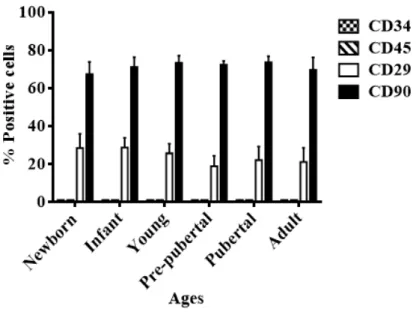
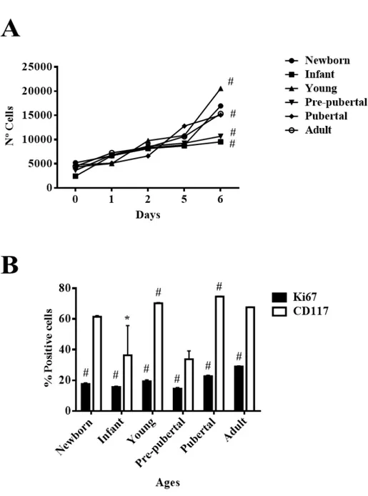
(16) 4. RESULTS 4.1 4.2 4.3 4.4 4.5 4.6 4.7 4.8 4.9 4.10 4.11 4.12 4.13 4.14 4.15. 43 Characterization of rBM MSCs Study of proliferation of rBM-MSCs at different ages Evaluation of biological capacity of rBM-MSCs at different ages Evaluation of pluripotency markers in rBM-MSCs at different ages Analysis of proteome in rBM-MSCs at different ages Mitochondrial function in rBM-MSCs in different age groups Glucolitic metabolism of rBM-MSCs at different ages mTOR pathway in rBM-MSCs at different ages Relationship of mTOR pathway with proliferation markers (CD117 and Ki67) in rBM-MSCs from adult group Transcriptome analysis using Next Generation Sequencing (NGS) of rBM-MSCs Analysis of pro-inflammatory potential in rBM-MSCs at different ages Characterization of rBM-MSC-derived EVs Detection of miRs relationship with Toll-like receptor in rBM-MSC-derived EVs at different ages miR-21-5p as regulator on pro-inflammatory and differentiation capacities of TLR4 in rBM-MSCs from pre-pubertal group Variation on rBM-MSC-derived EVs characteristic during aging. 47 47 49 51 51 56 58 60 61 67 72 74 77 79 85. 5. DISCUSSION. 93. 6. CONCLUSIONS. 103. 7. REFERENCES. 107. 8. SUPPLEMENTS. 123. 9. PUBLICATIONS. 143. 10 CURRICULUM VITAE.
(17)
(18) LIST OF FIGURES Figure 1.1. Definition of mesenchymal stem cells. 3. Figure 1.2. Several sources from MSCs. 4. Figure 1.3. Properties of MSCs. 6. Figure 1.4. Number and percentage of MSC-based clinical trials classified by disease type. 7. Figure 1.5. The Hallmarks of aging. 8. Figure 1.6. Extrinsic and intrinsic influences on stem cell aging. 11. Figure 1.7. Phenotype characterization of senescence MSCs. 12. Figure 1.8. Different types of EVs. 14. Figure 1.9. Protein composition of EVs. 15. Figure 1.10. Formation and sorting EVs. 17. Figure 1.11. Origin of EVs. 18. Figure 1.12. Process miRNAs biogenesis. 20. Figure 3.1. Workflow of iTRAQ-8plex. 37. Figure 3.2. Workflow of RNA-sequencing. 39. Figure 4.1. Characterization by flow cytometry. 47. Figure 4.2. Proliferation profile from rBM-MSCs at different age. 48. Figure 4.3. Evaluation of biological capacity of rBM-MSCs at different ages. 50. Figure 4.4. Pluripotency profile from rBM-MSCs at different ages. 51. Figure 4.5. Statistical analysis of iTRAQ-8plex. 52. Figure 4.6. iTRAQ of modulated proteins in rBM-MSCs. 53. Figure 4.7. Validation iTRAQ analysis. 55. Figure 4.8. Mitochondrial function in rBM-MSCs at different ages. 57. Figure 4.9. Glucolitic metabolism profile from rBM-MSCs at several ages. 59. Figure 4.10. mTOR pathway profile from rBM-MSCs at different ages. 60. Figure 4.11. Inhibition of proliferation markers (CD117 and Ki67) in rBM-MSCs from adult group. 62. Figure 4.12. PI3K/Akt pathway in rBM-MSCs from adult group treated with 5 µM or 10 µM IM and 0.1 ng/ml or 1 ng/ml JK184. 63.
(19) Figure 4.13. Level of p70S6k and AMPKα in rBM-MSCs from adult group treated with 5 µM or 10 µM IM and 0.1 ng/ml or 1 ng/ml JK184. 64. Figure 4.14. mTOR pathway in rBM-MSCs from adult group treated with 5 µM or 10 µM IM and 0.1 ng/ml or 1 ng/ml JK184. 65. Figure 4.15. mTOR pathway in rBM-MSCs from adult group treated with 5 µM or 10 µM IM and 0.1 ng/ml or 1 ng/ml JK184. 66. Figure 4.16. Proliferation markers (Ki67 and CD117) in rBM-MSCs from old group treated with 10 nM rapamycin. 67. Figure 4.17. Next Generation Sequencing study. 68. Figure 4.18. Metabolic pathways with statistically significant changes amog rBM-MSCs Part I. 70. Figure 4.19. Metabolic pathways with statistically significant changes amog rBM-MSCs Part II. 71. Figure4.20. Pro-inflammatory phenopype of rBM-MSCs at several ages. 73. Figure 4.21. Characterization of MSC-derived EVs. 75. Figure 4.22. NTA study of MSC-derived EVs at several ages. 76. Figure4.23. Pro-inflammatory profile of micro-RNAs contanined in MSC-derived EVs with age. 78. Figure 4.24. Effect of miR-21-5p on DAMPs and Nanog in mesenchymal stem cells from pre-pubertal group. 80. Figure 4.25. Effect of miR-21-5p on senescence and pro-inflammatory phenotype in rBM-MSCs from pre-pubertal group. 82. Figure 4.26. Effect of miR-21-5p on PI3K/Akt in rBM-MSCs from pre-pubertal group. 83. Figure 4.27. Effect of miR-21-5p on immune response in rBM-MSCs from pre-pubertal group. 84. Figure 4.28. Observation our in vitro model using fluorescence microscopy. 86. Figure 4.29. Nanog, Oct4 and Vinculin expression at genetic level in our in vitro model. 88. Figure 4.30. Analysis of isoforms of Lamin A using western-blot in our in vitro model. 90.
(20) Figure 4.31. Analysis of isoforms of Lamin A using western-blot in Our in vitro model. 91.
(21) LIST OF TABLES Table 1.1. Different types of EVs. 13. Table 8.1. List of antibodies to flow cytometry. 125. Table 8.2. List of antibodies to western-blot. 126. Table 8.3. Specific primers of differents rat genes for qRT-PCR. 127. Table 8.4. Specific primers of miRNAs for qRT-PCR. 127. Table 8.5. List of buffer to silver-stainning. 128. Table 8.6. List of modulated proteins (P<0.05) in rBM-MSCs at different ages classified according to their principal biological process using iTRAQ-8plex 129.
(22)
(23)
(24) ABBREVIATIONS AND ACRONYMS.
(25)
(26) µg µl µl/min µl/ml µM µm 11-β-HSD1 2D-LC-MALDI-TOF/TOF 53BP1 60S RP L10 60S RP L23 60S RP L24 60S RP L4 60S RP L6 60S RP L7 60S RP L9 6PGDH A Â A.U A/P aBM-MSCs AcN AD aEVs AMPK AMPKα aMSCs AP-1 Ar ATP bFGF BM-MSCs BMP-2 bp BSA C18-silice Ca2+ CD105 CD117 CD11b CD14 CD200 CD29 CD34. Micrograme Mililiter Microliter per minute Microliter per mililiter Micromolar Micrometer 11-βeta-hydroxysteroid dehydrogenase type 1 Two dimensional-liquid chromatography coupled offline to matrix-associated laser desorption ionization-time of flight Octomer-binding transcription factor 4 60S Ribosomal protein L10 60S Ribosomal protein L23 60S Ribosomal protein L24 60S Ribosomal protein L4 60S Ribosomal protein L6 60S Ribosomal protein L7 60S Ribosomal protein L9 6-phosphogluconate dehydrogenase, decarboxylating Adult Angström Arbitrary units Adult vs pubertal Bone marrow-mesenchymal stem cells from adult group Acetonitrile Adipogenic medium Mesenchymal stem cell-derived extracellular vesicles from adult group Adenosine monophosphate kinase Adenosine monophosphate kinase alpha Mesenchymal stem cells from adult group Activator protein-1 Alizarin red Adenosine triphosphate Basis fibroblast growth factor Bone marrow-mesenchymal stem cells Bone morphogenetic protein-2 bases pairs Bovine serum albumin Columne18-silice Calcium ion Endoglobin Mast/stem cell growth factor receptor Integrin alpha M Cluster of differentiation 14 or monocyte differentiation antigen CD14 Cluster of differentiation 200 or OX-2 membrane glycoprotein Integrin beta 1 Hematopoietic progenitor cell antigen cluster of differentiation 34.
(27) CD45 CD63 CD73 CD79α CD81 CD82 CD9 CD90 cDNA CH CID cm2 CM-CSF CMMs CNMs CO2. Protein tyrosine phosphatase, receptor type C Lysosome-associated membrane glycoprotein 3 5´-Nucleotidase Immunoglobulin associated alpha. Conf d DAMPs DAPI. Confidence Day(s) Damage-associated molecular pattern 4´,6-diamidino-2-phenylindole. DDR. DNA Damage Response Endoribonuclease Dicer-TAR RNA binding protein or helicase with RNase motif-TAR RNA binding protein 3-3´-diethylthiacarbocyanineiodide. Dicer-TRBP DiI DNA DROSHA RNase III. Target of the antiproliferative antibody 1 Metastasis suppressor Kangai-1 Motility-related protein Thy-1 cell surface antigen Complementary DNA Chondrogenic medium Collision-induced dissociation Square centimeter Granulocyte-macrophage colony-stimuling factor Células madre mesenquimales Células nai mesenquimais Carbon dioxide. Deoxyribonucleic acid Ribonuclease III enzyme. dsDNA ECM EDTA ESCRT ESCRT-0 ESCRT-3. Double-stranted DNA Extracellular matrix Ethylenediaminetetraacetic acid Endosomal sorting complex required for transport Endosomal sorting complex required for transport-0 Endosomal sorting complex required for transport-3. ESCRT-I ESCRT-II. Endosomal sorting complex required for transport-I Endosomal sorting complex required for transport-II Endosomal sorting complex required for transport-III et les autres personnes. ESCRT-III et al. EV EVs. FPKM G0. Extracellular vesicle Extracellular vesicles Fetal bovine serum False discovery rate Fragments per kilo base of exons per million Gap 0/Resting. G1. Gap 1. G2. Gap 2 phase. G6PDH GO. Glucose-6-phosphate-1-dehydrogenase Gene ontology. FBS FDR.
(28) GTPase Rab11 GTPase Rab7 GVHD Gβl h H1.5 H2B H2DCF-DA. Small GTPase rab11 Small GTPase rab7 Graft-versus-host disease Gbetal. H4 HDCF-DA. Histone variant H4 2´,7´-dichlorofluorescein diacetate Human embrionary stem cells Hepatocyte growth factor precursor High motility box 1 Human mesenchymal stem cells High-performer liquid chromatography Hypoxanthine-guanine phosphoribosyltransferase Horseradish peroxidase Homo sapiens. hESCs HGF HMGB1 hMSCs HPLC HPRT HRP hsa HSPs I. hour(s) Histone variant 1.5 Histone variant 2B 2´,7´-dichlorodihydrofluorescein diacetate. Heat shock proteins Infant. I/N ID IDO IFN IGFBP3 IGFBP4. Infant vs newborn Identification Indoleamine 2,3-dioxygenase 1 Type 1 interferon Insulin-like growth factor-binding protein 3 Insulin-like growth factor-binding protein 4. IGFBP7 IIS IL-1 IL-6 IL-8 ILV ILVs IM. Insulin-like growth factor-binding protein 7 Insulin/insulin-like growth factor signaling Interleukin-1 Interleukin-6 Interleukin-8 Intralumenal vesicle Intralumenal vesicles Imatinib mesylate Cyclin-dependent kinase inhibitor 2A, multiple tumor suppressor 1 loci. INK4/ARF loci iNOS iPSCs ISCT ISEV iTRAQ iTRAQ-8plex IU/ml kDa KO KRAS kV. Nitric oxide synthase Induced pluripotent stem cells International Society for Cellular Therapy International Society for Extracellular Vesicles Isobaric tag for relative and absolute quantitation Isobaric tag for relative and absolute quantitation-eightplex International units per mililiter Kilodalton(s) Knockout Kirsten rat sarcoma viral oncogene homolog Kilovoltie(s).
(29) LC-MALDI-TOF/TOF LDH LPS M M. Liquid chromatography coupled offline to matrix-associated laser desorption ionization-time of flight Lactate dehydrogenase Lipopolysaccharides. mg/ml MgCl2. Molar Mitosis Matrix-assisted laser desorption/ionization Monocyte chemoattractant protein-1 Miligrame per militer Magnesium chloride. MHC. Major histocompatibility complex. micro-ARNs microRNA microRNAs min miR. Micro-ácidos ribonucleicos. MALDI-TOF/TOF MCP-1. miRNA miRNAs miRs MM mM mm2 mRNA MSC-EV MSC-EVs MSCs mtDNA mTOR. Micro-RNA Micro-RNAs Minute(s) Micro-RNA Micro-RNA Micro-RNAs Micro-RNAs Modified Masson´s Milimolar Square milimeter MessengerRNA Mesenchymal stem cell-extracellular vesicle Mesenchymal stem cell-extracellular vesicles Mesenchymal stem cells. mTORC1 mTORC2 MVB MVBs. Mitochondrial DNA Mammalian target of rapamycin Mammalian target of rapamycin complex 1 Mammalian target of rapamycin complex 2 Microvesicular body Microvesicular bodies. N NAD(P)H NADP Nanog nanoHPLC ND NF-κB ng/ml NGF NGS nm nM nt NTA ºC. Newborn Nicotinamide adenine dinucleotide (phosphate) reductase Nicotinamide adenine dinucleotide phosphate Homeobox protein NANOG Nano-high-performer liquid chromatography Nodocazole Nuclear factor κappa-light-chain-enhancer of activated B cells Nanograme per militer Nerve growth factor Next Generation Sequencing Nanometer Nanomolar Nucleotide Nanoparticle tracking analysis º Celsius.
(30) Oct4 Or OS P P/PP P0 p53/p21 P70s6k p-Akt PBS PDDF PDGF PDIA1 PGE2 PI PI3K/Akt p-Mtor PP PP/Y pri-miRNAs. Octomer-binding transcription factor 4 Oil red Osteogenic medium Pubertal. PRKD1 qRT-PCR. rBM-MSCs Rex1. Protein kinase D1 Real time quantitative polymerase chain reaction Regulatory-associated protein of mTOR Retinoblastoma protein/cyclin-dependent kinase inhibitor 2A Rat bone marrow-mesenchymal stem cells Zinc finger protein 42 homolog. rhTGF-β3 Rictor RIN RISC RNA RNA Pol II. Recombinant human transforming growth factor-βeta 3 Rapamycin-insensitive companion of mTOR RNA integrity RNA-induced silencing complex Ribonucleic acid RNA Polymerase II. RNAi RNAs Rnase A RNA-seq rno RNU6 ROCKi ROS rpm rrTNFα RT s S S100. RNAinhibitor Ribonucleic acids Ribonuclease A RNA-sequencing Rattus novergicus. Raptor Rb/p16. Pubertal vs pre-pubertal Passage zero Tumor antigen p53/cyclin-dependent kinase inhibitor 1A Ribosomal protein S6 kinase beta-1 Phospho-Akt Phosphate-buffered saline Presence of characteristic enlarged Platelet-derived growth factor receptor A Protein disulfide-isomerase A1 Prostaglandin E2 Propidium iodide Phosphatidylinositol-3-kinase/Akt Phospho-mTOR Pre-pubertal Pre-pubertal vs young Long miRNA precursors. RNA, U6 small nuclear 1 Rho-associated protein kinase inhibitor Reactive oxygen species Revolutions per minute Recombinant rat tumor necrosis factor alpha Retrotranscption Second(s) Synthesis S100 calcium-binding protein.
(31) S100A4 S100A6 Saf O SASP SA-β-gal SDS-PAGE Sec1/Munc-18 Ser2448 Ser/thr SIPS SNAREs SOD-2 Sox2 SPRY229 St.Clara St.Louis T T cells TAB2 TAK1 TBS TBST TCEP TEAB TFA. S100 calcium-binding protein A4 S100 calcium-binding protein A6 Safranine O Senescence-associated Secretory Phenotype Senescence-associated β-galactosidase SDS polyacrylamide gel Protein transport protein SEC1/ Mammalian uncoordinated-18 Serine2448 Serine/threonine Stress-induced premature senescence (Soluble N-ethylmaleimide sensitive fusion proteins Attachment Protein) Receptors Superoxide dismutase-2 (Sex determining region Y)-box 2 SPRY domain-containing SOCS box protein 3 Saint Clara Saint Louis Temperature Tregs cells TGF-βeta activated kinase 1/MAP3K7 Binding Protein 2 TGF-βeta activated kinase 1 Tris buffered saline Standard buffer tris buffered saline with 0.1% (v/v) Tween® 20 Tris-(2-carboxyethy) phosphine Tryethylammonium bicarbonate Trifluoroacetic acid. TGF-β TGF-β1 TIMP-2 TLR. Transforming Growth Factor-beta Transforming growth factor-beta1. TLR4 TMRM. Toll-like receptor type 4 Tetramethylrhodamine methyl ester. TNF TSAP6. Tumour necrosis factor Tumour suppresour-activated pathway 6. Tsg101 V. Tumor susceptibility gene 101 protein Voltie(s). v/v VEGF w/v Wnt Wnt5a xg Y Y/I yBM-MSCs. Volume/volume Vascular endothelial growth factor Weight/volume Wingless-type MMTV integration site family Wnt family member type 5A G-force or relative centrifugal forces Young Young vs infant Bone marrow-mesenchymal stem cells from young group Mesenchymal stem cell-derived extracellular vesicles from young group. yEVs. TIMP metallopeptidase inhibitor-2 Toll-like receptor.
(32) yMSCs Y-RNA α-ciano β-actin γH2AX Δratio ΔΔCt. Mesenchymal stem cells from young group Small non-coding RNA Alpha-ciano beta-actin Histone variant gamma H2AX Differential of ratio Delta(delta(threshold cycles)).
(33)
(34) 1. INTRODUCTION.
(35)
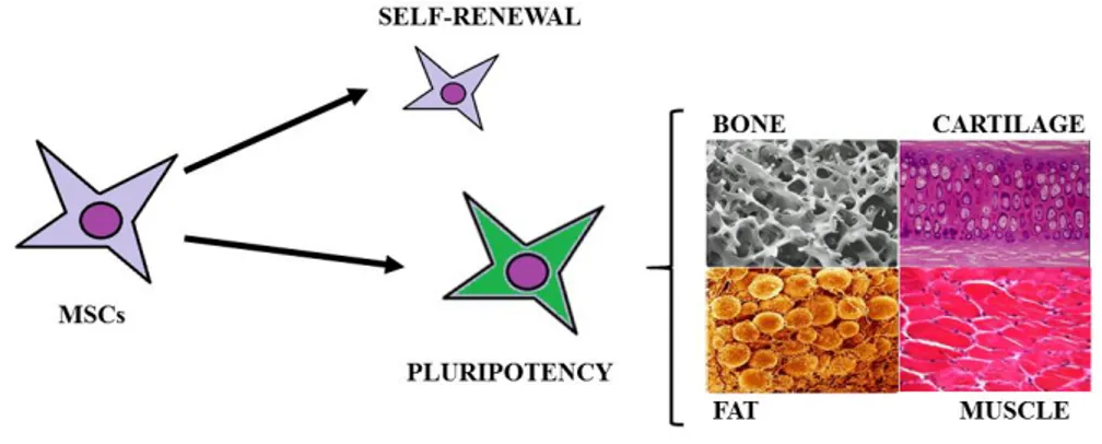
(36) 1. INTRODUCTION. 1.1. Mesenchymal stem cells. 1.1.1 Definition Mesenchymal stem cells (MSCs) are multipotent fibroblast-like cells that can be found in almost all tissues and they can differentiate into bone1, cartilage2, muscle3, tendon, ligament4, fat5, and a variety of other connectives tissues6,7. MSCs were first reportein in 1968 by Friendenstein et al.8 when human bone marrow cells were cultured in plastic dishes colonies of adhered fibroblastoid cells proliferative. MSCs are adult stem cells which have a great self-renewal capacity, is the process by which a stem cell divides asymmetrically or symmetrically to propagate one or two daughter stem cells with similar development potential as the mother cells 9 while maintaining pluripotency, namely the capacity to self-maintained in undifferentiated state10 (Figure 1.1). The International Society for Cellular Therapy (ISCT) suggested the following criteria for the identification of MSCs11: . Adherence to plastic. Differentiation into chondrocytes, osteoblasts and adipocytes under standard in vitro differentiating conditions. Expression of surface markers CD105, CD73, CD29 and CD90, in the absence of CD45, CD34, CD14, CD11b, CD79α.. Figure 1.1. Definition of mesenchymal stem cells.. 1.1.2 Sources for mesenchymal stem cells MSCs have been isolated from many different adult tissues, including bone marrow12, adipose tissue13, synovial membrane14, connectives tissues of dermis15, skeletal muscle16, peripheral blood17, liver18, lung19 and blood vessels20 and from rather “young sources” such as amniotic fluid21, amniotic membrane22, umbilical cord blood23, umbilical cord stroma24, or placenta25. In the last years the number of tissues with a potential for tissue engineering has increased6,26 (Figure1.2).. -3-.
(37) 1. INTRODUCTION. Figure 1.2. Several sources from MSCs.. Therefore, there are more studies about differences at cellular and molecular levels such as, cell morphology, surface markers27, differentiation28, proliferation27,1, transcriptomic29 and proteomic30 analyses among MSCs from several tissue sources.. 1.1.3 Properties of mesenchymal stem cells supporting therapeutic application To date, the clinical use of stem cells presents some disadvantages because stem cells from certain sources, such as somatic nuclear transfer, embryo destruction, or even induced pluripotent stem cells (iPSCs) obtained by reprogramming have raised deep ethical issues depending on the country work31,32. Besides, they could produce neoplastic disorders and immunologic rejection when they are injected in in vivo models32. For these reasons, MSCs are a good alternative because they don´t produce immunologic disorders due to their autologous origin and there are not ethical issues about their clinical use31. Additionally MSCs posses the following properties: 1.1.3.1. Differentiation. MSCs can differentiate both in vivo and in vitro, into various mesenchymal cells and exhibit remarkable plasticity given their ability to trans-differentiate, or undergo an abrupt alteration in phenotype, thereby giving rise to cells possessing the characteristics of different lineages33,34.. -4-.
(38) 1. INTRODUCTION. 1.1.3.2. Paracrine effects and immunomodulation. It is the capacity of MSCs to secrete a wide variety of cytokines, chemokines, and growth factors. Several studies based on examination of MSCs secretome in vivo and the strategies to modulate the secretion of molecules of MSCs have identified high levels of proteins involved in immune response such as interleukin-6 (IL-6), IL-8, monocyte chemoattractant protein (MCP-1), and transforming growth factor-β (TGF-β); extracellular matrix remodelers like TIMP metallopeptidase inhibitor 2 (TIMP-2), fibronectin, periostin, collagen, decorin, metalloproteinase inhibitors; growth factors and their regulators such as vascular endothelial growth factor (VEGF), granulocyte-macrophage colony-stimulating factor (CM-CSF), bone morphogenetic protein 2 (BMP-2), basis fibroblast growth factor (bFGF), and insulin-like growth factor-binding protein 3 (IGFBP3), IGFBP4, IGFBP735. Also, MSCs can the modulate immune response system and they were effective for treatment of various immune response disorders in both human and animal models36–38. However, the underlying mechanism of that modulation is not fully understood. The most accredited theory points to the important cell-to-cell contact and/or the release of soluble immunosuppressive factors. They interacted with a broad range of immune cells and displayed an ability to suppress the excessive response of T and B cells, dendritic cells, macrophages and natural killer cells39,38. Besides, MSCs can also induce regulatory T cells (Tregs) and maintain the capability of Tregs to suppress self-reactive T-effector responses40 (Figure 1.3). In the last years, it was proposed that MSCs interact with their environments both by negatively regulating the immune response in the case of major inflammation and by stimulating the immune response system by releasing pro-inflammatory molecules if the level of inflammatory cytokines is low 41. 1.1.3.3. Homing mechanism. The homing mechanism of MSCs lies in their ability to reach damaged tissue in response to a correct combination of signalling molecules from the injured tissue and corresponding receptors. Homing-related molecules in general can be upregulated by inflammatory cytokines such as tumour necrosis factor (TNF) and IL142, suggesting that different inflammation states might promote distinct MSC engraftment and therapeutic efficiencies43 (Figure 1.3).. -5-.
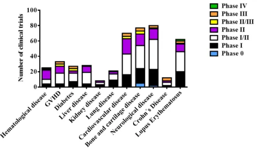
(39) 1. INTRODUCTION. Figure 1.3. Properties of MSCs. From http://www.iem.cas.cz/research/departments/transplantation-immunology.html at June 30, 2016.. 1.1.4 Applications in clinical use MSCs have emerged as a novel strategy to therapeutic application of the US National Institute of Health 493 MSC-based clinical trial have been reported as of June 15, 2015; most were performed to evaluate the biomedical potential of MSCs in treating haematological diseases including Graft-versus-host disease (GVHD), diabetes, inflammatory diseases, and disease in the liver, kidneys, and lungs, as well as cardiovascular, bone and cartilage, neurological (Figure 1.4). MSCs have the ability to differentiate into several mesenchymal linages27 and contribute to the replacement of the damaged tissue, but rather act as trophic mediators, promoting tissue repair by production and release of soluble factors that inhibit inflammation, reduce fibrosis, and induce angiogenesis44 among other functions. Phases of investigation of 493 MSC-based clinical trials and the most representative treated pathologies are shown in the figure 1.4 According to these data, most clinical trials occurred in an early phase (phase I, I/II, or II), demonstrating that more investigation about the therapeutic effectiveness of MSCs is required. Several studies indicate that donor heterogeneity, ex vivo expansion, immunogenicity, and cryopreservation can be considered the Achilles´-heel of MSC-base therapies. Therefore, it is necessary that researchers and clinical discoveries will address the mechanisms influencing their therapeutic use.. -6-.
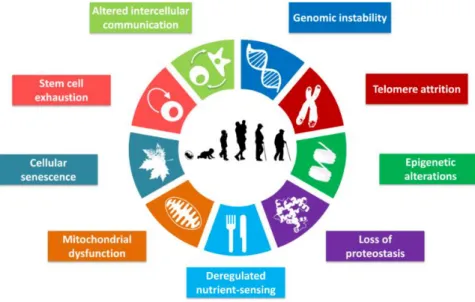
(40) 1. INTRODUCTION. Figure 1.4. Number and percentage of MSC-based clinical trials classified by disease type. Data from www.clinicaltrial.gov at June 30, 2015.. 1.2. Aging. 1.2.1 Definition Aging is the decline in the homeostasis and regenerative capacity of all tissues and organs and it is the greatest risk factor for the development of chronic diseases which comprise the majority of global disease burden and are the most common causes of mortality. Mammalian aging can be delayed with genetic, dietary, and pharmacological approaches. Given that the elderly population is dramatically increasing. (http://www.healthmetricsandevaluation.org/data-visualizations at July 9, 2016). Actually, in the world the number of people aged 65 or older will outnumber children under age 5. Driven by falling fertility rates and increasing in life expectancy, population aging will continue, even accelerate. The number of people aged 65 or older are projected to grow from an estimated 1.5 billion in 2050, with most of the increase in developing countries45. Actually, there is an urgent need to extend healthspan.. 1.2.2 The hallmarks of aging Defining the causes of aging is a difficult work because it is impeded by the complexity of the phenotype coupled with the costs and duration of longevity studies but in recent years, progress has accelerated, bringing geroscience to the forefront.. -7-.
(41) 1. INTRODUCTION. Firstly, genetic and environmental interventions and pathways that regulate longevity were studied in yeast (particularly the budding yeast, Saccharomyces cerevisiae)46,47 and invertebrate model organisms, such as Caenorhabditis elegans47,48. Conserved molecular pathways impacting aging have been identified, such as insulin/ insulinlike growth factor signalling (IIS). Secondly, mammalian studies have generated a more detailed understanding of age-associated pathologic changes. Additionally, studies about effects of environmental interventions on the lifespan have been performed. Those studies have revealed key genes and pathways for cellular ageing such as IIS and the mammalian target of rapamycin (mTOR) which are implicated in mediating the effects of dietary restriction49,50. In the last years, López-Otín et al.51 have identified and categorized the cellular and molecular hallmarks of aging. These hallmarks are not independent factors driving aging; rather, they were highly intertwined processes, and understanding the interplay among them is critical (Figure 1.5).. Figure 1.5. The Hallmarks of aging. From López-Otín et al.51.. 1.2.2.1. Genomic instability. The accumulation of genetic damage throughout life is the most studied cause of aging52. In this hallmark are included several areas of aging research such as mechanisms for the maintaining the appropriate length and functionality of telomeres53,54 and for ensuring the integrity of mitochondrial DNA (mtDNA) 55, as well as defects in the nuclear architecture known as laminopathies which causes premature aging syndromes56.. -8-.
(42) 1. INTRODUCTION. 1.2.2.2. Telomere attrition. Telomere attrition is associated with aging in mammal models because pathological telomerase dysfunction accelerates aging in mice and humans57, while experimental stimulation of telomerase can delay aging in mice58. 1.2.2.3. Epigenetic alterations. This hallmark contains a variety of epigenetic alterations affecting lifespan of cells and tissues such as DNA methylations patterns59, post-translational modification of histones60,61 and chromatin remodelling62,63. The important goals of this hallmark are biomarker development between chronological age and biologic aging, link agerelated environment inputs to epigenetic signatures and test small molecules that regulate enzymes controlling epigenetic events48,64,65. 1.2.2.4. Loss of proteostasis. Proteostasis involves mechanisms for the stabilization of correctly folded proteins, most prominently the heat-shock family of proteins, and mechanisms for the degradation of proteins by the proteasome or the lysosome66,67. Many studies have demonstrated that proteostasis is altered with aging66, also chronic expression of unfolded, misfolded or aggregated proteins contribute to the development of some age-related pathologies, such as Alzheimer´s disease, Parkinson´s disease and cataracts68. Some important studies to advance this hallmark through identification of proteostasis pathways that are owerwhelming in specific chronic disease states, examination crosstalk between proteostasis machinaries and understand non-cellautonomous signalling and activation of proteostasis pathways. 1.2.2.5. Deregulated nutrient-sensing. There are several conserved pathways related to deregulated nutrient-sensing such as IIS pathways, which is related to metabolism of glucose in the cells69, mTOR pathway which is also involved in anabolism metabolism and aging50,70 and AMPK and sirtuins, acting in the opposite direction to IIS and mTOR71. This hallmark have important focus as determination role of signal transduction pathways linked to metabolism in the aging process and pharmacological manipulation that mimics a state of limited nutrient availability like rapamycin 72. 1.2.2.6. Mitochondrial dysfunction. This detrimental process has been long associated with aging because the efficacy of the respiratory chain tends to diminish with increasing age, thus increasing electron leakage and reducing ATP generation73. But. there are less clear different aspects for example, knowledge about bridge continuum from physiological to molecular stresses, differentiation between toxic stress and hormesis, is the mechanism to response to harmless doses of toxins and other stressors. It could -9-.
(43) 1. INTRODUCTION. constitute to one of the mechanisms that allows stressed cells to avoid senescence and death74. 1.2.2.7. Cellular senescence. It is a stable arrest of the cell cycle coupled to stereotyped phenotypic changes 75. This phenomenon was described by Hayflick76. To date, it is known that the senescence observed by Hayflick et al.76 is caused by telomere shortening, but there are other aging-associated stimuli that trigger senescence independently of telomeric process77,78. The most famous non-telomeric DNA damage is derepression of INK4/ARF locus75. The controversy of cell senescence as a beneficial compensation on aging and cancer or deleterious and accelerate aging. 1.2.2.8. Stem cell exhaustion. The most obvious characteristic of aging is the decline in the regenerative potential of tissues. For example, hematopoiesis declines with age, resulting in a decreasing production of adaptive immune cells, a process termed immunosenescence 79. The important goals in this hallmark are: . To determine whether declining adult stem cell function drives to aging and chronic disease. To examine how aging and associated diseases impair adult stem cell function. To define how macromolecular damage accumulates in aging adult stem cells pools.. 1.2.2.9. Altered intercellular communication. A prominent aging-associated alteration in intercellular communication is “Inflammaging”, which may result from multiples causes such as the accumulation of pro-inflammatory tissue damage, the failure of an ever more dysfunctional immune system to effectively clear pathogens and dysfunctional host cells, the propensity of senescent cells to secrete pro-inflammatory cytokines through activation of the NF-kB transcription factor, chemokines and extracellular matrix (ECM) remodelling proteases, is named the senescence-associated secretory phenotype (SASP)80,81. Also, there are other processes related to cellular communication which induce senescence in neighbouring cells via gap junction-mediated cell-cell contacts and processes involving ROS82.. 1.2.3 Mechanisms of aging on mesenchymal stem cells Aging is associated with a marked decline in functionalities of adult stem cells, namely tissue homeostasis, repair and regeneration83. - 10 -.
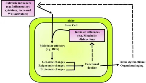
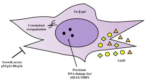
(44) 1. INTRODUCTION. The combination of cell-intrinsic changes leads to decline in cellular function, which in turn contributes to tissue dysfunction and organism aging (Figure 1.6). The intrinsic changes are: . . . Genomic changes: include accumulation measurable genomic lesions, including single- and double-strand DNA breaks, chromosomal translocations, telomere shortening84. Epigenetic changes: include DNA methylation and post-translation modification of histones, are dynamically maintained by a balance among chromatin-remodeling complexes and, thus, reversible85 and altered expression of cofactors of histones86. Proteomic changes: maintenance of the intracellular proteome requires timely removal of improperly folded or damaged proteins that can otherwise impede normal cellular function66.The machineries and cellular processes, which maintain protein homeostasis are autophagosomes, chaperones, lysosomes and the ubiquitin-proteosome system68.. Also the extrinsic influences, such as inflammatory cytokines and Wnt activators influence in the aging process of the tissue and organism. The niche is profoundly influenced by the systemic milieu and dynamically changing to regulate stem cell function, a feature that is especially relevant with regard to the process aging 87 (Figure 1.6).. Figure 1.6. Extrinsic and intrinsic influences on stem cell aging. Adapted from Liu et al.88.. MSCs have been reported to be highly resistant to apoptosis induced by different genotoxic insults and preferentially respond to injury with activation of stress-induced premature senescence (SIPS), which had been widely studied in MSCs, particularly for its clinical implications89. Also, it was demonstrated that the senescence activation pathway in MSCs is independent of the tissue source89–92. - 11 -.
(45) 1. INTRODUCTION. Senescent MSCs activate p53/p21 and Rb/p16 pathways to block the cell cycle and sustain growth arrest but they continue to be metabolically active93. The cells in a senescent state are characterized by a large, flat morphology, display changes in gene expression, typically exhibit a senescence-associated β-galactosidase (SA- β-gal), for persistent DNA damage response (DDR) activation, as highlighted by the presence of characteristic enlarged (PDDF), containing γH2AX and 53BP1 foci94 and SASP81,91 (Figure 1.7).. Figure 1.7. Phenotype characterization of senescence MSCs. Adapted from Turinetto V et al.95. Senescence impacts on migratory ability96,97, differentiation potential98, immunomodulation ability97, loss of proliferation capacity and tumour progression92.. 1.3. Extracellular vesicles 1.3.1 Definition Extracellular vesicles (EVs) are membrane-contained vesicles released in an evolutionally conserved manner by cells ranging from organisms such as prokaryotes. During the past decades, EVs have been recognized as potent vehicles of intercellular communication in different model systems with respect to other cell-to-cell communication strategies, such as quorum sensing, juxtacrine signalling, autocrine signalling, paracrine signalling, endocrine signalling and direct cell-to-cell communication (desmosomes, adherents and gap junctions).. - 12 -.
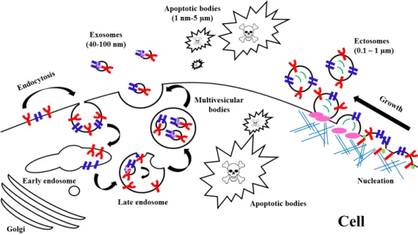
(46) 1. INTRODUCTION. 1.3.2 Types of extracellular vesicles EVs can be isolated from all types of body fluids including blood, urine, bronchoalveolar lavage fluid, breast milk, amniotic fluid, synovial fluid, pleural effusions and ascites99 and from several cell types100–104. The term EVs comprise a highly heterogeneous and dynamic group of nanoparticles. Therefore, International Society of Extracellular Vesicles (www.isev.org)105 have promoted the collaboration work since 2011 by the members to unify the nomenclature and the methodologies of EVs by the contents, size, membrane composition, cellular source, state and environmental conditions. Actually, three main subgroups of EVs have defined depend on size, sucrose gradient and origin (Table 1.1 and Figure 1.8). Vesicle. Size (Diameter)/nm. Sucrose gradient/ Origin g.ml-1. Luminal budding into MVBs Exosomes. 40-100. 1.13-1.19. Release by fusion of MVB. with. cell. membrane Microvesicles Microparticles. 50-1000. 1.04-1.07. 1-5000. 1.16 and 1.28. Ectosomes Apoptotic bodies. Table 1.1 Different types of EVs. Adapted from Rani et al.106.. - 13 -. Outward budding of cell membrane Outward. blebbing. of. apoptotic cell membrane.
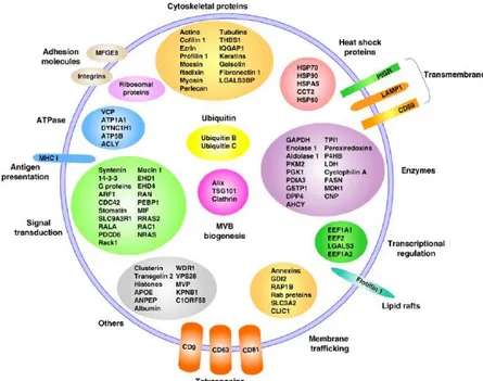
(47) 1. INTRODUCTION. Figure 1.8. Different types of EVs. Adapted from Cocucci et al.107. 1.3.3 Composition of Extracellular Vesicles Public on-line databases that catalogue EV-associated components, are available. These include Vesiclepedia (www.microvesicles.org/)108, EVpedia 109 110 (www.evpedia.info) and ExoCarta (www.exocarta.org) .. 1.3.3.1. Protein and protein-associated of EVs. Protemic studies of EVs released by primary cell cultures, cell lines, tissue cultures or isolated from biofluids have yielded extensive number of protein abundance in different types of EVs. In general, EVs are highly abundant in cytoskeletal, cytosolic, heat shock, plasma membrane proteins and proteins involved in vesicle trafficking. Also there are some studies where it have identifying some markers of EV subpopulations that are often used as markers, such as tetraspanins (CD9, CD63, CD81 and CD82) which are considered marker of exosomes, 14-3-3 protein, major histocompatibility complex (MHC) molecules and heat shock proteins (HSPs), Tsg101 and the Endosomal Sorting Complex Required for Transport (ESCRT-3) binding protein a Alix which are considered marker of exosomes111– 113 (Figure 1.9).. - 14 -.
(48) 1. INTRODUCTION. Figure 1.9. Protein composition of EVs. From Mathivanan et al.114.. Several studies reported about changes in the glycosylation patterns of EVs115–118, protein signature of different EVs which may be involved in biogenesis, sorting119,120, uptake110 and EV-associated cytokines121,122 (Figure 1.9). 1.3.3.2. RNA composition. EVs contain intact mRNA123, mRNA fragments124, long non-coding RNA125,126, miRNA127,128, piwi-interacting RNA125, ribosomal RNA125 and fragments of tRNA, vault- and Y-RNA129,130. It occurred an increased of studies about activity of RNA in EVs because they are more enriched in EVs with respect to parental cells101. EVs contained RNAs are involved in cell differentiation131–133, proliferation132,134, immune regulation135, modulation stress134 condition and other117,136–138. 1.3.3.3. DNA contain. The study of the DNA contained in EVs represents a relatively new approach to the field. Oncogenic DNA was found in apoptotic bodies139. Also, mitochondrial DNA (mtDNA), single-stranded DNA, double-stranded DNA (dsDNA) and oncogene amplifications have been detected in EVs140,141. 1.3.3.4. Lipid composition. EVs are generally enriched in sphingomyelin, cholesterol and glycosphingolipids similar to raft domain142. Some studies reported that the specific lipid that confers the stability of EVs may be used to improve liposomal drug delivery systems143,144, sorting, biogenesis142,145.. - 15 -.
(49) 1. INTRODUCTION. 1.3.4 Formation and sorting EVs 1.3.4.1. Exosome biogenesis. The membrane of late endosomes invaginates and forms small vesicles that are pinched off into the endosomal space. These are the intralumenal vesicles (ILVs) and the whole is the MVE. Notice that the internal face of an ILV membrane corresponds to the cytoplasmic face of the endosome limiting membrane, and the content of the ILV is originated from the cytosol prior to ILV formation. A set of MVEs fuse their limiting membranes to the plasma membrane and the ILVs with their cargo into the extracellular space146. Formation of ILVs in the late endosome involves the endosomal sorting complex required for transport (ESCRT) proteins. ESCRT proteins are components of four ESCRT complexes, ESCRT-0, ESCRT-I, ESCRT-II, and ESCRT-III. Each of these complexes is sequentially and transiently recruited to the forming MVE until a vesicle is fully shaped and released as an ILV into the endosomal space 146,147 (Figure 1.10). However, increasing evidences about the key role of some lipids such as ceramide in ILV formation, independently of ESCRT complexes146. As mentioned above, a set of MVE fuses with the plasma membrane while other MVEs follow a degradative route and fuse with lysosomes (Figure 1.10). Some studies identified the existence of different populations of MVEs: . MVEs rich in GTPase Rab7 and ILVs containing phosphatidylinositol-3phosphate and ubiquitinated proteins are sorted to lysosomes148. MVEs rich in GTPase Rab11 and ILVs with high amounts ceramide are sorted for exosome secretion149,150.. - 16 -.
(50) 1. INTRODUCTION. Figure 1.10. Formation and sorting EVs. Membrane-associated (triangles) and transmembrane proteins (rectangles) and RNAs (curved symbols) are selectively incorporated into the ILV of MVEs or into MVs budding from the plasma membrane. MVEs fuse with the plasma membrane to release exosomes into the extracellular milieu. MVs and exosomes may dock at the plasma membrane of a target cell (1). Bound vesicles may either fuse directly with the plasma membrane (2) or be endocytosed (3). Endocytosed vesicles may then fuse with the delimiting membrane of an endocytic compartment (4). Both pathways result in the delivery of proteins and RNA into the membrane or cytosol of the target cell. Fusion and endocytosis are only represented for exosomal vesicles, but plasma membrane-derived MVs may have similar fates. From Raposo et al.151.. MSC releases EVs differently depending on the external stimulation, such as hypoxia and inflammatory152. Tumour suppressor-activated pathway 6 (TSAP6) is found regulate EV formation153 and this pathway is regulated by p53 thereby enhancing EV production154,155. 1.3.4.2. Microvesicles biogenesis. Microvesicles result from outward budding and fusion of the plasma membrane. Membrane budding initiated by the activity of aminophospholipid translocase, responsible for placing phosphatidylserine to the outer membrane. ADPribosylation factor 6 plays an important role in enabling MV budding156,157 and contractile protein myosin light chain kinase 2 in release of MVs157–159 (Figure 1.11).. 1.3.5 EVs uptake 1.3.5.1. Endocytosis. It is the most evidence process of internalization because EVs are usually taken up into endosomal compartment via endocytosis. It was identified inside cells from as early as 15 minutes after initial introduction160,161. By using a range of inhibitor to block specific pathways and other experimental techniques such as - 17 -.
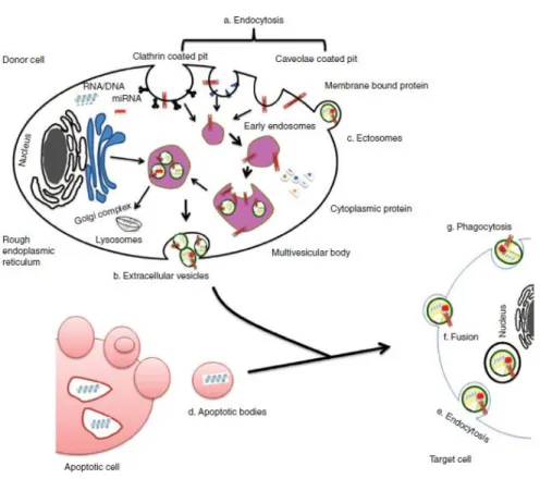
(51) 1. INTRODUCTION. RNAinhibitor (RNAi) to knockdown certain genes the role of the endocytosis processes responsible for EV uptake and they found several subtypes of endocytosis, such as clathrin-mediated endocytosis117,162,163, caveolin-dependent endocytosis162,164–167, macropinocytosis168, phagocytosis168,169 and involvement lipid raft170 (Figure 1.11). 1.3.5.2. Cell surface membrane fusion. It is via direct fusion between the EV membrane with cell plasma membrane171. Several proteins participate in this process including SNAREs, Rab proteins and Sec1/Munc-18 related proteins150,172,173. 1.3.5.3. Cell specific EV uptake. Results from some studies show that fluorescently labelled EVs can be taken up by virtually every cell type tested174,175, whereas other suggest that vesicular uptake is a highly specific process which can only occur if cell and EV share the right combination of ligand and receptor.. Figure 1.11. Origin of EVs. It is generally via (a) endocytosis or inward budding of plasma membrane that consist of lipid rafts and is mediated by clathrin-dependent or caveolae-dependent pathway, This gives rise to (b) early endosomes leading to the formation of numerous ILVs within a membrane maturing to MVBs. Finally MVBs fuse with plasma membrane releasing ILVs as exosomes. (c) Ectosomes are vesicles shed from the cell surface and (d) apoptotic bodies are also known as apobodies and are released by cells undergoing apoptosis. EVs are internalized by the target cells through several pathways including (e) endocytosis, (f) fusion, and (g) phagocytosis. From Rani et al.106.. - 18 -.
(52) 1. INTRODUCTION. 1.3.6 Applications in clinical use Recent animal model-based studies suggest that EVs have significant potential as a novel alternative to whole cell therapies106,176 and they were used to discovery biomarkers of diseases177,178. Compared to MSC, EVs may have a superior safety profile and can be safely stored without losing function106 but it is necessary to advance to clinical use of MSC-EVs for common human diseases because there are unresolved questions, such as definition, standardisation, cost-effective production, optimal dosing and, most importantly, safety. There are more studies about therapeutic effects of MSC-derived EVs in cardiovascular disease179, acute kidney injury135, liver disease180, lung diseases181, cutaneous wound healing182, Alzheimer´s disease183 and drug delivery143,184.. 1.4 miRNAs 1.4.1 Definition miRNAs are small, noncoding RNAs, 19-24 nucleotides in length, which regulate gene expression post transcriptionally185.. 1.4.2 Biogenesis of miRNAs miRNA biogenesis pathway starts in the nucleus186. Firstly, they are transcribed by RNA polymerase II (RNA Pol II) as an approximately 70-nucleotide (nt) long stemloop primary structure named primary-miRNA transcripts, pri-miRNAs (long miRNA precursors), which are processed by DROSHA RNase III enzyme into precursors to generate pre-miRNAs structure185. Finally, the two strands of the duplex are separated from each other by the Dicer– TRBP complex. Next, the RNA-induced silencing complex (RISC), which also consists of the Argonaute protein and the target mRNA, is complementary bound by specific miRNAs. Consequently, the target mRNAs translation is repressed resulting in translational silencing or induction of mRNA degradation by RNases187 (Figure 1.12).. - 19 -.
(53) 1. INTRODUCTION. Figure 1.12. Process miRNAs biogenesis. From Jin Jung et al.188. 1.4.3 miRNAs in EVs Secretion of miRNAs in EVs is a novel form of the intercellular communication. However, the mechanisms governing miRNA and miRNA-binding protein secretion into EVs remain largely unknown. Recently, mutant KRAS shown to regulate the content of RNA-binding protein in EVs189,190.. 1.4.4 Role in biological process miRNAs have crucial regulatory roles in development of hematopoietic linage, maturation, and differentiation of B,T lymphocytes191 and MSCs192, proliferation of neutrophils, monocytes193,194 and MSCs195, secretion of type 1 interferon (IFN) and inflammatory cytokine/chemokine193, and effectiveness of immune system response196,194, immunosenescence 197, inflammaging198, cancer199 and other200. In the last years, miRs have been suggested possible therapeutic approaches for agerelated life-threatening diseases201,202.. - 20 -.
(54)
(55)
(56) 2. HYPHOTESIS AND AIMS.
(57)
(58) 2. HYPOTHESIS AND METHODS. Mesenchymal stem cells have self-renewal capacity and multiple differentiation potentials, and a priori, could play important roles in regenerative medicine but the promising role of MSCs in cell-based therapies and tissue engineering appears to be limited due to a decline of their regenerative potential with increasing donor age. For that, we proposed the following aims to understand whether aging affects the properties of MSCs: 1. Determination of proliferation profile of rat bone marrow mesenchymal stem cells at different ages. 2. Determination of pluripotency profile of rat bone marrow mesenchymal stem cells at different ages. 3. Proteome and transcriptome descriptive study of rat bone marrow mesenchymal stem cells at different ages. 4. Pro-inflammatory phenotype of rat bone marrow mesenchymal stem cells at different ages. 5. Characterization of rat bone marrow mesenchymal stem cell-derived extracellular vesicles at different ages. 6. Evaluation of relationship between miRNAs and Toll like receptor 4 pathway in rat bone marrow mesenchymal stem cell-derived extracellular vesicles at different ages. 7. Effect of miR-21-5p on pro-inflammatory and pluripotent capabilities from Tolllike receptor 4 in rat bone marrow mesenchymal stem cells. 8. Influence of rat bone marrow mesenchymal stem cells-derived extracellular vesicles on their self-renewal using an in vitro model.. - 25 -.
(59)
(60) 3. MATERIAL AND METHODS.
(61)
(62) 3. MATERIAL AND METHODS. 3.1. Isolation and culture of rBM-MSCs. The animals were euthanized with Fluorane (Izasa, A Coruña, Spain) and sacrificed by cervical dislocation method. Femurs were dissected from male Wistar rats (Animal Service, Complejo Hospitalario Universitario de A Coruña, Spain) at different ages: newborn (0 days old), infant (7 days old), young (14 days old), pre-pubertal (35-38 days old), pubertal (45 days old) and adult (+2 months old). All methods were carried out in “accordance” with the approved guidelines of the Spanish law (32/2007). All experimental protocols were approved by The Animal Ethical Committee of Galicia. The protocol used by Karaoz et al.12 was followed in this work. Briefly, the end of the bones were cut away and a 21-gauge needle that was inserted into shaft of the bone marrow was extruded by flushing with 5 ml D-Hank´s solution supplemented with 100 IU/ml penicillin-100 mg/ml streptomycin (all from Life Technologies, Madrid, Spain). Marrow plug suspension was dispersed by pipetting, successively filtered through 70-µm mesh nylon filter (BD Biosciences, Bedford, United States) and centrifuged at 2000 xg for 10 minutes. Supernatant containing thrombocytes and erythrocytes was discarded, and the cell pellet was re-suspended in the RPMI supplemented with 10% (v/v) fetal bovine serum (FBS), 1% (v/v) penicillin, 1% (v/v) streptomycin (all from Life Technologies, Madrid, Spain). The cells from four rats were seeded into 100 cm2 dish plate (Corning Inc., New York, United States) and incubated at 37ºC with humidified atmosphere 5% CO 2. rBMMSCs were isolated on the basis of their ability to adhere to culture plates. On the third day, red blood cells and other non-adherent cells were removed by the pre-plating technique and fresh medium was added to allow further growth. The adherent cells grown to 70% confluence were defined passage zero (P0) cells. After 5 min of centrifugation, 1x10⁶ rBM-MSCs were seeded on two 100 cm2 dish plates (Corning Inc., New York, United States) in RPMI supplemented with 10% (v/v) FBS, 1% (v/v) penicillin and 1% (v/v) streptomycin (all from Life Technologies, Madrid, Spain). The culture medium was added and replaced every 3 or 4 days for 2 weeks. rBM-MSCs have been expanded for 2 passages to use in the following techniques.. 3.2. Characterization of rBM-MSCs by flow cytometry. To characterize the populations of rBM-MSCs from chronologically different animals, their rBM-MSCs were washed twice in phosphate-buffered saline (PBS) (MP Biomedicals, Illkrich, France), then pre-blocked with 2% (v/v) rat serum (Life Technologies, Madrid, Spain) in PBS (MP Biomedicals, Illkrich, France). The following direct antibodies were used at different dilutions and wavelenght detection windows (Table 8.1) to check mesenchymal and hematopoietic markers of the different populations of rBM-MSCs from chronologically different animals. 2x10⁵ cells were analyzed by FACSAria flow cytometer (BD Science, Madrid, Spain). FACS data was generated by DIVA software (BD Science, Madrid, Spain).. - 29 -.
(63) 3. MATERIAL AND METHODS. 3.3. Proliferation analysis by flow cytometry. rBM-MSCs from adult cultured with medium RPMI supplemented with 10% (v/v) FBS, 1% (v/v) penicillin, 1% (v/v) streptomycin (all from Life Technologies, Madrid, Spain) with 10 nM rapamycin (Sigma-Aldrich, St.Louis, United States) for 2 days. After incubation with the drug, the cells were washed with PBS (MP Biomedicals, Illkrich, France), then fixed in 4% (w/v) (Sigma-Aldrich, St.Louis, United States) for 10 min. After the fixation, the cells were washed twice in phosphate-buffered saline (PBS) (MP Biomedicals, Illkrich, France), then pre-blocked with 2% (v/v) rat serum (Life Technologies, Madrid, Spain) in PBS (MP Biomedicals, Illkrich, France). The following direct antibodies against CD117 and Ki67 were used at different dilutions and wavelenght detection windows (Table 8.1) to check proliferation profile of the different populations of rBM-MSCs from chronological different animals. The stained cells were washed twice with PBS (MP Biomedicals, Illkrich, France) and 2x10⁵ cells were analyzed by FACSAria flow cytometer (BD Science, Madrid, Spain). FACS data was generated by DIVA software (BD Science, Madrid, Spain).. 3.4. Reactive oxygen species analysis by flow cytometry. Intracellular reactive oxygen species (ROS) accumulation was measured using 2´,7´dichlorodihydrofluorescein diacetate (H2DCF-DA) (Thermo Fisher, Waltham, United States). Upon oxidation by ROS, the non-fluorescent H2DCF-DA is converted to the highly fluorescent 2´,7´-dichlorofluorescein diacetate (HDCF-DA)203. MitoSOXTM Red mitochondrial superoxide indicator *for live-cell imaging* (Life Technologies, Madrid, Spain) was used to determine mitochondrial ROS, including superoxide dismutase activity204. Tetramethylrhodamine methyl ester (TMRM) (Life Technologies, Madrid, Spain), a permanent dye that accumulates in active mitochondria with intact potential205, was used to detect functional mitochondria in the rBM-MSCs at different ages following functional mitochondrial staining protocol from commercial.. 3.5. Cell cycle analysis. Cell cycle analysis of rBM-MSCs from adult cultured with medium RPMI supplemented with 10% (v/v) FBS, 1% (v/v) penicillin, 1% (v/v) streptomycin (all from Life Technologies, Madrid, Spain) with different concentrations of imatinib mesylate (5µM and 10µM) or JK184 (0.1 ng/ml and 1 ng/ml) (all from Sigma-Aldrich, St.Louis, United States) for 2 days. After incubation with the drug, the cells were washed with PBS (MP Biomedicals, Illkrich, France), then fixed in 70% (v/v) ethanol (Panreac, Darmstadt, Germany). Posteriorly, the cells were incubated with 1 mg/ml RNase A (Sigma-Aldrich, St.Louis, United States) and 100 µg/ml propidium iodide (PI) (Sigma-Aldrich, St.Louis, United States). The cells cultured in RPMI 0% for 48 h were used such positive control. - 30 -.
(64) 3. MATERIAL AND METHODS. and the cells cultured in RPMI supplemented with 10% FBS (v/v), 1% (v/v) penicillin, 1% (v/v) streptomycin and 1.5 mg/ml methyl-(5-[2-thienylcarboyl]-1-H-benzimidazol-2YL) carbamate) (Nodocazole) (all from Sigma-Aldrich, St.Louis, United States) overnight. 2x10⁵ cells were analyzed by FACSAria flow cytometer (BD Science, Madrid, Spain). FACS data was generated by DIVA software (BD Science, Madrid, Spain).. 3.6. Pro-inflammatory phenotype analysis 3.6.1 Determination expression of CD200 by flow cytometry rBM-MSCs at different ages were cultured with RPMI supplemented with 10% (v/v) FBS, 1% (v/v) penicillin and 1% (v/v) streptomycin (all from Life Technologies, Madrid, Spain) and 1 ng/ml recombinant rat tumor necrosis factorα (rrTNFα) (Immunotools, Gladiolenweg, Germany) for 2 days. After that, cells were washed with Hank´s balanced salt solution (HBSS) (Life Thecnologies, Madrid, Spain) and they were stained with anti-CD200 (Table 8.1). The stained cells were washed twice with PBS (MP Biomedicals, Illkrich, France) and 2x10⁵ cells were analyzed by FACSAria flow cytometer (BD Science, Madrid, Spain). FACS data was generated by DIVA software (BD Science, Madrid, Spain). 3.6.2 Activation TLR4 in rBM-MSCs rBM-MSCs at different ages were cultured with RPMI supplemented with 10% (v/v) FBS, 1% (v/v) penicillin and 1% (v/v) streptomycin (all from Life Technologies, Madrid, Spain) and 10 ng/ml lipopolysaccharides (LPS) (SigmaAldrich, St. Louis, United States) for 4 hours.. 3.7. Characterization MSC-derived EVs by flow cytometry. MSC-derived EVs were stained with 10µM 3-3-Diethylthiadicarbocyanineiodide (DiI) (all from Sigma-Aldrich, St.Louis, United States). MSC-EVs were incubated using antiCD63 Dynabeads (Thermo Fisher, Waltham, United States) overnight at 4ºC and they were detected by FACs (Becton Dickinson, Mountain View, United States). MSCderived EVs with dynabeads were washed twice with PBS (MP Biomedicals, Illkrich, France) and 2x10⁵ cells were analyzed by flow cytometry. Anti-CD63 Dynabeads (Thermo Fisher, Waltham, United States) alone were used as negative control.. 3.8. Proliferation assay. Different numbers of cells (0, 1000, 2000, 4000, 8000 and 16000 cells), were plated for triplicate at 96-well plates (Corning Inc., New York, United States) and allowed to adhere. - 31 -.
(65) 3. MATERIAL AND METHODS. for 8 h to calculate the proliferation curve. The number of cells was calculated using CellTiter 96® Aqueous Non-Radiactive Cell Proliferation Assay (Promega, Madison, United States) following manufacturer´s instructions. 4000 cells were plated for each cell line in triplicate at 96-well plates (Corning Inc., New York, United States), and the total number of cells was calculated at different points (0, 1, 2, 5 and 6 days).. 3.9. Cytotoxicity assay. Cell Counting Kit-8 (Dojindo Molecular Technologies, Rockville, United States) was used to check cytotoxicity in our cell cultures when they were supplemented with imatinib mesylate or JK184. Briefly 5000 cells/well were cultured with RPMI supplemented with 10% (v/v) FBS, 1% (v/v) penicillin, 1% (v/v) streptomycin (all from Life Technologies, Madrid, Spain) and imatinib mesylate (5µM and 10µM) or JK184 (0.1 ng/ml and 1 ng/ml) (all from Sigma-Aldrich, St. Louis, United States) at 96-well plate (Corning Inc., New York, United States) at 37ºC, 5% CO2 for 2 days. After the incubation with 10 µl of CCK8 solution in each well for 2 h, the absorbance was measured at 450 nm using a SUNRISE spectrophotometer (TECAN, Mannedorf, Switzerland). It was used as negative control cells cultured with RPMI supplemented with 10% (v/v) FBS, 1% (v/v) penicillin, 1% (v/v) streptomycin (all from Life Technologies, Madrid, Spain) for 2 days.. 3.10 Biological characterization rBM-MSCs from different ages were cultured with RPMI supplemented with 10% (v/v) FBS, 1% (v/v) penicillin, 1% (v/v) streptomycin (all from Life Technologies, Madrid, Spain) in cell culture chambers (Millipore, Billeica, United States) until reaching 80% confluency. 3.10.1 Adipogenic differentiation Cells at 80% confluency were incubated with RPMI supplemented with 1 µM dexamethasone, 10 µg/ml insulin, 0.5 mM of 3-isobutyl-1-methylxantine (all from Sigma-Aldrich, St. Louis, United States). After 2 days, cells were incubated with RPMI supplemented with 10% (v/v) FBS (all from Life Technologies, Madrid, Spain) and 5 µg/ml insulin (Sigma-Aldrich, St.Louis, United States). This medium was replaced every 3 days for 14 days. 3.10.2 Chondrogenic differentiation rBM-MSCs from different ages were cultured with RPMI supplemented with 15% (v/v) knockout (KO) serum, 1% (v/v) penicillin, 1% (v/v) streptomycin (all from Life Technologies, Madrid, Spain), 10 µl/ml ascorbic acid (Sigma-Aldrich, St.Louis, United States), 10µM dexasomehasone (Sigma-Aldrich, St.Louis, United States), 1 ng/ml recombinant human transforming growth factor-beta 3. - 32 -.
(66) 3. MATERIAL AND METHODS. (rhTGF-β3) (ProsSpec-Tany TechnoGene Ltd., Ness Ziona, Israel), 10-7 M retinoic acid (Sigma-Aldrich, St.Louis, United States), 6 µl/ml transferrine (Sigma-Aldrich, St.Louis, United States) in chambers (Millipore, Billeica, United States) for 14 days. The medium was replaced every 3 days. 3.10.3 Osteogenic differentiation rBM-MSCs from different ages were cultured in chamber (Millipore, Billeica, United States) with osteogenic commercial medium (Lonza, A Coruña, Spain) for 14 days. The medium was replaced every 3 days.. 3.11 Histochemical analysis All the cell cultures were fixed with 4 % (w/v) paraformaldehyde (Sigma-Aldrich, St.Louis, United States) for 10 min. After the fixation, cells were washed with PBS (MP Biomedicals, Illkrich, France) and they were incubated with 60% (w/w) isopropyl alcohol (PANREAC, Barcelona, Spain). Adipogenic cultures were stained with 0.5% (w/v) oil red O (Sigma-Aldrich, St.Louise, United States) solution for 20 min to check lipid drops formation in cells differentiated towards adipocyte-like cells. After that, cells were washed with 1% (v/v) isopropyl alcohol (PANREAC, Barcelona, Spain) and distilled water (LABESFAL, Santiago de Besteiros, Portugal). After the fixation, osteogenic cultures were washed with PBS (MP Biomedicals, Illkrich, France). After they were stained with 2% (v/v) alizarin red aqueous solution at pH 4.2 (Sigma-Aldrich, St.Louise, United States) to check alkaline deposits in cell differentiated towards osteocyte-like cells. Then the slides were air dried and mounted with glicerol mounting medium (Dako, Glostrup, Denmark). Chondrogenic cultures were fixed with 4% (w/v) paraformaldehyde (Sigma-Aldrich, St.Louise, United States) for 10 min. Then cells were washed with PBS (MP Biomedicals, Illkrich, France) and they were stained with safranin O (Sigma-Aldrich, St.Louise, United States) for 30 min to evaluate the distribution of proteoglycan in the extracellular matrix generated by cells differentiated towards chondrocyte-like cells. Also chondrogenic cultures were washed with PBS (MP Biomedicals, Illkrich, France) at pH 7.4. Then they were incubated with 5% (w/v) ferric ammonium sulfate (MERCK, Darmstadt, Germany) for 30 min. After, they were washed twice with distilled water (LABESFAL, Santiago de Besteiros, Portugal). Cells were incubated with weirgert´s hematoxylin (1% (w/v) ferric hematoxylin (Sigma-Aldrich, St.Louis, United States) in absolute alcohol (PANREAC, Barcelona, Spain) for 10 min. Later, they were washed twice with distilled water (LABESFAL, Santiago de Besteiros, Portugal) and the cells were incubated with picric acid satured in ethanol at 96% (v/v) (all from Sigma-Aldrich, St. Louis, United States) for 6 min and then they were washed five times with distilled water (LABESFAL, Santiago de Besteiros, Portugal). The cells were stained with Ponceau-fuchsin (Masson) (MERCK, Darmstadt, Germany) for 8 min. After, cells were. - 33 -.
Figure
+7
Documento similar
Shahdadfar A, Fronsdal K, Haug T (2005) In vitro expansion of human mesenchymal stem cells: choice of serum is a determinant of cell proliferation, differentiation, gene expression,
Percutaneous intramyocardial delivery of mesenchymal stem cells induces superior improvement in regional left ventricular function compared with bone marrow
In this article we present a complete set of novel numerical results for the confining potential in the 3D U(1) theory, and show that they can be described for all β values by
EP-GRN arose by co-option (Nanog, Sox2, Fgf4), duplication (Pou2-r) and the appearance of novel genes (Dppas, Utf1), as well as new regulatory interactions that recruited
To make a precise evaluation of the influence of incorporating nanoparticles into the structure of a dental restorative material on their antibacterial capacity, three
Using unperturbed neural stem cells (NSCs) derived from a murine conditional model of loss-of-function of RING1B, we unveil roles of RING1B in cell proliferation,
We aimed to investigate whether intravenous administration of MSC-derived EVs could induce functional recovery, promote oligodendrogenesis and aid white matter fiber repair when
La coordinación vertical debe apo11ar coherencia al conjunto del plan de estudios, estableciendo bien sus objetivos y adecuando a ellos todas las actividades docentes
