ORIGINAL PAPER
Clarisa Simon Perez&Belén García Medrano&
Jose Ignacio Rodriguez Mateos&Begoña Coco Martin& Omar Faour Martin&Miguel Angel Martin Ferrero
Received: 2 June 2014 / Accepted: 20 June 2014 / Published online: 20 July 2014
#SICOT aisbl 2014
Abstract
PurposeOur purpose is to describe the results obtained in surgical treatment of a series of patients with symptoms of radial tunnel syndrome.
Methods We performed a prospective study on 42 patients (43 limbs) operated for radial tunnel syndrome between 1996 and 2010, using a posterior-external approach.
ResultsUsing the Roles and Maudsley criteria, 21 patients had excellent results (48.8%), 16 good (37.2%) and six fair results (13.9%). Most patients were satisfied with the surgery, reporting symptom relief and improved functionality. Conclusions Radial tunnel syndrome consists of intermittent compression of the posterior interosseous nerve in the fore-arm, with pain and functional disability of the forefore-arm, with-out motor or sensory electromyogram alterations. Because it is often confused with enthesitis of the epicondyle muscle inser-tions (an entity often occurring simultaneously), differential diagnosis is necessary with treatment-resistant epicondylitis. The most effective treatment is surgical, releasing all possible nerve compression sites.
Keywords Radial tunnel syndrome . Distal neurolysis of the arcade of Frohse . Resistant epicondylitis
Introduction
Radial tunnel syndrome (RTS) is a painful syndrome in the proximal forearm area caused by compression of the posterior interosseous nerve (PIN), the motor branch of the radial nerve, at forearm level [1–8]. It is sometimes called resistant epicondylitis
because it has been confused on multiple occasions with epicondylitis resistant to medical and surgical treatment and because of the interrelation between both pathologies [3,6,7,9]. The syndrome was characterised for the first time by Roles and Maudsley in 1972. However, some authors indicate that the first to describe this pathology were Michele and Krueger in 1956, who called it radial pronator syndrome [3,6,7,10]. In 1966 Sharrard published the first series of patients with RTS treated surgically [8].
It is characterised by a dynamic compression of the PIN brought about by repetitive pronation-supination forearm movements [11, 12]. The most frequent causes [3, 4,6,7, 13–16] are: fibrous bands located above the radial head; recurrent radial vessels jumping over the nerve in the area most proximal to the lateral epicondyle, which increase blood flow to the extensor, supinator and brachialis muscles during exercise, compressing the PIN; proximal aponeurotic inser-tion of the second radial; and the arcade of Frohse, formed by the two muscle bellies of the supinator, which surround the nerve and can also produce compression inside the supinator or at its distal border (Fig.1).
The most normal clinical presentation of this entity is the presence of pain in the proximal radial area of the forearm at the level of the radial tunnel, in the external and posterior side of the forearm, at some 5 cm distal to the epicondyle region following the lateral epicondyle-radial styloid process; the pain increases with pressure in this area and with other pro-voking manoeuvres: (1) pain upon supination against resis-tance with the elbow in extension, to avoid the supinator force of the brachial biceps and the wrist in dorsiflexion, which provokes the contraction of the supinator muscle, and (2) pain upon extension against resistance of the third finger with the elbow extended and the wrist in neutral position, because it produces the contraction of the extensor carpi radialis brevis (ECRB) (second radial) that inserts in the base of the third metacarpal [3–7,13–15,17,18].
C. Simon Perez (*)
:
B. García Medrano:
J. I. Rodriguez Mateos:
B. Coco Martin:O. Faour Martin:
M. A. Martin FerreroHospital Clínico Universitario de Valladolid, Avenida Ramón y Cajal s/n, 47005 Valladolid, Spain
e-mail: [email protected]
The neurophysiological study, static electromyogram (EMG), is negative because there is no involvement of nerve conduction [3,9,14,15]. This makes diagnosing the condi-tion more difficult [5,10,14,17]. A decrease in the speed of motor conduction has been shown in patients with RTS during contraction against resistance of the supinator [5]. A reliable test is infiltration of a local nerve anaesthetic at the level of the radial tunnel, which makes the symptoms disappear [4–6,8, 15].
Differential diagnosis is necessary with multiple entities. The most important of these are lateral epicondylitis (enthesitis of the insertion of the epicondyle muscles) and PIN syndrome, characterised by painless motor alterations, normally provoked by the compression of the nerve by masses and including EMG alterations [19]; multiple authors describe the relation with other nerve compression syndromes such as carpal tunnel syndrome or cubital tunnel syndrome [18].
The treatment of choice is surgery. There are two main approaches: anterior and posterior or transmuscular, freeing all the possible sites of PIN compression [3–5,8]. Postopera-tive results are satisfactory in the majority of the cases, al-though there are certain differences among authors [3,5,6,8, 9,13].
The objective of this article was the study of a cohort of 42 consecutive patients treated for radial tunnel syndrome in the Hospital Clínico Universitario in Valladolid (HCUV) (Spain).
Material and methods
From 1996 to 2010, 42 patients (43 upper limbs) were treated surgically for RTS. We performed a prospective study of the patients, with the next criteria for inclusion being pain in the proximal posterior-external area of the forearm (arcade of Frohse) without presence of any masses or sensory or motor alterations of the radial nerve on electroneuromyography.
For each patient we gathered the following information for variables studied (Tables1and2): personal patient data (age, gender, occupation, etc.), prior antecedents, relation with re-sistant epicondylitis and previous treatments, clinical symp-toms and results of physical and complementary examina-tions, surgical technique used, and postoperative follow-ups.
Results were assessed using the criteria of Roles and Maudsley and the degree and patient satisfaction.
Roles and Maudsley criteria:
& Excellent: no pain, complete mobility and full range of activity.
& Good: occasional symptoms of discomfort, complete mo-bility and full range of activity.
& Fair: some symptoms after prolonged activity.
& Poor: pain that limits activity.
The same surgeon, using the same surgical technique, carried out all the surgical procedures. This surgical technique involved a posterior-external incision at the line that joins the lateral epicondyle with the radial styloid, 2–3 cm distal to the epicondyle and 8–9 cm in length. Dissection was between the second radial and the common extensor of the fingers after opening the fascia (Fig.2). Release of the nerve was at the level of the insertion of the second radial. There was section of the superficial part of the proximal edge of the supinator muscle (Fig. 3) and of all the muscle belly until the distal border of the supinator (Fig.4). There was visualisation of all possible areas of compression of the PIN and complete release of the nerve (Fig. 5). Closure of the fascia with transversal incisions as used to decrease the strength of the epicondyle muscles. A plaster splint was placed for two weeks.
Results
Clinical characteristics
Thirty-one patients (32 procedures) were female (74.4 %) and 11 (25.6 %) were male, with ages ranging from 19 to 59 years (mean age of 42 years) at the time of intervention. Most of the patients had jobs involving repetitive manual work (house-wife, hairdresser, mechanic, nursing assistant, etc.).
Table 1 Patient data
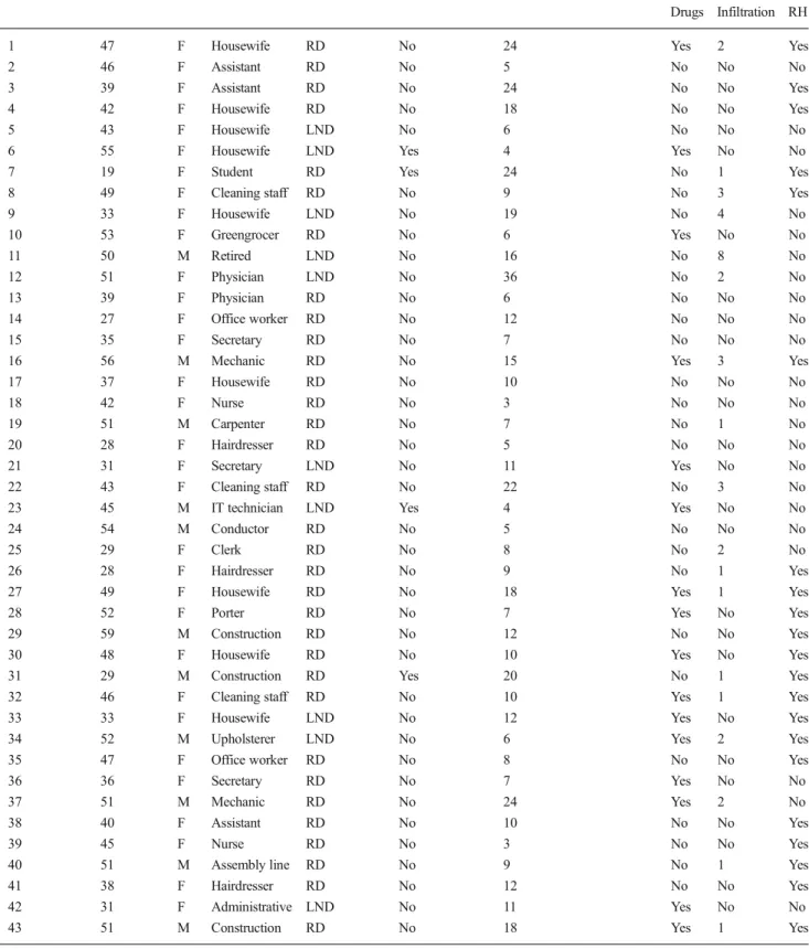
Upper limb (UL) Age (years) Sex Occupation Limb operated Prior traumatism Symptom duration (months) Previous treatments
Drugs Infiltration RH
1 47 F Housewife RD No 24 Yes 2 Yes
2 46 F Assistant RD No 5 No No No
3 39 F Assistant RD No 24 No No Yes
4 42 F Housewife RD No 18 No No Yes
5 43 F Housewife LND No 6 No No No
6 55 F Housewife LND Yes 4 Yes No No
7 19 F Student RD Yes 24 No 1 Yes
8 49 F Cleaning staff RD No 9 No 3 Yes
9 33 F Housewife LND No 19 No 4 No
10 53 F Greengrocer RD No 6 Yes No No
11 50 M Retired LND No 16 No 8 No
12 51 F Physician LND No 36 No 2 No
13 39 F Physician RD No 6 No No No
14 27 F Office worker RD No 12 No No No
15 35 F Secretary RD No 7 No No No
16 56 M Mechanic RD No 15 Yes 3 Yes
17 37 F Housewife RD No 10 No No No
18 42 F Nurse RD No 3 No No No
19 51 M Carpenter RD No 7 No 1 No
20 28 F Hairdresser RD No 5 No No No
21 31 F Secretary LND No 11 Yes No No
22 43 F Cleaning staff RD No 22 No 3 No
23 45 M IT technician LND Yes 4 Yes No No
24 54 M Conductor RD No 5 No No No
25 29 F Clerk RD No 8 No 2 No
26 28 F Hairdresser RD No 9 No 1 Yes
27 49 F Housewife RD No 18 Yes 1 Yes
28 52 F Porter RD No 7 Yes No Yes
29 59 M Construction RD No 12 No No Yes
30 48 F Housewife RD No 10 Yes No Yes
31 29 M Construction RD Yes 20 No 1 Yes
32 46 F Cleaning staff RD No 10 Yes 1 Yes
33 33 F Housewife LND No 12 Yes No Yes
34 52 M Upholsterer LND No 6 Yes 2 Yes
35 47 F Office worker RD No 8 No No Yes
36 36 F Secretary RD No 7 Yes No No
37 51 M Mechanic RD No 24 Yes 2 No
38 40 F Assistant RD No 10 No No Yes
39 45 F Nurse RD No 3 No No Yes
40 51 M Assembly line RD No 9 No 1 Yes
41 38 F Hairdresser RD No 12 No No Yes
42 31 F Administrative LND No 11 Yes No No
43 51 M Construction RD No 18 Yes 1 Yes
Table 2 Patient data
Upper limb (UL)
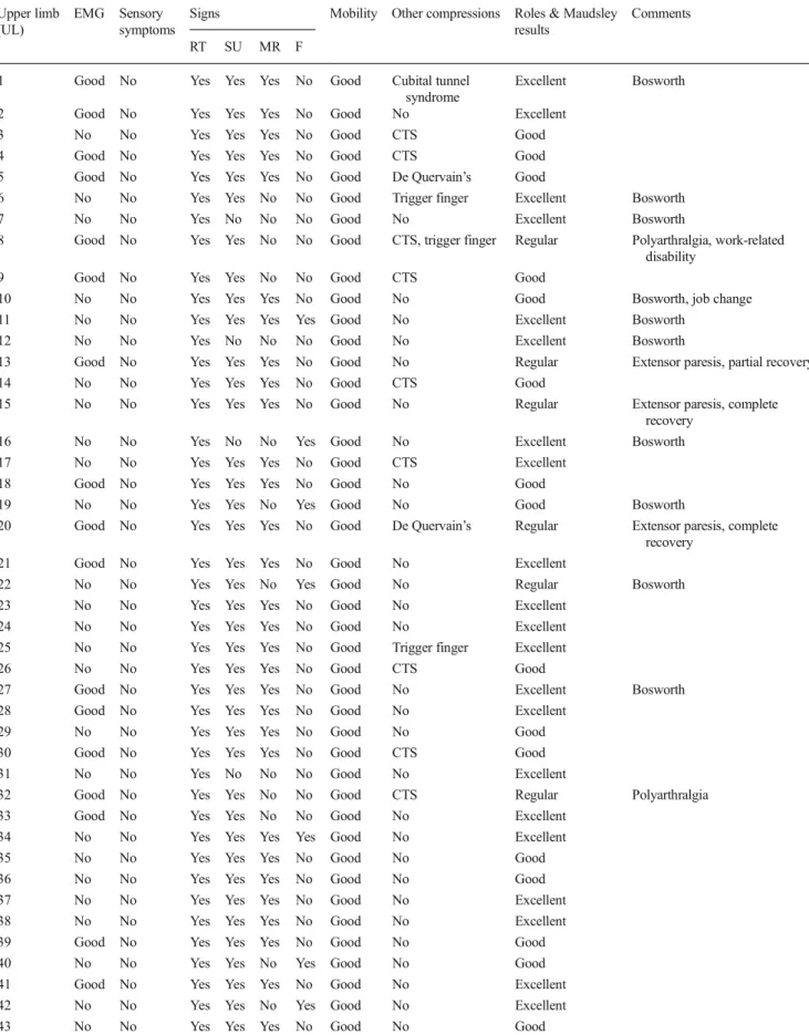
EMG Sensory symptoms
Signs Mobility Other compressions Roles & Maudsley results
Comments
RT SU MR F
1 Good No Yes Yes Yes No Good Cubital tunnel syndrome
Excellent Bosworth
2 Good No Yes Yes Yes No Good No Excellent
3 No No Yes Yes Yes No Good CTS Good
4 Good No Yes Yes Yes No Good CTS Good
5 Good No Yes Yes Yes No Good De Quervain’s Good
6 No No Yes Yes No No Good Trigger finger Excellent Bosworth
7 No No Yes No No No Good No Excellent Bosworth
8 Good No Yes Yes No No Good CTS, trigger finger Regular Polyarthralgia, work-related disability
9 Good No Yes Yes No No Good CTS Good
10 No No Yes Yes Yes No Good No Good Bosworth, job change
11 No No Yes Yes Yes Yes Good No Excellent Bosworth
12 No No Yes No No No Good No Excellent Bosworth
13 Good No Yes Yes Yes No Good No Regular Extensor paresis, partial recovery
14 No No Yes Yes Yes No Good CTS Good
15 No No Yes Yes Yes No Good No Regular Extensor paresis, complete recovery
16 No No Yes No No Yes Good No Excellent Bosworth
17 No No Yes Yes Yes No Good CTS Excellent
18 Good No Yes Yes Yes No Good No Good
19 No No Yes Yes No Yes Good No Good Bosworth
20 Good No Yes Yes Yes No Good De Quervain’s Regular Extensor paresis, complete recovery
21 Good No Yes Yes Yes No Good No Excellent
22 No No Yes Yes No Yes Good No Regular Bosworth
23 No No Yes Yes Yes No Good No Excellent
24 No No Yes Yes Yes No Good No Excellent
25 No No Yes Yes Yes No Good Trigger finger Excellent
26 No No Yes Yes Yes No Good CTS Good
27 Good No Yes Yes Yes No Good No Excellent Bosworth
28 Good No Yes Yes Yes No Good No Excellent
29 No No Yes Yes Yes No Good No Good
30 Good No Yes Yes Yes No Good CTS Good
31 No No Yes No No No Good No Excellent
32 Good No Yes Yes No No Good CTS Regular Polyarthralgia
33 Good No Yes Yes No No Good No Excellent
34 No No Yes Yes Yes Yes Good No Excellent
35 No No Yes Yes Yes No Good No Good
36 No No Yes Yes Yes No Good No Good
37 No No Yes Yes Yes No Good No Excellent
38 No No Yes Yes Yes No Good No Excellent
39 Good No Yes Yes Yes No Good No Good
40 No No Yes Yes No Yes Good No Good
41 Good No Yes Yes Yes No Good No Excellent
42 No No Yes Yes No Yes Good No Excellent
43 No No Yes Yes Yes No Good No Good
suffered a prior traumatism. The duration of the symptoms ranged from three months to 36 months (mean of 13.9 months).
Before the surgical procedure, multiple conservative treat-ments were carried out (Table1). One patient (2.3 %) was previously operated on for lateral epicondylitis, without im-proving the symptoms.
The test of supination against resistance was positive in 40 procedures (93 %) and the middle finger extension resistance test was positive in 30 (69.7 %). A loss of wrist force due to pain was observed in eight procedures (18.6 %).
No patient presented paresthesias or dysesthesias in the radial innervation area. In eight procedures there were CTS (18.6 %), and one patient cubital tunnel syndrome (2.3 %) associated to RTS. Elbow mobility was complete in all the arcs of motion in all the patients studied (100 %).
Eleven patients also presented selective pain at the level of the lateral epicondyle, related with enthesitis of the epicondyle muscles at their insertion, associated with a radial tunnel (25.6 %).
Elbow X-rays were taken in 18 patients; the results were normal in 17 patients, while one patient presented sclerosis of the lateral epicondyle secondary to the multiple infiltrations carried out. In ten patients (23.2 %) a NMR was performed;
normal results were reported for six patients (13.9 %) and four patients (9.3 %) showed non-specific inflammatory changes (Table2).
An EMG study was carried out on 17 patients, with the nerve conduction velocity for the PIN being normal in all of them (39.5 %).
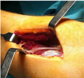
Fig. 2 Dissection between the second radial and the common extensor of the fingers and section of the supinator muscle
Fig. 3 Section of the superficial part of the proximal border of the supinator muscle
Fig. 4 PIN decompression in the distal border of the supinator
Fig. 5 Visualisation of compression of the PIN
The approach described previously was used in all patients, releasing the nerve from all the possible compression sites. No masses or anatomical alterations were observed in any pa-tients. In the majority of the patients, signs of possible PIN thinning were found.
All of them presented greater nerve compression at the level of the proximal border of the supinator in the intra-operative provocation procedures. Likewise, such compres-sion was also observed at the level of the distal border of that nerve in nine patients (20.9 %).
(53.4 %). There were fibrous adhesions of the superficial bundle of the supinator muscle in ten patients (23.2 %). In seven patients that had received multiple infiltrations (16.2 %), great fibrosis was observed under the lateral epicondyle in the extensor muscle insertion. We performed a simultaneous modified Bosworth technique in 11 patients (25.6 %), as they presented symptoms of lateral epicondylitis in addition to RTS.
Mean follow-up time for the patients was 22 months (min-imum, four months; max(min-imum, six years and four months).
At present, 21 patients (48.8 %) present no symptoms, 16 patients (37.2 %) present occasional symptoms and six pa-tients (13.95 %), symptoms with repetitive activities. Elbow mobility is complete in all patients, while all of the patients except one (2.3 %) recovered wrist force.
As for complications associated with the intervention, we observed a paresis of the common extensor of the fingers (lack of complete extension of the middle and ring fingers) in three patients (6.9 %). Two of them recovered complete extension in less than three months; one patient recovered only partial extension, with a lack of 15° in the metacarpophalangeal (MCP) joints of the middle and ring fingers (although the later E M G s t u d y d i d n o t s h o w a n y ne r v e c o n d u c t i o n abnormalities).
All of the patients reported that they had improved after the intervention, although one patient would not choose to be operated again knowing the results if the same thing should happen once more.
Using the criteria of Roles and Maudsley, the results were excellent in 48.8 % of the patients, good in 37.2 % and fair in 13.9 % of the patients. One patient with polyarthritis who achieved only a fair result requested work-related disability and two patients asked for a change in their job positions due to the impossibility of performing strong repetitive move-ments (7.14 %).
Discussion
Radial tunnel syndrome is a nerve compression syndrome with special characteristics, given that in spite of compressing a motor nerve (the PIN), it does not produce motor or nerve alterations [3,4,7,8,10,14,15].
Roles and Maudsley described this syndrome in 1972, with excellent results following surgical treatment of radial tunnel decompression; however, since that time, many authors have published results that are also good, but not as successful as those published by Roles and Maudsley [3,6,7,13].
The diagnosis of RTS is chiefly clinical, with the most characteristic symptoms being pain at the level of the radial tunnel that increases with pressure, as well as weakness and heaviness in the forearm, especially after exertion. None of
our patients presented motor anomalies, but an elevated per-centage of them positive results in the provocation manoeu-vres, supination against resistance and the middle finger test [4,6,7,9,14,15,18].
The appearance of paresthesias and limitations in elbow and hand mobility are infrequent [13,14]. A significant number of patients complain of loss of wrist force, probably due to the pain that picking up weight causes them from the contraction of the extensor muscles. There are no alterations in the normal static neurophysiological state with a normal nerve conduction velocity [3,5,6,9,10,13–15]. This is all due to the fact that the PIN is compressed intermittently by the muscles that form the radial tunnel, causing pain [6,11,12,14].
In general, RTS is a mainly dynamic nerve compression syndrome. This was demonstrated in several studies in which a decrease in nerve conduction velocity was observed in the EMG during supination against resistance [3,5,8,14,17]. It is often mistaken for enthesitis of the insertion of the epicondyle muscles, which is why some authors call this syndrome treatment-resistant epicondylitis [4, 13, 18, 19]. These two pathologies are sometimes difficult to differentiate. In addi-tion, they can even be associated occasionally; if so, both processes should be handled at the same time.
In our series there was a relationship with other nerve compression syndromes, such as carpal tunnel syndrome, cubital syndrome and repeated tendonitis, as other authors have indicated previously [3,7,9,13,18].
In all cases we used a posterior-external approach, follow-ing the radial epicondyle-styloid line because it made it pos-sible for us to visualise the entire PIN, even in the border distal to the supinator [14]. The anterior approach allows us to see the proximal area more easily, but not the distal area of the arcade of Frohse and it has a greater risk of injuring the radial superficial sensory branch [7,14,18].
It is important to consider the variability of the pattern of radial nerve innervation at the distal level, as we have been able to observe in our study [1,11,12]. This explains the three pareses of the common finger extensors seen in our patients caused by excessive distal dissections. We did not observe the presence of tumours or anatomical abnormalities, which are characteristics that are more typical of the PIN syndrome [4,6, 7,10,14].
The site in the surgical field where we suspected that PIN compression could exist was the proximal entry of the radial tunnel, although compression at the level of the distal border was observed in nine patients; for that reason, we believe that the posterior approach is better [3,7,9,14]. In a few patients we found fibrosis between the second radial and the supinator, while fibrosis was present in the superficial bundle of the supinator in others.
patients; most patients returned to daily activities in two or three months [3,7,8,13,14].
Conflict of interest There is no conflict of interest and the paper has not been submitted elsewhere.
References
1. Branovacki G, Hanson R, Cash R, Gonzalez M (1998) The innerva-tion pattern of radial nerve at the elbow and in the forearm. J Hand Surg 23 B:167–169
2. Crawford GP (1998) Late radial tunnel syndrome after excision of radial head. J Bone Joint Surg 1416–1418
3. Jebson PJL, Arbor A, Enger WD, Madison W (1997) Radial tunnel syndrome: long-term results of surgical decompression. J Hand Surg 22A:889–896
4. Kleinert JM, Metha S (1996) Radial nerve entrapment. Clin Orthop 27:305–315
5. Kupfer DM, Bronson J, Lee G, Beck J, Gillet J, Diego S (1998) Differential latency testing: a more sensitive test for radial tunnel syndrome. J Hand Surg 23A:859–864
6. Sarhadi NS, Korday SN, Bainbridge LC (1998) Radial tunnel syn-drome: diagnosis and management. J Hand Surg 23A:617–619 7. Sotereanos DG, Varitimidis SE, Giannakopoulos PN et al (1999)
Results of surgical treatment for radial tunnel syndrome. J Hand Surg 24A:566–570
8. Naam NH, Nemani S (2012) Radial tunnel syndrome. Orthop Clin N Am 43(4):529–536
9. Ritts GD, Wood MB, Linscheid L (1987) Radial tunnel syndrome. Clin Orthop Relat Res 219:201–205
10. Van Rossum J, Buruma JS, Kamphuisen HAC, Onvlee GJ (1978) Tennis elbow—a radial tunnel syndrome? J Bone Joint Surg 60-B: 197–198
11. Clavert P, Lutz JC, Adam P, Wolfram-Gabel R, Liverneaux P, Kahn JL (2009) Frohse’s arcade is not the exclusive compression site of the radial nerve in its tunnel. Orthop Traumatol Surg Res 95(2):114–118 12. Berton C, Wavreille G, Lecomte F, Miletic B, Kim HJ, Fontaine C (2013) The supinator muscle: anatomical bases for deep branch of the radial nerve entrapment. Surg Radiol Anat 35(3):217–224
13. Lawrence T, Mobbs P, Fortems Y, Stanley JK (1995) Radial tunnel syndrome. J Hand Surg 20B:454–459
14. Lister GD (1991) Radial tunnel syndrome. In: Gelberman RH (ed) Operative nerve repair and reconstruction. Lippincott Company, Philadelphia, pp 1023–1037
15. Martín Ferrero MA (2001) Síndromes de compresión nerviosa. In: Sánchez Martín MA (ed) Traumatología y Ortopedia, Universidad de Valladolid, p 483–494
16. Portilla Molina AE, Bour C, Oberlin C, Nzeusseu A, Vanwijck R (1998) The posterior interosseous nerve and the radial tunnel syn-drome: an anatomical study. Int Orthop 22(2):102–106
17. Verhaar J, Spaans F (1991) Radial tunnel syndrome. J Bone Joint Surg 73-A:539–554
18. Stanley J (2006) Radial tunnel syndrome: a surgeon’s perspective. J Hand Ther 19(2):180–184