Autophagy and oxidative stress in non communicable diseases: A matter of the inflammatory state?
18
0
0
Texto completo
(2) Free Radical Biology and Medicine 124 (2018) 61–78. D. Peña-Oyarzun et al.. Fig. 1. Statistics of non-communicable diseases worldwide. (A) Non communicable diseases (NCDs) account for approximately 40 million of the 56 million deaths worldwide. (B) Of the total deaths as a result of NCDs, most of them are caused by cardiovascular diseases (CVD) (32%), followed by cancer (Ca) (16%), respiratory chronic diseases (Resp) (7%) and diabetes mellitus (DM) (3%). (C) According to World Health Organization (WHO) reports total deaths derived from NCDs represent over 80% in North and South America, the West Pacific and Europe; approximately 60% in Eastern Mediterranean and South-East Asia, and around 34% in Africa. (D) NCDs-related deaths increase with age (15 million at 30–69 years of age; 22 million persons over 70 years old), with equal contributions among sexes. (E) Most NCDs associated deaths in higher income groups are due to CVD and cancer.. disruption of one results in alterations of the other [7]. Additionally, inflammation is also altered in NCDs [8]. The level of inflammation in the organism is highly dependent on the cellular redox and autophagic state [9,10]. In the present review, our aims are: a) To describe the role of inflammatory cytokines and immune cells in NCDs. b) To discuss the apparent dual role of autophagy, which can both promote or inhibit NCDs progression. c) To explain how oxidative imbalance results in oxidative stress, thereby inducing NCDs. d) To analyze how cross-talk between autophagy and oxidative stress modifies the inflammatory context of tissues, ultimately leading to NCDs development. There are other chronic conditions such as mental health problems and oral diseases that are associated with a significant degree of morbidity worldwide, affecting the quality of life of large segments of the population, and increasing the complexity of global and national responses. stages. During aging, excessive production of specific oxidative agents or an impairment in protective reductive systems occur. Indeed, the aberrant formation or accumulation of different reactive oxygen or nitrogen species (ROS or RNS, respectively) involved in specific signaling pathways with organelle-specific functions are severely altered in older organisms. The aforementioned condition can generate oxidative stress in the cell, and together with the dysregulation of autophagy, a mechanism that maintains cellular proteostasis, represent a common feature of NCDs, such as cancer, CVD, respiratory chronic diseases and T2DM [5,6]. It is important to note that many other cellular processes contribute to NCDs progression, like mitochondrial dysfunction, altered gene expression and impaired proteosomal activity. However, this article will focus in autophagy modulation, as an altered cellular process during NCDs. Interestingly, oxidative stress and autophagy are connected by several crosstalk pathways, whereby 62.
(3) Free Radical Biology and Medicine 124 (2018) 61–78. D. Peña-Oyarzun et al.. Fig. 2. Inflammation and non-communicable diseases. Type 2 diabetes mellitus (T2DM), cardiovascular diseases (CVD), cancer and obesity begin with persistent damage (i.e. lipid/glucose overload, hypoxia or mechanical stress, among others) that stress cells and provoke the release of molecular markers. The markers released include several cytokines, interleukins and adhesion molecules, thereby determining a new inflammatory set point that is both systemic and tissue specific. Importantly, inflammation is strongly associated with altered tissue/organ function, like diminished response to insulin, increased fibrosis, augmented arterial pressure, reduced lipid storage and increased tumor cell proliferation. All these events ultimately lead to hyperglycemia, lipid spill, hypertension, tumor progression, atherosclerosis, myocardial infarction and heart failure.. against the epidemic of NCDs. However, their study is beyond the scope of this review [4].. stimulated by systemically increased levels of pro-inflammatory cytokines [20].. 2. Inflammation and non-communicable diseases. 2.2. Inflammation and type 2 diabetes mellitus. 2.1. Inflammation: general aspects. The obesity epidemic promoted a dramatic increase in the incidence of T2DM, a pathological condition characterized by insulin resistance and/or β-cell dysfunction. As previously mentioned, various studies consistently reported increased levels of pro-inflammatory cytokines and acute-phase proteins in patients with T2DM [21–23]. Thus, excessive consumption of nutrients rich in sugars and fatty acids activates cellular stress responses, promoting local production of pro-inflammatory markers together with the accumulation of IL-1β, IL-6 and C-reactive protein (CRP) in plasma and tissues, which in turn correlates with T2DM onset [24–27]. Indeed, fatty acids can activate Toll-like receptors (TLR), such as TLR4 and TLR2, which stimulate the myeloid differentiation primary-response protein 88 (MYD88) [28] and thus the nuclear factor-kappa B (NFκB) signaling pathway, leading to pro-inflammatory cytokine production [29]. In addition, studies have shown that high glucose concentrations can activate the inflammasome, and therefore stimulate the production of IL-1β, in different cell types [30,31]. Specifically, thioredoxin-interacting protein (TXNIP) can dissociate from its inhibitor, the protein thioredoxin (TXR), when glucose levels are high, thus activating the NOD-, LRR‑ and pyrin domain containing 3 (NLRP3) inflammasome, which activates caspase 1 [32,33] that cleaves pro-IL-1β to generate IL-1β [34]. IL-1β, in turn, amplifies the pro-inflammatory signal through the production of pro-. Chronic inflammation refers to a prolonged inflammatory response or condition in which the production of pro-inflammatory cytokines persists over time. This process might follow an acute inflammatory event; however, in most of the cases, in the context of NCDs, it begins as a low-grade inflammatory response with no presence of an acute reaction. An increasing body of evidence shows that this sterile, lowgrade inflammatory response, is involved in the development of NCDs [11]. Indeed, elevated levels of pro-inflammatory cytokines, such as interleukin (IL)−1β, IL-6 and tumor necrosis factor alpha (TNFα) not only have been identified in adipose tissue, muscle, brain, liver and heart in animal models and humans affected by NCDs, but also their levels positively correlate with the severity of these diseases [12–15]. Thus, studies have shown that T2DM is correlated both with neuronal inflammation in the central nervous system (CNS) [16] and immune cell infiltration with subsequent production of pro-inflammatory cytokines in peripheral adipose tissue [17]. These data reveal the importance of inflammation at the level of the CNS and its impact in NCDs, such as T2DM. Chronic inflammation also plays a critical role in the development of atherosclerosis, predisposing to CVD [18,19]. Consistently, tumoral initiation, promotion and progression are 63.
(4) Free Radical Biology and Medicine 124 (2018) 61–78. D. Peña-Oyarzun et al.. progression by activation of the NF-κB and signal transducer and activator of transcription 3 (STAT3) pathways [64,65]. Activation of STAT3 suppresses the expression of major histocompatibility complex class II, impairing the activation of T CD4 + T cells [66]. Importantly, these pathways are self-regulated by a feed-forward mechanism. Thus, in addition to inducing pro-survival and proliferative signaling in premalignant cells, the system becomes self-sustainable over the time [67]. The latter is also valid for TNFα, which, when released as a result of an increased inflammatory response, exacerbates the ROS-dependent genetic instability due to p53 tumor suppressor protein mutations and DNA damage [68]. Also, TNFα plays a crucial role in colon cancer development where it promotes β-catenin translocation to the nucleus to increase the expression of genes that promote cancer cell growth and survival [69]. Macrophages and myeloid precursors work together to induce tumor angiogenesis. The macrophages sense hypoxia and activate the transcription factor hypoxia-inducible factor 1 alpha (HIF-1α), which promotes the expression of stromal cell-derived factor 1 (SDF1), also known as C-X-C motif chemokine 12 (CXCL12), to favor endothelial cell recruitment, while myeloid precursors produce vascular endothelial growth factor (VEGF) [70]. Both macrophages and myeloid cells also participate in metastasis. CCr1 + myeloid cells release metalloprotease (MMP) 2 and MMP-9, which enhance degradation of components of the basal membrane [71]. CCr1 + myeloid cells are recruited by the C-C motif ligand 9 (CCL9) chemokine released by cancer cells [72]. On the other hand, macrophages increase the levels of TNFα, which decrease E-cadherin expression by stabilizing Snail, an E-cadherin transcription repressor [73]. In conclusion, localized and chronic inflammation promote tumor initiation and progression, and therefore, complementary anti-cancer therapies should also focus on the communication between cancer cells and immune cells.. inflammatory cytokines and chemokines [32,33]. Different cytokines have been linked to insulin resistance. Hotamisligil et al. showed that increased levels of TNFα reduce insulin sensitivity in the adipose tissue of obese rodents and humans, while TNFα knockout in mice was shown to be sufficient to protect from obesity-associated insulin resistance [35]. Since then, additional studies revealed that inhibition or depletion of pro-inflammatory cytokines, such as IL-1β and IL-6, enhances insulin sensitivity in different tissues, thus preventing the development of T2DM [27,36–39]. Altogether, these studies indicate that the administration of anti-inflammatory drugs represents a potentially beneficial approach in the treatment of T2DM [40,41]. Hyperglycemia has also been associated with an increased production of ROS in the mitochondria in various cell types [42–44]. Importantly, ROS can activate the NFκB signaling pathway [45], promoting inflammation and inhibiting insulin signaling [46]. The mechanisms by which ROS promote inflammation and therefore lead to the onset of metabolic diseases will be discussed in the next sections (Fig. 2). 2.3. Inflammation and cardiovascular diseases CVD are caused by disorders that affect the blood vessels and/or the heart, such as, atherosclerosis, heart failure and myocardial infarction, among others [1]. Hypertension (HT) is a major risk factor for premature CVD [1,47]. There is evidence that links inflammation and CVD in both ways: on the one hand, low-grade chronic inflammation plays a crucial role in HT and CVD development [48,49]; on the other hand, neurohumoral activation, such as via upregulation of the renin-angiotensin-aldosterone system, enhances the synthesis of proteins involved in inflammation, cell death, and fibroblast proliferation [50]. Also, clinical studies have suggested that elevated serum CRP and plasminogen activator inhibitor-1 [49,51] are determining factors for the development of HT and myocardial pressure overload, which then induces systemic inflammation through IL-33 produced by endothelial cells [52]. In addition, alterations in the TNFα system are also associated with chronic inflammation [53]. Heart failure (HF) is defined as the inability to meet the metabolic demand of the tissue due to a structural or functional impairment of ventricular filling or ejection [54]. Disturbances in the inflammationrelated TNFα system have been implicated in the pathogenesis of HF [53], as well as changes in IL-6 [55], vascular cell adhesion protein 1(VCAM-1) [56] and galectin-3 [57]. Other ILs play a key role in myocardial ischemia and reperfusion (I/R) injury. In this context, IL-23 may promote myocardial I/R injury by increasing the inflammatory and oxidative stress response [58]. However, it is worth noting that the main inflammatory component in myocardial infarction is the acute post-injury inflammatory response [59] and it is not clear yet whether there is a systemic inflammatory component prior to the damage. In summary, numerous studies have consistently shown an association between CVD and systemic inflammatory biomarkers, but the molecular mechanisms remain to be elucidated.. 2.5. Inflammation and obesity Overweight and obesity are defined as abnormal or excessive adipose tissue accumulation that may impair health. The World Health Organization (WHO) uses a definition based on the body mass index (BMI), a calculation of the weight divided by the square of the height (Kg/m2). Based on this criteria, a person with BMI ≥ 25 is considered overweight and is obese with BMI ≥ 30. According to the WHO, over 650 million adults were estimated to be obese worldwide in 2016 and prevalence has almost tripled since 1975. Noteworthy, a high BMI by itself does not completely determine the pathologic nature of the disease, and a subgroup of obese individuals has been defined as metabolically healthy [74,75]. Even though this concept is still controversial, there is agreement in that cardiometabolic problems in obese subjects are closely linked to the inflammatory status in their adipose tissue. Adipose tissue used to be considered metabolically inert, with its only purpose being the storage of triglycerides. This vision is now considered obsolete and adipose tissue is currently the object of great interest due to its relevance as a metabolic, endocrine, and homeostasisregulating organ, able to secrete a vast number of active factors (adipokines and cytokines) with pro-inflammatory, anti-inflammatory and immunomodulating properties [76–78]. The link between increased adipose tissue mass and the development of obesity-related disorders is largely determined by the pro-inflammatory status of the tissue. The resulting local inflammation and ensuing adipose tissue dysfunction has a substantial systemic impact, by inducing insulin resistance, hyperglycemia, hyperlipidemia, and damaging fat infiltration in key metabolic organs (such as liver and pancreas), a consequence known as lipotoxicity [79,80]. The accepted pathophysiological model of dysfunctional adipose tissue in obesity suggests that excess energy intake and the ensuing metabolic challenge lead to an evolutionarily-conserved adaptive. 2.4. Inflammation and cancer The relevance of inflammation in cancer development has become increasingly appreciated over the past decade, since approximately 90% of cancers are not caused by inherited mutations, but rather by environmental factors that increase inflammation, e.g., smoking and consumption of western-style diets [60,61]. Among the tumor cell populations, several immune cells can be found, whereby the majority of them are macrophages, T cells and myeloid precursors, reflecting the existence of an inflammatory microenvironment [62,63]. This microenvironment is controlled by cytokine release, with important repercussions in the different tumor stages. The pro-inflammatory cytokines IL-6 and IL-1β are released as a consequence of genotoxic stress, thereby favoring tumor initiation and 64.
(5) Free Radical Biology and Medicine 124 (2018) 61–78. D. Peña-Oyarzun et al.. Fig. 3. Autophagy and non-communicable diseases. (A) Autophagy is divided into five stages. First, stimuli like stress and metabolic deficiency transduce signals to activate AMPK and inactivate mTORC1, which in turn activates the ULK1 complex (“Initiation”). Activated ULK1 translocates to pre-autophagosomal sites and phosphorylates Beclin 1 to induce formation of the PI3K-III complex. This results in the production of PI3P, a lipid molecule that recruits ATG proteins implicated in the autophagome formation (“Nucleation”). LC3 protein is cleaved proteolytically, conjugated with the lipid phosphatidyethanolamine and incorporated into the autophagosome. LC3 assists with the autophagosome extension (“Elongation”) and targeting of cargoes by associating with p62/polyubiquitinated proteins. Finally, the autophagosome fuses with a lysosome (“Fusion”), allowing the recycling of the cargoes by lysosomal enzymes (“Degradation”). (B) The hypoxic and low-nutrient environment of cancer stem cells increases autophagy to promote proliferation, differentiation, survival and resistance to chemotherapy. (C) Autophagy plays a dual role of cardiac tissue: after ischemia, autophagy protects the cardiomyocyte; however, after ischemia/reperfusion (I/R), autophagy is detrimental, leading to hypertrophy and cell death. (D) Under conditions of cholesterol overload, vascular smooth muscle cells (VSMC) augment autophagy provoking their dedifferentiation and proliferation, which ultimately aids in the formation of the atherosclerotic plaque. (E) Chronic gluco/lipotoxicity of pancreatic β-cells alters insulin processing and induces autophagy-dependent cell death. (F) Autophagy is induced by excess nutrition in adipocytes produces hypertrophy and liberation of cytokines. (G) High fat diet reduces autophagy-dependent degradation of lipids in hepatocytes, thereby leading to accumulation of fatty acids inside the cell and insulin receptor damage. (H) Augmented inflammation in macrophages reduces autophagy, one of the major degradation pathways for non-self molecules in immune cells.. positive feedback loop that involves pro-inflammatory macrophage infiltration and worsening of the inflammatory profile, together with local insulin resistance, impaired extracellular matrix remodeling, fibrosis and impaired lipid handling capacity. These events in turn result in elevated circulating free fatty acids and ectopic fat accumulation that will trigger insulin resistance in other organs. Chronic over-nutrition thus results in the classic low grade inflammatory state associated with obesity, accompanied by impaired lipid metabolism and both local and. response, which, under the current environmental conditions, results in deleterious inflammation and insulin resistance [81–83]. The energy surplus is dealt with either by inducing adipocyte formation through adipogenesis (hyperplasia) or by enlarging existing adipocytes by increasing triglyceride storage (hypertrophy). While hyperplasia is considered a healthy process, excess adipocyte hypertrophy is associated with insulin resistance and endocrine dysregulation [78,84,85]. Upon chronically exacerbated energy intake, these alterations generate a 65.
(6) Free Radical Biology and Medicine 124 (2018) 61–78. D. Peña-Oyarzun et al.. spontaneous tumor formation, i.e. liver dysplasia, lung carcinoma and hepatocellular carcinoma [98]. However, it is worth noting that recent data propose additional, autophagy-independent roles of Beclin 1 in breast cancer tumorigenesis by controlling EGF receptor maturation [99]. Cancer stem cells (CSC) are a specialized subtype of tumor cell with self-renewal capability that are directly considered responsible for tumor cell growth. Since they are mainly encountered in nutrient and oxygen- deficient regions, autophagy modulation is reportedly considered an important factor for CSC proliferation and differentiation into daughter tumor cells. In mammospheres, increased Beclin 1 expression and accelerated autophagy are observed in response to starvation [100]. Beclin 1 has also been shown to be required for breast CSC maintenance, because Beclin 1 knockdown decreases mammosphere formation and ultimately, reduces tumor volume in vivo [100]. Similar results are observed with the potassium ionophore salinomycin, which has been reported to decrease autophagy flux in breast CSC [101]. Accordingly, autophagy is required to maintain the CD44+/ CD24low phenotype in breast CSCs and is blocked by LC3/ATG12 knockdown or chloroquine treatment [102]. Hypoxia-dependent autophagy is also necessary for transforming pancreatic cancer cells into CSC-like (CD133+) cells, a mechanism that relies on HIF-1α activation [103]. Furthermore, abrogation of autophagy with 3-methyladenine (3MA) increases apoptosis under hypoxia in pancreatic CSC [104]. Similarly, liver CSC CD133+ sub-populations have increased expression of Beclin 1, ATG5, ATG7 and LC3, as well as augmented resistance to apoptosis under hypoxia, when compared with CD133- cells. This phenomenon is reversed when CD133+ liver cells are treated with chloroquine [105]. All together, these studies suggest that autophagy is not only required to acquire CSC markers, but also to improve CSC survival under unfavorable conditions. Interestingly, cancer cells use autophagy as a resistance mechanism against chemotherapy. In cancer cell lines, such as LoVo and HeLa, treatment with doxorubicin, a non-selective class 1 anthracycline that induces cell death by inhibiting topoisomerases I and II and by inducing ROS generation [106], increases in LC3-II levels, which are exacerbated by bafilomycin A1, showing elevated autophagic flux. These changes correlate with reduced activation of mTORC1 and increased nuclear translocation of the transcription factor EB (TFEB) [107]. Notably, increased autophagy seems to be a defensive mechanism against the treatment with doxorubicin, since downregulation of TFEB or ATG5 with specific siRNAs reduces viability of doxorubicin-treated cells [107]. Also, the adipokine resistin induces autophagy via AMPK and mTORC1 in the breast cancer cell lines MCF-7 and MDA-MB-231, thereby decreasing the antineoplastic effects of doxorubicin. Conversely, the osteosarcoma cells U2OS, MG-63 and SAOS-2 have been shown to be more prone to doxorubicin-induced cell death when autophagy is reduced upon treatment with the long non-coding RNA CTA by a mechanism implicating mTORC1 activation [108]. A well-known secondary effect of chemotherapy with doxorubicin is the cardiotoxicity. In fact, cardiomyocytes exposed to doxorubicin are sensitized to cell death when autophagy is impaired with 3-MA or by down regulation of UV radiation resistance-associated gene protein (UVRAG) [109,110]. Similar effects have been observed with other chemotherapy agents, such as cisplatin and oxaliplatin, which promote DNA instability and high ROS production, leading to tumor cell death [106]. Cisplatin induces LC3 II accumulation and p62/SQSTM1 reduction in HOS and OSR osteosarcoma cells, both indicative of increased autophagy flux. In the same study, induction of autophagy and JNK signaling were shown to activate a pro survival response in tumor cells treated with cisplatin [111]. In this regard, SP600125, a chemical inhibitor of JNK, reduces autophagy and increases oxaliplatin-induced apoptosis in colorectal cancer cell lines [112], indicating that tumor cells able to undergo autophagy are more resistant to chemotherapeutic agents. Indeed, reducing autophagy using the long non-coding RNA MEG3 or by down. systemic insulin resistance [78,84,85]. 3. Autophagy and non-communicable diseases 3.1. General mechanism of autophagy Macroautophagy, hereafter refereed as autophagy, involves the formation of double membrane vesicles, called “autophagosomes”, which sequester intra-cellular components such as old organelles, protein aggregates, and misfolded proteins, among others. Then, autophagosomes fuse with lysosomes and the luminal content is degraded by hydrolytic lysosomal enzymes [86]. This process is highly conserved from yeast to mammals, and it is required to maintain cellular homeostasis. The autophagy-related proteins (ATG) are implicated in the different steps of the autophagic process [87], which can be divided into initiation, nucleation, elongation, fusion and degradation. The stage of “initiation” describes the on- and off-switch of autophagic signaling pathways. Thus, the canonical ways that control autophagy initiation are the mechanistic target of rapamycin complex 1 (mTORC1) and the AMP-activated protein kinase (AMPK), both stress sensors. While mTORC1 is an autophagy inhibitor activated by serum, nutrients, growth factors, etc. AMPK is an autophagy inducer activated by low energy conditions. Both mTORC1 and AMPK phosphorylate Unc-51 like autophagy activating kinase ULK1 (ATG1), and while mTORC1 inhibits ULK1 by phosphorylation at S737, AMPK activates ULK1 by phosphorylation at S317 and S777 [88]. The active ULK1 complex relocates to the endoplasmic reticulum to phosphorylate Beclin 1 at S14 [89]. At this point the process referred to as “nucleation” starts: phosphorylated Beclin 1 recruits VPS34 and several co-activators, forming a new complex with a PtdsIns3-kinase activity. The lipid phosphatidylinositol 3-phosphate (the product of the active VPS34/Beclin 1 complex) serves as “re-localization flags” for PX- and FYVE- domain containing ATGs [90]. The “elongation stage” refers to the extension of the autophagosome membrane, a process mainly directed by the microtubule-associated protein light chain 3 (MAP1LC3, also known as LC3), a protein that undergoes post-translational proteolysis by ATG4 to form LC3-I and then incorporation of phosphatidylethanolamine to generate LC3II, which permits association with the autophagosome membrane [91]. The LC3-I to LC3-II conversion is catalyzed by the ATG5/ATG12ATG16L complex, which functions as an E3-enzyme [92]. Since LC3II localizes to the autophagosome membranes, it is widely used as an autophagy marker [93]. The two final stages of the autophagy process are the “fusion” of the autophagosome with the lysosome, which then permits “degradation” of the cargos [94]. Other additional ATGs participate in the selectivity for cargo degradation. In this regard, p62/sequestosome 1 (SQSTM1), a bimodular protein that recognizes both poly-ubiquitinated proteins and LC3 by a LC3-interacting domain (LIR), binds poly-ubiquitinated proteins to the autophagosome for degradation [95]. Since p62/SQSTM1 is also degraded by the lysosome, it is commonly used as an autophagic flux marker, indicative of the lysosomal capacity to degrade autophagosome cargos [96]. Thus, chemical compounds, such as chloroquine and bafilomycin-A1, block lysosomal degradation and provoke both p62/ SQSTM1 and LC3-II accumulation associated with reduced autophagy flux [93] (Fig. 3). 3.2. Autophagy in cancer The role of autophagy in spontaneous tumor initiation has been reported in studies around the 2000's. Levine's group demonstrated that overexpression of Beclin 1 in MCF7 breast cancer cells increased autophagy and reduced tumor formation when injected to mice, therefore suggesting that Beclin 1 acts as a tumor suppressor [97]. To support this hypothesis, the same group also showed that heterozygous disruption of Beclin 1 in mice decreased autophagy and increased the frequency of 66.
(7) Free Radical Biology and Medicine 124 (2018) 61–78. D. Peña-Oyarzun et al.. secretion in a mouse model of streptozotocin-induced T1DM [127], showing that insulin secretion is an autophagy-dependent process [127].. regulation of the co-chaperone BAG3 increased the sensitivity of human glioblastoma cells U87 to cisplatin-induced cell death [113,114]. However, in contrast to the effects seen with doxorubicin, inhibition of the mTORC1 pathway increases cisplatin-dependent cell death of lung adenocarcinoma and atypical teratoid rhabdoid tumors [115,116]. This is in line with the observation that oxaliplatin induces mTORC1 activation in different colorectal cancer cell lines such as HCT15, HCT116 and HT29 in vitro and in xenograft models, therefore suggesting that co-treatment with oxaliplatin and everolimus, a chemical inhibitor of mTORC1, could effectively improve the survival of colorectal cancer patients [117]. Indeed, mTORC1 inhibitors everolimus and sirolimus are currently being tested in multiple clinical trials for treatment of cancers, such as colorectal cancer, pancreatic cancer, melanoma, glioblastoma, small cell lung cancer, among others [118]. Interestingly, responses of these types of cancer to treatments with AMPK activators, like metformin and resveratrol, have also been evaluated [118].. 3.4. Autophagy in cardiovascular diseases Different studies have shown that autophagy is involved in the development and progression of different CVD, such as atrial fibrillation, I/R, cardiac hypertrophy, HF and atherosclerosis, among others [54,128–133]. Indeed, several studies show that autophagy might have beneficial or detrimental roles depending on the stage and type of cardiovascular disease considered [8,131,132]. A beneficial function of autophagy has been observed in I/R, cardiac hypertrophy and atrial fibrillation. In conditions of in vitro hypoxia (simulated ischemia), autophagy can be activated as an adaptive mechanism providing essential nutrients and removing damaged mitochondria [134]. In fact, inhibition of Beclin 1 by the cardiac peptide urocortin, increases cardiomyocyte death upon ischemia, suggesting a pro-survival role of autophagy [135]. In chronic ischemia, cardiac myocytes express increased levels of lysosomal enzymes, like cathepsins B and D, along with autophagosome vesicles, reflecting a higher autophagic state. Augmented autophagy in chronic ischemia is required to sustain cell viability by increasing damaged mitochondrial recycling, thereby diminishing apoptosis and tissue damage [136]. Induction of autophagy has also been observed during ischemic preconditioning. Moreover, when autophagy is inhibited with an ATG5 dominant negative (ATG5 K130R), the protective effects of preconditioning are blunted [136,137]. In cardiac hypertrophy induced by transverse aortic constriction (TAC), a reduction in the number of autophagic vacuoles can be observed, suggesting that basal autophagy is required to maintain cardiac homeostasis in rats [138]. On the other hand, cardiacspecific deficiency of ATG5 promotes cardiac hypertrophy, left ventricular dilatation and contractile dysfunction in mice [139]. Moreover, Beclin 1 haploinsufficient mice, submitted to TAC-induced overload stress, show decreased cardiac pathological remodeling compared to control animals. Thus, this study suggests that autophagy is required for cardiac hypertrophy [140]. Post-operatory atrial fibrillation (POAF) occurs in 20–30% of patients undergoing coronary artery bypass surgery [141]. It has been reported that autophagy is activated in human right atrial appendages collected during cardiac surgery [142]. However, impairment of the autophagic flux was associated with increased occurrence of POAF [143]. All together, these studies suggest that autophagic flux has a protective role in POAF patients. On the other hand, detrimental actions of autophagy have been described during reperfusion after ischemia. In cultured neonatal cardiomyocytes exposed to simulated I/R, inhibition of autophagy with 3MA enhances cell viability [135]. Moreover, Beclin 1 knockdown impaired autophagosome formation and increased cell death in a mouse model of I/R [144]. In a model of pressure overload, the level of autophagic activity correlated with the magnitude of hypertrophy and the rate of transition to HF [140]. Indeed, cardiomyocyte-specific overexpression of Beclin 1 amplified the pathological remodeling response [140,145]. Conversely, Beclin 1 haploinsufficiency partially protected against HF [140]. These studies suggest that autophagy can be a maladaptive response in conditions of severe pressure overload. Analysis of human cardiac tissue confirmed that autophagic cell death contributes to the pathogenesis of HF [146,147]. Patients with dilated cardiomyopathy show proteasomal dysfunction, which redirects autophagy to degrade preferentially the sarcomere structure, leading to autophagic cardiomyocyte death [146]. Autophagy activation also enhances the development of atherosclerotic plaques by promoting survival and dedifferentiation of vascular smooth muscle cells (VSMC) [148–151]. Moreover, excessive autophagic activity can provoke plaque destabilization, thrombosis and acute clinical events [152]. These data suggest that autophagy contributes to the development of atherosclerosis.. 3.3. Autophagy in diabetes Obesity-related hyperglycemia leads to T2DM. Under this condition, autophagy has been proposed as an anti-apoptotic process, counteracting glucotoxicity. For instance, when the rat pancreatic beta cell line INS-1 is cultured in high glucose conditions, increased caspase 3 activation and reduced viability were observed in cells treated with cathepsin inhibitors, which disrupt autophagic/lysosomal degradation [119]. Nevertheless, studies have shown that autophagy plays a dual role in the survival of beta cells, which may depend of the levels of glucose that the cells are exposed to. Down regulation of the regulatoryassociated protein of mTOR (RAPTOR), a protein required for mTORC1 activity, increases TUNEL labeling of β-cells as a consequence of increased autophagy [120]. In addition, augmented autophagy by inhibition of pancreatic mTORC1 produces alterations in insulin processing, leading to decreased insulin serum levels in mice [120]. However, pancreatic beta cells from ATG7/ATG5 knockout mice show a higher proinsulin/insulin ratio, which has been attributed to autophagic degradation of pro-insulin [121]. Therefore, under normoglycemic conditions, mTORC1 is required to maintain the homeostasis of β-cells in an autophagy-independent manner. Notwithstanding, under hyperglycemia conditions, activation of autophagy has been attributed a protective role. Similarly to glucotoxicity, autophagy is also implicated in conditions of lipotoxicity. Pancreatic β-cells treated with cholesterol show increased LC3-I to LC3-II conversion, along with a higher accumulation of autophagosomes when cells are treated with E64d/pepstatin A [122]. Other studies have shown that autophagy is induced by the ERstress pathway, but not the mTORC1 pathway [123], as a rapid and sustained response mechanism that favors β-cell survival [122]. Following ER stress, glucolipotoxicity, induced by a combination of glucose and palmitate, induces autophagy via TFEB in primary β-cells [124]. Since obesity-derived glucolipotoxicity causes death of pancreatic in β-cells, autophagy impairment may be a crucial factor to promote T2DM to type-I diabetes (T1DM) conversion, which is characterized by hyperglycemia as a result of serum insulin deficiency. Indeed, an example of this is the development of diabetic nephropathy, which is associated with decreased levels of autophagy via arrestindependent inhibition of ATG7 [125]. A mutation that activates the Kir6.2 channel generates a T1DM mouse model unable to secrete insulin. This model is characterized by marked hyperglycemia and diminished autophagy, as reflected in accumulation both of LC3-II and p62/SQSTM1 and reduction of Beclin 1. Interestingly, the number of autophagic vacuoles is restored when βV59M mice are treated with sulphonylurea, which blocks the Kir6.2 channel and normalizes glycemia [126], thereby suggesting that decreased levels of autophagy are a direct result of hyperglycemia in T1DM. Furthermore, studies have shown that vitamin D-induced autophagy increases Beclin 1 levels and prevents the decrease in insulin 67.
(8) Free Radical Biology and Medicine 124 (2018) 61–78. D. Peña-Oyarzun et al.. with impaired metabolic indicators (due to their crossing with genetically obese mice) enhances their autophagic flux and improves their metabolic profile [170]. Other commonly-used supplements in obesity and related disorders include caffeine, that inhibits mTOR signaling [171,172] and resveratrol, a polyphenolic compound that activates sirtuin 1 [173,174]. Notably, the two main strategies in obesity management, namely caloric restriction and exercise, have also been shown to induce autophagy [175,176]. Martinez-Lopez et al. recently reported a number of beneficial metabolic and body composition effects in a mouse model of fasting between meals, that were associated with increased autophagy [177]. Overall, more research is needed to determine if the effects of these compounds or approaches involve autophagy modulation and thus whether we should focus on more direct modulation of autophagy pathways specifically oriented to wards obesity-related diseases. In general terms, autophagy is expected to be an adaptative mechanism for cells to dispose of damaged structures arising from obesity-related stress, mainly lipotoxicity. Autophagy may be induced with this purpose; however, the continuous presence of the stress situation may surpass the beneficial effect and induce cell death, given the extensive crosstalk between autophagy and the numerous cell damage and death pathways [178].. So far, there are no clinical trials showing intervention of autophagy to prevent CVD or decrease the injury associated with CVD. However, in a double blind randomized study, hydroxychloroquine (a well-known autophagy inhibitor) potentiated atorvastatin effects on dyslipidemia [153]. In another study hydroxychloroquine also showed antiplatelet properties [154]. However, the role of autophagy was not assessed in those studies. Further research is required to investigate the role of autophagy as a potential target to treat CVD. 3.5. Autophagy in obesity The role of autophagy in obesity is currently controversial and appears to be tissue and cell-specific. Consistent with its role in the clearance of dysfunctional mitochondria and protein aggregates, the most frequently reported association between autophagy and disease (including obesity) implies an impairment or deficiency in the process [155]. However, in adipose tissue from obese subjects upregulation of autophagy genes is observed, and it has been proposed that adipose tissue dysfunction is associated with activated autophagy [156]. In support of this view, mice with adipocyte-specific autophagy inhibition are protected from high fat diet-induced metabolic impairment [157]. In addition, several studies have determined that elevated autophagy is associated with adipose tissue features involved in enhanced obesityrelated morbidity, such as visceral distribution, adipocyte hypertrophy, inflammation and presence of T2DM [158–160]. However, as mentioned above, not all studies agree, and autophagy downregulation has been observed in the adipose tissue of mice on high fat diet, with autophagy suppression being linked to elevated inflammatory gene expression and ER stress [161]. The controversy may be related to the heterogeneous cell composition of adipose tissue and the type of cell that undergoes the autophagy changes. As described in previous sections, a relevant causal factor in the development of adipose tissue dysfunction is the infiltration of proinflammatory macrophages. Contrary to what is described for adipocytes, the work by Kang et al. observed that inflammatory stimuli downregulate autophagy in macrophages and autophagy inhibition elevates oxidative stress in these cells [162]. This study also shows that, in contrast to the effect of adipocyte autophagy inhibition mentioned above, macrophage-specific autophagy gene knockout exacerbates high fat diet-induced insulin resistance and metabolic derangements. Autophagy plays a distinct role in other organs that are relevant to the cardiometabolic consequences of obesity. For HepG2 cells treated with saturated fatty acids, an in vitro model of obesity-induced liver damage, a decrease in autophagy flux is observed, which is confirmed in high fat-diet and genetically obese mice [163,164]. Autophagy restoration in livers of obese mice improves obesity-induced impairment in insulin signaling and sensitivity and reduces obesity-induced elevated liver ER stress, liver fatty acid infiltration and fat content, as well as serum insulin levels and glucose tolerance [163]. Autophagic flux is also inhibited in pancreatic β-cells exposed to fatty acids, thus suppressing insulin secretion [165,166]; however, an in vivo study in high fat-diet mice revealed elevated autophagic flux in these cells [167]. Altogether these studies indicate that further research is needed to better understand the role of autophagy in obesity in the different tissues. The impact of obesity on autophagy, or the effects of autophagy modulation on obesity-related diseases is organ- and context- dependent. Therefore, formulating a direct autophagy-based therapy in obesity or obesity-induced disorders is highly complex. Interestingly, a number of pharmacological agents already available on the market have been shown to modulate autophagy or its associated signaling pathways [118]. One such case related to obesity-associated disorders is the indirect AMPK activator metformin, a widely used drug in diabetes [168]. Moreover, the carbohydrate trehalose, used in the pharmaceutical and food industries, has been shown to increase autophagy [169] and its administration to systemically autophagy haploinsufficient mice. 4. Oxidative stress and non-communicable diseases 4.1. Intracellular production of oxidant molecules ROS are formed upon incomplete reduction of molecular oxygen and they are characterized by their elevated chemical reactivity [179]. They include free radical (i.e. O2·-) and non-radical species (i.e·H2O2) [180]. In addition to ROS, further important reactive species are involved in the reduction-oxidation (redox) homeostasis including RNS, such as nitric oxide (·NO) and peroxynitrite (ONOO-) [179]. Accumulation of oxidative species is controlled by antioxidant systems, enzymes and substances, such as catalase, glutathione, vitamin A, vitamin C and vitamin E, that chemically react with ROS/RNS to quench and inactivate these radical species [179]. Mitochondria have been proposed to represent the largest source of intracellular oxidant species [181]. Mitochondrial ROS (mtROS) are generated by the partial reduction of molecular oxygen to O2·- and these events occur mainly within complex I and III of the electron transport chain, which is located in the inner mitochondrial membrane [182]. Under physiological conditions, mitochondria produce very low levels of ROS. However, alterations in the electron transport chain integrity or activity results in a robust increases in mtROS generation, leading to different diseases, such as cancer and T2DM [183]. Another source of ROS is generated by the NADPH oxidase, NOX, family. NOXs catalyze the transfer of electrons from NADPH to molecular oxygen, generating O2·-, primarily used by phagocytic cells as a defense mechanism against pathogens [184]. The NOX family contributes to several physiological processes, including skeletal muscle adaptation to physical exercise [185,186], learning, memory [187] and vascular regeneration [188], among others. Additionally, xanthine oxidase (XO), which has been linked to western diet-induced aortic stiffness [189] and a prothrombotic state in mice [190], catalyzes the conversion of hypoxanthine to uric acid, generating O2·- as a byproduct [191]. Nitric oxide synthase (NOS) enzymes catalyze the oxidation of L-arginine to L-citrulline and release nitric oxide (NO) [192], which is important in several biological functions, such as cardiac contractility and vasodilatation [193]. However, O2·- can rapidly react with NO to form ONOO−, a very powerful oxidant and nitrating agent that can damage DNA, proteins and other cellular structures and in doing so is implicated in inflammation, arteriosclerosis and CVD [194] (Fig. 4). 4.2. Oxidative stress: breaking the ROS/RNS balance Oxidative stress arises as the consequence of a perturbation in the 68.
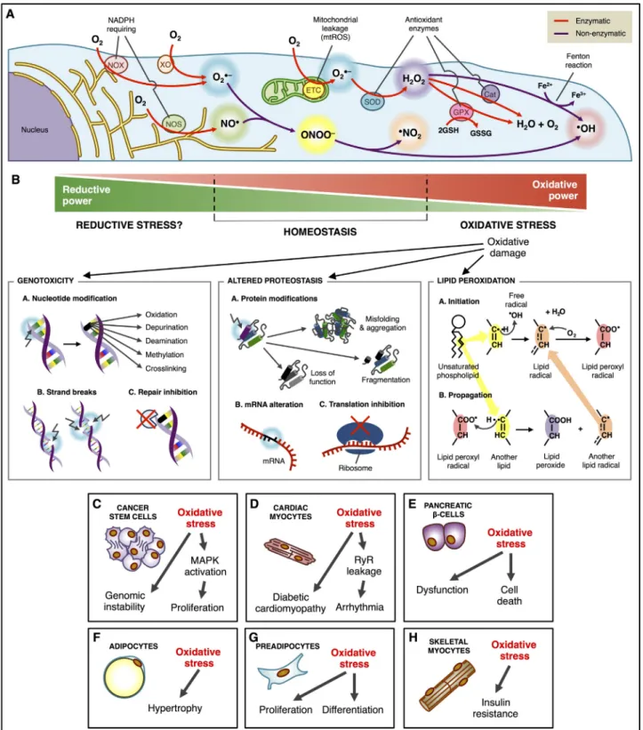
(9) Free Radical Biology and Medicine 124 (2018) 61–78. D. Peña-Oyarzun et al.. Fig. 4. Oxidative stress and non-communicable diseases. (A) Intracellular sources of oxidant molecules involve both enzymatic and non-enzymatic processes. Nitric oxide synthase (NOS) uses O2 for the oxidation of arginine to citrulline, releasing the gas nitric oxide (NO). NADPH oxidase (NOX) and xanthine oxidase (XO) catalyze the formation of superoxide (O2·-) from O2, while the mitochondria release O2·- as a byproduct of the electron transport chain. O2·- can react nonenzymatically with NO to form peroxynitrite (ONOO-), which in turn dissociates into the radicals NO2- and OH-. However, O2·- can also be enzymatically dismutated by the superoxide dismutase (SOD) to generate H2O2·H2O2 may either be converted into the radical OH- by the Fenton reaction involving the oxidation of Fe2+ to Fe3+, or be reduced into H2O by the catalase and the glutathione systems. (B) High oxidant levels disrupt redox homeostasis, which generates genotoxicity (nucleotide modifications, DNA single and double strand breaks), and inhibition of DNA repair by inactivation of the FAPY glycosylase, proteotoxicity and lipotoxicity. Also, lipid peroxidation generates a chain reaction by which the oxidized unsaturated phospholipids generate a second lipid peroxyl radical. (C) Oxidative stress participates in tumor growth by altering the genome and increasing proliferation via enhanced MAPK signaling in cancer stem cells. (D) Cardiomyocytes with oxidized ryanodine receptors (RyR) constantly release Ca2+ leading to uncontrolled contractions and arrhythmia. (E) Oxidation in pancreatic β cells alters insulin release and renders the cells more susceptible to death. (F) Oxidative stress induces hypertrophy of adipocytes that reduce GLUT4 translocation and increase release of adipokines. (G) Excessive oxidation leads to proliferation and differentiation in preadipocytes, increasing fat mass. (H) High oxidant levels in skeletal myocytes produce insulin resistance by inactivation of the Akt pathway.. 69.
(10) Free Radical Biology and Medicine 124 (2018) 61–78. D. Peña-Oyarzun et al.. membrane. The attenuation of mitochondrial H2O2 by overexpression of catalase in the mitochondria completely preserved insulin sensitivity [217]. These studies suggest that exacerbated levels of ROS/RNS are involved in the onset of obesity and T2DM. Given the evidence of an association between oxidative stress and the development of obesity-related diseases, many have proposed the use of antioxidant therapies. The study of this alternative is highly complex given the wide diversity of candidate antioxidant agents, different antioxidant mechanisms (that in many cases involve not only antioxidant but also anti-inflammatory and other effects), and different possible outcomes. In support of such therapies, a recent study by Okuno et al. [218] evaluated the metabolic features of obese mice in which ROS production was either eliminated or exacerbated locally in white adipose tissue. The results revealed that inhibition of oxidative stress was associated with a favorable metabolic profile and healthy adipose expansion, whereas fat ROS-augmented mice exhibited the opposite phenotype, with insulin resistance and lipid accumulation in the liver. However, other studies have reported controversial results. Alcala et al. [219] studied the effect of antioxidant (vitamin E) supplementation during mild weight gain in mice, and observed that the intervention induced a number of undesired effects. The authors argued that inhibiting ROS impaired physiological signaling events normally triggered in response to weight gain, rendering the animals insulin resistant and with deleterious ectopic lipid accumulation. Based on these and other negative results [220], the authors caution against antioxidants as a preventive therapy (before the actual oxidative insult is established) due to the relevance of the oxidative balance for maintaining homeostasis. Clinical trials using antioxidant therapies have failed to improve insulin resistance and other obesity-related disorders, and even induce adverse outcomes [220]. Together with the controversial observations in animal studies, these results suggest that there is still no evidence to support the use of antioxidant therapy for the treatment of insulin resistance and other obesity-related metabolic disorders in individuals without nutrient deficiencies [221,222].. pro-oxidant/anti-oxidant equilibrium that tilts the balance to favor the accumulation of the pro-oxidant species and promote oxidative damage to different biomolecules [179]. Oxidative stress directly induces DNA damage by several mechanisms, including nucleotide base modification, single strand break, double strand break, and indirectly by inhibiting DNA-formamidopyrimidine (FAPY) glycosylase [195]. Furthermore, oxidative stress alters the mRNA translational process and impairs protein synthesis, altering cellular proteostasis [196]. In addition, oxidative stress can impair protein folding, causing fragmentation and loss of protein function [7]. These oxidized proteins are recognized and degraded by the cell; however, when exacerbated protein oxidation occurs, the toxic products can accumulate leading to cellular dysfunction [7]. Finally, lipid oxidation by ROS leads to the formation of lipid hydroperoxides and aldehydes, which contribute to cellular toxicity [197]. Lipid oxidation of plasma and organelle membranes alters membrane permeability and fluidity [197]. The intracellular accumulation of these oxidized biomolecules has been implicated in the onset of different NCDs, including cancer, diabetes and CVD [6]. 4.3. Oxidative stress in cancer Oxidative species have been related to the initiation, development and maintenance of cancer [198]. Exacerbated ROS/RNS production modifies nucleotides and chromatin-bound proteins, inducing mutation in DNA and genomic instability, which ultimately leads to cancer initiation [199]. It has been suggested that oxidative species contribute to cancer by either acting as a second messenger or by promoting mutation of genomic DNA [199]. Under oxidative stress, the mitogen activated protein kinase (MAPK) is activated, thereby promoting tumor growth [200]. While reactive species have been associated with cancer, an exacerbated antioxidant system might also favor cancer development [201]. Indeed, inhibition of glutathione and thioredoxin antioxidant pathways leads to cancer cell death, suggesting an important role of these antioxidant systems in tumor progression [202]. However, the molecular mechanisms involved in this process are still poorly understood. Administration of the antioxidant vitamin C, in combination with cisplatin chemotherapy, in non-small cell lung cancer patients, shows a slightly improved response to chemotherapy that, however, is not statistically significant [203]. Same experiments performed with β-carotenoids and vitamin E showed similar results [204]. Better results were obtained with vitamin A co-treatment in breast cancer, resulting in nearly two-fold improved responses response to chemotherapy [205]. Despite that the role of antioxidants in slowing tumor growth is still controversial, the major contribution of antioxidants seems to be the protection of non-cancer cells against chemotherapy side effects [206,207].. 4.5. Oxidative stress in cardiovascular diseases Relevant studies have proposed that ROS/RNS induce permanent oxidation of different intracellular proteins, such as ion channels and transporters including the L-type channel, Na+/K+ exchanger and ryanodine receptor, all of which play crucial roles in pathological cardiac remodeling [223]. For example, redox modification of the ryanodine receptor type 2 (RyR2) Ca2+ channel contributes to chronic sarcoplasmic reticulum Ca2+ leakage, arrhythmia and systolic force reduction [224]. Also, the interplay between ROS and Ca2+ signals contribute to angiotensin II-induced hypertrophy in adult rat cardiomyocytes [225]. Furthermore, recent studies have shown that overexpression of specific antioxidants in cardiac mitochondria decreases diabetic cardiomyopathy [226] and prevents ventricular remodeling after myocardial infarction [227]. Indeed, NOX2 contributes to the development of myocardial contractile dysfunction and interstitial fibrosis during pressure overload in mice [228]. Other studies have shown that down-regulation of NOX4 ameliorates cardiac dysfunction induced by lipopolysaccharides [229] and prevents angiotensin-II-induced cardiac fibroblast proliferation and migration in adult mice, suggesting a critical role of NOX4 in the maintenance of heart homeostasis [230]. The pathophysiological roles of the NOX family and mtROS might overlap given that different ROS sources contribute to cardiac dysfunction and crosstalk between the different NOX family members and mtROS might occur in pathological conditions. Interestingly, crosstalk between NOX family and mitochondria has been proposed in others cell types [231]. Altogether these studies suggest that oxidative stress is implicated in functional and structural cardiac changes, which culminate in cardiac pathological remodeling, fibrosis and contractile dysfunction. The Prevencion-con-Dieta-Mediterranea (PREDIMED) study. 4.4. Oxidative stress in obesity and diabetes Overweight and obesity are the main causes of insulin resistance, T2DM and metabolic syndrome [208]. Oxidative stress plays a causal role in obesity development [209]. Indeed, oxidative stress increases the mass of adipose tissue by inducing proliferation, differentiation and hypertrophy of pre-adipocytes [210–212]. Studies have shown that local oxidative stress can induce insulin resistance. Oxidative stress triggered by H2O2 treatment generates insulin resistance in skeletal muscle cells isolated from lean Zucker rats [213], while oxidative stress induces cell dysfunction and death in pancreatic β-cells [214,215]. Both insulin-resistance and β-cell dysfunction lead to T2DM [208]. Moreover, insulin-resistant mice exhibit higher levels of NOX2 and a decrease in the reduced/oxidized glutathione ratio (GSH/GSSG) [216]. Intriguingly, the treatment with apocynin, an inhibitor of NOX2, prevents insulin-resistance induced by diet [216]. On the other hand, skeletal muscle from mice exposed to high fat diet exhibit insulin resistance and increased generation of H2O2 in the mitochondrial inner 70.
(11) Free Radical Biology and Medicine 124 (2018) 61–78. D. Peña-Oyarzun et al.. 5.2. Autophagy and oxidative stress-dependent cross-talk with inflammation in cancer. [232,233] as well as other clinical trials [234–236] showed that Mediterranean-style diets can cause a significant decline in CVD. The beneficial effects of Mediterranean-style diets may be because of the increased intake of polyphenolic flavonoids, carotenoids, omega-3 fatty acids, antioxidants, vitamins and minerals as well as essential and nonessential amino acids [237,238]. In small clinical trials, administration of antioxidants showed beneficial cardiovascular effects. Administration of vitamin E improves peripheral vascular function in patients with diabetes mellitus and Haptoglobin 2-2 genotype [239], resveratrol decreases arterial stiffness in patients with T2DM [240], and N-acetylcysteine (NAC) with nitrate therapy reduces myocardial infarct in patients undergoing primary percutaneous coronary intervention [241]. However, when large clinical trials or meta-analysis was performed, a lack of positive clinical evidence in the prevention of CVD was found. A meta-analysis study of 13 randomized controlled trials evaluated the effect of vitamin E in the prevention of stroke. The study concluded that the administration of vitamin E showed was benefitial in preventing stroke of any type, including ischemic stroke, hemorrhagic stroke, fatal stroke and non-fatal stroke [242]. Moreover, other metaanalysis studies showed that vitamin E and beta carotene have no effect on the cardiovascular mortality and morbidity [243,244]. In two randomized controlled trials, Vitamin E, vitamin C or grape-seed polyphenols did not significantly alter the rate of blood pressure variation [245]. Therefore, the available evidence suggests that the positive effects of antioxidants in the prevention of CVD was associated with the consumption of antioxidant rich foods rather the supplementation of antioxidants.. Recent studies show that tumor cells promote microenvironmental changes that favor autophagy and are required for sustained tumor growth. Ras-depletion in Drosophila promotes generation of LC3 positive dots not only in tumor surrounding tissues, but also in distant tissues like muscle, gut and adipose tissue, indicative of non-cell-autonomous autophagy induction [256]. Autophagy sustains tumor growth, since the volume of the tumor is effectively reduced when treated with the autophagic flux inhibitor chloroquine [256]. The pro-survival role of autophagy may involve the simultaneous activation of NRF2 (oxidative stress pathway) and NF-κB (Inflammatory pathway), which increase p62/SQSTM1 levels, thereby promoting the autophagic response [257]. Again, transcriptional increases in p62/SQSTM1 are indicative of a higher autophagic state, and should not to be confused with p62/ SQSTM1 accumulation by lysosomal dysfunction [93]. High-mobility group box 1 (HMGB1) is also upregulated under oxidative stress, as the result of activation of the NF-κB inflammatory pathway to prevent cell death [258,259]. HMGB1 stimulates autophagy by competing with Bcl2 for interaction with Beclin 1, thereby increasing resistance of leukemia cells to chemotherapy [260,261]. Cancer cells further amplify this autophagy mediated-survival state by releasing a substantial amount of superoxide (O2·-) to the local surroundings [256]. However, it should be noted that oxidative stress is tolerated to a certain threshold level, above which autophagy functions in the opposite sense to favor cancer cell death. For instance, excessive ROS production by H2O2 or 2-methoxyestradiol drives ATG5/ATG7/Beclin 1 dependent death of transformed cells [262]. Additional studies regarding the crosstalk between autophagy and ROS in the context of cancer involve immunogenic cell death (ICD). ICD refers to the capability of dying cells to attract immune cells to promote their degradation. This process is characterized by surface exposure of calreticulin, ATP release and late apoptotic protein liberation [263,264]. Over the past few years, Kroemer's group published a series of articles that suggest a crucial role for autophagy in cancer cells by modulating these processes. Syngeneic transplantable tumors treated with mitoxantrone, an anthracycline used as chemotherapeutic agent, release ATP as a “death signal” to recruit dendritic cells and cytotoxic Tcells, ultimately leading to tumor cell death [265]. However, autophagy impairment by silencing ATG5 and ATG7 abrogates ATP release, increasing the survival of tumor cells in the presence of mitoxantrone [265]. These results have been replicated also in a spontaneous melanoma model (not transplantable tumor) since the activation of the oncogene braf in WT mice provokes a strong ICD response when treated with mitoxantrone, a response that is abolished in Atg7-deficient mice [266]. However, other studies show that oxidative stress-dependent autophagy caused by treatment with hypericin, inhibits dendritic cell maturation in contact with melanoma cancer cells, as reflected in decreased IL-6 release [267]. These apparently contradictory conclusions may be due to differences between photodynamic therapy and chemotherapy on ICD. Altogether, the proposed pro-survival role of ROS-induced autophagy acts in two coordinated manners: inside the cancer cell, autophagy increases recycling of damaged molecules; while, outside the cancer cell, autophagy modulates the immune response with the objective of eliminating malignant cells.. 5. Crosstalk between inflammation, oxidative stress and autophagy 5.1. Autophagy and oxidative stress Studies have suggested that ROS could modulate the classic AMPKmTOR autophagy signaling axis. It was shown that p53 promotes the expression of antioxidant proteins, such as sestrins, to alleviate oxidative stress [246]. ROS interact with AMPK, which phosphorylates and activates the TSC1/TSC2 complex to inhibit mTORC1 and thereby induce autophagy [247]. AMPK activity can be reduced by the treatment with NAC, a ROS scavenger [248]. Furthermore, the ser/thr-kinase ataxia telangiectasia mutated protein (ATM) is also involved in the activation of AMPK and inhibition of mTORC1, thus stimulating autophagy in response to NO [249]. ROS also activate Beclin 1 dependent autophagy, as BNIP3, a protein that is upregulated by HIF-1α, prevents Bcl-2-mediated inhibition of Beclin 1 [250]. In addition, the autophagic substrate p62/SQSTM1 is over-expressed (by increased synthesis, not decreased degradation) in response to NRF2 activation, an anti-oxidant transcription factor. p62/SQSTM1 promotes the autophagy-dependent degradation of the Kelch-like ECHassociated protein 1 (KEAP1) [251], an adaptor protein required for the proteasome-dependent degradation of NRF2 [252]. Therefore, p62/ SQSTM1 is proposed to participate in a positive-feedback loop to maintain the NRF2 anti-oxidant effect by increasing autophagy [251]. It is important to consider that mitochondria (and particularly dysfunctional mitochondria) are the main source of ROS in the cell [253]. Thus, this p62/SQSTM1 positive-feedback loop may help protect against oxidative stress-dependent cell death by increasing mitophagy. ROS could also induce mitophagy in a direct manner by provoking mitochondrial oxidative damage [254]. Furthermore, H2O2 levels increase after starvation-induced autophagy, allowing direct oxidation of ATG4 at Cys-81 [255]. This modification on ATG4 decreases its activity and leads to accumulation of LC3-II, due to diminished removal of PE [255].. 5.3. Autophagy and oxidative stress-dependent cross-talk with inflammation in cardiovascular diseases Oxidative stress is involved in cardiac tissue damage during I/R and ethanol exposition. In mice, oxidative stress in ischemic conditions leads to a “protective” autophagy [268]. However under I/R injury conditions, oxidative stress upregulated autophagy exacerbates cell death [269]. By contrast, when the HL-1 cardiac muscle cell line is 71.
(12) Free Radical Biology and Medicine 124 (2018) 61–78. D. Peña-Oyarzun et al.. clearance by impairment of the lysosomal vacuolar ATPase function through oxidative modifications, which prevent lysosomal acidification and pH-dependent enzymatic activity [283]. Other studies have suggested that saturated fatty acid exposure, particularly to palmitic acid, induces autophagy through a mTOR-independent pathway in mouse embryonic fibroblasts (MEFs), HepG2 and human gastric cancer cells (MKN45) [284,285]. Fatty acid-induced mtROS production and/or protein kinase C (PKC) activation may mediate autophagy activation, which targets dysfunctional mitochondria or other damaged cell structures as an acute mechanism for cell survival [284,286]. However, chronically elevated autophagy can exceed the lysosomal capacity, impairing autophagic flux. The mechanism by which this impairment occurs has not been elucidated yet. In obesity-induced inflammation and insulin resistance, elevated free fatty acid availability and dysregulated adipokine synthesis may act as an ER stress signal within the adipocyte. ER stress is directly related to induction of autophagy, as revealed by higher levels of ER stress markers upon exposure to the saturated fatty acid palmitate in 3T3L1 adipocytes, along with increased levels of autophagic flux [287]. Consistent with this finding, impaired autophagy has been observed in different cell lines with elevated pro-inflammatory markers, ER stress [287] and elevated mitochondrial ROS [27]. As a unifying model, it has been proposed that the induction of autophagy may act in response to ER stress as a protective mechanism to attenuate the damaging response in metabolically challenged cells. Other studies support the hypothesis that autophagy elevation is an adaptive mechanism in the metabolic deregulation that occurs within the inflammatory and lipotoxic environment of obesity and T2DM. In the heart, lipid deposition leads to deleterious effects on myocardial structure and function. Lipid overload-induced oxidative stress and decreased autophagic turnover may contribute to cardiac dysfunction in obesity [288]. In skeletal muscle, excess lipid availability decreases insulin sensitivity, an effect that has important consequences for whole body glucose metabolism [289]. The preferential use of fat as an energy source by skeletal and cardiac muscle tissues generates more H2O2 and a number of free radical-producing lipophilic fatty acid-derived intermediates as compared to the glycolytic pathway [281], which represents a short-term adaptive response to the reduction in insulin signaling. However, this different “redox signature” generated by fat as opposed to carbohydrate utilization becomes detrimental if the stimulus is chronic, as is the case for obesity and T2DM. Defects in pancreatic autophagic flux and elevated oxidative stress are observed in diabetic patients [290]. Autophagy is also relevant in the maintenance of normal pancreatic β-cell function, and additional studies indicate that it may prevent the effects of inflammatory mediators in obesity and T2DM [291,292]. Increased autophagy, observed upon exposure to high fat diet-induced insulin resistance and T2DM represents an adaptive response to reduce ER stress [167,291]. Lipotoxicity in the liver becomes apparent as hepatic steatosis and non-alcoholic fatty liver disease, both highly relevant pathologies associated with obesity-induced metabolic disorders and with the development of insulin resistance and T2DM [293,294]. The liver is particularly susceptible to visceral obesity, since the release of free fatty acids and pro-inflammatory factors drains directly into the portal circulation. Numerous studies have observed that hepatic autophagy flux is downregulated, while oxidative and ER stress is elevated in models of obesity and lipotoxicity [295,296]. Mice with liver impaired autophagy show insulin resistance and elevated ER stress [163]. High fat diet-induced lipotoxicity also results in autophagy impairment in the kidneys, yielding an accumulation of ubiquitin-positive protein aggregates [297]. These mice are unable to induce autophagy upon lipid overload, elevating mitochondrial dysfunction and inflammasome activation. Autophagy protects kidneys from lipotoxicity in obesity, since high fat-diet mice with kidney-specific autophagy inhibition showed mitochondrial dysfunction, macrophage infiltration, inflammation and fibrosis not seen in control mice. Interestingly,. submitted to I/R conditions, increased rates of autophagosome formation are observed concomitant with a reduction in apoptosis [270]. On the other hand, the acute exposition of mice to ethanol, which stimulated autophagy in the heart, has been associated with cardiac apoptosis and malfunction [271]. In these conditions, pretreatment with NAC improves cardiac function by reducing autophagy [271]. Damaged mitochondria can be removed only by autophagy. As mentioned before, mitochondria are thought to be the source of > 90% of intracellular ROS [272]. Thus, any cardiac condition that affects the removal of damaged mitochondria represents a potential link between ROS and CVD. Parkin-deficient flies develop cardiomyopathy associated with dysmorphic mitochondria and elevated levels of ROS [273]. Parkindeficient mice do not develop cardiomyopathy at least until 12 months of age; however, they have a reduced survival and they develop larger myocardial infarction [274]. In addition, cardiomyocyte-specific deletion of mitofusin 2 (Mfn2) in mice disrupts the degradation of damaged mitochondria by mitophagy inducing cardiomyopathy [275]. Moreover, the expression of the ROS scavenger catalase, at lower levels, protects the heart, while expression at high levels exacerbates dilated cardiomyopathy [276]. An interesting mechanism of crosstalk between autophagy, ROS and inflammation in CVD has been proposed in a study showing that DNA from defective mitochondria, which escape from lysosomal degradation by autophagy leads to a Toll-like receptor 9mediated inflammatory response causing heart failure [277]. In addition, the exposure to LPS in THP-1 human macrophages induced LOX-1 expression, mitochondrial DNA damage, ROS, autophagy and the NLRP3 inflammasome. Both the use of ROS inhibitors and autophagy inducers decrease the expression of the NLRP3 inflammasome. By contrast, autophagy inhibition increases the expression of the NLPR3 inflammasome [278]. Another study, showed that ATG5 haplodeficiency in a model of angiotensin II-induced cardiac injury decreased mitophagy and increased mitochondrial ROS production associated with NF-κB activation in macrophages, which increased both the expression of inflammatory cytokines and macrophage infiltration [279]. Recently, TLR4 activity, which is important in innate immunity and inflammation, has been associated with the development of cardiac dysfunction. Thus, TLR4-deficient mice subjected to high fat diet showed decreased levels of ROS, cell death, intracellular anomalies in Ca2+ signaling and improved cardiac function compared with wild type animals. This improvement was associated with an increased level of autophagy in a NF-κB/JNK-dependent manner [280]. 5.4. Autophagy and oxidative stress-dependent cross-talk with inflammation in diabetes and obesity Inflammation is a common feature in obesity, and is triggered by a positive energy balance. At first this outcome is thought to be physiological in order to restore a new homeostatic state [82]. However, upon chronic over nutrition, the inflammatory response becomes maladaptive, leading to complications, such as insulin resistance and ultimately T2DM [81]. Obesity and inflammation are closely associated with the production of ROS and elevated markers of oxidative stress. As reviewed elsewhere [281], oxidative stress further activates inflammatory mediators, such as NF-κB and JNK, and interferes with insulin signaling pathways, also generating insulin resistance and T2DM, in addition to damage in multiple tissues as observed in animal models and humans. The excessive production of ROS in obesity is thought to be caused by adipose tissue macrophage infiltration and the ensuing increased release of pro-inflammatory cytokines, as well as greater expression and activity of NOX and mitochondrial H2O2 production. One of the main mechanisms through which obesity and T2DM lead to cardiometabolic derangements is via the increase in circulating free fatty acids, which leads to fat deposition and lipotoxicity in metabolically relevant organs. Thus, studies showed that a high exposure to lipids elevates the production of ROS and induces NOX2 activity [282]. Importantly, NOX2-derived O2·- may decrease autophagosome 72.
Figure
Documento similar
